understanding the ductus arteriosus. are we ... - sap.org.ar · understanding the ductus...
TRANSCRIPT
Understanding the DuctusArteriosus. Are we
hemodynamically naive?
Patrick J McNamaraAssociate Professor of Pediatrics
Hospital for Sick Children, Toronto
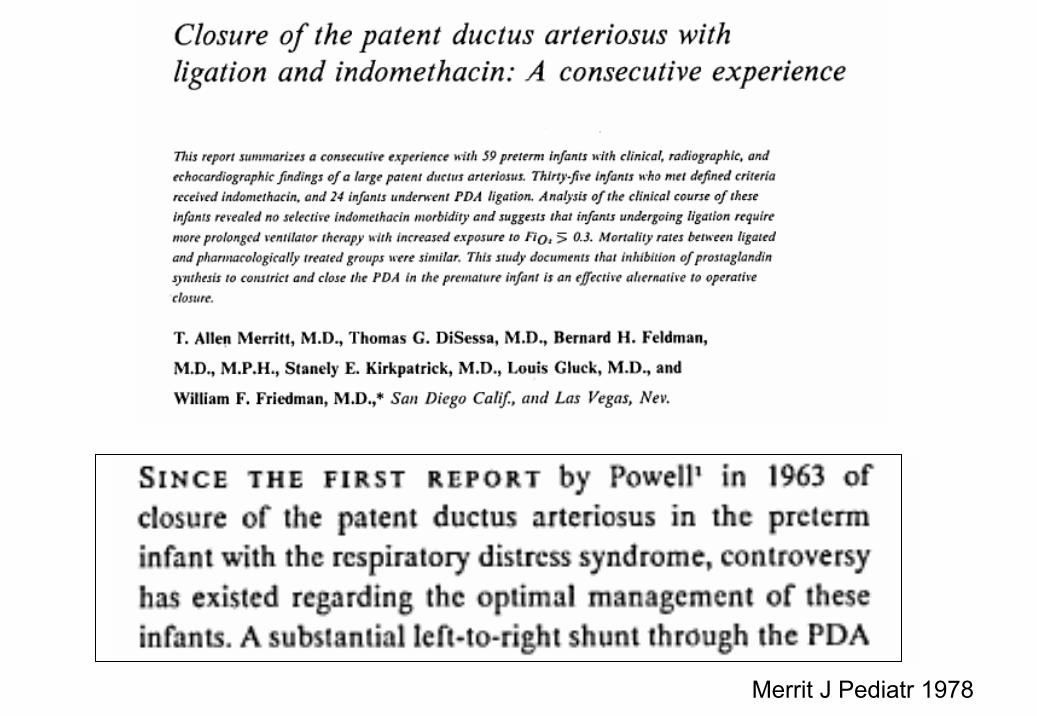
Merrit J Pediatr 1978
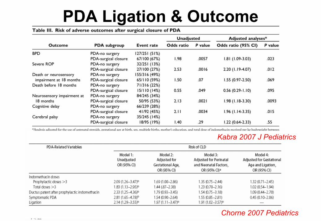
PDA Ligation & Outcome
Kabra 2007 J Pediatrics
Chorne 2007 Pediatrics
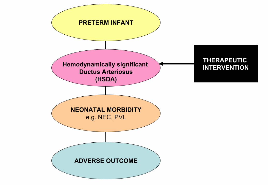
NEONATAL MORBIDITYe.g. NEC, PVL
ADVERSE OUTCOME
Hemodynamically significant Ductus Arteriosus
(HSDA)
PRETERM INFANT
THERAPEUTIC INTERVENTION
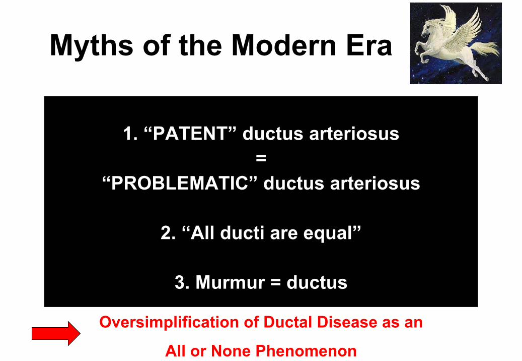
Myths of the Modern Era
1. “PATENT” ductus arteriosus=
“PROBLEMATIC” ductus arteriosus
2. “All ducti are equal”
3. Murmur = ductus
Oversimplification of Ductal Disease as an
All or None Phenomenon
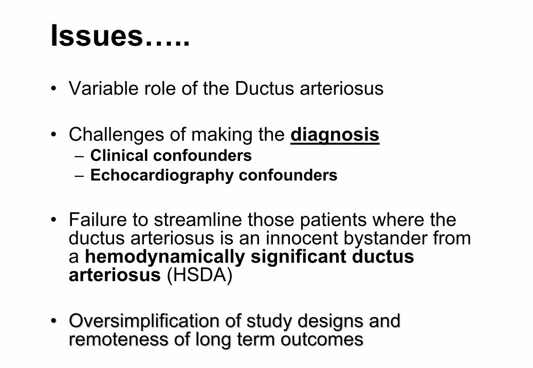
Issues…..• Variable role of the Ductus arteriosus
• Challenges of making the diagnosis– Clinical confounders– Echocardiography confounders
• Failure to streamline those patients where the ductus arteriosus is an innocent bystander from a hemodynamically significant ductusarteriosus (HSDA)
•• Oversimplification of study designs and Oversimplification of study designs and remoteness of long term outcomes remoteness of long term outcomes
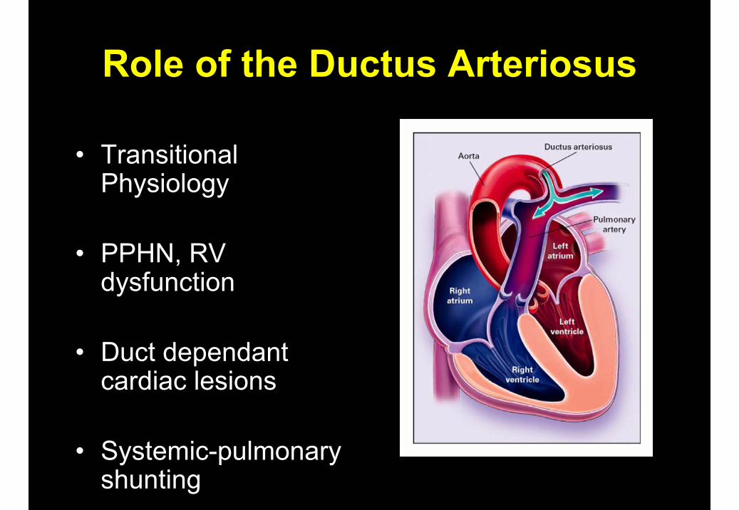
Role of the Ductus Arteriosus
• Transitional Physiology
• PPHN, RV dysfunction
• Duct dependant cardiac lesions
• Systemic-pulmonary shunting
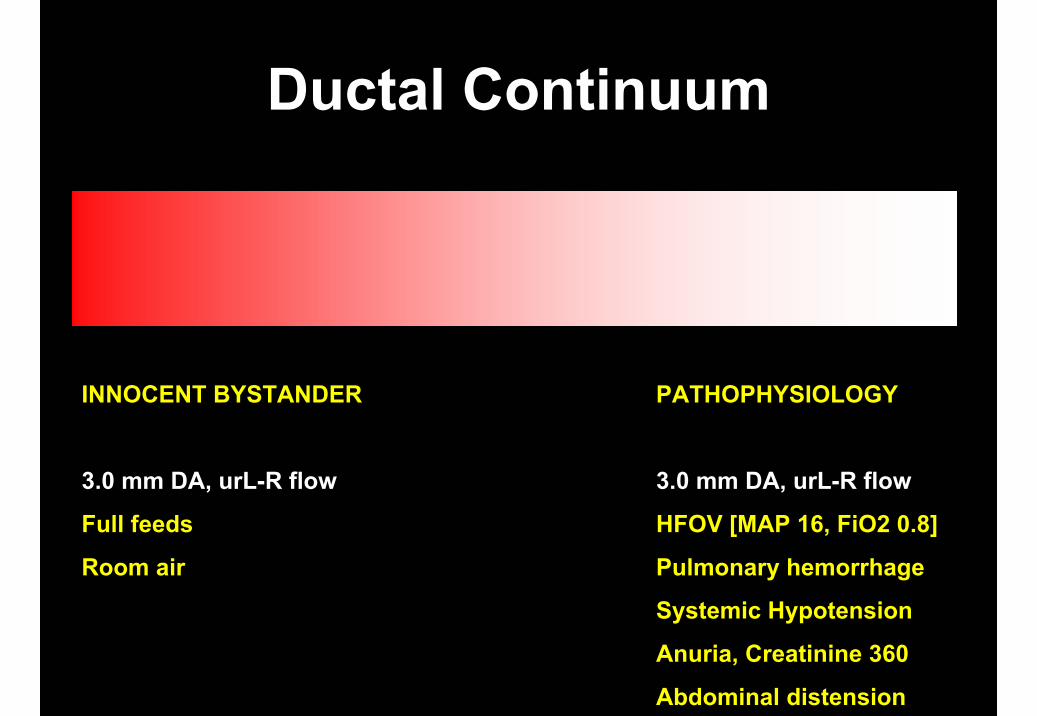
Ductal Continuum
INNOCENT BYSTANDER PATHOPHYSIOLOGY
3.0 mm DA, urL-R flow 3.0 mm DA, urL-R flow
Full feeds HFOV [MAP 16, FiO2 0.8]
Room air Pulmonary hemorrhage
Systemic Hypotension
Anuria, Creatinine 360
Abdominal distension
Is their hemodynamic impact?
• Is the clinical and/or physiologic instability related to increased ductal severity?
• Does the clinical and/or physiologic alteration resolve after ductal treatment?
If YES, then the DA is likely to be contributing to ongoing patient instability
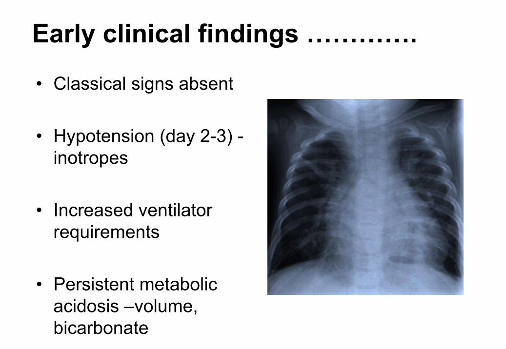
Early clinical findings ………….
• Classical signs absent
• Hypotension (day 2-3) -inotropes
• Increased ventilator requirements
• Persistent metabolic acidosis –volume, bicarbonate
Quantification of the volume of blood flow across the Ductus Arteriosus would
provide the best measure of hemodynamicsignificant

Is the ductus patent?
What is transductal diameter?
Issues: Measurement error, Variability in architecture and longitudinal diameter of the ductus arteriosus, Size is NOT STATIC
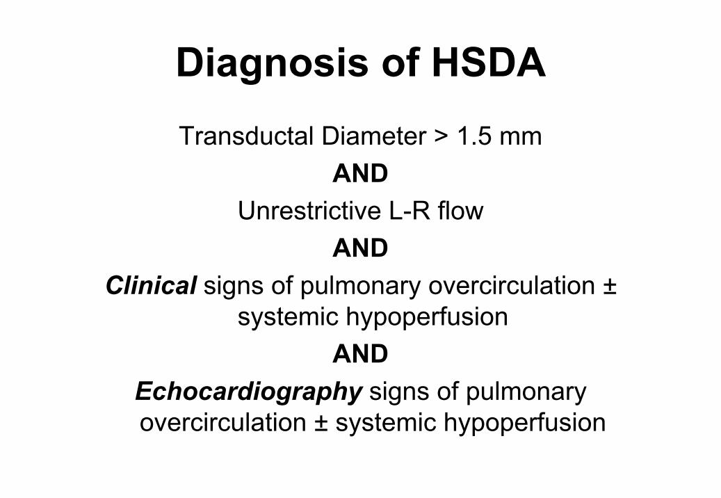
Diagnosis of HSDATransductal Diameter > 1.5 mm
ANDUnrestrictive L-R flow
ANDClinical signs of pulmonary overcirculation ±
systemic hypoperfusionAND
Echocardiography signs of pulmonary overcirculation ± systemic hypoperfusion
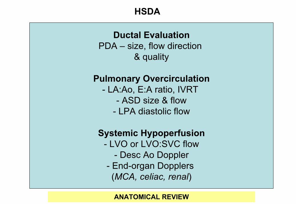
Ductal EvaluationPDA – size, flow direction
& quality
Pulmonary Overcirculation- LA:Ao, E:A ratio, IVRT
- ASD size & flow- LPA diastolic flow
Systemic Hypoperfusion- LVO or LVO:SVC flow
- Desc Ao Doppler- End-organ Dopplers(MCA, celiac, renal)
HSDA
ANATOMICAL REVIEW

IVCT IVRT
E wave
A wave
Transmitral flow Aortic flow Transmitral flow
1 2 3 4 5
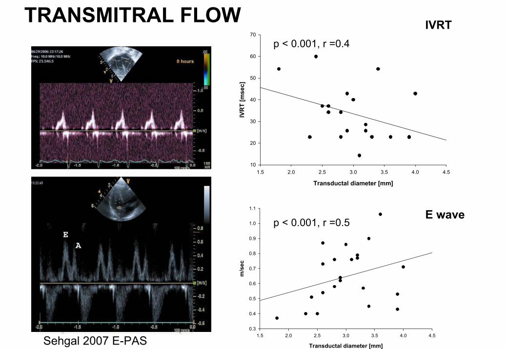
Transmitral Flow
CA
Sehgal 2007 E-PAS
TRANSMITRAL FLOW
Transductal diameter [mm]
1.5 2.0 2.5 3.0 3.5 4.0 4.5
IVR
T [m
sec]
10
20
30
40
50
60
70
Transductal diameter [mm]
1.5 2.0 2.5 3.0 3.5 4.0 4.5
m/s
ec
0.3
0.4
0.5
0.6
0.7
0.8
0.9
1.0
1.1
IVRT
E wave
p < 0.001, r =0.4
p < 0.001, r =0.5

End-organ flow and Ductal size
Lipman 1982 Pediatrics
50/58(86.3%)1/58(1.7%)>1.7
058/61(95%)< 1.5
RetrogradeAnterogradeSize (mm)
Evans 1995 Arch Dis Child
Increased transductal diameter leads to absence or reversal of diastolic flow to vital organs
Middle Cerebral Artery
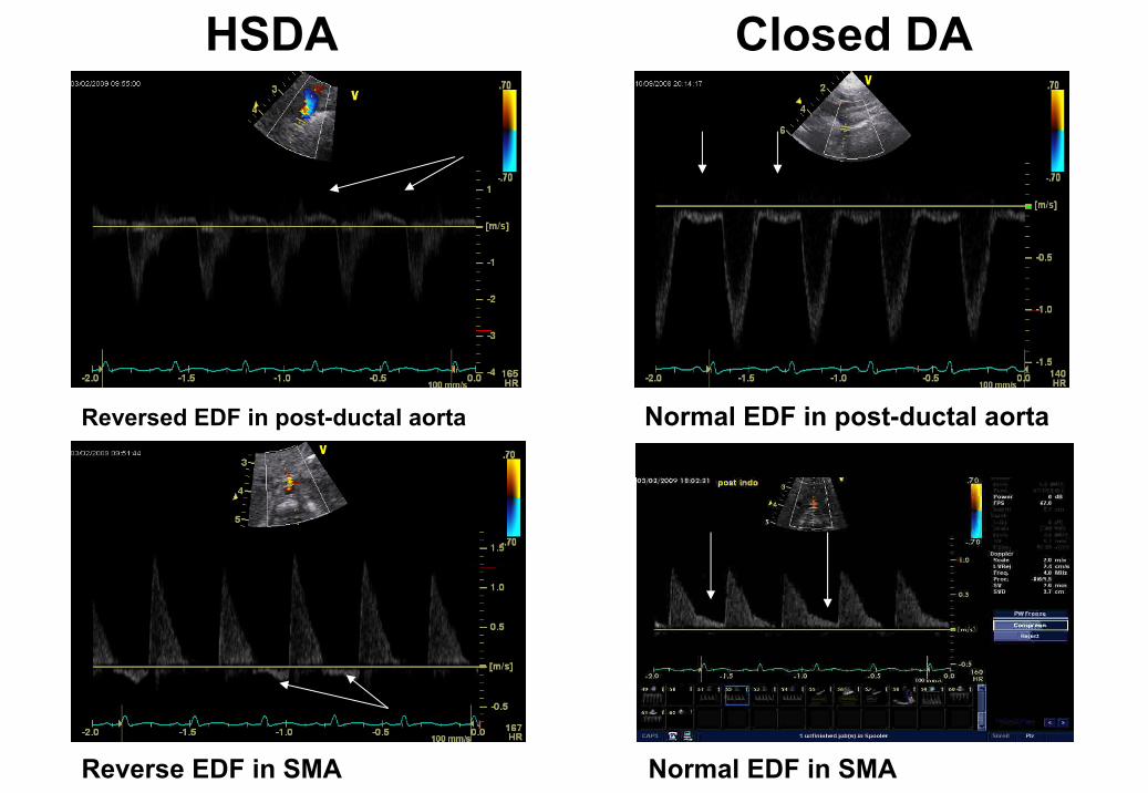
Reversed EDF in post-ductal aorta Normal EDF in post-ductal aorta
Reverse EDF in SMA Normal EDF in SMA
HSDA Closed DA
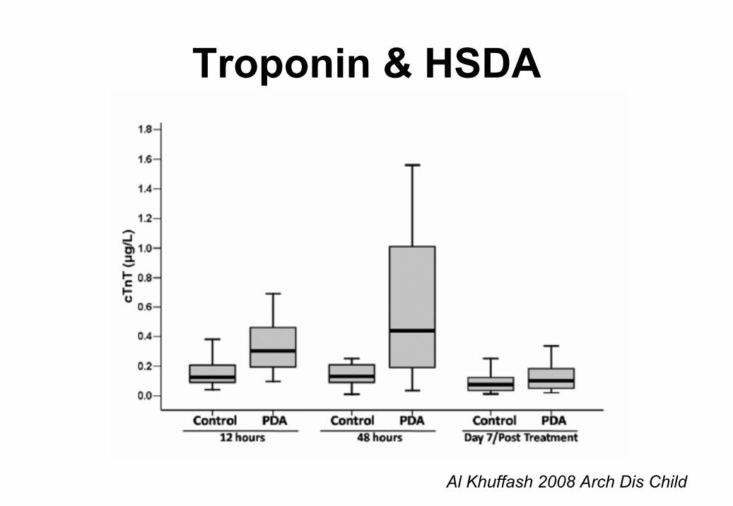
Al Khuffash 2008 Arch Dis Child
Troponin & HSDA
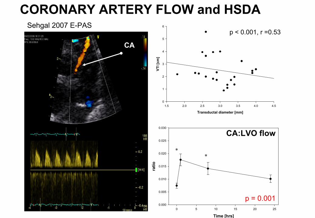
CA
Sehgal 2007 E-PAS
CORONARY ARTERY FLOW and HSDA
Transductal diameter [mm]
1.5 2.0 2.5 3.0 3.5 4.0 4.5
VTI [
cm]
0
1
2
3
4
5
6
p < 0.001, r =0.53
Time [hrs]0 5 10 15 20 25
ratio
0.000
0.005
0.010
0.015
0.020
0.025
0.030
∗∗
p = 0.001
CA:LVO flow
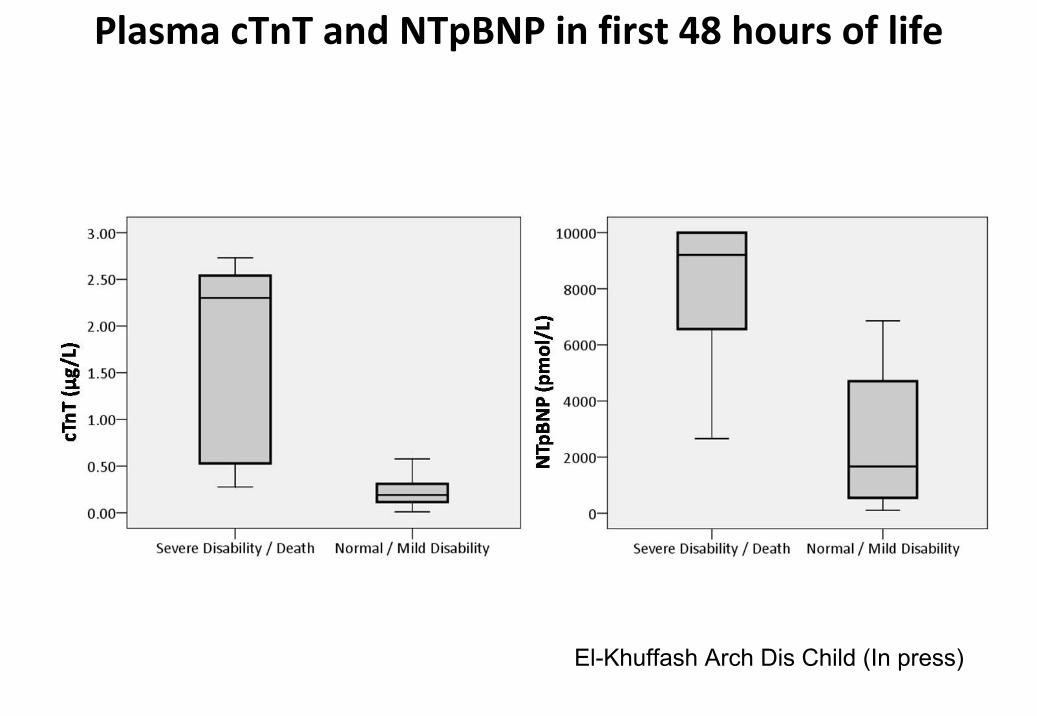
Plasma cTnT and NTpBNP in first 48 hours of life
El-Khuffash Arch Dis Child (In press)
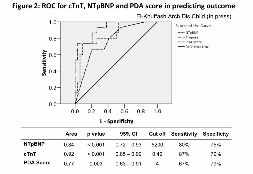
Area p value 95% CI Cut off Sensitivity Specificity
NTpBNP 0.84 < 0.001 0.72 – 0.93 5200 80% 75%
cTnT 0.92 < 0.001 0.85 – 0.99 0.49 87% 79%PDA Score 0.77 0.003 0.63 – 0.91 4 67% 79%
Figure 2: ROC for cTnT, NTpBNP and PDA score in predicting outcomeEl-Khuffash Arch Dis Child (In press)
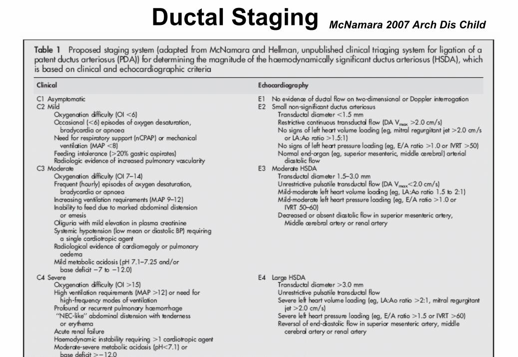
Ductal Staging McNamara 2007 Arch Dis Child
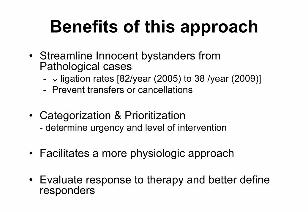
Benefits of this approach• Streamline Innocent bystanders from
Pathological cases- ↓ ligation rates [82/year (2005) to 38 /year (2009)]- Prevent transfers or cancellations
• Categorization & Prioritization - determine urgency and level of intervention
• Facilitates a more physiologic approach
• Evaluate response to therapy and better define responders

A hemodynamically significant ductusarteriosus is associated with acute reversible physiologic disturbance……
• BUT what about neonatal morbidities?

Teixeira 2006 Acta Paed
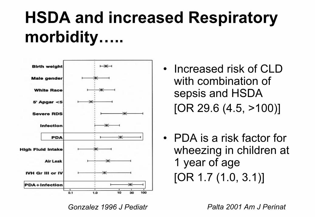
HSDA and increased Respiratory morbidity…..
• Increased risk of CLD with combination of sepsis and HSDA [OR 29.6 (4.5, >100)]
• PDA is a risk factor for wheezing in children at 1 year of age [OR 1.7 (1.0, 3.1)]
Gonzalez 1996 J Pediatr Palta 2001 Am J Perinat
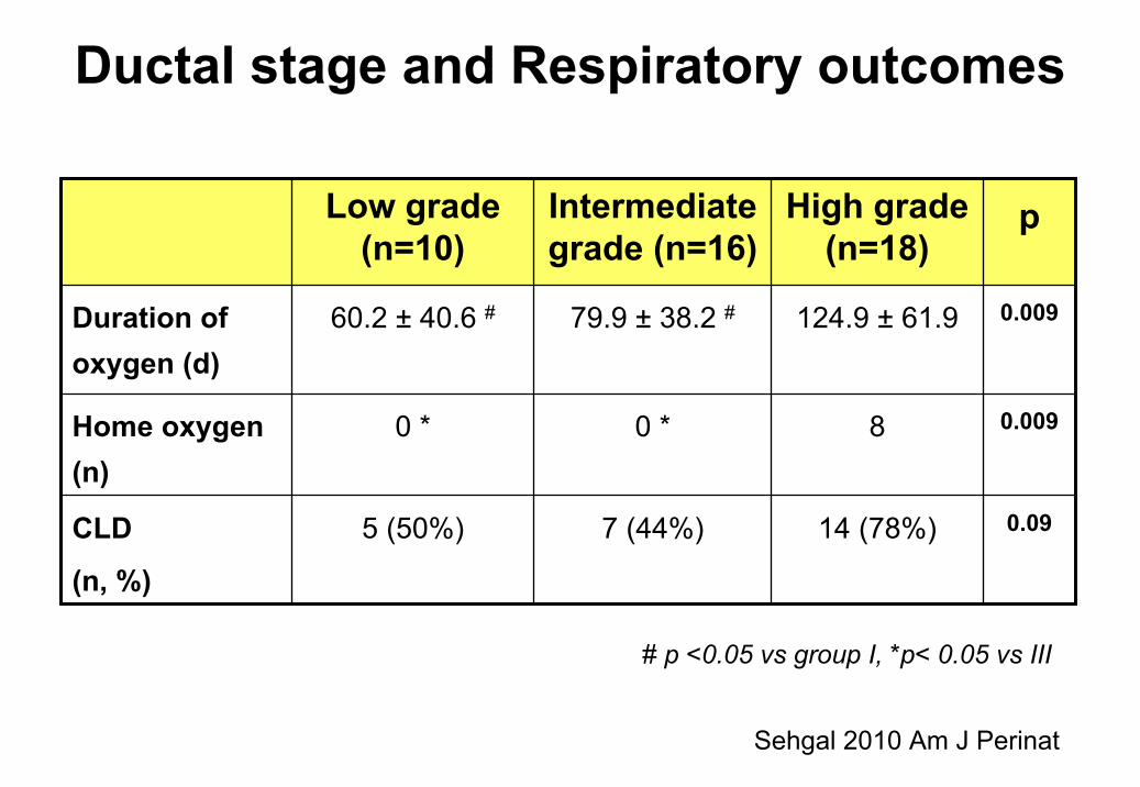
Ductal stage and Respiratory outcomes
0.00980 *0 *Home oxygen (n)
0.0914 (78%)7 (44%)5 (50%)CLD
(n, %)
0.009124.9 ± 61.979.9 ± 38.2 #60.2 ± 40.6 #Duration of oxygen (d)
pHigh grade (n=18)
Intermediate grade (n=16)
Low grade (n=10)
# p <0.05 vs group I, *p< 0.05 vs III
Sehgal 2010 Am J Perinat
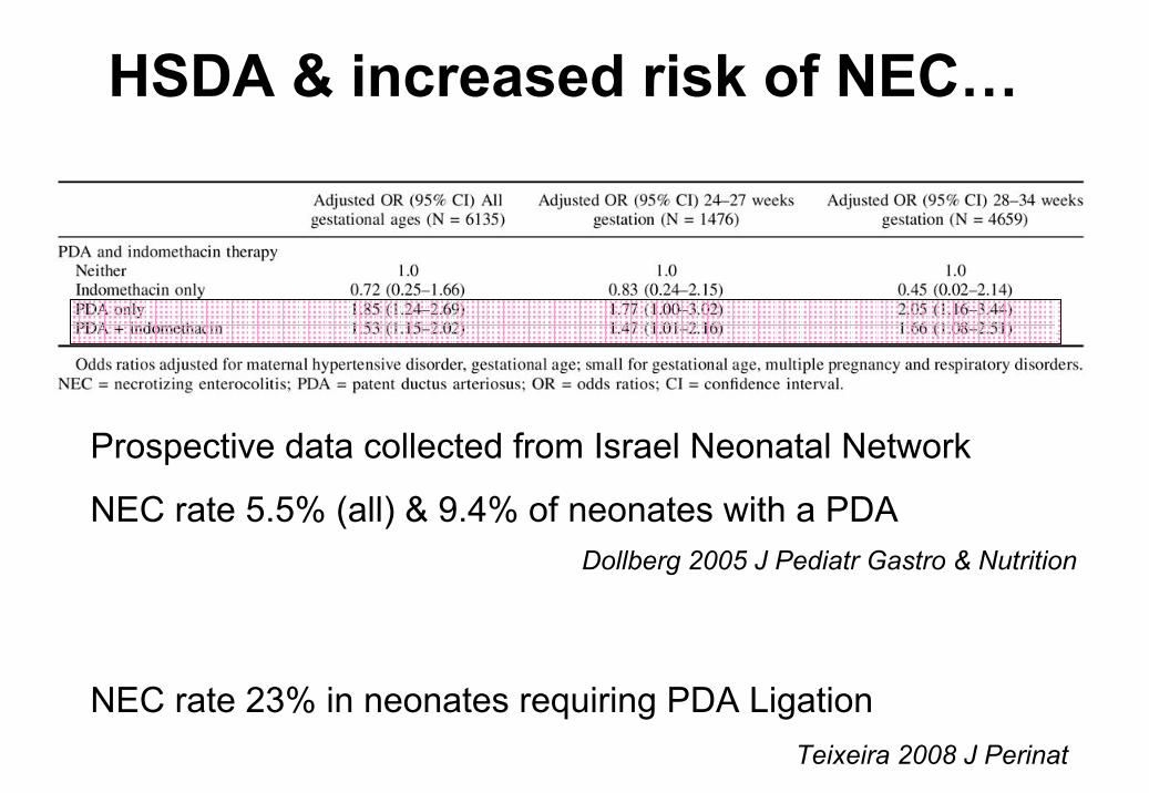
HSDA & increased risk of NEC…
Teixeira 2008 J Perinat
Prospective data collected from Israel Neonatal Network
NEC rate 5.5% (all) & 9.4% of neonates with a PDA
NEC rate 23% in neonates requiring PDA Ligation
Dollberg 2005 J Pediatr Gastro & Nutrition
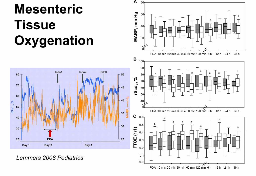
Lemmers 2008 Pediatrics
Mesenteric Tissue Oxygenation
Is there evidence that intervention is beneficial?
The viewpoint of the “permissivist”
“there is NO evidence that treatment of the DA improves long term outcomes”
No placebo controlled trials of therapeutic intervention
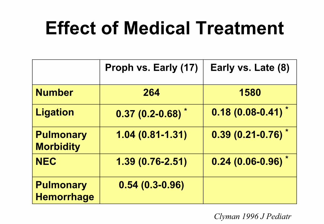
Effect of Medical Treatment
1580264Number
0.54 (0.3-0.96)Pulmonary Hemorrhage
0.24 (0.06-0.96) *1.39 (0.76-2.51)NEC
0.39 (0.21-0.76) *1.04 (0.81-1.31)Pulmonary Morbidity
0.18 (0.08-0.41) *0.37 (0.2-0.68) *Ligation
Early vs. Late (8)Proph vs. Early (17)
Clyman 1996 J Pediatr
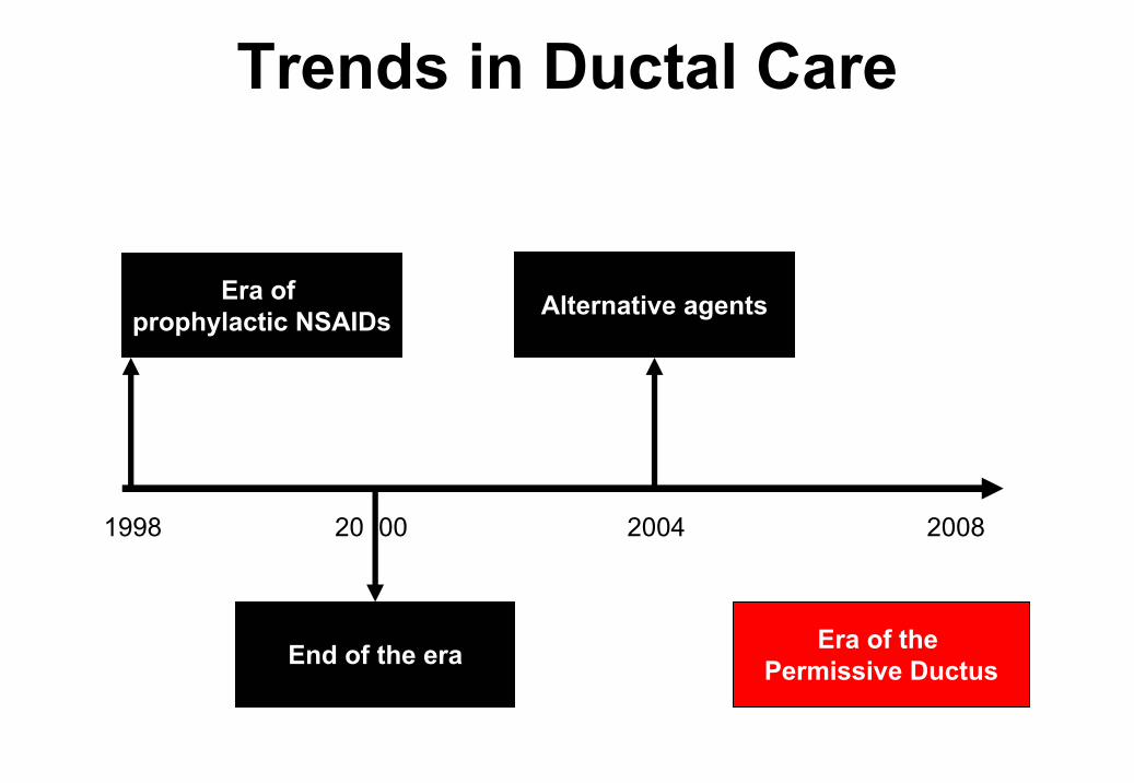
Era of prophylactic NSAIDs
End of the era
Alternative agents
Era of the Permissive Ductus
Trends in Ductal Care
1998 20 00 2004 2008
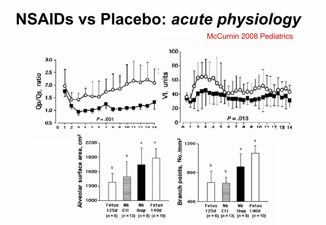
NSAIDs vs Placebo: acute physiologyMcCurnin 2008 Pediatrics
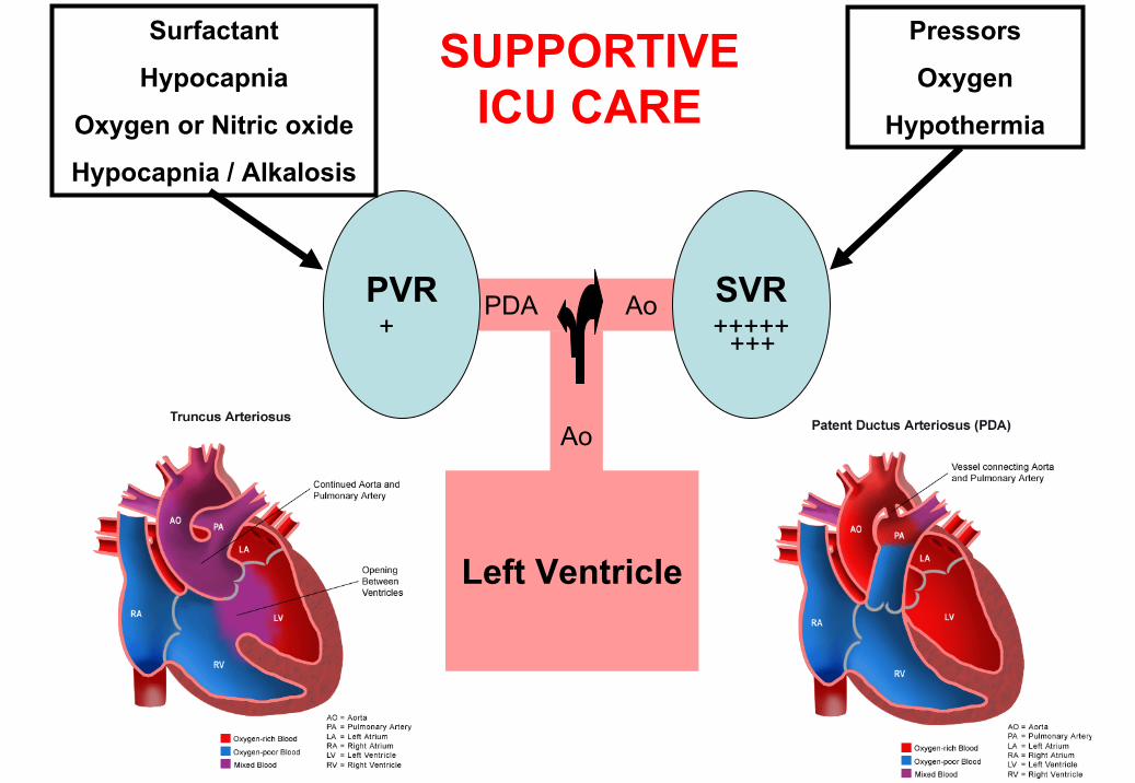
Left Ventricle
Ao
PDA Ao SVR+++++
PVR+++
+++
Surfactant
Hypocapnia
Oxygen or Nitric oxide
Hypocapnia / Alkalosis
Pressors
Oxygen
Hypothermia
SUPPORTIVE ICU CARE

HSDA
Therapeutic
Indomethacin
(fECHO guided)
PDA Ligation
SupportivePermissive acidosis
(pH 7.25-7.3)
Permissive Hypercapnemia
(50-60 mmHg)
Minimize oxygen exposure
(SpO2 85-92%)
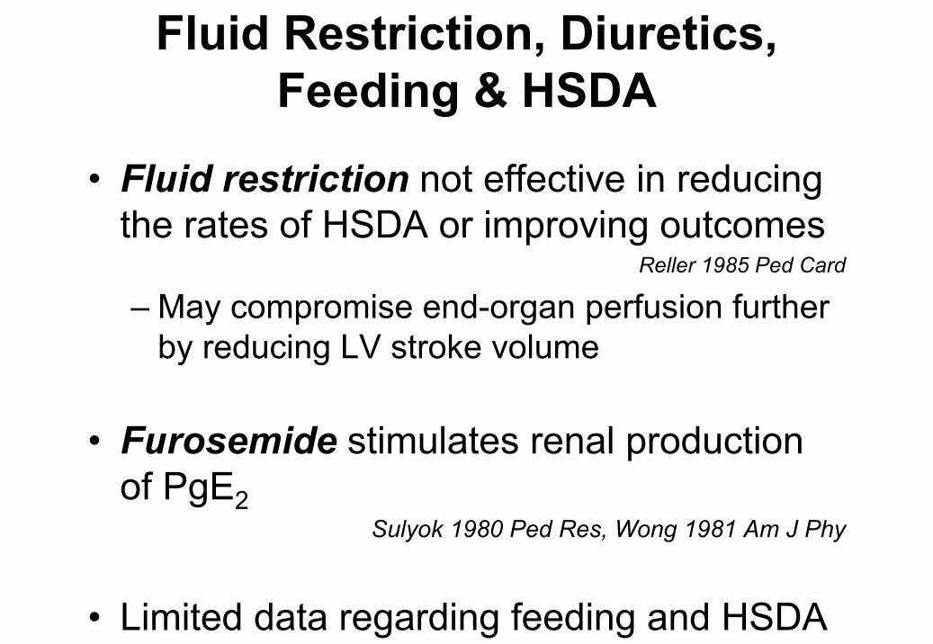
Fluid Restriction, Diuretics, Feeding & HSDA
• Fluid restriction not effective in reducing the rates of HSDA or improving outcomes
Reller 1985 Ped Card
– May compromise end-organ perfusion further by reducing LV stroke volume
• Furosemide stimulates renal production of PgE2
Sulyok 1980 Ped Res, Wong 1981 Am J Phy
• Limited data regarding feeding and HSDA
• Is surgical intervention preferable?
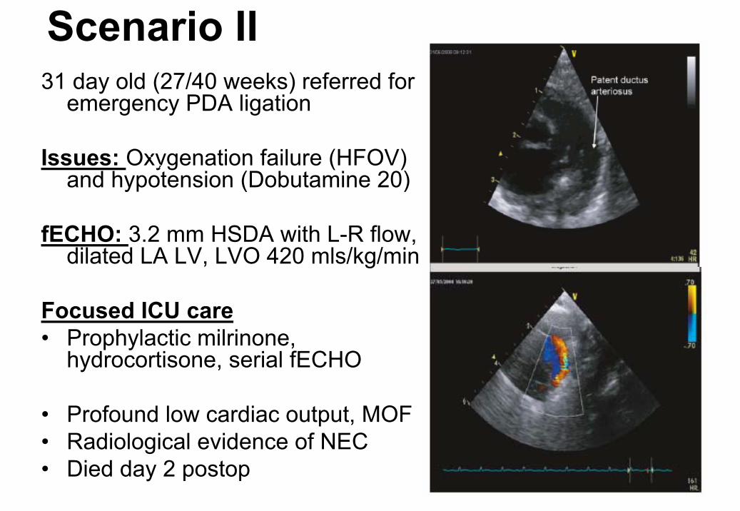
Scenario II31 day old (27/40 weeks) referred for
emergency PDA ligation
Issues: Oxygenation failure (HFOV) and hypotension (Dobutamine 20)
fECHO: 3.2 mm HSDA with L-R flow, dilated LA LV, LVO 420 mls/kg/min
Focused ICU care• Prophylactic milrinone,
hydrocortisone, serial fECHO
• Profound low cardiac output, MOF• Radiological evidence of NEC • Died day 2 postop
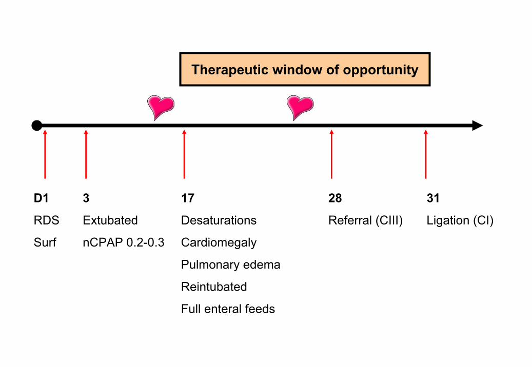
D1 3 17 28 31
RDS Extubated Desaturations Referral (CIII) Ligation (CI)
Surf nCPAP 0.2-0.3 Cardiomegaly
Pulmonary edema
Reintubated
Full enteral feeds
Therapeutic window of opportunity
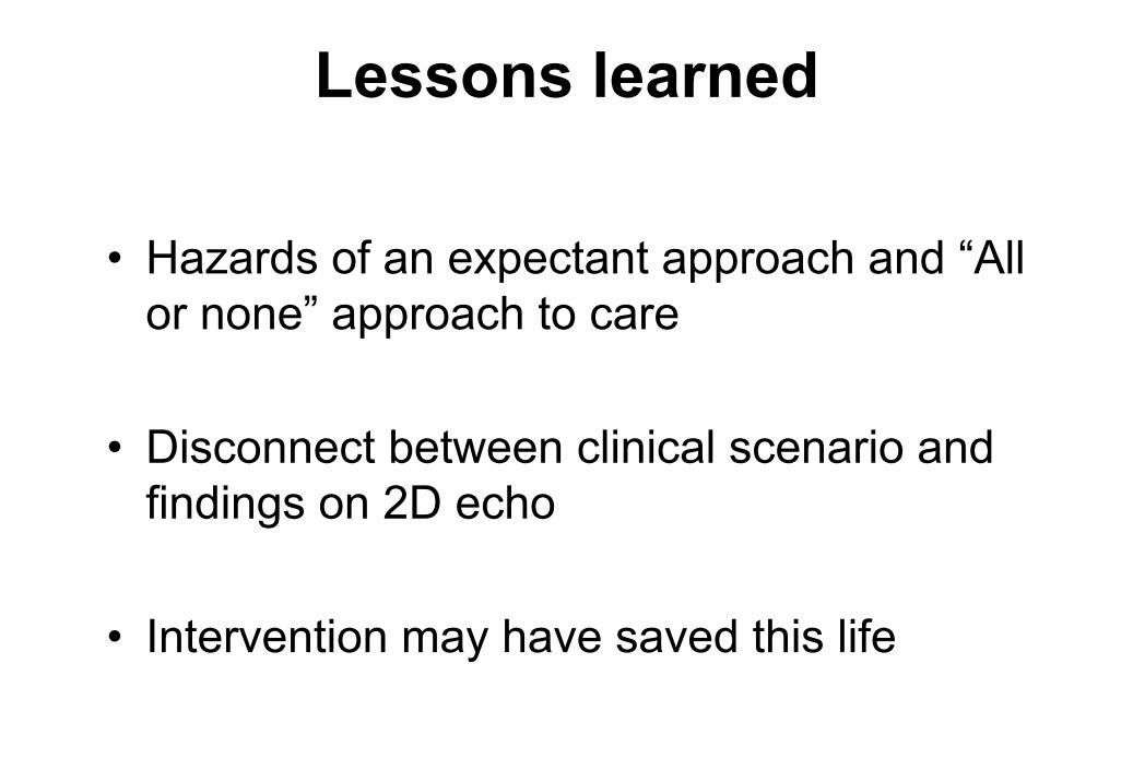
Lessons learned
• Hazards of an expectant approach and “All or none” approach to care
• Disconnect between clinical scenario and findings on 2D echo
• Intervention may have saved this life
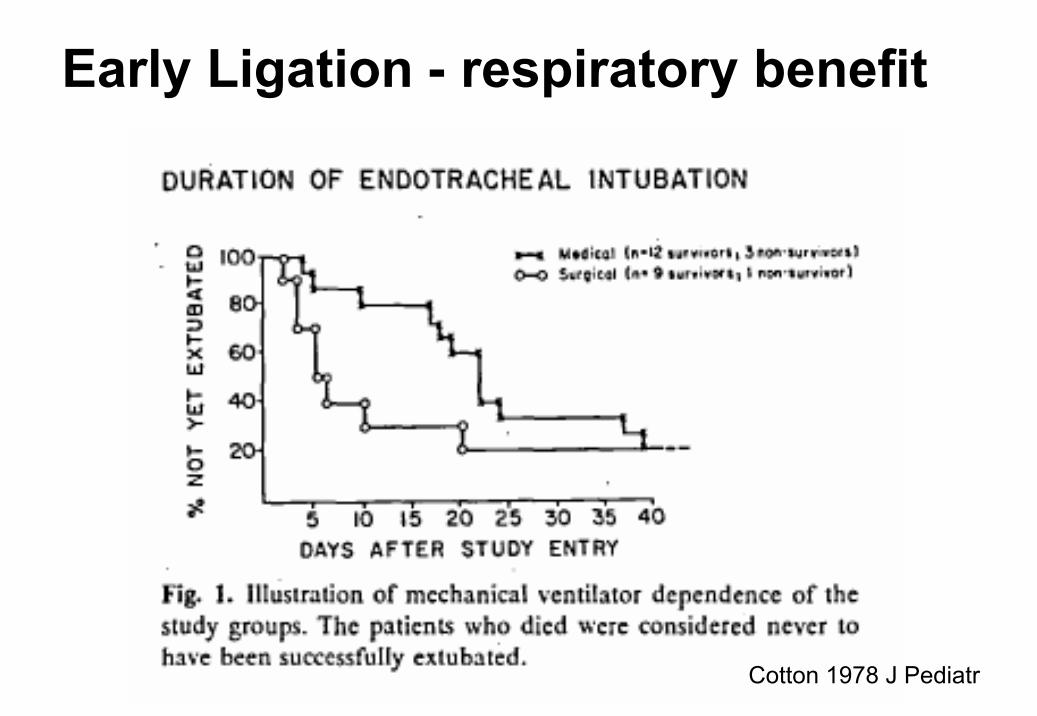
Cotton 1978 J Pediatr
Early Ligation - respiratory benefit

Early Ligation reduces NEC rates….
Neonates < 1000 g (n=84) with ⇓rate of NEC (30 vs 8%)
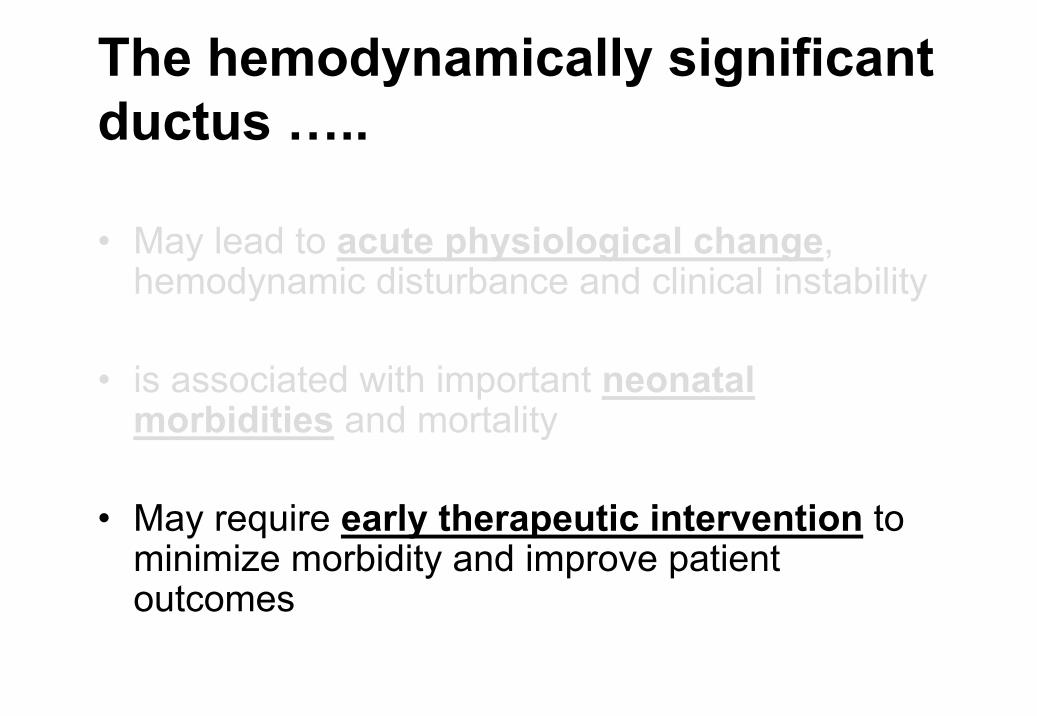
The hemodynamically significant ductus …..
• May lead to acute physiological change, hemodynamic disturbance and clinical instability
• is associated with important neonatal morbidities and mortality
• May require early therapeutic intervention to minimize morbidity and improve patient outcomes

Treatment is BAD?Treatment doesn’t work!
Benefit
Harm
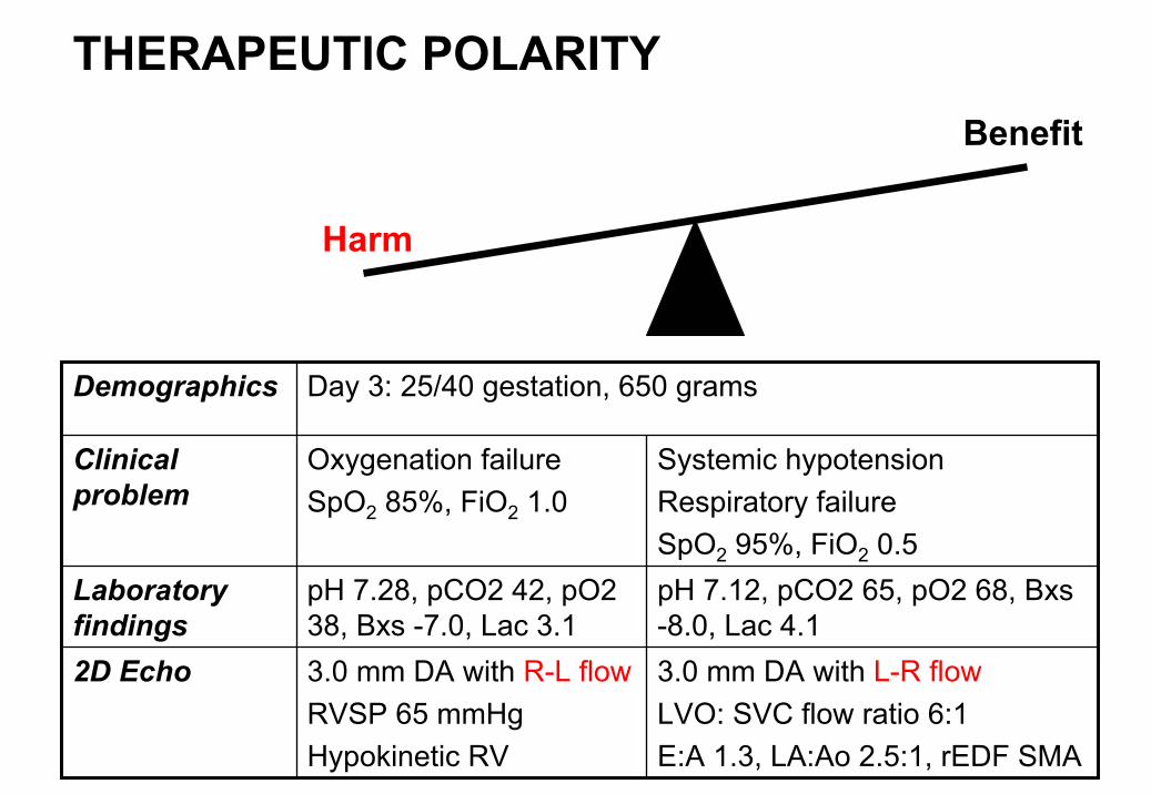
3.0 mm DA with L-R flowLVO: SVC flow ratio 6:1E:A 1.3, LA:Ao 2.5:1, rEDF SMA
3.0 mm DA with R-L flowRVSP 65 mmHgHypokinetic RV
2D Echo
pH 7.12, pCO2 65, pO2 68, Bxs-8.0, Lac 4.1
pH 7.28, pCO2 42, pO2 38, Bxs -7.0, Lac 3.1
Laboratory findings
Systemic hypotensionRespiratory failureSpO2 95%, FiO2 0.5
Oxygenation failureSpO2 85%, FiO2 1.0
Clinical problem
Day 3: 25/40 gestation, 650 gramsDemographics
THERAPEUTIC POLARITY
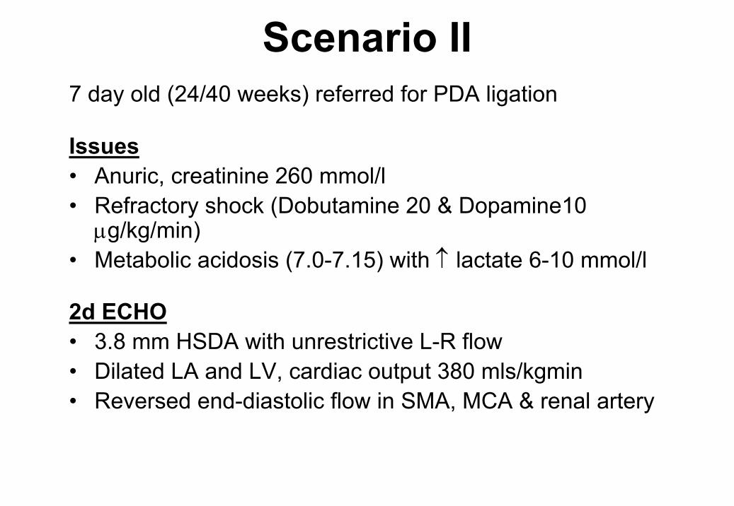
Scenario II7 day old (24/40 weeks) referred for PDA ligation
Issues• Anuric, creatinine 260 mmol/l• Refractory shock (Dobutamine 20 & Dopamine10
µg/kg/min)• Metabolic acidosis (7.0-7.15) with ↑ lactate 6-10 mmol/l
2d ECHO• 3.8 mm HSDA with unrestrictive L-R flow• Dilated LA and LV, cardiac output 380 mls/kgmin• Reversed end-diastolic flow in SMA, MCA & renal artery
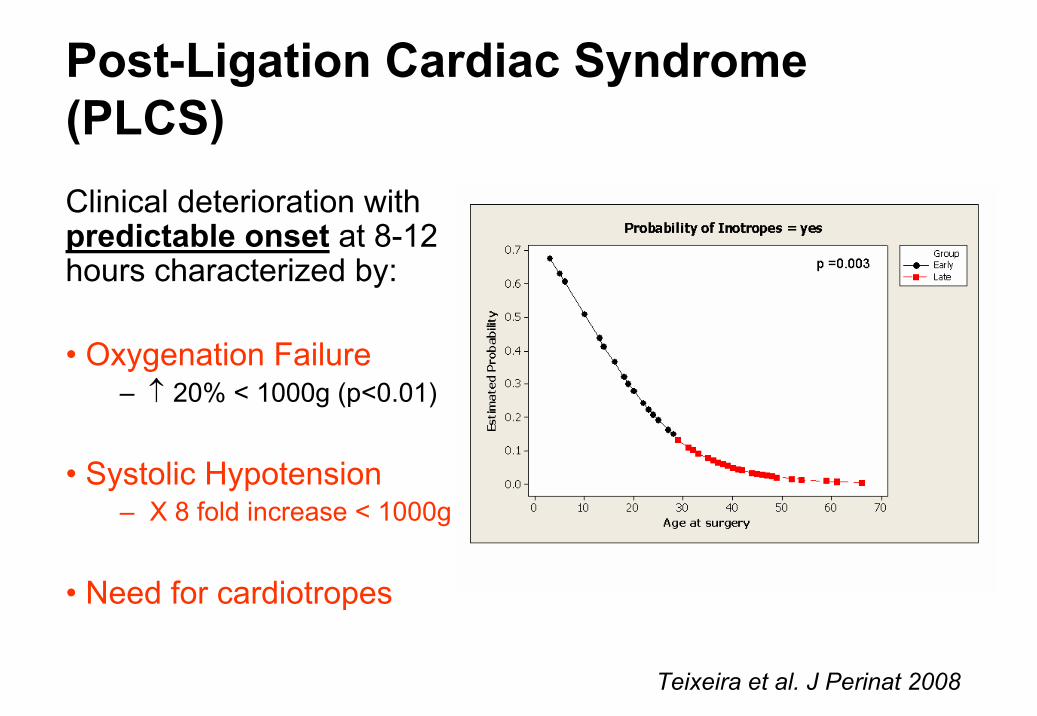
Post-Ligation Cardiac Syndrome (PLCS)Clinical deterioration with predictable onset at 8-12 hours characterized by:
• Oxygenation Failure– ↑ 20% < 1000g (p<0.01)
• Systolic Hypotension– X 8 fold increase < 1000g
• Need for cardiotropes
Teixeira et al. J Perinat 2008
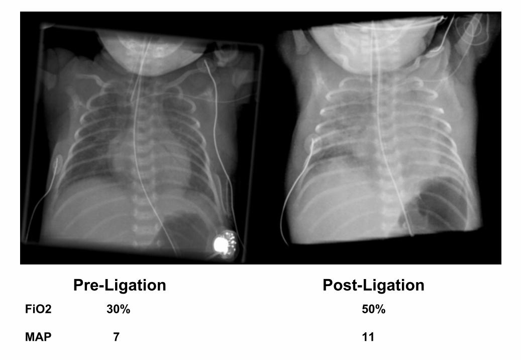
Pre-Ligation Post-LigationFiO2 30% 50%
MAP 7 11
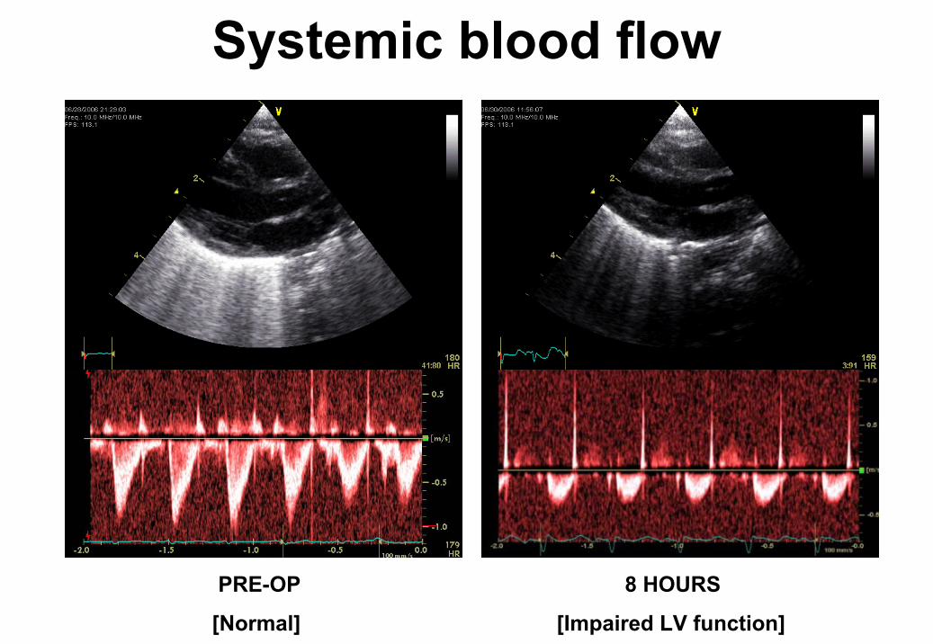
Systemic blood flow
PRE-OP 8 HOURS
[Normal] [Impaired LV function]
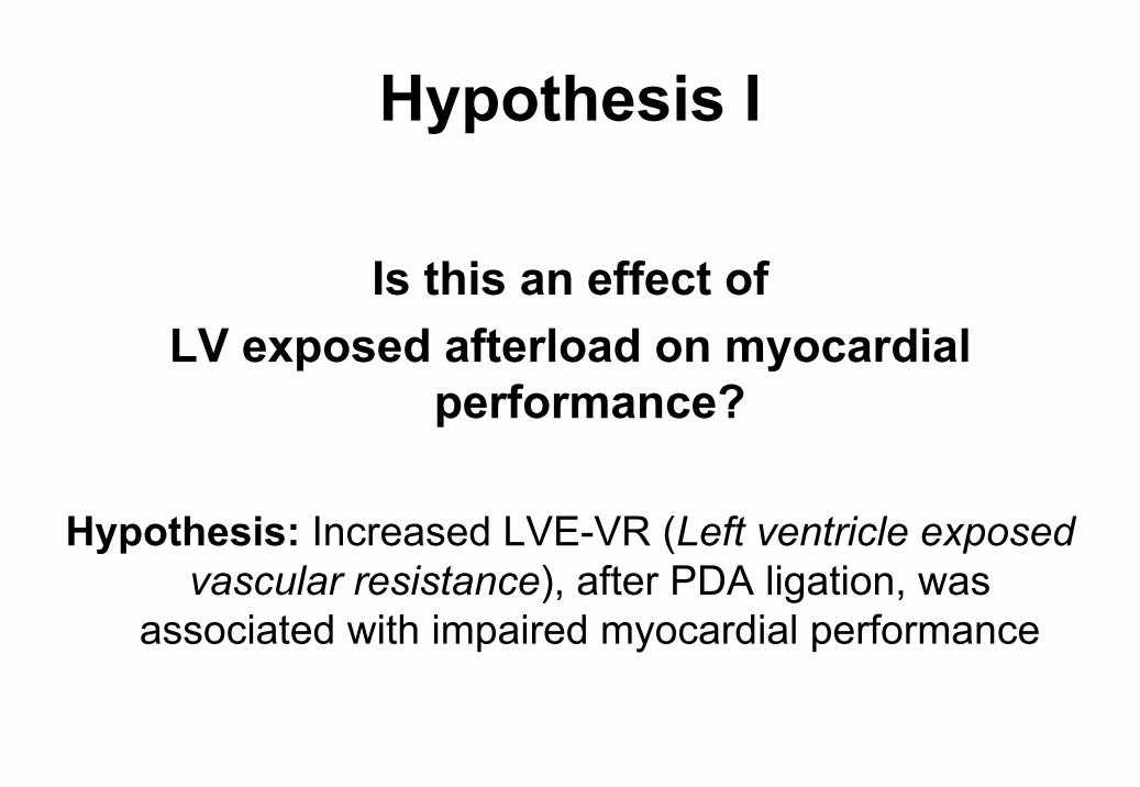
Is this an effect of LV exposed afterload on myocardial
performance?
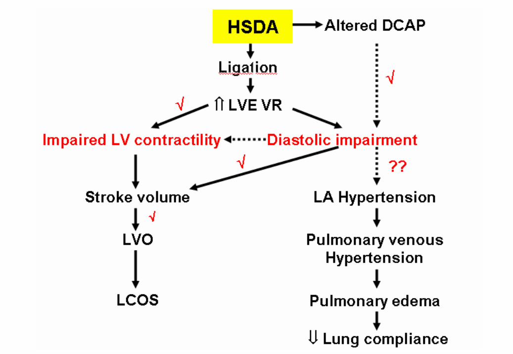
Hypothesis: Increased LVE-VR (Left ventricle exposed vascular resistance), after PDA ligation, was
associated with impaired myocardial performance
Hypothesis I
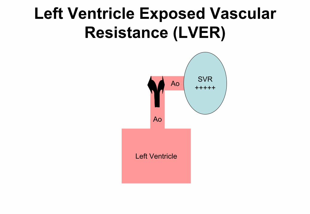
Left Ventricle
Ao
PDA Ao SVR+++++
PVR++
Left Ventricle Exposed Vascular Resistance (LVER)
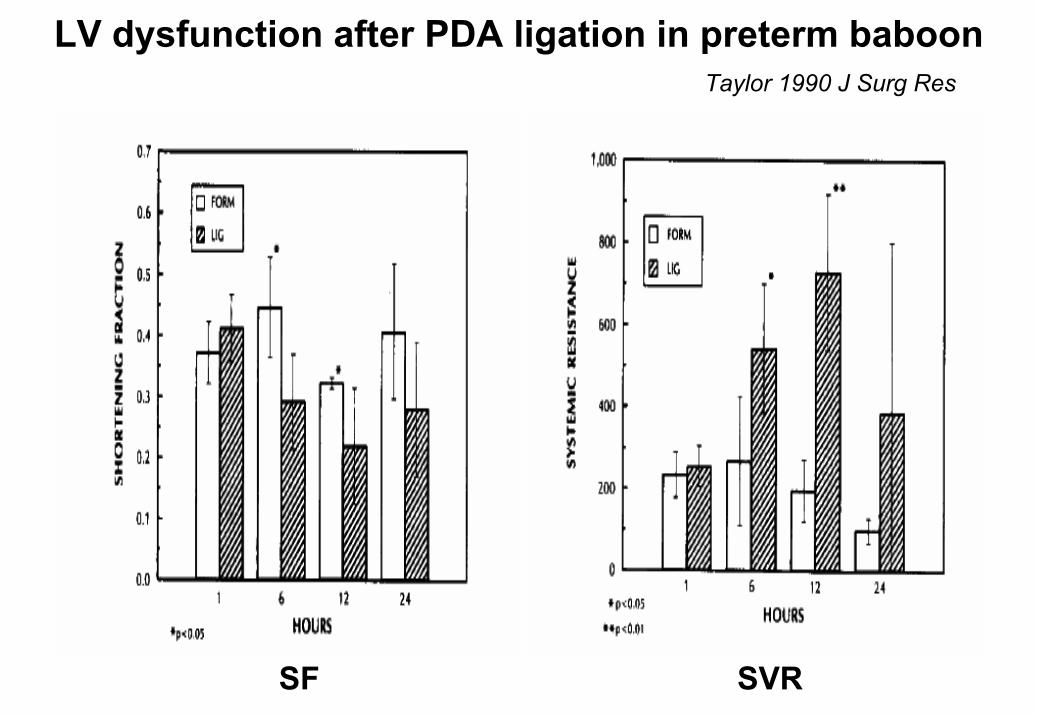
LV dysfunction after PDA ligation in preterm baboon
SF SVR
Taylor 1990 J Surg Res
# p < 0.05 vs baseline
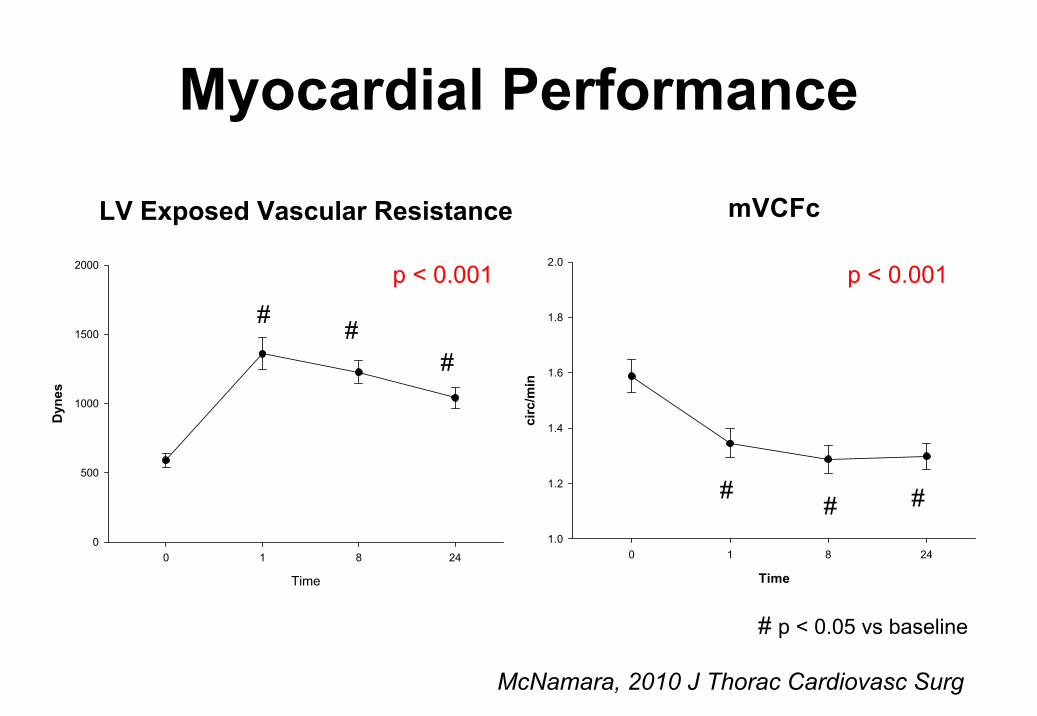
mVCFc
Time
0 1 8 24
circ
/min
1.0
1.2
1.4
1.6
1.8
2.0
** *
LV Exposed Vascular Resistance
Time
0 1 8 24
Dyn
es
0
500
1000
1500
2000
**
*
Myocardial Performance
p < 0.001 p < 0.001
McNamara, 2010 J Thorac Cardiovasc Surg
##
#
## #
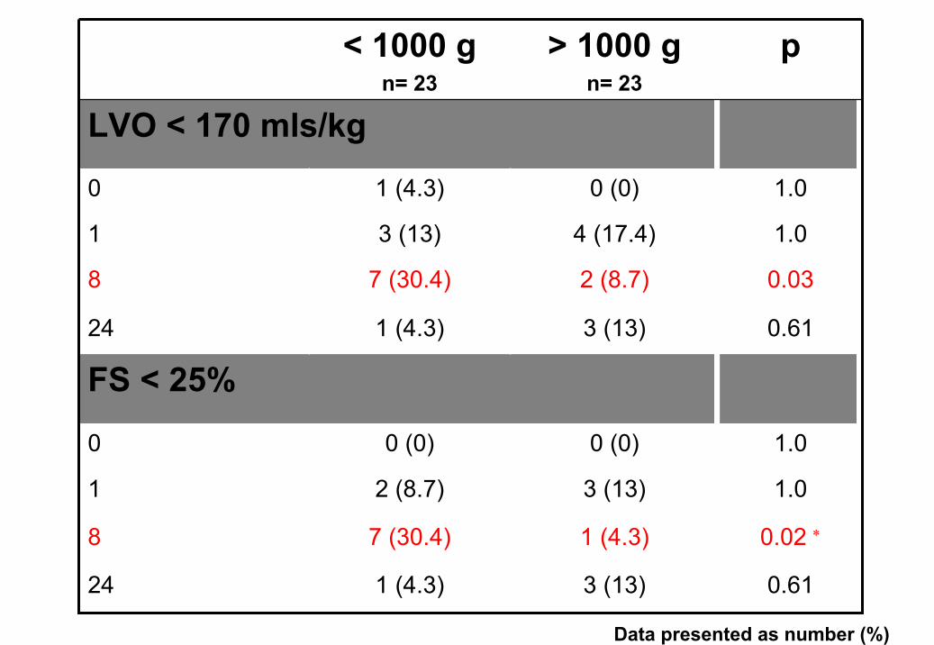
0.02 ∗1 (4.3)7 (30.4)8
1.03 (13)2 (8.7)1
FS < 25%
1.00 (0)0 (0)0
0.613 (13)1 (4.3)24
0.613 (13)1 (4.3)24
0.032 (8.7)7 (30.4)8
1.04 (17.4)3 (13)1
1.00 (0)1 (4.3)0
LVO < 170 mls/kg
p> 1000 gn= 23
< 1000 gn= 23
Data presented as number (%)
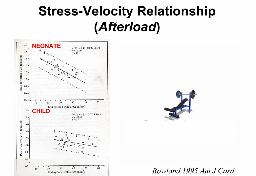
Rowland 1995 Am J Card
Stress-Velocity Relationship (Afterload)
NEONATE
CHILD
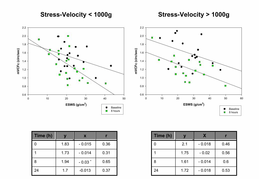
Stress-Velocity < 1000g
ESWS (g/cm2)
0 10 20 30 40 50
mVC
Fc (c
irc/s
ec)
0.6
0.8
1.0
1.2
1.4
1.6
1.8
2.0
2.2
Baseline8 hours
Stress-Velocity > 1000g
ESWS (g/cm2)
0 10 20 30 40 50 60
mVC
Fc (c
irc/s
ec)
0.6
0.8
1.0
1.2
1.4
1.6
1.8
2.0
2.2
Baseline8 hours
-0.013
- 0.03 *
- 0.014
- 0.015
x
0.371.724
0.651.948
0.311.731
0.361.830
ryTime (h)
- 0.018
- 0.014
- 0.02
- 0.018
X
0.531.7224
0.61.618
0.561.751
0.462.10
ryTime (h)
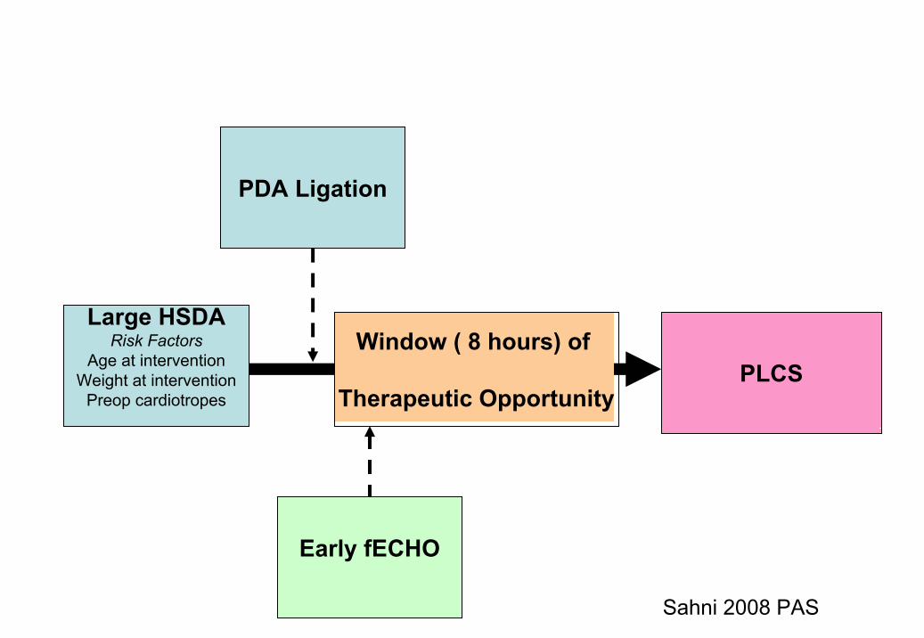
Large HSDARisk Factors
Age at interventionWeight at intervention
Preop cardiotropesPLCS
Window ( 8 hours) of
Therapeutic Opportunity
PDA Ligation
Early fECHO
Sahni 2008 PAS
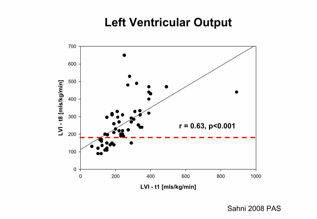
Left Ventricular Output
LVI - t1 [mls/kg/min]
0 200 400 600 800 1000
LVI -
t8 [m
ls/k
g/m
in]
0
100
200
300
400
500
600
700
r = 0.63, p<0.001
Sahni 2008 PAS
#
p < 0.05
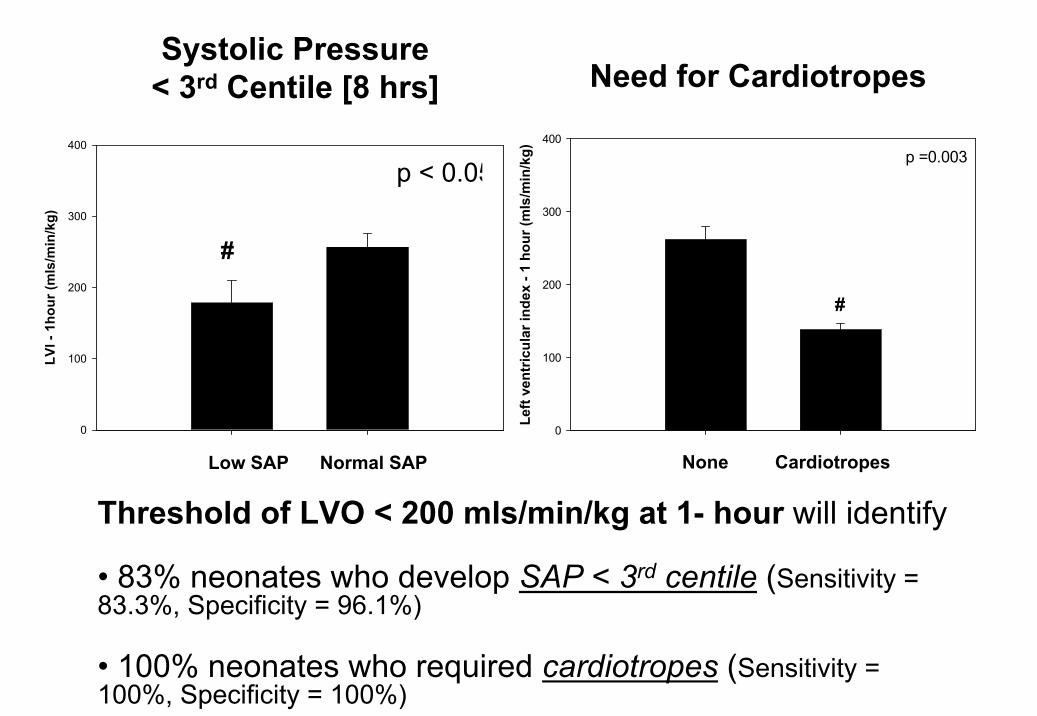
Systolic Pressure < 3rd Centile [8 hrs]
Threshold of LVO < 200 mls/min/kg at 1- hour will identify
• 83% neonates who develop SAP < 3rd centile (Sensitivity = 83.3%, Specificity = 96.1%)
• 100% neonates who required cardiotropes (Sensitivity = 100%, Specificity = 100%)
Low SAP Normal SAP
Left
vent
ricul
ar in
dex
- 1 h
our (
mls
/min
/kg)
0
100
200
300
400
#
p =0.003
None Cardiotropes
LVI -
1ho
ur (m
ls/m
in/k
g)
0
100
200
300
400
Need for Cardiotropes

Summary
• Early fECHO may help anticipate postoperative cardiorespiratory instability
• LVO < 200 mls/min/kg is the best marker of clinical and echo indices of PLCS
Targeted neonatal ECHO directed therapy program
– introduced in January 2009
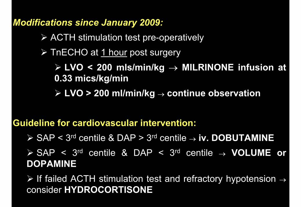
Modifications since January 2009:ACTH stimulation test pre-operativelyTnECHO at 1 hour post surgery
LVO < 200 mls/min/kg → MILRINONE infusion at 0.33 mics/kg/min
LVO > 200 ml/min/kg → continue observation
Guideline for cardiovascular intervention:SAP < 3rd centile & DAP > 3rd centile → iv. DOBUTAMINESAP < 3rd centile & DAP < 3rd centile → VOLUME or
DOPAMINE If failed ACTH stimulation test and refractory hypotension →
consider HYDROCORTISONE
Study Objective
To compare the rate and components of PLCS in infants who have undergone PDA ligation before and after the introduction of targeted neonatal echocardiography (TnECHO) directed therapy program
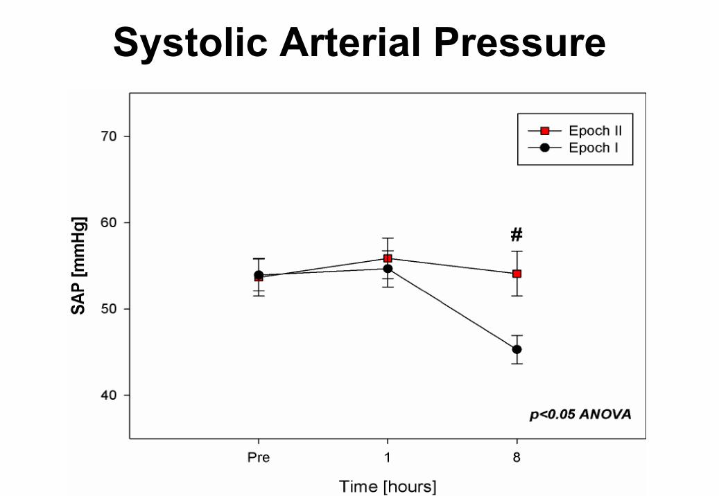
Systolic Arterial Pressure
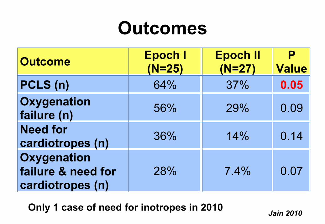
0.0929%56%Oxygenation failure (n)
Outcome Epoch I (N=25)
Epoch II (N=27)
P Value
PCLS (n) 64% 37% 0.05
Need for cardiotropes (n) 36% 14% 0.14
Oxygenation failure & need for cardiotropes (n)
28% 7.4% 0.07
Outcomes
Jain 2010 Only 1 case of need for inotropes in 2010
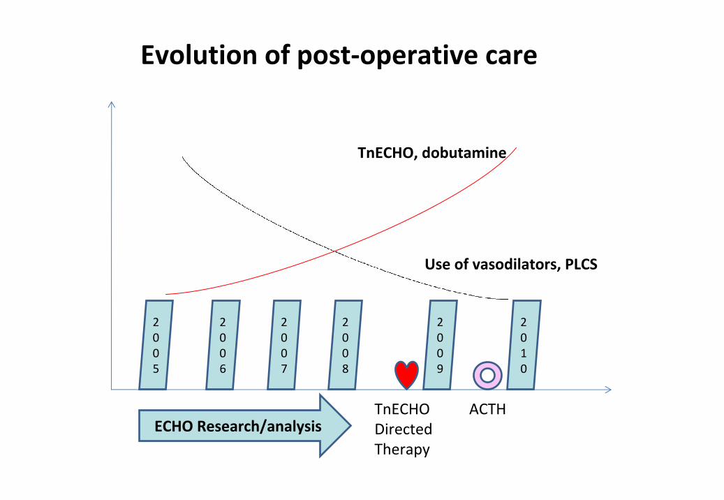
2005
2006
2007
2008
2009
2010
ECHO Research/analysis
Use of vasodilators, PLCS
TnECHO, dobutamine
TnECHODirectedTherapy
ACTH
Evolution of post‐operative care
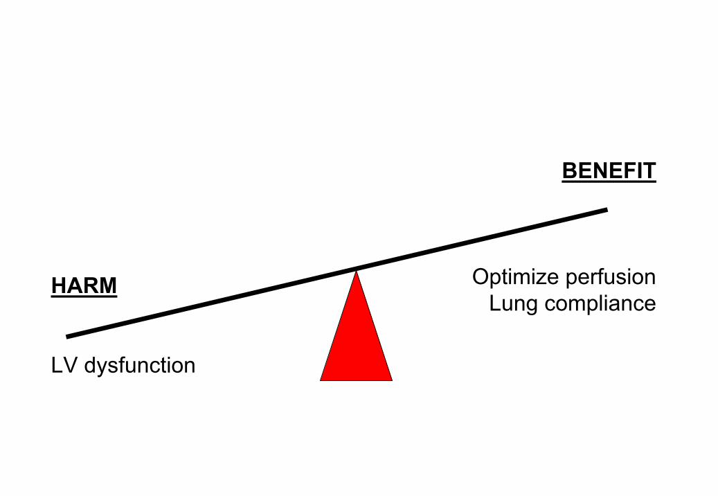
HARM
LV dysfunction
BENEFIT
Optimize perfusionLung compliance
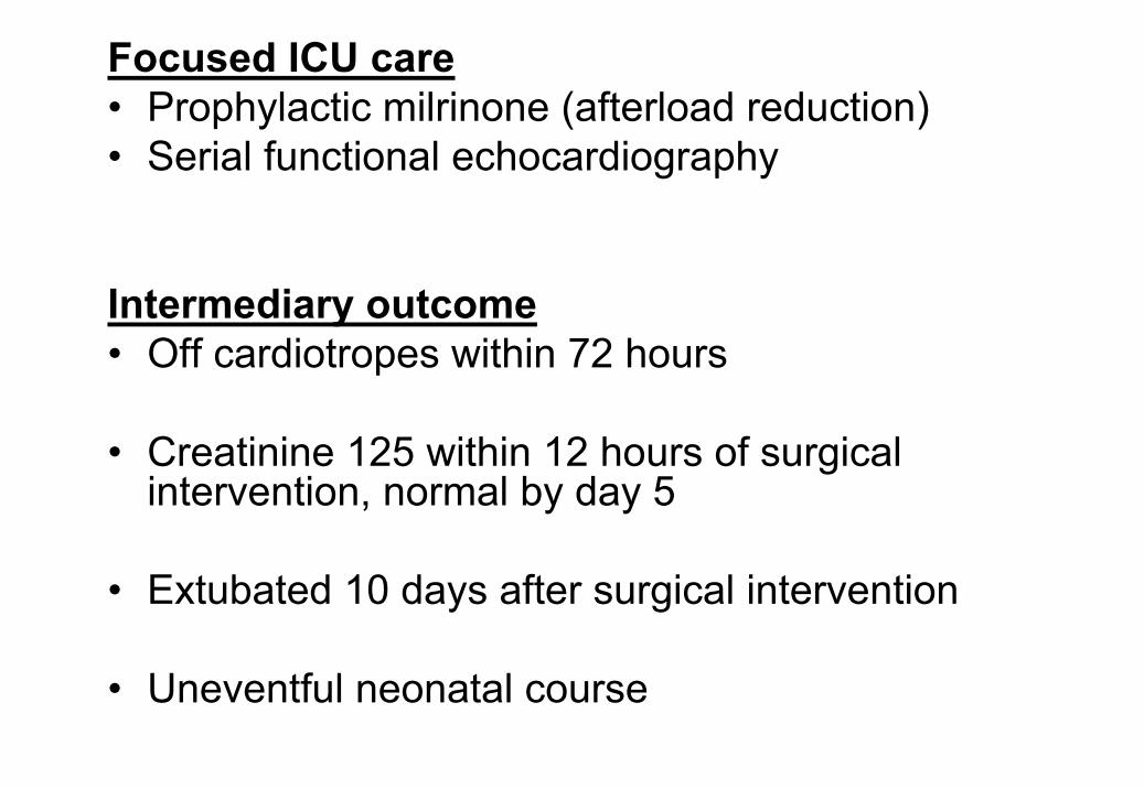
Focused ICU care• Prophylactic milrinone (afterload reduction)• Serial functional echocardiography
Intermediary outcome• Off cardiotropes within 72 hours
• Creatinine 125 within 12 hours of surgical intervention, normal by day 5
• Extubated 10 days after surgical intervention
• Uneventful neonatal course
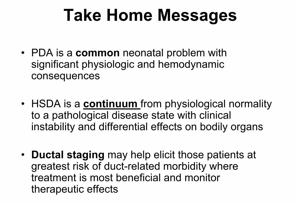
Take Home Messages
• PDA is a common neonatal problem with significant physiologic and hemodynamic consequences
• HSDA is a continuum from physiological normality to a pathological disease state with clinical instability and differential effects on bodily organs
• Ductal staging may help elicit those patients at greatest risk of duct-related morbidity where treatment is most beneficial and monitor therapeutic effects
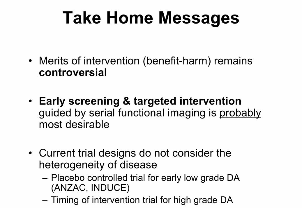
Take Home Messages
• Merits of intervention (benefit-harm) remains controversial
• Early screening & targeted interventionguided by serial functional imaging is probablymost desirable
• Current trial designs do not consider the heterogeneity of disease– Placebo controlled trial for early low grade DA
(ANZAC, INDUCE)– Timing of intervention trial for high grade DA

Special ThanksNeonatal Research FellowsArvind Sehgal Lilian TeixeiraSandesh Shivananda Emer Finan
Research AssistantsWendy Mak
Derek Stephens (Statistical support)Glen Van Arsdell & CVS team

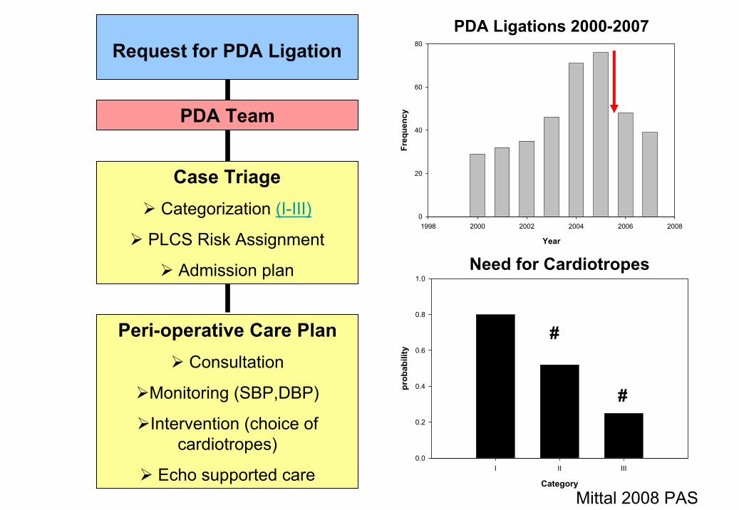
Request for PDA Ligation
PDA Team
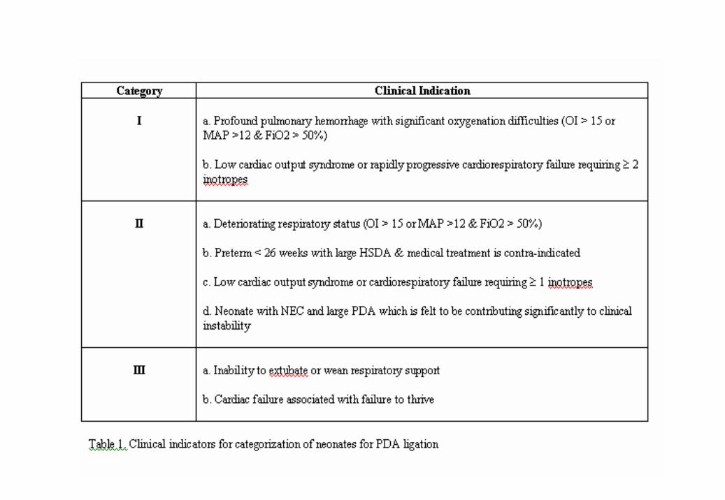
Case TriageCategorization (I-III)
PLCS Risk Assignment
Admission plan
Peri-operative Care PlanConsultation
Monitoring (SBP,DBP)
Intervention (choice of cardiotropes)
Echo supported care
PDA Ligations 2000-2007
Year
1998 2000 2002 2004 2006 2008
Freq
uenc
y
0
20
40
60
80
Need for Cardiotropes
Category
I II III
prob
abili
ty
0.0
0.2
0.4
0.6
0.8
1.0
Mittal 2008 PAS
#
#
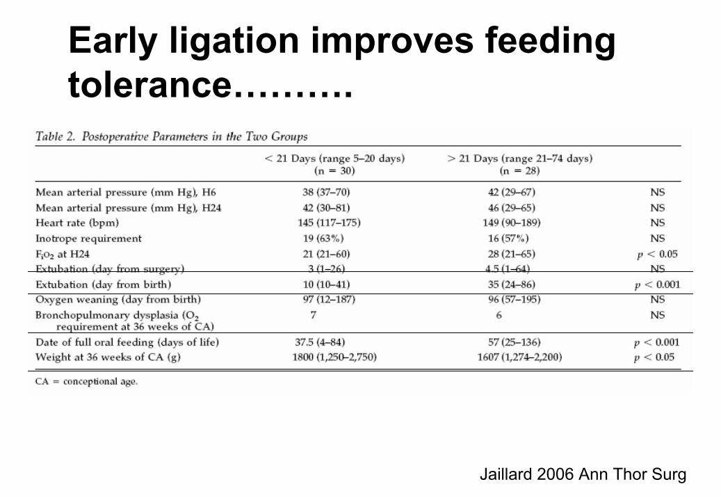
Jaillard 2006 Ann Thor Surg
Early ligation improves feeding tolerance……….
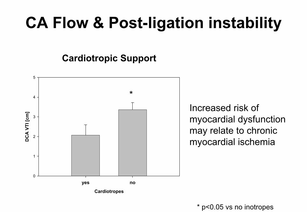
CA Flow & Post-ligation instability
Cardiotropic Support
Cardiotropes
yes no
DC
A V
TI [c
m]
0
1
2
3
4
5
*
* p<0.05 vs no inotropes
Increased risk of myocardial dysfunction may relate to chronic myocardial ischemia
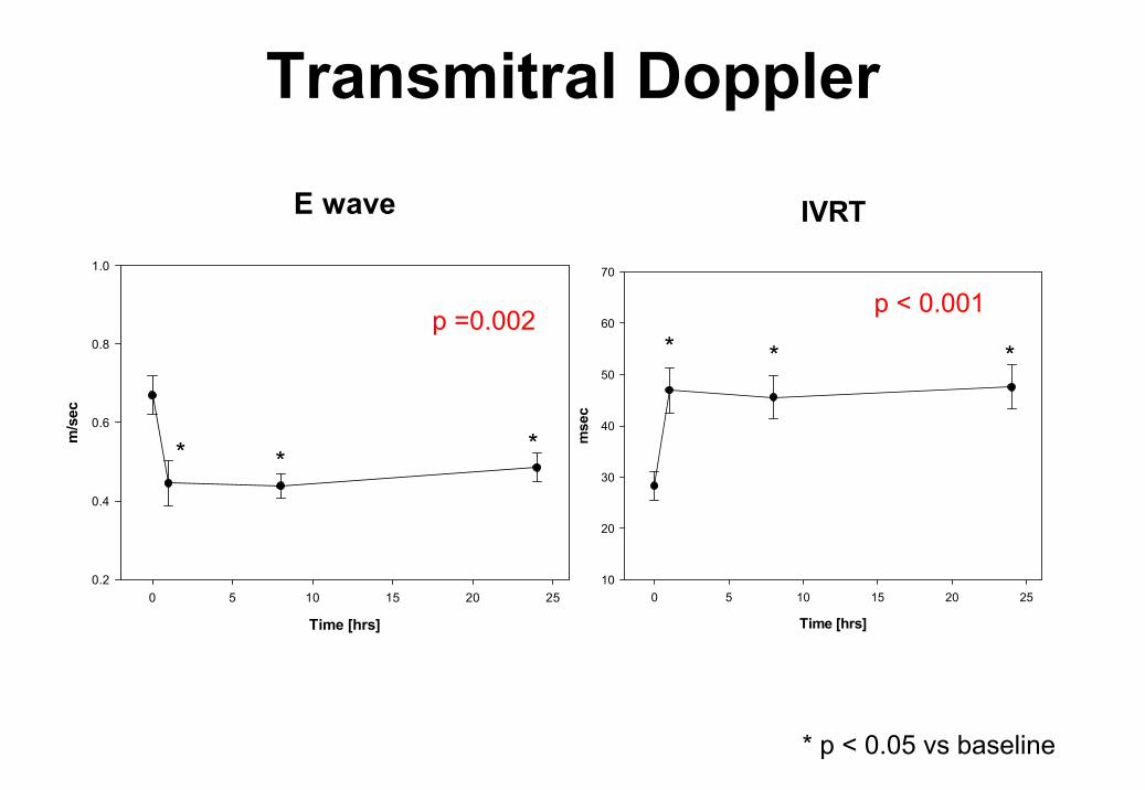
E wave
Time [hrs]
0 5 10 15 20 25
m/s
ec
0.2
0.4
0.6
0.8
1.0
* p < 0.05 vs baseline
p =0.002
***
Transmitral Doppler
IVRT
Time [hrs]
0 5 10 15 20 25m
sec
10
20
30
40
50
60
70
* * *
p < 0.001
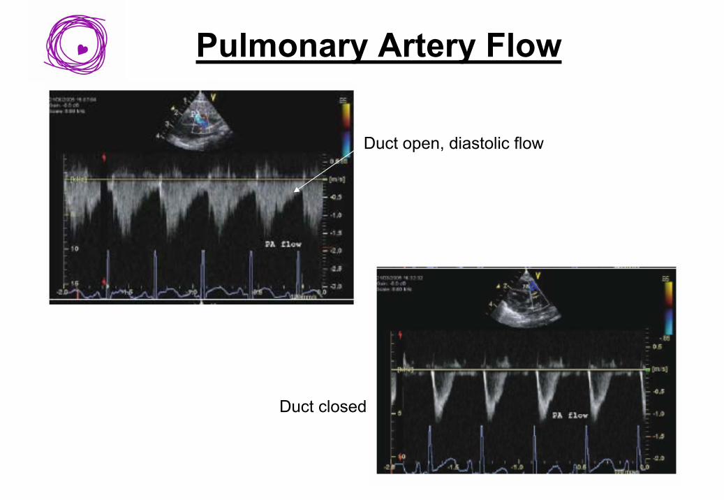
Pulmonary Artery Flow
Duct open, diastolic flow
Duct closed
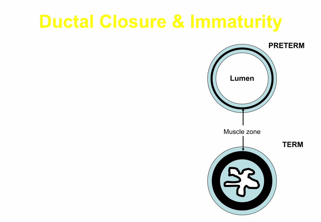
Ductal Closure & Immaturity
Suboptimal Functional Closure• Less responsive to oxygen (Murphy 1972 Ped
Res) & more responsive to PgE2 (Clyman 1980 J Pediatr) & iNO
• Lacks intimal folds
Failure of Anatomic Remodeling• DA wall hypoxia only if complete obliteration
of intraluminal flow
• Thin walled lacking musc. so limited ability to ↑ avascular zone
• No intramural vasa vasorum
Muscle zone
Lumen
TERM
PRETERM
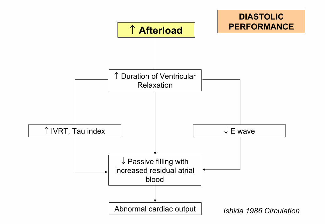
↑ Afterload
↑ IVRT, Tau index
↑ Duration of Ventricular Relaxation
↓ E wave
Abnormal cardiac output
↓ Passive filling with increased residual atrial
blood
Ishida 1986 Circulation
DIASTOLIC PERFORMANCE
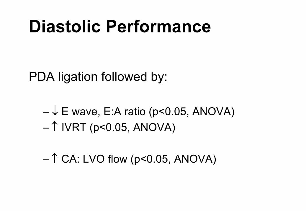
Diastolic Performance
PDA ligation followed by:
– ↓ E wave, E:A ratio (p<0.05, ANOVA)– ↑ IVRT (p<0.05, ANOVA)
– ↑ CA: LVO flow (p<0.05, ANOVA)

LA : Ao ratio
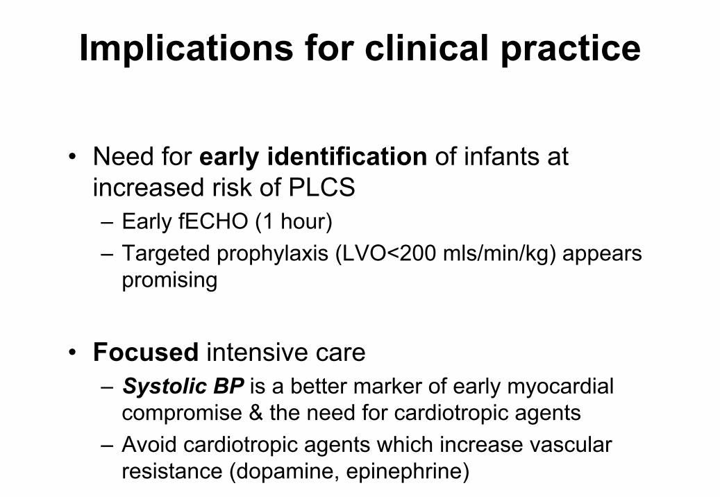
Implications for clinical practice
• Need for early identification of infants at increased risk of PLCS– Early fECHO (1 hour) – Targeted prophylaxis (LVO<200 mls/min/kg) appears
promising
• Focused intensive care– Systolic BP is a better marker of early myocardial
compromise & the need for cardiotropic agents– Avoid cardiotropic agents which increase vascular
resistance (dopamine, epinephrine)
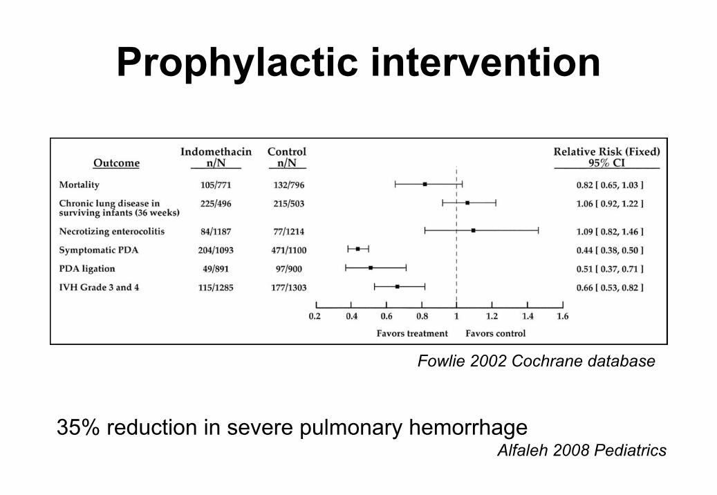
Prophylactic intervention
Fowlie 2002 Cochrane database
35% reduction in severe pulmonary hemorrhage Alfaleh 2008 Pediatrics