unconventional serine proteases: variations on the catalytic
TRANSCRIPT

10.1110/ps.035436.108Access the most recent version at doi: 2008 17: 2023-2037; originally published online Sep 29, 2008; Protein Sci.
Özlem Dogan Ekici, Mark Paetzel and Ross E. Dalbey
Ser/His/Asp triad configurationUnconventional serine proteases: Variations on the catalytic
References
http://www.proteinscience.org/cgi/content/full/17/12/2023#References
This article cites 126 articles, 59 of which can be accessed free at:
serviceEmail alerting
click heretop right corner of the article or Receive free email alerts when new articles cite this article - sign up in the box at the
Notes
http://www.proteinscience.org/subscriptions/ go to: Protein ScienceTo subscribe to
© 2008 Cold Spring Harbor Laboratory Press
Cold Spring Harbor Laboratory Press on November 20, 2008 - Published by www.proteinscience.orgDownloaded from

REVIEW
Unconventional serine proteases: Variationson the catalytic Ser/His/Asp triad configuration
OZLEM DOGAN EKICI,1 MARK PAETZEL,2 AND ROSS E. DALBEY1
1Department of Chemistry, The Ohio State University, Columbus, Ohio 43210, USA2Department of Molecular Biology and Biochemistry, Simon Fraser University, Burnaby, British Columbia,V5A 1S6 Canada
(RECEIVED March 18, 2008; FINAL REVISION September 17, 2008; ACCEPTED September 17, 2008)
Abstract
Serine proteases comprise nearly one-third of all known proteases identified to date and play crucialroles in a wide variety of cellular as well as extracellular functions, including the process of bloodclotting, protein digestion, cell signaling, inflammation, and protein processing. Their hallmark is thatthey contain the so-called ‘‘classical’’ catalytic Ser/His/Asp triad. Although the classical serineproteases are the most widespread in nature, there exist a variety of ‘‘nonclassical’’ serine proteaseswhere variations to the catalytic triad are observed. Such variations include the triads Ser/His/Glu, Ser/His/His, and Ser/Glu/Asp, and include the dyads Ser/Lys and Ser/His. Other variations are seen withcertain serine and threonine peptidases of the Ntn hydrolase superfamily that carry out catalysis with asingle active site residue. This work discusses the structure and function of these novel serine proteasesand threonine proteases and how their catalytic machinery differs from the prototypic serine proteaseclass.
Keywords: enzymes; active sites; structure/function studies; protein turnover; structure; serine proteases;threonine proteases
Proteases play indispensable functions in all living cells.In mammalian cells, proteases function in angiogenesis,apoptosis, differentiation, immune response, matrixremodeling, and protein activation. They are found inevery organelle and compartment of most, if not all,eukaryotic cells. There are over 500 proteases in humancells as determined by sequencing of the human genome(Puente et al. 2005). They are associated with a number ofdiseases, including cardiovascular and Alzheimer’s dis-ease, cancer, autoimmune diseases, inflammation, andhypertension.
Proteases are typically grouped into four mechanisticclasses: the cysteine, serine proteases, metallo, and
aspartic acid proteases. In 2004, the protease field washonored by a Nobel Prize in chemistry presented toAvram Hershko, Aaron Ciechanover, and Irwin Rose fortheir pioneering work on the ubiquitin-proteasome systemin which the proteasome is a novel threonine protease.The proteasome, like the proteases in the other fourclasses, is a target for drug discovery programs. It isessential to understand the specificity, catalytic mecha-nism, and structure of proteases in order to facilitate drugdesign efforts.
The best-known class of proteases is the serine proteaseclass (E.C. 3.4.21) that uses the classical Ser/His/Aspcatalytic triad mechanism, where serine is the nucleo-phile, histidine is the general base and acid, and theaspartate helps orient the histidine residue and neutralizethe charge that develops on the histidine during thetransition states. Well-studied members of this class arechymotrypsin, trypsin, elastase, and subtilisin. The three-dimensional structure of chymotrypsin was first solved in
ps035436 Ekici et al. REVIEW RA
Reprint requests to: Ross E. Dalbey, Department of Chemistry, TheOhio State University, 100 West 18th Avenue, Columbus, OH 43210,USA; e-mail: [email protected]; fax: (614) 292-1532.
Article and publication are at http://www.proteinscience.org/cgi/doi/10.1110/ps.035436.108.
Protein Science (2008), 17:2023–2037. Published by Cold Spring Harbor Laboratory Press. Copyright � 2008 The Protein Society 2023
JOBNAME: PROSCI 17#12 2008 PAGE: 1 OUTPUT: Monday November 3 16:04:21 2008
csh/PROSCI/170217/ps035436
Cold Spring Harbor Laboratory Press on November 20, 2008 - Published by www.proteinscience.orgDownloaded from

1967 (Matthews et al. 1967), and the first trypsinstructure was solved in 1974 (Huber et al. 1974; Stroudet al. 1974).
John Northrop made a major advance in the serineprotease area in the 1930s by successfully crystallizingproteases, including trypsin and chymotrypsin (Northropand Kunitz 1931). Other early important discoveriesincluded the identification of the specific amino acid thatfunctioned as the nucleophile by modification with DFP(disopropyl fluorophosphates) in both trypsin and chy-motrypsin (Dixon et al. 1958). The determination of theamino acid sequence of trypsinogen (Walsh and Neurath1964) and chymotrypsinogen (Hartley 1964) revealedthey are homologous proteases. In 1969, David Blowproposed the famous charge-relay mechanism where twoproton exchanges were thought to occur (from Ser195 toHis57 and from His57 to Asp102) (Blow et al. 1969). LaterNMR data (Robillard and Shulman 1974; Bachovchin1985) and neutron diffraction structural data (Kossiakoffand Spencer 1981) suggest a single proton exchange fromSer195 Og to the His57Ne2. More recently, ultra-high-resolution structures of serine protease intermediates havebeen performed to provide mechanistic insights into catal-ysis (Fodor et al. 2006).
In addition to the Ser/His/Asp serine proteases, thereare serine proteases that use catalytic residue arrange-ments other than the canonical triad (Fig. 1; Table 1).These atypical serine proteases use novel triads such asSer/His/Glu, Ser/His/His, or Ser/Glu/Asp, dyads such asSer/Lys or Ser/His, or a single Ser catalytic residue. Thereare also proteases in which the nucleophilic hydroxyl isderived from threonine rather than a serine residue.
In this work, we describe serine proteases with non-canonical active site arrangements. We will discuss andcompare atypical serine proteases with the classicalserine protease class in terms of the structure–functionand catalytic mechanism. Also, we will present someideas about why there are different types of active siteconfigurations within serine/threonine proteases.
Different clans of Ser/Thr proteases
Serine/threonine proteases have been classified into clansand families (see Merops database, http://merops.sanger.ac.uk) (Rawlings et al. 2006). Members of the same clanare proteases that have evolved from a common ancestorand share a common protein fold. Proteases in the samefamily are related based on the sequence homology oftheir amino acid sequences. Most, but not all, clansconsist of one active site arrangement. The prefix ‘‘S’’is used for clans formed entirely from families which areserine peptidases, whereas ‘‘P’’ is used for clans in whichthe homologous families are of several catalytic types(e.g., serine and cysteine), despite the fact that they have
diverged from a common ancestor. Table 1 shows thatproteases with a Ser/His/Asp triad fall within fourof these clans (PA, SB, SC, and SK), while the Ser/Lysproteases fall within five clans (SE, SF, SJ, SK, and SR).This shows that different ancestors can converge on thesame Ser/His/Asp or Ser/Lys mechanism.
Since some members of the same clan can use differentactive site architectures, this indicates that the tertiarystructure is not always related to the active site configuration.For example, clan PB has family members that use a Ser/His/Glu active site configuration or Ser- or Thr-only activesite architecture (Table 1). Why do different serine/threonineproteases use different active site configurations? The differ-ent active site arrangements may allow for activity in adifferent cellular environment. For instance, the proteaseswith Ser/Lys active sites typically carry out catalysis with apH optimum that is higher than Ser/His/Asp proteases. Incontrast, the pH optimum is lower for serine proteases withSer/Glu/Asp active sites than those with Ser/His/Asp activesites. In large part, the different pH optimums reflectdifferences in the pKa values of the different general baseresidues that are employed in catalysis. In addition, varia-tions in the active site architecture of proteases mayinfluence what cellular inhibitor they are susceptible to.There are other possibilities as well that we discuss below.
Figure 1. The catalytic residues used in serine and threonine proteases.
Ekici et al.
2024 Protein Science, vol. 17
JOBNAME: PROSCI 17#12 2008 PAGE: 2 OUTPUT: Monday November 3 16:04:22 2008
csh/PROSCI/170217/ps035436
Cold Spring Harbor Laboratory Press on November 20, 2008 - Published by www.proteinscience.orgDownloaded from
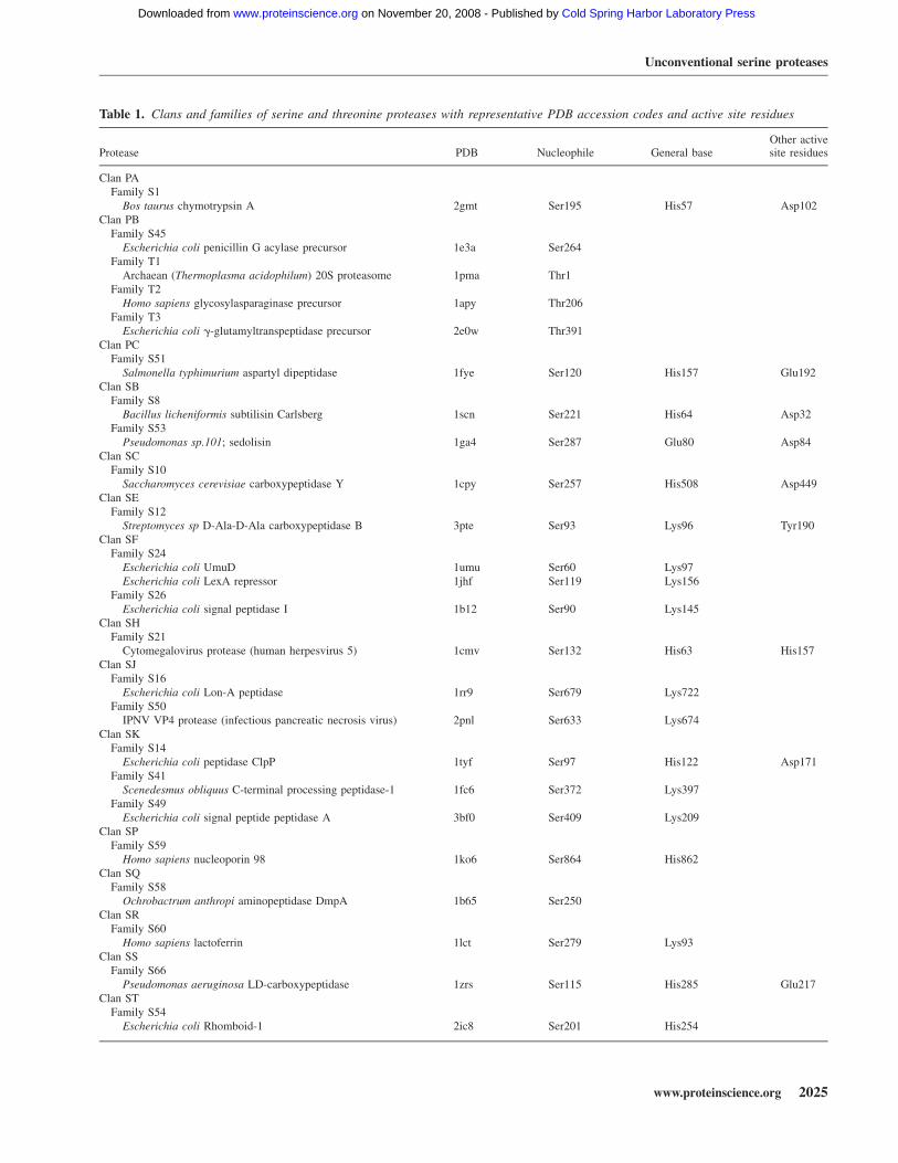
Table 1. Clans and families of serine and threonine proteases with representative PDB accession codes and active site residues
Protease PDB Nucleophile General baseOther activesite residues
Clan PAFamily S1
Bos taurus chymotrypsin A 2gmt Ser195 His57 Asp102Clan PB
Family S45Escherichia coli penicillin G acylase precursor 1e3a Ser264
Family T1Archaean (Thermoplasma acidophilum) 20S proteasome 1pma Thr1
Family T2Homo sapiens glycosylasparaginase precursor 1apy Thr206
Family T3Escherichia coli g-glutamyltranspeptidase precursor 2e0w Thr391
Clan PCFamily S51
Salmonella typhimurium aspartyl dipeptidase 1fye Ser120 His157 Glu192Clan SB
Family S8Bacillus licheniformis subtilisin Carlsberg 1scn Ser221 His64 Asp32
Family S53Pseudomonas sp.101; sedolisin 1ga4 Ser287 Glu80 Asp84
Clan SCFamily S10
Saccharomyces cerevisiae carboxypeptidase Y 1cpy Ser257 His508 Asp449Clan SE
Family S12Streptomyces sp D-Ala-D-Ala carboxypeptidase B 3pte Ser93 Lys96 Tyr190
Clan SFFamily S24
Escherichia coli UmuD 1umu Ser60 Lys97Escherichia coli LexA repressor 1jhf Ser119 Lys156
Family S26Escherichia coli signal peptidase I 1b12 Ser90 Lys145
Clan SHFamily S21
Cytomegalovirus protease (human herpesvirus 5) 1cmv Ser132 His63 His157Clan SJ
Family S16Escherichia coli Lon-A peptidase 1rr9 Ser679 Lys722
Family S50IPNV VP4 protease (infectious pancreatic necrosis virus) 2pnl Ser633 Lys674
Clan SKFamily S14
Escherichia coli peptidase ClpP 1tyf Ser97 His122 Asp171Family S41
Scenedesmus obliquus C-terminal processing peptidase-1 1fc6 Ser372 Lys397Family S49
Escherichia coli signal peptide peptidase A 3bf0 Ser409 Lys209Clan SP
Family S59Homo sapiens nucleoporin 98 1ko6 Ser864 His862
Clan SQFamily S58
Ochrobactrum anthropi aminopeptidase DmpA 1b65 Ser250Clan SR
Family S60Homo sapiens lactoferrin 1lct Ser279 Lys93
Clan SSFamily S66
Pseudomonas aeruginosa LD-carboxypeptidase 1zrs Ser115 His285 Glu217Clan ST
Family S54Escherichia coli Rhomboid-1 2ic8 Ser201 His254
Unconventional serine proteases
www.proteinscience.org 2025
JOBNAME: PROSCI 17#12 2008 PAGE: 3 OUTPUT: Monday November 3 16:04:36 2008
csh/PROSCI/170217/ps035436
Cold Spring Harbor Laboratory Press on November 20, 2008 - Published by www.proteinscience.orgDownloaded from
Ser/His/Asp triad
Chymotrypsin- and trypsin-like proteases
Two of the best-known serine proteases that utilize theSer/His/Asp triad are chymotrypsin and trypsin. Theyshare the same protein fold and have their catalyticresidues in the order of His/Asp/Ser from the N to Cterminus. Their active site regions are composed of (1)the substrate binding groove where nonspecific main-chain hydrogen bond interactions occur between theenzyme and substrate, (2) the substrate specificity bind-ing pockets, (3) the catalytic Ser/His/Asp triad, and (4)the oxyanion hole (for conventions regarding peptides,proteases, protease nomenclature, see http://www.chem.qmul.ac.uk/iubmb/enzyme/EC3/intro.html#EC34) (for acomprehensive review on the catalytic mechanism ofclassical serine proteases, see Hedstrom [2002]).
The substrate binding pocket of the protease allows theprotease to bind its substrates and determines the sub-strate specificity of the enzyme. For example, trypsincleaves its substrates at the C-terminal side of arginine orlysine (P1 position, Schechter and Berger nomenclature)(Schechter and Berger 1967). This specificity for a P1basic side chain of substrates is stabilized by an acidicresidue near the bottom of the sterically complementarydeep S1 binding pocket. In some proteases, the specificitycan go to the opposite side of the cleavage site (scissilebond) as well, P9 side, or also further down the chain ofthe substrate to the P3, P4, P5 site, etc. The substrateregion near the cleavage site needs to be in an extendedconformation (not protected by hydrogen bonds withinthe substrate itself) to bind to the protease in order for thecarbonyl of the scissile bond to be attacked by the serinehydroxyl nucleophile of the protease.
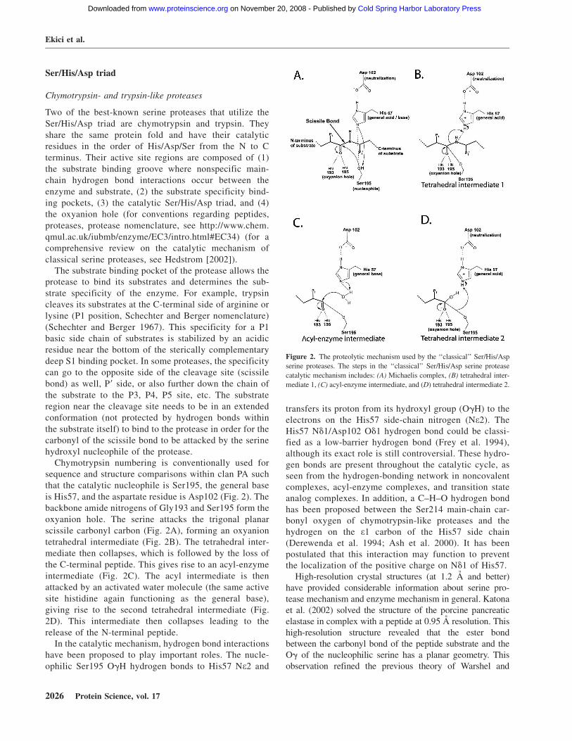
Chymotrypsin numbering is conventionally used forsequence and structure comparisons within clan PA suchthat the catalytic nucleophile is Ser195, the general baseis His57, and the aspartate residue is Asp102 (Fig. 2). Thebackbone amide nitrogens of Gly193 and Ser195 form theoxyanion hole. The serine attacks the trigonal planarscissile carbonyl carbon (Fig. 2A), forming an oxyaniontetrahedral intermediate (Fig. 2B). The tetrahedral inter-mediate then collapses, which is followed by the loss ofthe C-terminal peptide. This gives rise to an acyl-enzymeintermediate (Fig. 2C). The acyl intermediate is thenattacked by an activated water molecule (the same activesite histidine again functioning as the general base),giving rise to the second tetrahedral intermediate (Fig.2D). This intermediate then collapses leading to therelease of the N-terminal peptide.
In the catalytic mechanism, hydrogen bond interactionshave been proposed to play important roles. The nucle-ophilic Ser195 OgH hydrogen bonds to His57 Ne2 and
transfers its proton from its hydroxyl group (OgH) to theelectrons on the His57 side-chain nitrogen (Ne2). TheHis57 Nd1/Asp102 Od1 hydrogen bond could be classi-fied as a low-barrier hydrogen bond (Frey et al. 1994),although its exact role is still controversial. These hydro-gen bonds are present throughout the catalytic cycle, asseen from the hydrogen-bonding network in noncovalentcomplexes, acyl-enzyme complexes, and transition stateanalog complexes. In addition, a C–H–O hydrogen bondhas been proposed between the Ser214 main-chain car-bonyl oxygen of chymotrypsin-like proteases and thehydrogen on the e1 carbon of the His57 side chain(Derewenda et al. 1994; Ash et al. 2000). It has beenpostulated that this interaction may function to preventthe localization of the positive charge on Nd1 of His57.
High-resolution crystal structures (at 1.2 A and better)have provided considerable information about serine pro-tease mechanism and enzyme mechanism in general. Katonaet al. (2002) solved the structure of the porcine pancreaticelastase in complex with a peptide at 0.95 A resolution. Thishigh-resolution structure revealed that the ester bondbetween the carbonyl bond of the peptide substrate and theOg of the nucleophilic serine has a planar geometry. Thisobservation refined the previous theory of Warshel and
Figure 2. The proteolytic mechanism used by the ‘‘classical’’ Ser/His/Asp
serine proteases. The steps in the ‘‘classical’’ Ser/His/Asp serine protease
catalytic mechanism includes: (A) Michaelis complex, (B) tetrahedral inter-
mediate 1, (C) acyl-enzyme intermediate, and (D) tetrahedral intermediate 2.
Ekici et al.
2026 Protein Science, vol. 17
JOBNAME: PROSCI 17#12 2008 PAGE: 4 OUTPUT: Monday November 3 16:04:37 2008
csh/PROSCI/170217/ps035436
Cold Spring Harbor Laboratory Press on November 20, 2008 - Published by www.proteinscience.orgDownloaded from

Russell (1986), who proposed that the H-bonds between theester carbonyl oxygen and the oxyanion hole induced strain onthe ester bond and distorted it toward sp3 hybridization, whichwould enhance the formation rate of the second tetrahedralintermediate. A recent comparison of the acyl-enzyme struc-ture and a high-resolution Michaelis complex structure sug-gests that the b-strand H-bond type interactions betweenthe enzyme binding site and substrate become shorter duringthe transition from the Michaelis complex to the acyl–enzymecomplex (Fodor et al. 2006). This result suggests that theenzyme may be able to utilize the energy released from theshorter, stronger hydrogen bonds to help overcome the energybarrier in the nucleophilic attack steps.
Mutagenesis studies on the related trypsin proteinrevealed that Ser195 and His57 contribute ;1 million-fold toward the rate of catalysis for trypsin (Corey et al.1992). The Asp102 contributes ;10,000-fold as deter-mined by mutagenesis experiments (Craik et al. 1987;Corey et al. 1992).
Subtilisin-like proteases
Like chymotrypsin (Fig. 3A) and trypsin, subtilisin (Fig.3B) utilizes a Ser/His/Asp triad (Fig. 3B), but it has nosequence similarity to chymotrypsin-like proteases. Infact, it adopts the a/b-twisted open sheet structure(Wright et al. 1969) rather than the double b-barrelstructure seen in the chymotrypsin-like proteases. Sub-tilisin and chymotrypsin provide excellent examples ofconvergent evolution (Wallace et al. 1996). The proteinfolds of these proteases are completely different, althoughthey both converged on a similar Ser/His/Asp mechanismto carry out proteolysis. In the subtilisin-like proteases,the order of the active site residues is Asp/His/Ser inthe primary sequence, from N to C terminus, which isdifferent than the chymotrypsin-like proteases (His/Asp/Ser). The serine and histidine residues contribute 106-foldtoward catalysis, and the aspartic acid contributes 104-fold residue (Carter and Wells 1988). The active siteregion of subtilisin is shown in Figure 3B. The subtilisinAsn155, which forms a hydrogen bond to the oxyanionintermediate, contributes ;100- to 1000-fold (Wells et al.1986; Pantoliano et al. 1987). Subtilisin is engineered forthermal and pH stability for use in laundry detergents.These subtilisin mutants were among the first patentsgranted for engineered proteins.
Other Ser/His/Asp proteases with different folds thanthe chymotrypsin- and subtilisin-like proteases includecarboxypeptidase Y (Jung et al. 1999) and ClpP (Wanget al. 1997) proteases (Table 1). Interestingly, the ClpPprotease is within the SK clan that has members thatpossess alternative active site arrangements such as theSer/Lys dyad configuration.
Ser/His/Glu triad
Aspartyl dipeptidase
A variation of the classical Ser/His/Asp triad is found inthe aspartyl dipeptidase protease, where the aspartate issubstituted by a glutamate residue (Rawlings and Barrett1999). Aspartyl dipeptidase is not inactivated by the conven-tional serine protease inhibitor DFP or PMSF (phenylmethane sulfonyl fluoride) (Conlin et al. 1994). While initialsite-directed mutagenesis studies suggested that aspartyldipeptidase was a classical serine protease, the structure ofaspartyl dipeptidase from Salmonella typhimurium revealedthat it in fact belongs to a new serine peptidase family with aSer/His/Glu triad (Hakansson et al. 2000). The Ser120 Og
atom is 2.7 A away from the His157 Ne2 atom. The
Figure 3. Structures of the ‘‘classical’’ Ser/His/Asp serine proteases. (A)
Chymotrypsin from Bos taurus (PDB 2gmt). (B). Subtilisin Carlsberg from
Bacillus licheniformis (PDB 1scn). The subtilisin protease family is
different from the chymotrypsin protease family in protein fold, and it
utilizes the side chain of an asparagine in its oxyanion hole (Asn155). The
proteases are shown in standard orientation. The ‘‘standard orientation’’
presents the enzyme such that the reader is looking down onto the substrate
binding groove so that the catalytic residues (the Ser-His-Asp catalytic
triad) are located on the right side of the figure. The N-terminal side of the
substrate would bind on the left-hand side of the binding groove, and the
C-terminal side of the substrate (P1/P19 residues, the scissile bond) would
bind the right side of the binding groove, nearest the catalytic residues.
The length of the substrate binding groove varies from protease to
protease.
Unconventional serine proteases
www.proteinscience.org 2027
JOBNAME: PROSCI 17#12 2008 PAGE: 5 OUTPUT: Monday November 3 16:04:50 2008
csh/PROSCI/170217/ps035436
Fig. 3 live 4/C
Cold Spring Harbor Laboratory Press on November 20, 2008 - Published by www.proteinscience.orgDownloaded from

Glu192 Oe2 is within 2.7 A of the His Nd1 atom. Thisglutamate carboxylate/histidine imidazole hydrogen bondadopts an unusual anti-conformation rather that the syn-conformation usually seen with other serine proteases(Ippolito et al. 1990).
Mutagenesis of the glutamate residue of the S. typhi-murium aspartyl dipeptidase resulted in a 100-fold dropin activity. Typically, mutagenesis of the aspartate resi-due, the third member of the catalytic triad (Ser/His/Asp),leads to a 10,000-fold loss in activity (Craik et al.1987; Carter and Wells 1988; Corey et al. 1992).Interestingly, the aspartyl dipeptidase has a similarprotein fold as the cysteine proteases g-glutamyl hydro-lase and PfpI peptidase, suggesting they evolved from acommon ancestor.
LD-carboxypeptidase
The Ser/His/Glu active site arrangement has evolvedmore than once. The LD-carboxypeptidase from Pseudo-monas aeruginosa that functions in peptidoglycan recy-cling uses a Ser/His/Glu triad (Fig. 4A). The crystalstructure of the LD-carboxypeptidase from P. aeruginosawas solved to 1.5 A resolution (Korza and Bochtler2005). Its protein fold consists of an N-terminal b-sheetand a C-terminal b-barrel domain that is different fromthe aspartyl dipeptidase fold. This indicates that conver-gent evolution has taken place to give rise to the Ser/His/Glu triad. It is not clear why most serine proteases utilizean aspartate rather than the glutamate observed with theLD-carboxypeptidase. Notably, the Ser/His/Glu activesite is observed in lipases (Schrag et al. 1991), and insome esterases as well (Nachon et al. 2005).
Ser/His/His triad: Cytomegalovirus protease
Another unconventional active site triad, Ser/His/His, isfound in herpes virus proteases that plays a key role in thematuration of the assembly protein. The cytomegalovirusprotease is inactivated by DFP (Stevens et al. 1994).Ser132 was identified as the active site serine of thehuman cytomegalovirus protease as it was modified bythe inhibitor. The invariant His63 is critical for activity asdetermined by site-directed mutagenesis (Welch et al.1993).
The Ser/His/His catalytic triad was validated by thecrystal structure of the cytomegalovirus protease (Qiuet al. 1996; Shieh et al. 1996; Tong et al. 1996). Thestructure revealed that the protein has a unique fold withthe Ser132 Og atom and bridging His63 Ne2 nitrogenbeing within hydrogen-bonding distance (Fig. 4B). Inaddition, the His63 Nd1 is within hydrogen-bondingdistance to the His157 Ne2 that replaces the aspartateresidue within the classic serine proteases.
Site-directed mutagenesis studies showed the non-bridging histidine residue (His157) of the triad contrib-utes only ;10-fold to the catalytic rate of the enzyme(Khayat et al. 2001) in comparison to 1000- to 10,000-fold for the contribution of aspartate in the classical triadprotease (Carter and Wells 1988; Corey et al. 1992).Therefore, the cytomegalovirus protease functions morelike a Ser/His dyad. Herpes virus proteases, in compar-ison to Ser/His/Asp proteases such as trypsin and sub-tilisin, are kinetically less efficient enzymes. Thissuggests that this alternate active site is a way to tunethe catalytic activity of the serine protease.
Ser/Glu/Asp triad: Sedolisin proteases
The sedolisin proteases carry out catalysis using a novelSer/Glu/Asp triad (Wlodawer et al. 2001a). These pro-teases take their name from their catalytic groups serine(S), glutamate (E), and aspartate (D). Due to the low pKa
Figure 4. Structures of serine proteases that use variations on the active
site triad. (A) LD-carboxypeptidase from Pseudomonas aeruginosa (PDB
1zrs) utilizes a Ser/His/Glu triad. (B) Cytomegalovirus protease (PDB
1cmv) utilizes a Ser/His/His triad. (C) Sedolisin from Pseudomonas sp.
(PDB 1ga4) uses a Ser/Glu/Asp triad. A zoomed-in view of the catalytic
residues is shown to the right of each structure. The side chains are shown
as sticks with nitrogens in blue, oxygens in red, and carbons in green. LD-
carboxypeptidase (A) and cytomegalovirus protease (B) exist as homo-
dimers in solution; therefore, one monomer is shown in white and the other
monomer is shown in black.
Ekici et al.
2028 Protein Science, vol. 17
JOBNAME: PROSCI 17#12 2008 PAGE: 6 OUTPUT: Monday November 3 16:05:05 2008
csh/PROSCI/170217/ps035436
Fig. 4 live 4/C
Cold Spring Harbor Laboratory Press on November 20, 2008 - Published by www.proteinscience.orgDownloaded from
of the carboxylate that plays the role of the generalbase, these proteases are active at low pH, which isunusual for serine proteases. Proteases in this clan includekumamolysin that is found in acidic hot springs andhuman tripeptidyl peptidase that is found in the low pHenvironment of the lysosome. This is an excellentdemonstration of how the different active site architec-tures allow the protease to be active in different cellularenvironments.
Because they operate at low pH, these proteases werethought to be aspartic acid proteases. However, theseproteases are not inactivated by pepstatin, which is anatural inhibitor of the aspartic acid class of proteases.Rather they are inactivated by inhibitors with aldehydefunctional groups such as chymostatin, which typicallymodify the serine nucleophile within serine proteases(Wlodawer et al. 2001b).
The X-ray structure of the sedolisin protease fromPseudomonas revealed that these proteases have thesubtilisin protein fold (Wlodawer et al. 2001a). Structuresof the enzyme with the iodotyrostatin inhibitor revealedthat Ser287 is the catalytic residue. Ser287 is covalentlyattached to the inhibitor and forms a hemiacetal linkageto the Ser278 Og side chain. Within hydrogen-bondingdistance of the Ser278 side chain is the side-chaincarboxylate of Glu80 (Fig. 4C), suggesting that the acidicresidue acts as the general base in catalysis. The thirdmember of the triad is Asp84, which interacts with theGlu80 side chain. It is the Glu80/Asp84 pair that allowsthis protease class to have optimum activity at acidic pH.The active site geometry was also confirmed in a 1.4 AX-ray structure of kumamolysin, a serine-carboxyl-typeprotease from Bacillus (Comellas-Bigler et al. 2002).Site-directed mutagenesis studies confirmed the impor-tance of these active site serine, glutamate, and aspartateresidues (Oyama et al. 1999, 2005).
Ser/Lys dyad
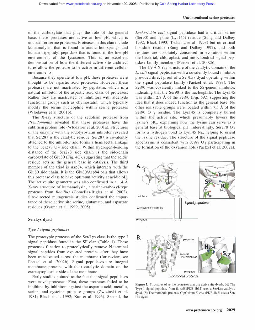
Type I signal peptidases
The prototypic protease of the Ser/Lys class is the type Isignal peptidase found in the SF clan (Table 1). Theseproteases function to proteolytically remove N-terminalsignal peptides from exported proteins after they havebeen translocated across the membrane (for review, seePaetzel et al. 2002b). Signal peptidases are integralmembrane proteins with their catalytic domain on theextracytoplasmic side of the membrane.
Early studies pointed to the fact that signal peptidaseswere novel proteases. First, these proteases failed to beinhibited by inhibitors against the aspartic acid, metallo,serine, and cysteine protease groups (Zwizinski et al.1981; Black et al. 1992; Kuo et al. 1993). Second, the
Escherichia coli signal peptidase had a critical serine(Ser90) and lysine (Lys145) residue (Sung and Dalbey1992; Black 1993; Tschantz et al. 1993) but no criticalhistidine residue (Sung and Dalbey 1992), and bothresidues are absolutely conserved in evolution withinthe bacterial, chloroplast, and mitochondrial signal pep-tidase family members (Paetzel et al. 2002b).
The 1.9 A X-ray structure of the catalytic domain of theE. coli signal peptidase with a covalently bound inhibitorprovided direct proof of a Ser/Lys dyad operating withinthe signal peptidase family (Paetzel et al. 1998). TheSer90 was covalently linked to the 5S-penem inhibitor,indicating that the Ser90 is the nucleophile. The Lys145was within 2.8 A of the Ser90 (Fig. 5A), supporting theidea that it does indeed function as the general base. Noother ionizable groups were located within 7.5 A of theSer90 O g residue. The Lys145 is completely buriedwithin the active site, which presumably lowers thelysine’s pKa, explaining how the lysine can serve as ageneral base at biological pH. Interestingly, Ser278 Og
forms a hydrogen bond to Lys145 Nz, helping to orientthe lysine residue. The structure of the signal peptidaseapoenzyme is consistent with Ser88 Og participating inthe formation of the oxyanion hole (Paetzel et al. 2002a).
Figure 5. Structures of serine proteases that use active site dyads. (A) The
Type 1 signal peptidase from E. coli (PDB 1b12) uses a Ser/Lys catalytic
dyad. (B) The rhomboid protease GlpG from E. coli (PDB 2ic8) uses a Ser/
His dyad.
Unconventional serine proteases
www.proteinscience.org 2029
JOBNAME: PROSCI 17#12 2008 PAGE: 7 OUTPUT: Monday November 3 16:05:24 2008
csh/PROSCI/170217/ps035436
Fig. 5 live 4/C
Cold Spring Harbor Laboratory Press on November 20, 2008 - Published by www.proteinscience.orgDownloaded from
Mutagenesis studies have shown Ser88 contributes;1000-fold to catalysis by stabilizing the oxyanionintermediate (Carlos et al. 2000).
The active site lysine residue has an apparent pKa of8.7 as determined from the pH rate profile of the E. colisignal peptidase (Paetzel et al. 1997). Typically, Ser/Lysproteases show optimum activity at a higher pH such as 9compared with Ser/His/Asp proteases that have optimumactivity near pH 7.
The UmuD family of peptidases
Other Ser/Lys dyad proteases are found within the UmuDfamily of peptidases that include UmuD, LexA, and the l
repressor (Slilaty and Little 1987; Little 2004; Paetzeland Woodgate 2004). These proteases are involved in theSOS response and function as repressors. These proteinsundergo autoproteolysis in vivo in a manner dependent onRecA, a protein involved in homologous recombination.The biological function of autocleavage is to inactivatethese proteins (Mustard and Little 2000).
UmuD was the first protease in the UmuD familywhose structure was solved to high resolution. Its proteinfold consists of a b-sheet structure (Peat et al. 1996), andthe critical serine and lysine residues that were implicatedin autocatalysis (McDonald et al. 1998) are within hydrogen-bonding distance. The likely role of the RecA binding in thein vivo autoproteolysis reaction is to decrease the pKa of thegeneral base by burying the e-amino group of the lysine in ahydrophobic environment.
LexA protease is proposed to use an active site Ser/Lysdyad that is not inactivated by a classical proteaseinhibitor (Slilaty and Little 1987). Autodigestion of LexAis pH-dependent. In the absence of RecA, a basic residuewith a pKa of 10 is necessary for autodigestion (Slilatyet al. 1986). A likely candidate for this basic group is alysine residue. Further evidence came from structuralstudies. Briefly, the structure of LexA was solved at 2.0 Awith two conformations of the protein present (Luo et al.2001). One of the conformations, the ‘‘cleavable form(C),’’ revealed the scissile bond of LexA within the activesite region. The other conformation, the ‘‘noncleavableform (NC)’’of the protein had the scissile bond of LexAlocated ;20 A from the Ser/Lys dyad. A 1.8 A resolutioncrystal structure revealed that the catalytic serine andlysine are within hydrogen-bonding distance. Consistentwith the lysine functioning as a general base in theproteolytic mechanism, the lysine side chain is com-pletely buried in a hydrophobic pocket in the cleavedform. Similar to signal peptidase, this hydrophobic en-vironment is expected to lower the pKa of the catalyticlysine. The residues that contribute to the oxyanion holeare the main-chain amide nitrogens of Met118 andSer119.
The l repressor, which also has a Ser/Lys dyad, has aThr190 residue that may also help orient the catalyticLys192 residue as it is hydrogen bonded to this residue(Bell et al. 2000). Most of the Ser/Lys proteases (suchas UmuD, LexA, Lon, VP4, signal peptidase I, and signalpeptide peptidase SppA) have a second (non-nucleo-philic) hydroxyl group coordinating to the lysine, sug-gesting this interaction has a functional role (Paetzel et al.2002a). This seems to be a common theme with Ser/Lysproteases, in general.
Comparison of the active site regions of the signalpeptidase and UmuD family proteases reveals that theorientation of the serine and lysine are quite similar(Paetzel and Strynadka 1999; Paetzel et al. 2002b). Inthese proteases, the serine hydroxyl groups attack thesi-face of the scissile amide bond. In contrast, serineproteases that contain the Ser/His/Asp triad attack the re-face of the substrate (James 1994).
Lon protease
Another Ser/Lys dyad serine protease is the Lon protease,which is involved in quality control and degradation ofmisfolded proteins. It has a peptidase and an AAAdomain. The latter domain uses the energy of ATPhydrolysis to actively unfold proteins (Chung 2004).However, ATP hydrolysis is not required for cleavage ofshort peptides.
In 2003, the E. coli Lon protease was shown to containan essential serine and lysine residue (Rotanova et al.2003). Like many Ser/Lys proteases (Tschantz andDalbey 1994), the Lon protease is not inhibited by theclassical serine protease inhibitors such as PMSF or DFP.The X-ray crystal structure of the peptidase domain wasachieved using a mutant with the catalytic serine residuemutated to an alanine. Modeling studies where theAla679 was replaced with Ser679 are consistent with aSer/Lys dyad mechanism and revealed that the e-aminogroup of Lys722 is in position to hydrogen bond to thecatalytic Ser679 residue. In addition, the conservedThr704 residue could hydrogen bond to the Lys722 sidechain, helping to position the general base. The proteinfold of the E. coli Lon protease domain is completelydifferent from that found in the signal peptidase andUmuD family members and contain 10 b-strands and sixa-helices (Botos et al. 2004).
C-terminal processing peptidases
The Ser/Lys dyad C-terminal processing peptidases(Cpps) cleave precursor proteins in eubacteria and plantsand show no sequence homology with signal pepti-dases and UmuD members or to the D-Ala-D-Ala
Ekici et al.
2030 Protein Science, vol. 17
JOBNAME: PROSCI 17#12 2008 PAGE: 8 OUTPUT: Monday November 3 16:05:42 2008
csh/PROSCI/170217/ps035436
Cold Spring Harbor Laboratory Press on November 20, 2008 - Published by www.proteinscience.orgDownloaded from

carboxypeptidases. E. coli tail specific protease (Tsp)exhibits C-terminal processing activity and is implicatedin the degradation of truncated proteins that are tagged bythe SsrA pathway (Keiler et al. 1996). Tsp is located inthe periplasm of Gram-negative bacteria and in the extra-cellular/cell wall region of Gram-positive bacteria.
The E. coli Tsp, as seen with the E. coli signal pep-tidase, is not inactivated by inhibitors against thefour groups of proteases (Silber et al. 1992). Mutagenesisstudies showed that the protein had a critical serineand lysine residue (Keiler and Sauer 1995). Sequencealignment studies of Cpp members revealed that theseresidues are absolutely conserved (Keiler and Sauer2001). Interestingly, N-ethylmaleimide could inactivateTsp when the critical serine is changed to a cysteine,supporting the notion that the serine is an active siteresidue (Keiler and Sauer 1995). Tsp has activity over abroad pH range, from pH 5 to 9.
Currently, the only Cpp whose structure is solved is thephotosystem II D1 C-terminal processing protease fromthe green algae Scenedesmus obliquus (Liao et al. 2000),which is essential for the assembly of the manganesecluster involved in water oxidation (Taylor et al. 1988).The structure reveals the C-terminal protease domainwith the active site Ser372 and Lys397 residues, a middlePDZ-like domain, and a mainly a-helical N-terminaldomain. The active site Lys397 is hydrogen bonded to athreonine residue. The active site Ser372 interacts withseveral water molecules. In order for Lys397 to act as ageneral base, a conformational change has to take placeupon substrate binding. In addition, substrate bindingmost likely would cause the Lys397 to become buried in ahydrophobic environment so that its pKa is lowered.
Other Ser/Lys peptidases include the penicillin bindingproteins 5 D-Ala-D-Ala carboxypeptidase B (Davies et al.2001), the viral VP4 protease of the Birnaviridae family(Feldman et al. 2006; Lee et al. 2007), signal peptidepeptidase A (Kim et al. 2007), and Lactoferrin (Table 1;Anderson et al. 1989). The Ser/Lys dyad active sitearrangement has arisen many times during evolution.
Ser/His Dyad
Rhomboid protease
Rhomboid proteases have a Ser/His active site dyad.These proteases play roles in signal transduction andparticipate in regulated intramembrane proteolysis (Selkoeand Kopan 2003; Wolfe and Kopan 2004). They have sixtransmembrane helices, and their catalytic groups arelocalized within the membrane plane (Wolfe and Kopan2004; Maegawa et al. 2005). Initial site-directed muta-genesis studies suggested that the rhomboid proteaseutilized a Ser/His/Asn catalytic triad (Urban et al. 2001).
However, further studies showed that the asparagine,the third member of the proposed triad, is not alwaysnecessary for catalysis (Lemberg et al. 2001; Maegawaet al. 2005).
Recently, the X-ray structure of the E. coli andHaemophilus influenzae rhomboid protease GlpG wassolved (Wang et al. 2006; Wu et al. 2006; Ben-Shem et al.2007; Lemieux et al. 2007)). The E. coli structurerevealed that the protease does use a Ser/His dyad incatalysis where Ser201 and His254 are within hydrogen-bonding distance (Fig. 5B). The structure showed that itscatalytic groups are in a hydrophilic environment, ;10 Abelow the periplasmic membrane surface. In addition, theGlpG protease has a proposed gate that controls access tothe hydrophilic active site region of GlpG (Baker et al.2007; Wang and Ha 2007). Although the gating mecha-nism is controversial because none of the structurescontain an extended substrate, this mechanism allowsfor the substrate to enter the substrate binding site of theprotease laterally from the lipid phase.
Nucleoporin
Another Ser/His peptidase is the nucleoporin proteins thatundergo autoprocessing to produce a dimer. The crystalstructure of the human nucleoporin Nup98 revealed anactive site Ser/His dyad (Hodel et al. 2002). The structurewas determined using a catalytic deficient mutant wherethe serine nucleophile was substituted with an alanine.The overall structure revealed a b-sandwich-like proteinfold that is half open. Modeling studies where thecatalytic Ser864 hydroxyl side chain is modeled fromthe solved mutant Ser864Ala structure show that it canform a hydrogen bond to the His862. Surprisingly, muta-genesis of the His862 to alanine only slightly inhibits theenzyme with over 50% activity remaining. This is veryunusual for serine proteases as a similar mutation in sub-tilisin causes a million-fold inhibition (Hodel et al. 2002).
‘‘Ser-only’’ configuration
L-aminopeptidase D-Ala-esterase/amidase
The simplest active site arrangement uses a ‘‘Ser-only’’configuration. Proteases that use this configuration aremade in a precursor form and undergo autoprocessing togenerate the active site residue at position 1 in the matureprotein. This active site arrangement is found in theOchrobactrum anthropi L-aminopeptidase D-Ala-esterase/amidase. After autocleavage, Ser250 becomes the firstresidue of the mature protein and the a-amino group ofthe newly generated N terminus of the protein functions asthe general base in catalysis abstracting a proton from the
Unconventional serine proteases
www.proteinscience.org 2031
JOBNAME: PROSCI 17#12 2008 PAGE: 9 OUTPUT: Monday November 3 16:05:43 2008
csh/PROSCI/170217/ps035436
Cold Spring Harbor Laboratory Press on November 20, 2008 - Published by www.proteinscience.orgDownloaded from

Ser250 hydroxyl group. The protein fold of this protease isnovel and characterized by a four-layered abba topology(Bompard-Gilles et al. 2000) called the N-terminal nucle-ophile hydrolase fold (Brannigan et al. 1995).
Penicillin G acylase precursor
The penicillin G acylase precursor uses a ‘‘Ser-only’’catalytic configuration to undergo autoproteolysis. Itundergoes self-processing using Ser264 as a nucleophile(Fig. 6A) in order to produce the enzymatically activemature acylase. The autoproteolysis generates a dimericenzyme with the serine nucleophile located at the Nterminus of one of the subunits (Ser264 becomes Ser1 inone of the newly generated protein subunits). Thispenicillin G acylase protein has an abba-core proteinfold (Duggleby et al. 1995), similar to the L-amino-peptidase D-Ala-esterase/amidase L. This fold is alsofound in the proteasome and glutamine PRPP amido-
transferase that contain a catalytic threonine located atresidue 1 in the polypeptide chain (Brannigan et al. 1995;Lowe et al. 1995; Oinonen and Rouvinen 2000). In theglutamine PRPP enzyme, a cysteine nucleophile is found atthis position (Smith et al. 1994). Therefore, proteins thathave the N-terminal nucleophile hydrolase fold alwayscarry out catalysis with an N-terminal active site residue(Brannigan et al. 1995), although the specific active siteresidue (serine, threonine, or cysteine) can vary.
Insights into the catalytic mechanism of these pepti-dases emerged from the structure of the penicillin Gacylase precursor from E. coli (Hewitt et al. 2000). Thestructure revealed two conformations of the active site.One of the conformations revealed that Ser264 in theactive site is positioned such that its Og group couldattack the appropriate carbonyl carbon atom of thescissile bond. Moreover, this active site Ser264 is abso-lutely essential for activity (Choi et al. 1992). Strikingly,from the structure, there was no obvious candidate for thegeneral base in the proteolytic reaction (Hewitt et al.2000).
Glutaryl 7-aminocephalosporanic acid acylaseprecursor (GCA precursor)
There is another member of the acylase family calledGCA, glutaryl 7-aminocephalo-sporanic acid acylasewith a Ser-only catalytic configuration. The structure ofthe precursor GCA at 2.5 A resolution suggests that awater molecule is the general base that activates theactive site serine residue (Kim et al. 2002; Yoon et al.2004). The water molecule is coordinated by the main-chain NHs of Thr69 and Gly168 and by the amide sidechain of Asn244.
‘‘Thr-only’’ configuration
Proteasome
The proteasome is a threonine protease that plays akey role in protein degradation and quality control.Proteolysis involves a Thr/a-amine configuration. Inarchaea and prokaryotes, the proteasome forms a 20Sstructure comprised of four 7-membered rings (a7b
7b7a7) (for review, see Baumeister et al. 1998; Vogeset al. 1999). The two outer rings are comprised of iden-tical a-subunits and the two inner rings contain theidentical b-subunits. The b-subunits contain the activesite. In eukaryotic cells, the 20S proteolytic core complexassociates with a regulatory domain complex, forming a26S proteinase particle. This 26S particle functions inthe ubiquitin-dependent degradation pathway. It uses ATPhydrolysis to unfold protein substrates and promotingtranslocation of the substrate peptide chain into theprotease degradation chamber.
Figure 6. Structures of serine and threonine proteases that use single
active site residues. (A) Serine-only peptidase. The penicillin acylase from
E. coli. The electron density for the nucleophilic Ser264 revealed alternate
conformations for the side-chain hydroxyl. One of the conformations
directs the Og toward the scissile carbonyl between residues 263 and 264.
(PDB 1e3a) (Hewitt et al. 2000). Chain A is shown in black, chain B is in
white. (B) ‘‘Thr-only’’ peptidase. The 20S proteasome from the Archaeon
thermoplasma acidophilum (PDB 1pma). The active site threonine for each
b-subunit is shown as red spheres. For clarity, one b-subunit is highlighted
as a ribbon diagram; all other protein chains are rendered as Ca trace.
Ekici et al.
2032 Protein Science, vol. 17
JOBNAME: PROSCI 17#12 2008 PAGE: 10 OUTPUT: Monday November 3 16:05:43 2008
csh/PROSCI/170217/ps035436
Fig. 6 live 4/C
Cold Spring Harbor Laboratory Press on November 20, 2008 - Published by www.proteinscience.orgDownloaded from
In contrast to eubacterial and archaeal proteasomes, theeukaryotic enzyme contains two copies each of sevendistinct a- and b-subunits. The protease chamber contains28 subunits that form the 20S particle. The a-subunits arelocated on the outside of the 20S particle, and the b
polypeptides are on the inside. Briefly, of the seven differenttypes of b-subunits, three of them are proteolytically active(for review, see Voges et al. 1999). The eukaryotic protea-some exhibits several distinct substrate specificities cleavingafter basic, hydrophobic, or acidic amino acids.
The active site of the proteasome is comprised of an N-terminal threonine residue. To form the proteasome activesite, it is necessary for the b-subunit to be autoprocessedsuch that the N-terminal propeptide is removed from theprecursor polypeptide as the 20S proteosome particle isbeing assembled (Seemuller et al. 2001). Evidence thatthe active site threonine is involved in catalysis comes fromsite-directed mutagenesis and protease inhibitor studies.Mutagenesis of the threonine to an alanine residue blocksprocessing of the archaeal (Seemuller et al. 1995) and yeastproteasome (Chen and Hochstrasser 1996; Arendt andHochstrasser 1997; Heinemeyer et al. 1997). Modificationof the threonine residue of the proteasome by proteaseinhibitors completely inactivates the protease (Fenteany etal. 1995; Bogyo et al. 1997; Groll et al. 1997).
The crystal structure of the Thermoplasma acidophilumproteasome showed that the catalytic machinery is com-posed of the Thr1, Glu17, and Lys33 (Fig. 6B; Lowe et al.1995). The structure was solved in the presence of thecalpain inhibitor I, acetyl-Leu-Leu-norleucinal (LLnL).The structure revealed that the Thr1 hydroxyl group islocalized very close to the aldehyde functional group ofLLnL and may form a hemiacetal. Thus, the structuresupports the notion that the Thr1 Og residue is thenucleophile involved in catalysis. Also in proximity tothe Thr1 residue is the amino group of Lys33 and theGlu17 carboxylate. The Lys33 e-amino group is coordi-nated to the Glu17 carboxylate and the threonine side-chain oxygen. Based on the coordination partners, it islikely that the Lys33 is positively charged. Initially, theamino group of Thr1 was proposed to function as thegeneral base. However, a bound water molecule, locatedin between the Thr1 amino group and its alcohol side-chain hydroxyl most likely acts as the general base todeprotonate the Thr1 hydroxyl group. The N terminus iscoordinated to the carbonyl groups of Arg19 and Ala168residues. A similar active site architecture is seen withinthe enzymatically active b subunit of the yeast protea-some solved at 2.4 A resolution (Groll et al. 1997).
Proteasome precursor
A ‘‘Thr-only’’ catalytic mechanism is involved in auto-proteolysis to generate mature proteasome (Seemuller
et al. 1996). A water molecule bound to the proteasomeprecursor b-subunit is proposed to function as a generalbase in activating the threonine hydroxyl group (Ditzelet al. 1998; for review, see Seemuller et al. 2001).
Glycosylasparaginase and g-glutamyltransferaseI protein precursors
Other ‘‘Thr-only’’ active site peptidases are the glyco-sylasparaginase and g-glutamyltransferase I protein inwhich a threonine residue is the catalytic nucleophileinvolved in autocleavage. A threonine residue is impli-cated in autoproteolysis of the glycosylasparaginase(Aronson 2004). The inhibitor 5-diazo-4-oxonorvalinehas been shown to react with the N-terminal threonineamino acid of the b subunit (Kaartinen et al. 1991). Theg-glutamyltransferase I protein (Sakai et al. 1996), whichundergoes self processing, also requires a threonineresidue (Suzuki and Kumagai 2002; Okada et al. 2007).Both the glycosylasparaginase and g-glutamyltransferaseI protein have the N-terminal nucleophile hydrolase fold(Brannigan et al. 1995; Suzuki and Kumagai 2002; Okadaet al. 2007). To date, the structural studies did not clearlydetermine the general base that activates the threoninehydroxyl group in the precursor.
Variation on a theme: Ser/Ser/Lys triad
A general base lysine residue is not only used in serineproteases but also employed in certain amidases thatcontain a Ser/Ser/Lys triad active site. The amidases arefound in archaea, eubacteria, fungi, plants, and mammalsand rival the number of proteins in the Ser/His/Asp serineprotease families (for review, see McKinney and Cravatt2005). The signature domain is ;130 residues in lengthand includes the conserved motif with the active site Ser/Ser/Lys residues. Initially, it was unclear what type ofmechanism was used by the amidases since they wereinactivated by both serine and cysteine types of proteaseinhibitors (Ueda et al. 1995; Patterson et al. 1996).
By using the inhibitor ethoxy oleoyl fluorophospho-nate, it was shown that serine is the nucleophilic residuein this amidase family (Patricelli et al. 1999). Ser241 wasestablished to be the active site nucleophile of the fattyacid amide hydrolase (FAAH). While no histidine resi-dues were found to be required for activity, the invariantSer217, Ser241, and Lys142 residues were discovered tobe indispensable for catalysis (Patricelli and Cravatt1999; Patricelli et al. 1999).
The pH rate profiles for the FAAH enzyme indicated anincrease in activity going from pH 5 to 9, revealing atitratable group with a pKa of ;7.9. This titratablegroup most likely corresponds to the Lys142. Mutation
Unconventional serine proteases
www.proteinscience.org 2033
JOBNAME: PROSCI 17#12 2008 PAGE: 11 OUTPUT: Monday November 3 16:06:02 2008
csh/PROSCI/170217/ps035436
Cold Spring Harbor Laboratory Press on November 20, 2008 - Published by www.proteinscience.orgDownloaded from

of the lysine creates altered pH rate profiles thatare similar to those of histidine mutants in classical Ser/His/Asp proteases (Patricelli et al. 1999). The combineddata suggested that the amidase family employed a Ser/Lys dyad mechanism (Patricelli and Cravatt 2000).
The Ser/Ser/Lys triad active site was established by thecrystal structures of several amidases (Fig. 7; Braceyet al. 2002; Shin et al. 2002). The structure of the mal-onamidase E2 demonstrated that the catalytic serineresidue was separated from the lysine by a bridgingserine residue (Shin et al. 2002). The bridging serinewas in an unusual conformation, in which the serinehydroxyl side chain and the amide nitrogen polarize thecatalytic serine. The lysine e-amino group was hydrogenbonded to the bridging serine residue hydroxyl. The Ser/Ser/Lys triad was also seen in the structure of the FAAHenzyme (Bracey et al. 2002). The Ser241 was covalentlyattached to a phosphonate inhibitor while the bridgingSer217 was hydrogen bonded to Lys142.
The catalytic mechanism of the FAAH amidases isunusual. In addition to using a lysine as a general base,the residue (Ser217) that normally removes the protonfrom the catalytic nucleophile (Ser241) is not the ‘‘cata-lytic base.’’ It is Lys217 that interacts only with thebridging serine that initiates the entire proton transferprocess (McKinney and Cravatt 2003).
Conclusion
The results collected over the past two decades indicatethat there are a wide variety of active site configurationsin serine and threonine proteases. Variations are observedin each of the residues that comprise the Ser/His/Asptriad. Serine can be substituted by a threonine residue; his-tidine can be replaced with glutamic acid and lysine; andthe aspartic acid can be substituted by a glutamic acid or ahistidine residue or in some cases eliminated all together.
Ser/Glu/Asp, Ser/His/Glu, and Ser/His/His triads arefound in serine proteases, and a Ser/Ser/Lys triad is foundin the amidase family. Glutamate and even serine (acti-vated by a lysine) can function as a general base residuewithin serine proteases/amidases. Furthermore, the thirdmember of the triad can include a histidine, glutamate, ora lysine residue. Here histidine and glutamate are pro-posed to function in the orientation of the catalyticgeneral base similar to the way aspartic acid orientshistidine in the conventional serine proteases. However,in the amidases, the general base lysine is believed tofunction via a bridging serine residue to activate theserine hydroxyl oxygen nucleophile. The lysine alsoassists in the protonation step of the substrate, allowingit to break down and release one of the products.
In addition to catalytic triads, active site dyads arefound to function in serine and threonine proteases. Themost common dyad is the Ser/Lys dyad, where lysinefunctions as a general base. Some of these proteases alsocontain a third residue (usually a serine or threonine) thathelps to orient the lysine side chain such that it canfunction properly as a general base.
The simplest catalytic center that was discoveredamong serine/threonine peptidases utilizes a single cata-lytic residue. A Thr/a-amine configuration, which isgenerated by autoproteolysis, is utilized in the protea-some. In the proteasome, a water molecule that isactivated by the amine may function as the general base.A serine-only catalytic residue is found in the glutaryl7-aminocephalosporanic acid acylase precursor. The ser-ine is the nucleophile, and a bound water is believed to bethe general base. The mechanism is not fully understood.A similar single catalytic residue is found in threonineprotease precursors that undergo self processing. Theactive site reveals a threonine catalytic residue with nocandidate general base protein residue except for a boundwater. How the threonine nucleophile is activated suchthat it can catalyze the reaction is not yet certain.
So far, serine and threonine peptidases have been foundin 15 clans. Some of these clans contain mixed catalytictypes. The mixed catalytic types include the PB clan withserine or threonine proteases, the PC clan with serine andcysteine peptidases, and the PA clan that contain theserine, cysteine, or threonine peptidases. All peptidase
Figure 7. Structure of the Ser/Ser/Lys amidase FAAH from Rattus
norvegicus (PDB 1mt5). This enzyme exists as homodimers in solution;
therefore, one monomer is shown in white and the other monomer is shown
in black.
Ekici et al.
2034 Protein Science, vol. 17
JOBNAME: PROSCI 17#12 2008 PAGE: 12 OUTPUT: Monday November 3 16:06:02 2008
csh/PROSCI/170217/ps035436
Fig. 7 live 4/C
Cold Spring Harbor Laboratory Press on November 20, 2008 - Published by www.proteinscience.orgDownloaded from
members of a clan have similar protein folds. While manyof the clans contain peptidase families that carry outcatalysis using the same active site catalytic residues,some family members within a clan use different activesite residues. This indicates that there is not necessarily aconnection between clans and the catalytic residues.
Why are different active site arrangements used inserine proteases across the three kingdoms of life? Onepossibility is that they allow these proteases to work indifferent environments as seen with the sedolisin proteasewith the Ser/Glu/Asp triad that can function at low pH.Also, alternate active site geometries might allow the cellto regulate proteases that use specific types of active sitegeometries without interfering with other configurations.Future studies will provide insight into the reasons whyalternative active site constellations have arisen duringevolution.
Acknowledgments
We thank Alan Barrett for discussions of clans. This work wassupported by the National Sciences Foundation Grant MCB-0316670 (to R.E.D.), a Canadian Institute of Health Researchoperating grant (to M.P.), National Science and EngineeringResearch Council of Canada discovery grants (to M.P.), and aMichael Smith Foundation for Health Research Senior Scholaraward (to M.P.).
References
Anderson, B.F., Baker, H.M., Norris, G.E., Rice, D.W., and Baker, E.N. 1989.Structure of human lactoferrin: Crystallographic structure analysis andrefinement at 2.8 A resolution. J. Mol. Biol. 209: 711–734.
Arendt, C.S. and Hochstrasser, M. 1997. Identification of the yeast 20Sproteasome catalytic centers and subunit interactions required for active-site formation. Proc. Natl. Acad. Sci. 94: 7156–7161.
Aronson, N.N. 2004. Glycosylasparaginase precursor and other self-processingN-terminal nucleophile amidohydrolases. In Handbook of proteolyticenzymes, 2nd ed. (eds. A. Barret et al.), pp. 2086–2089. Elsevier, London,UK.
Ash, E.L., Sudmeier, J.L., Day, R.M., Vincent, M., Torchilin, E.V.,Haddad, K.C., Bradshaw, E.M., Sanford, D.G., and Bachovchin, W.W.2000. Unusual 1H NMR chemical shifts support (His) Ce1–H���O¼¼C H-bond: Proposal for reaction-driven ring flip mechanism in serine proteasecatalysis. Proc. Natl. Acad. Sci. 97: 10371–10376.
Bachovchin, W.W. 1985. Confirmation of the assignment of the low-fieldproton resonance of serine proteases by using specifically nitrogen-15labeled enzyme. Proc. Natl. Acad. Sci. 82: 7948–7951.
Baker, R.P., Young, K., Feng, L., Shi, Y., and Urban, S. 2007. Enzymaticanalysis of a rhomboid intramembrane protease implicates transmembranehelix 5 as the lateral substrate gate. Proc. Natl. Acad. Sci. 104: 8257–8262.
Baumeister, W., Walz, J., Zuhl, F., and Seemuller, E. 1998. The proteasome:Paradigm of a self-compartmentalizing protease. Cell 92: 367–380.
Bell, C.E., Frescura, P., Hochschild, A., and Lewis, M. 2000. Crystal structureof the l repressor C-terminal domain provides a model for cooperativeoperator binding. Cell 101: 801–811.
Ben-Shem, A., Fass, D., and Bibi, E. 2007. Structural basis for intramembraneproteolysis by rhomboid serine proteases. Proc. Natl. Acad. Sci. 104: 462–466.
Black, M.T. 1993. Evidence that the catalytic activity of prokaryote leaderpeptidase depends upon the operation of a serine-lysine catalytic dyad. J.Bacteriol. 175: 4957–4961.
Black, M.T., Munn, J.G., and Allsop, A.E. 1992. On the catalytic mechanism ofprokaryotic leader peptidase 1. Biochem. J. 282: 539–543.
Blow, D.M., Birktoft, J.J., and Hartley, B.S. 1969. Role of a buried acid groupin the mechanism of action of chymotrypsin. Nature 221: 337–340.
Bogyo, M., McMaster, J.S., Gaczynska, M., Tortorella, D., Goldberg, A.L., andPloegh, H. 1997. Covalent modification of the active site threonine ofproteasomal b subunits and the Escherichia coli homolog HslV by a newclass of inhibitors. Proc. Natl. Acad. Sci. 94: 6629–6634.
Bompard-Gilles, C., Villeret, V., Davies, G.J., Fanuel, L., Joris, B., Frere, J.M.,and Van Beeumen, J. 2000. A new variant of the Ntn hydrolase foldrevealed by the crystal structure of L-aminopeptidase D-ala-esterase/amidase from Ochrobactrum anthropi. Structure 8: 153–162.
Botos, I., Melnikov, E.E., Cherry, S., Tropea, J.E., Khalatova, A.G.,Rasulova, F., Dauter, Z., Maurizi, M.R., Rotanova, T.V., Wlodawer, A.,et al. 2004. The catalytic domain of Escherichia coli Lon protease has aunique fold and a Ser-Lys dyad in the active site. J. Biol. Chem. 279: 8140–8148.
Bracey, M.H., Hanson, M.A., Masuda, K.R., Stevens, R.C., and Cravatt, B.F.2002. Structural adaptations in a membrane enzyme that terminatesendocannabinoid signaling. Science 298: 1793–1796.
Brannigan, J.A., Dodson, G., Duggleby, H.J., Moody, P.C., Smith, J.L.,Tomchick, D.R., and Murzin, A.G. 1995. A protein catalytic frameworkwith an N-terminal nucleophile is capable of self-activation. Nature 378:416–419.
Carlos, J.L., Klenotic, P.A., Paetzel, M., Strynadka, N.C., and Dalbey, R.E.2000. Mutational evidence of transition state stabilization by serine 88 inEscherichia coli type I signal peptidase. Biochemistry 39: 7276–7283.
Carter, P. and Wells, J.A. 1988. Dissecting the catalytic triad of a serineprotease. Nature 332: 564–568.
Chen, P. and Hochstrasser, M. 1996. Autocatalytic subunit processing couplesactive site formation in the 20S proteasome to completion of assembly. Cell86: 961–972.
Choi, K.S., Kim, J.A., and Kang, H.S. 1992. Effects of site-directed mutationson processing and activities of penicillin G acylase from Escherichia coliATCC 11105. J. Bacteriol. 174: 6270–6276.
Chung, C.H. and Goldberg, A.L. 2004. Endopeptidase La. In Handbook ofproteolytic enzymes, 2nd ed. (eds. A. Barret et al.), pp. 1998–2002. Elsevier,London, UK.
Comellas-Bigler, M., Fuentes-Prior, P., Maskos, K., Huber, R., Oyama, H.,Uchida, K., Dunn, B.M., Oda, K., and Bode, W. 2002. The 1.4 A crystalstructure of kumamolysin: A thermostable serine-carboxyl-type proteinase.Structure 10: 865–876.
Conlin, C.A., Hakensson, K., Liljas, A., and Miller, C.G. 1994. Cloning andnucleotide sequence of the cyclic AMP receptor protein-regulated Salmo-nella typhimurium pepE gene and crystallization of its product, an a-aspartyl dipeptidase. J. Bacteriol. 176: 166–172.
Corey, D., McGrath, M.E., Vasquez, J.R., Fletterick, R.J., and Craik, C.S. 1992.An alternate geometry for the catalytic triad of serine proteases. J. Am.Chem. Soc. 114: 4905–4907.
Craik, C.S., Roczniak, S., Largman, C., and Rutter, W.J. 1987. The catalyticrole of the active site aspartic acid in serine proteases. Science 237: 909–913.
Davies, C., White, S.W., and Nicholas, R.A. 2001. Crystal structure of adeacylation-defective mutant of penicillin-binding protein 5 at 2.3 Aresolution. J. Biol. Chem. 276: 616–623.
Derewenda, Z.S., Derewenda, U., and Kobos, P.M. 1994. (His)Ce-H���O¼C <hydrogen bond in the active sites of serine hydrolases. J. Mol. Biol. 241:83–93.
Ditzel, L., Huber, R., Mann, K., Heinemeyer, W., Wolf, D.H., and Groll, M.1998. Conformational constraints for protein self-cleavage in the protea-some. J. Mol. Biol. 279: 1187–1191.
Dixon, G.H., Kauffman, D.L., and Neurath, H. 1958. Amino acid sequence inthe region of diisopropylphosphoryl binding in diisopropylphosphoryl-trypsin. J. Biol. Chem. 233: 1373–1381.
Duggleby, H.J., Tolley, S.P., Hill, C.P., Dodson, E.J., Dodson, G., andMoody, P.C. 1995. Penicillin acylase has a single-amino-acid catalyticcentre. Nature 373: 264–268.
Feldman, A.R., Lee, J., Delmas, B., and Paetzel, M. 2006. Crystal structure ofa novel viral protease with a serine/lysine catalytic dyad mechanism. J. Mol.Biol. 358: 1378–1389.
Fenteany, G., Standaert, R.F., Lane, W.S., Choi, S., Corey, E.J., andSchreiber, S.L. 1995. Inhibition of proteasome activities and subunit-specific amino-terminal threonine modification by lactacystin. Science268: 726–731.
Fodor, K., Harmat, V., Neutze, R., Szilagyi, L., Graf, L., and Katona, G. 2006.Enzyme:substrate hydrogen bond shortening during the acylation phase ofserine protease catalysis. Biochemistry 45: 2114–2121.
Frey, P.A., Whitt, S.A., and Tobin, J.B. 1994. A low-barrier hydrogen bond inthe catalytic triad of serine proteases. Science 264: 1927–1930.
Unconventional serine proteases
www.proteinscience.org 2035
JOBNAME: PROSCI 17#12 2008 PAGE: 13 OUTPUT: Monday November 3 16:06:14 2008
csh/PROSCI/170217/ps035436
Cold Spring Harbor Laboratory Press on November 20, 2008 - Published by www.proteinscience.orgDownloaded from

Groll, M., Ditzel, L., Lowe, J., Stock, D., Bochtler, M., Bartunik, H.D., andHuber, R. 1997. Structure of 20S proteasome from yeast at 2.4 A resolution.Nature 386: 463–471.
Hakansson, K., Wang, A.H., and Miller, C.G. 2000. The structure of aspartyldipeptidase reveals a unique fold with a Ser-His-Glu catalytic triad. Proc.Natl. Acad. Sci. 97: 14097–14102.
Hartley, B.S. 1964. Amino-acid sequence of bovine chymotrypsinogen-A.Nature 201: 1284–1287.
Hedstrom, L. 2002. Serine protease mechanism and specificity. Chem. Rev.102: 4501–4524.
Heinemeyer, W., Fischer, M., Krimmer, T., Stachon, U., and Wolf, D.H. 1997.The active sites of the eukaryotic 20 S proteasome and their involvement insubunit precursor processing. J. Biol. Chem. 272: 25200–25209.
Hewitt, L., Kasche, V., Lummer, K., Lewis, R.J., Murshudov, G.N.,Verma, C.S., Dodson, G.G., and Wilson, K.S. 2000. Structure of a slowprocessing precursor penicillin acylase from Escherichia coli revealsthe linker peptide blocking the active-site cleft. J. Mol. Biol. 302: 887–898.
Hodel, A.E., Hodel, M.R., Griffis, E.R., Hennig, K.A., Ratner, G.A., Xu, S., andPowers, M.A. 2002. The three-dimensional structure of the autoproteolytic,nuclear pore-targeting domain of the human nucleoporin Nup98. Mol. Cell10: 347–358.
Huber, R., Kukla, D., Bode, W., Schwager, P., Bartels, K., Deisenhofer, J., andSteigemann, W. 1974. Structure of the complex formed by bovine trypsinand bovine pancreatic trypsin inhibitor. II. Crystallographic refinement at1.9 A resolution. J. Mol. Biol. 89: 73–101.
Ippolito, J.A., Alexander, R.S., and Christianson, D.W. 1990. Hydrogen bondstereochemistry in protein structure and function. J. Mol. Biol. 215: 457–471.
James, M.N.G. 1994. Proteolysis and protein turnover (eds. J.S. Bond and A.J.Barrett), pp. 1–8. Portland, Brookfield, VT.
Jung, G., Ueno, H., and Hayashi, R. 1999. Carboxypeptidase Y: Structural basisfor protein sorting and catalytic triad. J. Biochem. 126: 1–6.
Kaartinen, V., Williams, J.C., Tomich, J., Yates 3rd, J.R., Hood, L.E., andMononen, I. 1991. Glycosaparaginase from human leukocytes. Inactivationand covalent modification with diazo-oxonorvaline. J. Biol. Chem. 266:5860–5869.
Katona, G., Wilmouth, R.C., Wright, P.A., Berglund, G.I., Hajdu, J., Neutze, R.,and Schofield, C.J. 2002. X-ray structure of a serine protease acyl–enzymecomplex at 0.95 A resolution. J. Biol. Chem. 277: 21962–21970.
Keiler, K.C. and Sauer, R.T. 1995. Identification of active site residues of theTsp protease. J. Biol. Chem. 270: 28864–28868.
Keiler, K.C. and Sauer, R.T. 2001. Tsp and related C-terminal proteases. In Theenzymes, co- and posttranslational proteolysis of proteins (eds. R.E. Dalbeyand D.S. Sigman), Vol. 22, pp. 373–386. Academic Press, San Diego, CA.
Keiler, K.C., Waller, P.R., and Sauer, R.T. 1996. Role of a peptide taggingsystem in degradation of proteins synthesized from damaged messengerRNA. Science 271: 990–993.
Khayat, R., Batra, R., Massariol, M.J., Lagace, L., and Tong, L. 2001.Investigating the role of histidine 157 in the catalytic activity of humancytomegalovirus protease. Biochemistry 40: 6344–6351.
Kim, Y., Kim, S., Earnest, T.N., and Hol, W.G. 2002. Precursor structure ofcephalosporin acylase. Insights into autoproteolytic activation in a newN-terminal hydrolase family. J. Biol. Chem. 277: 2823–2829.
Kim, A.C., Oliver, D.C., and Paetzel, M. 2007. Crystal structure of a bacterialsignal peptide peptidase. J. Mol. Biol. 376: 352–366.
Korza, H.J. and Bochtler, M. 2005. Pseudomonas aeruginosa LD-carboxypep-tidase, a serine peptidase with a Ser-His-Glu triad and a nucleophilic elbow.J. Biol. Chem. 280: 40802–40812.
Kossiakoff, A.A. and Spencer, S.A. 1981. Direct determination of the proto-nation states of aspartic acid-102 and histidine-57 in the tetrahedralintermediate of the serine proteases: Neutron structure of trypsin. Bio-chemistry 20: 6462–6474.
Kuo, D.W., Chan, H.K., Wilson, C.J., Griffin, P.R., Williams, H., andKnight, W.B. 1993. Escherichia coli leader peptidase: Production of anactive form lacking a requirement for detergent and development of peptidesubstrates. Arch. Biochem. Biophys. 303: 274–280.
Lee, J., Feldman, A.R., Delmas, B., and Paetzel, M. 2007. Crystal structure ofthe VP4 protease from infectious pancreatic necrosis virus reveals the acyl–enzyme complex for an intermolecular self-cleavage reaction. J. Biol.Chem. 282: 24928–24937.
Lemberg, M.K., Bland, F.A., Weihofen, A., Braud, V.M., and Martoglio, B.2001. Intramembrane proteolysis of signal peptides: An essential step in thegeneration of HLA-E epitopes. J. Immunol. 167: 6441–6446.
Lemieux, M.J., Fischer, S.J., Cherney, M.M., Bateman, K.S., and James, M.N.2007. The crystal structure of the rhomboid peptidase from Haemophilus
influenzae provides insight into intramembrane proteolysis. Proc. Natl.Acad. Sci. 104: 750–754.
Liao, D.I., Qian, J., Chisholm, D.A., Jordan, D.B., and Diner, B.A. 2000.Crystal structures of the photosystem II D1 C-terminal processing protease.Nat. Struct. Biol. 7: 749–753.
Little, J.W. 2004. Repressor LexA. In Handbook of proteolytic enzymes, 2nd ed.(eds. A. Barret, et al.), pp. 2086–2089. Elsevier Academic Press, London, UK.
Lowe, J., Stock, D., Jap, B., Zwickl, P., Baumeister, W., and Huber, R. 1995.Crystal structure of the 20S proteasome from the archaeon T. acidophilumat 3.4 A resolution. Science 268: 533–539.
Luo, Y., Pfuetzner, R.A., Mosimann, S., Paetzel, M., Frey, E.A., Cherney, M.,Kim, B., Little, J.W., and Strynadka, N.C. 2001. Crystal structure of LexA:A conformational switch for regulation of self-cleavage. Cell 106: 585–594.
Maegawa, S., Ito, K., and Akiyama, Y. 2005. Proteolytic action of GlpG, arhomboid protease in the Escherichia coli cytoplasmic membrane. Bio-chemistry 44: 13543–13552.
Matthews, B.W., Sigler, P.B., Henderson, R., and Blow, D.M. 1967. Three-dimensional structure of tosyl-a-chymotrypsin. Nature 214: 652–656.
McDonald, J.P., Frank, E.G., Levine, A.S., and Woodgate, R. 1998. Intermo-lecular cleavage by UmuD-like mutagenesis proteins. Proc. Natl. Acad. Sci.95: 1478–1483.
McKinney, M.K. and Cravatt, B.F. 2003. Evidence for distinct roles in catalysisfor residues of the serine-serine-lysine catalytic triad of fatty acid amidehydrolase. J. Biol. Chem. 278: 37393–37399.
McKinney, M.K. and Cravatt, B.F. 2005. Structure and function of fatty acidamide hydrolase. Annu. Rev. Biochem. 74: 411–432.
Mustard, J.A. and Little, J.W. 2000. Analysis of Escherichia coli RecAinteractions with LexA, l CI, and UmuD by site-directed mutagenesis ofrecA. J. Bacteriol. 182: 1659–1670.
Nachon, F., Asojo, O.A., Borgstahl, G.E., Masson, P., and Lockridge, O. 2005.Role of water in aging of human butyrylcholinesterase inhibited byechothiophate: The crystal structure suggests two alternative mechanismsof aging. Biochemistry 44: 1154–1162.
Northrop, J.H. and Kunitz, M. 1931. Isolation of protein crystals possessingtryptic activity. Science 73: 262–263.
Oinonen, C. and Rouvinen, J. 2000. Structural comparison of Ntn-hydrolases.Protein Sci. 9: 2329–2337.
Okada, T., Suzuki, H., Wada, K., Kumagai, H., and Fukuyama, K. 2007. Crystalstructure of the g-glutamyltranspeptidase precursor protein from Escherichiacoli. Structural changes upon autocatalytic processing and implications forthe maturation mechanism. J. Biol. Chem. 282: 2433–2439.
Oyama, H., Abe, S., Ushiyama, S., Takahashi, S., and Oda, K. 1999.Identification of catalytic residues of pepstatin-insensitive carboxyl protein-ases from prokaryotes by site-directed mutagenesis. J. Biol. Chem. 274:27815–27822.
Oyama, H., Fujisawa, T., Suzuki, T., Dunn, B.M., Wlodawer, A., and Oda, K.2005. Catalytic residues and substrate specificity of recombinant humantripeptidyl peptidase I (CLN2). J. Biochem. 138: 127–134.
Paetzel, M. and Strynadka, N.C. 1999. Common protein architecture andbinding sites in proteases utilizing a Ser/Lys dyad mechanism. Protein Sci.8: 2533–2536.
Paetzel, M. and Woodgate, R. 2004. UmuD and UmuD9 proteins. In Handbookof proteolytic enzymes, 2nd ed. (eds. A. Barret, et al.), pp. 1976–1981.Elsevier Academic Press, London, UK.
Paetzel, M., Strynadka, N.C., Tschantz, W.R., Casareno, R., Bullinger, P.R., andDalbey, R.E. 1997. Use of site-directed chemical modification to study anessential lysine in Escherichia coli leader peptidase. J. Biol. Chem. 272:9994–10003.
Paetzel, M., Dalbey, R.E., and Strynadka, N.C. 1998. Crystal structure of abacterial signal peptidase in complex with a b-lactam inhibitor. Nature 396:186–190.
Paetzel, M., Dalbey, R.E., and Strynadka, N.C. 2002a. Crystal structure of abacterial signal peptidase apoenzyme: Implications for signal peptidebinding and the Ser-Lys dyad mechanism. J. Biol. Chem. 277: 9512–9519.
Paetzel, M., Karla, A., Strynadka, N.C., and Dalbey, R.E. 2002b. Signalpeptidases. Chem. Rev. 102: 4549–4580.
Pantoliano, M.W., Ladner, R.C., Bryan, P.N., Rollence, M.L., Wood, J.F., andPoulos, T.L. 1987. Protein engineering of subtilisin BPN9: Enhancedstabilization through the introduction of two cysteines to form a disulfidebond. Biochemistry 26: 2077–2082.
Patricelli, M.P. and Cravatt, B.F. 1999. Fatty acid amide hydrolase compet-itively degrades bioactive amides and esters through a nonconventionalcatalytic mechanism. Biochemistry 38: 14125–14130.
Ekici et al.
2036 Protein Science, vol. 17
JOBNAME: PROSCI 17#12 2008 PAGE: 14 OUTPUT: Monday November 3 16:06:14 2008
csh/PROSCI/170217/ps035436
Cold Spring Harbor Laboratory Press on November 20, 2008 - Published by www.proteinscience.orgDownloaded from
Patricelli, M.P. and Cravatt, B.F. 2000. Clarifying the catalytic roles ofconserved residues in the amidase signature family. J. Biol. Chem. 275:19177–19184.
Patricelli, M.P., Lovato, M.A., and Cravatt, B.F. 1999. Chemical and mutagenicinvestigations of fatty acid amide hydrolase: Evidence for a family of serinehydrolases with distinct catalytic properties. Biochemistry 38: 9804–9812.
Patterson, J.E., Ollmann, I.R., Cravatt, B.F., Boger, D.L., Wong, C.-H., andLerner, R.A. 1996. Inhibition of oleamide hydrolase catalyzed hydrolysis ofthe endogenous sleep-induced lipid cis-9-octadecenamide. J. Am. Chem.Soc. 118: 5938–5945.
Peat, T.S., Frank, E.G., McDonald, J.P., Levine, A.S., Woodgate, R., andHendrickson, W.A. 1996. Structure of the UmuD9 protein and its regulationin response to DNA damage. Nature 380: 727–730.
Puente, X.S., Sanchez, L.M., Gutierrez-Fernandez, A., Velasco, G., and Lopez-Otin, C. 2005. A genomic view of the complexity of mammalianproteolytic systems. Biochem. Soc. Trans. 33: 331–334.
Qiu, X., Culp, J.S., DiLella, A.G., Hellmig, B., Hoog, S.S., Janson, C.A.,Smith, W.W., and Abdel-Meguid, S.S. 1996. Unique fold and active site incytomegalovirus protease. Nature 383: 275–279.
Rawlings, N.D. and Barrett, A.J. 1999. MEROPS: The peptidase database.Nucleic Acids Res. 27: 325–331.
Rawlings, N.D., Morton, F.R., and Barrett, A.J. 2006. MEROPS: The peptidasedatabase. Nucleic Acids Res. 34: D270–D272.
Robillard, G. and Shulman, R.G. 1974. High resolution nuclear magneticresonance studies of the active site of chymotrypsin. II. Polarization ofhistidine 57 by substrate analogues and competitive inhibitors. J. Mol. Biol.86: 541–558.
Rotanova, T.V., Mel’nikov, E.E., and Tsirul’nikov, K.B. 2003. Catalytic dyadSer-Lys at the active site of Escherichia coli ATP-dependent Lon-proteinase.Bioorg. Khim. 29: 97–99.
Sakai, H., Sakabe, N., Sasaki, K., Hashimoto, W., Suzuki, H., Tachi, H.,Kumagai, H., and Sakabe, K. 1996. A preliminary description of the crystalstructure of g-glutamyltranspeptidase from E. coli K-12. J. Biochem. 120:26–28.
Schechter, I. and Berger, A. 1967. On the size of the active site in proteases. I.Papain. Biochem. Biophys. Res. Commun. 27: 157–162.
Schrag, J.D., Li, Y.G., Wu, S., and Cygler, M. 1991. Ser-His-Glu triad formsthe catalytic site of the lipase from Geotrichum candidum. Nature 351:761–764.
Seemuller, E., Lupas, A., Stock, D., Lowe, J., Huber, R., and Baumeister, W.1995. Proteasome from Thermoplasma acidophilum: A threonine protease.Science 268: 579–582.
Seemuller, E., Lupas, A., and Baumeister, W. 1996. Autocatalytic processing ofthe 20S proteasome. Nature 382: 468–471.
Seemuller, E., Zwickl, P., and Baumeister, W. 2001. Self-processing of subunitsof the proteasome. In The enzymes, Vol. 22, 3rd ed. (eds. R.E. Dalbey andD.S. Sigman), pp. 335–371. Academic Press, San Diego, CA.
Selkoe, D. and Kopan, R. 2003. Notch and presenilin: Regulated intramem-brane proteolysis links development and degeneration. Annu. Rev. Neurosci.26: 565–597.
Shieh, H.S., Kurumbail, R.G., Stevens, A.M., Stegeman, R.A., Sturman, E.J.,Pak, J.Y., Wittwer, A.J., Palmier, M.O., Wiegand, R.C., Holwerda, B.C.,et al. 1996. Three-dimensional structure of human cytomegalovirusprotease. Nature 383: 279–282.
Shin, S., Lee, T.H., Ha, N.C., Koo, H.M., Kim, S.Y., Lee, H.S., Kim, Y.S., andOh, B.H. 2002. Structure of malonamidase E2 reveals a novel Ser-cisSer-Lys catalytic triad in a new serine hydrolase fold that is prevalent in nature.EMBO J. 21: 2509–2516.
Silber, K.R., Keiler, K.C., and Sauer, R.T. 1992. Tsp: A tail-specific proteasethat selectively degrades proteins with nonpolar C termini. Proc. Natl.Acad. Sci. 89: 295–299.
Slilaty, S.N. and Little, J.W. 1987. Lysine-156 and serine-119 are required forLexA repressor cleavage: A possible mechanism. Proc. Natl. Acad. Sci. 84:3987–3991.
Slilaty, S.N., Rupley, J.A., and Little, J.W. 1986. Intramolecular cleavage ofLexA and phage l repressors: Dependence of kinetics on repressorconcentration, pH, temperature, and solvent. Biochemistry 25: 6866–6875.
Smith, J.L., Zaluzec, E.J., Wery, J.P., Niu, L., Switzer, R.L., Zalkin, H., andSatow, Y. 1994. Structure of the allosteric regulatory enzyme of purinebiosynthesis. Science 264: 1427–1433.
Stevens, J.T., Mapelli, C., Tsao, J., Hail, M., O’Boyle II., D., Weinheimer, S.P.,and Diianni, C.L. 1994. In vitro proteolytic activity and active-site
identification of the human cytomegalovirus protease. Eur. J. Biochem.226: 361–367.
Stroud, R.M., Kay, L.M., and Dickerson, R.E. 1974. The structure of bovinetrypsin: Electron density maps of the inhibited enzyme at 5 A and at 2-7 Aresolution. J. Mol. Biol. 83: 185–208.
Sung, M. and Dalbey, R.E. 1992. Identification of potential active-site residuesin the Escherichia coli leader peptidase. J. Biol. Chem. 267: 13154–13159.
Suzuki, H. and Kumagai, H. 2002. Autocatalytic processing of g-glutamyl-transpeptidase. J. Biol. Chem. 277: 43536–43543.
Taylor, M.A., Packer, J.C.L., and Bowyer, J.R. 1988. Processing of the D1polypeptide of the photosystem II reaction centre and photoactivation of alow fluorescence mutant (LF-1) of Scenedesmus obliquus. FEBS Lett. 237:229–233.
Tong, L., Qian, C., Massariol, M.J., Bonneau, P.R., Cordingley, M.G., andLagace, L. 1996. A new serine-protease fold revealed by the crystalstructure of human cytomegalovirus protease. Nature 383: 272–275.
Tschantz, W.R. and Dalbey, R.E. 1994. Bacterial leader peptidase 1. MethodsEnzymol. 244: 285–301.
Tschantz, W.R., Sung, M., Delgado-Partin, V.M., and Dalbey, R.E. 1993. Aserine and a lysine residue implicated in the catalytic mechanism of theEscherichia coli leader peptidase. J. Biol. Chem. 268: 27349–27354.
Ueda, N., Kurahashi, Y., Yamamoto, S., and Tokunaga, T. 1995. Partialpurification and characterization of the porcine brain enzyme hydrolyzingand synthesizing anandamide. J. Biol. Chem. 270: 23823–23827.
Urban, S., Lee, J.R., and Freeman, M. 2001. Drosophila rhomboid-1 defines afamily of putative intramembrane serine proteases. Cell 107: 173–182.
Voges, D., Zwickl, P., and Baumeister, W. 1999. The 26S proteasome: Amolecular machine designed for controlled proteolysis. Annu. Rev. Bio-chem. 68: 1015–1068.
Wallace, A.C., Laskowski, R.A., and Thornton, J.M. 1996. Derivation of 3Dcoordinate templates for searching structural databases: Application toSer-His-Asp catalytic triads in the serine proteinases and lipases. ProteinSci. 5: 1001–1013.
Walsh, K.A. and Neurath, H. 1964. Trypsinogen and chymotrypsinogen ashomologous proteins. Proc. Natl. Acad. Sci. 52: 884–889.
Wang, Y. and Ha, Y. 2007. Open-cap conformation of intramembrane proteaseGlpG. Proc. Natl. Acad. Sci. 104: 2098–2102.
Wang, J., Hartling, J.A., and Flanagan, J.M. 1997. The structure of ClpP at2.3 A resolution suggests a model for ATP-dependent proteolysis. Cell91: 447–456.
Wang, Y., Zhang, Y., and Ha, Y. 2006. Crystal structure of a rhomboid familyintramembrane protease. Nature 444: 179–180.
Warshel, A. and Russell, S. 1986. Theoretical correlation of structure andenergetics in the catalytic reaction of trypsin. J. Am. Chem. Soc. 108: 6569–6579.
Welch, A.R., McNally, L.M., Hall, M.R., and Gibson, W. 1993. Herpesvirusproteinase: Site-directed mutagenesis used to study maturational, release,and inactivation cleavage sites of precursor and to identify a possiblecatalytic site serine and histidine. J. Virol. 67: 7360–7372.
Wells, J.A., Cunningham, D.C., Graycar, T.P., and Estell, D.A. 1986. Impor-tance of hydrogen-bond formation in stabilizing the transition state ofsubtilisin. Philos. Trans. R. Soc. Lond. A 317: 415–423.
Wlodawer, A., Li, M., Dauter, Z., Gustchina, A., Uchida, K., Oyama, H.,Dunn, B.M., and Oda, K. 2001a. Carboxyl proteinase from Pseudomonasdefines a novel family of subtilisin-like enzymes. Nat. Struct. Biol. 8: 442–446.
Wlodawer, A., Li, M., Gustchina, A., Dauter, Z., Uchida, K., Oyama, H.,Goldfarb, N.E., Dunn, B.M., and Oda, K. 2001b. Inhibitor complexes of thePseudomonas serine-carboxyl proteinase. Biochemistry 40: 15602–15611.
Wolfe, M.S. and Kopan, R. 2004. Intramembrane proteolysis: Theme andvariations. Science 305: 1119–1123.
Wright, C.S., Alden, R.A., and Kraut, J. 1969. Structure of subtilisin BPN’ at2.5 A resolution. Nature 221: 235–242.
Wu, Z., Yan, N., Feng, L., Oberstein, A., Yan, H., Baker, R.P., Gu, L.,Jeffrey, P.D., Urban, S., and Shi, Y. 2006. Structural analysis of a rhomboidfamily intramembrane protease reveals a gating mechanism for substrateentry. Nat. Struct. Mol. Biol. 13: 1084–1091.
Yoon, J., Oh, B., Kim, K., Park, J., Han, D., Kim, K.K., Cha, S.S., Lee, D., andKim, Y. 2004. A bound water molecule is crucial in initiating autocatalyticprecursor activation in an N-terminal hydrolase. J. Biol. Chem. 279: 341–347.
Zwizinski, C., Date, T., and Wickner, W. 1981. Leader peptidase is found inboth the inner and outer membranes of Escherichia coli. J. Biol. Chem.256: 3593–3597.
Unconventional serine proteases
www.proteinscience.org 2037
JOBNAME: PROSCI 17#12 2008 PAGE: 15 OUTPUT: Monday November 3 16:06:18 2008
csh/PROSCI/170217/ps035436
Cold Spring Harbor Laboratory Press on November 20, 2008 - Published by www.proteinscience.orgDownloaded from