ultrastructure of frenkelia microti in prairie voles...
TRANSCRIPT
J. Helminthol. Soc. Wash.59(2), 1992, 170-176
infrastructure of Frenkelia microti in Prairie Voles Inoculated withSporocysts from Red-tailed Hawks
DAVI D S. LINDSAY,1 STEVE J. UPTON,2 MARIA Toivio-KiNNUCAN,1
RICHARD D. McKowN,3 AND BYRON L. BLAGBURN1
1 Department of Pathobiology, 166 Greene Hall, College of Veterinary Medicine,Auburn University, Alabama 36849-5519,2 Division of Biology, Ackert Hall, Kansas State University, Manhattan, Kansas 66506, and3 Department of Laboratory Medicine, College of Veterinary Medicine,Kansas State University, Manhattan, Kansas 66506
ABSTRACT: The ultrastructure of Frenkelia microti tissue cysts was examined in experimentally inoculatedprairie voles (Microtus ochrogaster). Tissue cysts were lobate, thin-walled, and divided into compartments bysepta. The host cell nucleus was often hypertrophic. Tissue cysts were enclosed by a primary cyst wall composedof the parasitophorous vacuole membrane that was highly ornamented with 0.2-^m, electron-dense, knoblikeprojections. The primary cyst wall was supported by granular ground substance. The entire tissue cyst wall wasabout 0.7 /urn in thickness. Ground substance formed septa that extended into the cyst to produce compartments.Osmiophilic bodies about 60 nm in diameter and associated with microfilaments were observed in the groundsubstance and septa. Membrane fragments and membranous bodies were often observed within compartments.Metrocytes and mature bradyzoites were present in compartments both centrally and at the periphery of tissuecysts. Metrocytes divided by endodyogeny to produce bradyzoites. Rhoptries and micronemes were present inthe anterior Vi to '/? of each bradyzoite. The bradyzoite nucleus was often elongate and located in the posterior'/2 of the parasite. Amylopectin granules were most abundant in the posterior portion of the bradyzoite.
KEY WORDS: ultrastructure, tissue cyst, prairie vole, Microtus ochrogaster, red-tailed hawk, Buteo jamaicensis,Apicomplexa, Sarcocystidae, Frenkelia microti.
Frenkelia microti (Findlay and Middleton,1934) Biocca, 1968, is an obligatory, heterox-enous coccidium that produces tissue cysts in thebrains of a variety of rodents (Dubey et al., 1989).Recently, Upton and McKown (1992) reportedthat sporocysts isolated from the feces of a red-tailed hawk (Buteo jamaicensis) collected inKansas, U.S.A., induced infections in orally in-oculated prairie voles (Microtus ochrogaster). Thetissue cysts, found only in the brain, resembledF. microti when examined with light microscopy.They were unable to demonstrate infection inwhite-footed mice (Peromyscus leucopus) orallyinoculated with sporocysts of the same isolate.
Previous studies of the ultrastructure of tissuecysts of F. microti have been done on naturallyinfected hosts, often using poorly fixed material(Tadros et al., 1972; Hayden et al., 1976; Ken-nedy and Frelier, 1986). The present study wasconducted to examine the ultrastructure of thetissue cysts of F. microti in an experimentallyinfected prairie vole.
Materials and Methods
Tissue cysts were obtained from the brain of a prairievole inoculated orally 114 days previously with Fren-kelia-like sporocysts obtained from a red-tailed hawk
(SAR-13 of Upton and McKown, 1992). Small por-tions (about 2-3 mm3) of brain were fixed in 2.5% (v/v) glutaraldehyde in phosphate-buffered saline, post-fixed in 1% osmium tetroxide, dehydrated in ethanols,and embedded in Spurr's plastic (Polysciences, Inc.,Warrington, Pennsylvania). Thin sections were stainedwith uranyl acetate and lead citrate and examined witha Philips 301 transmission electron microscope oper-ating at 60 kV. One-micrometer thick sections werestained with toluidine blue and examined with lightmicroscopy. Additional portions of the brain were fixedin 10% (v/v) neutral buffered formalin, embedded inparaffin, sectioned at 8 jitm and stained with hematox-ylin and eosin (H&E) or periodic acid-Schiff (PAS) forlight microscopic examination.
Results
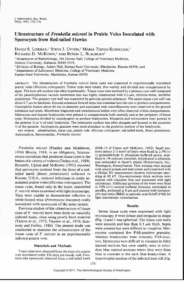
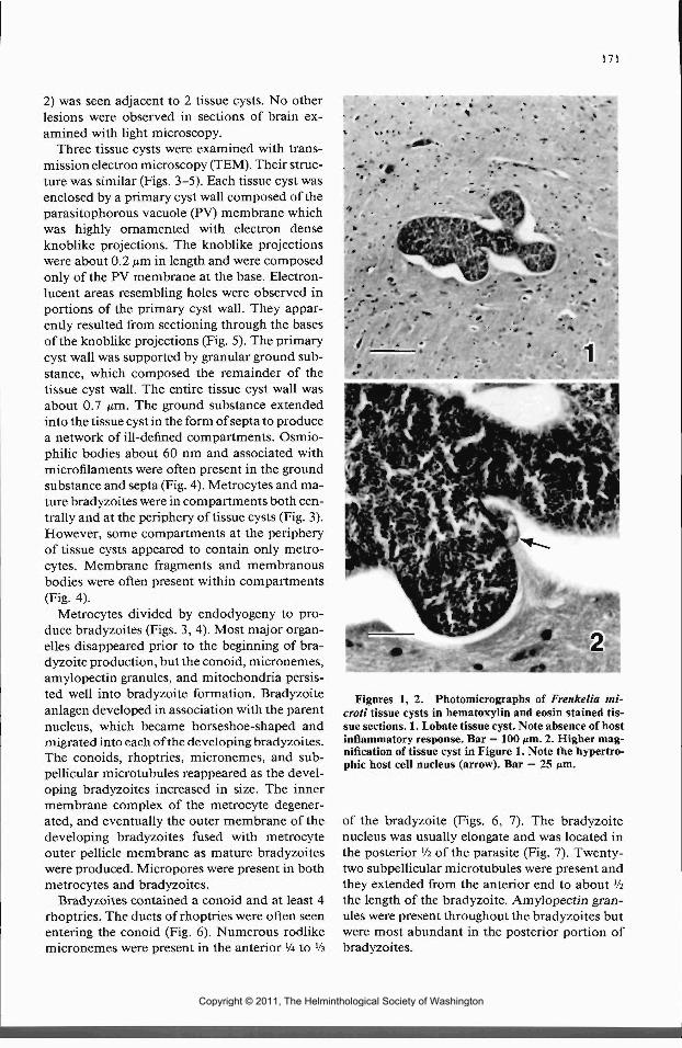
Seven tissue cysts were examined with lightmicroscopy; 6 were lobate and irregular in shape(Fig. 1) and 1 was spherical. The tissue cyst wallswere smooth and less than 0.5 nm. thick. Septawere present but were difficul t to visualize. Met-rocytes contained few PAS-positive granules,whereas bradyzoites were intensely PAS-posi-tive. Metrocytes were difficult to visualize in H&Estained sections but were readily seen in tolui-dine blue stained sections where they were faintblue in contrast to the dark blue bradyzoites. Ahypertrophic nucleus of the infected host cell (Fig.
170
Copyright © 2011, The Helminthological Society of Washington
171
2) was seen adjacent to 2 tissue cysts. No otherlesions were observed in sections of brain ex-amined with light microscopy.
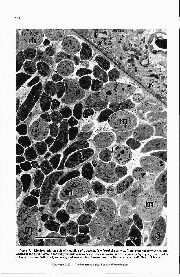
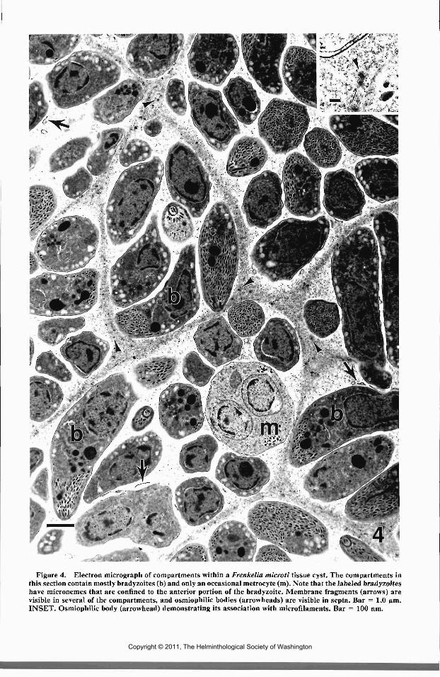
Three tissue cysts were examined with trans-mission electron microscopy (TEM). Their struc-ture was similar (Figs. 3-5). Each tissue cyst wasenclosed by a primary cyst wall composed of theparasitophorous vacuole (PV) membrane whichwas highly ornamented with electron denseknoblike projections. The knoblike projectionswere about 0.2 nm in length and were composedonly of the PV membrane at the base. Electron-lucent areas resembling holes were observed inportions of the primary cyst wall. They appar-ently resulted from sectioning through the basesof the knoblike projections (Fig. 5). The primarycyst wall was supported by granular ground sub-stance, which composed the remainder of thetissue cyst wall. The entire tissue cyst wall wasabout 0.7 jim. The ground substance extendedinto the tissue cyst in the form of septa to producea network of ill-defined compartments. Osmio-philic bodies about 60 nm and associated withmicrofilaments were often present in the groundsubstance and septa (Fig. 4). Metrocytes and ma-ture bradyzoites were in compartments both cen-trally and at the periphery of tissue cysts (Fig. 3).However, some compartments at the peripheryof tissue cysts appeared to contain only metro-cytes. Membrane fragments and membranousbodies were often present within compartments(Fig. 4).
Metrocytes divided by endodyogeny to pro-duce bradyzoites (Figs. 3, 4). Most major organ-elles disappeared prior to the beginning of bra-dyzoite production, but the conoid, micronemes,amylopectin granules, and mitochondria persis-ted well into bradyzoite formation. Bradyzoiteanlagen developed in association with the parentnucleus, which became horseshoe-shaped andmigrated into each of the developing bradyzoites.The conoids, rhoptries, micronemes, and sub-pellicular microtubules reappeared as the devel-oping bradyzoites increased in size. The innermembrane complex of the metrocyte degener-ated, and eventually the outer membrane of thedeveloping bradyzoites fused with metrocyteouter pellicle membrane as mature bradyzoiteswere produced. Micropores were present in bothmetrocytes and bradyzoites.
Bradyzoites contained a conoid and at least 4rhoptries. The ducts of rhoptries were often seenentering the conoid (Fig. 6). Numerous rodlikemicronemes were present in the anterior 14 to lh
Figures 1, 2. Photomicrographs of Frenkelia mi-croti tissue cysts in hematoxylin and eosin stained tis-sue sections. 1. Lobate tissue cyst. Note absence of hostinflammatory response. Bar = 100 nm. 2. Higher mag-nification of tissue cyst in Figure 1. Note the hypertro-phic host cell nucleus (arrow). Bar = 25 nm.
of the bradyzoite (Figs. 6, 7). The bradyzoitenucleus was usually elongate and was located inthe posterior l/i of the parasite (Fig. 7). Twenty-two subpellicular microtubules were present andthey extended from the anterior end to about lhthe length of the bradyzoite. Amylopectin gran-ules were present throughout the bradyzoites butwere most abundant in the posterior portion ofbradyzoites.
Copyright © 2011, The Helminthological Society of Washington
172
Figure 3. Electron micrograph of a portion of a Frenkelia microti tissue cyst. Numerous metrocytes (m) arelocated at the periphery and centrally within the tissue cyst. The compartments are separated by septa (arrowheads)and most contain both bradyzoites (b) and metrocytes. Arrows point to the tissue cyst wall. Bar = 5.0 ftm.
Copyright © 2011, The Helminthological Society of Washington
v-m'-'m^-w^-'^jK^'' i*P'JwM^?^^Sm^i^R
.;**3*5i?r«<>>^< Ifi ^
Figure 4. Electron micrograph of compartments within a Frenkelia microti tissue cyst. The compartments inthis section contain mostly bradyzoites (b) and only an occasional metrocyte (m). Note that the labeled bradyzoiteshave micronemes that are confined to the anterior portion of the bradyzoite. Membrane fragments (arrows) arevisible in several of the compartments, and osmiophilic bodies (arrowheads) are visible in septa. Bar = 1.0 pm.INSET. Osmiophilic body (arrowhead) demonstrating its association with microfilaments. Bar = 100 nm.
Copyright © 2011, The Helminthological Society of Washington
174 JOURNAL OF THE HELMINTHOLOGICAL SOCIETY
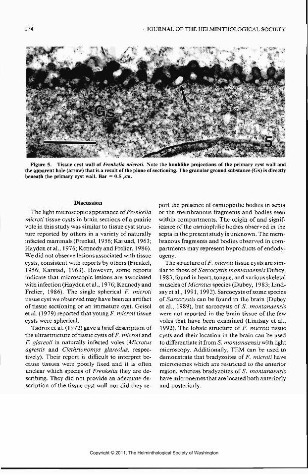
Figure 5. Tissue cyst wall of Frenkelia microti. Note the knoblike projections of the primary cyst wall andthe apparent hole (arrow) that is a result of the plane of sectioning. The granular ground substance (Gs) is directlybeneath the primary cyst wall. Bar = 0.5 ^m.
Discussion
The light microscopic appearance of Frenkeliamicroti tissue cysts in brain sections of a prairievole in this study was similar to tissue cyst struc-ture reported by others in a variety of naturallyinfected mammals (Frenkel, 1956; Karstad, 1963;Haydenetal., 1976; Kennedy and Frelier, 1986).We did not observe lesions associated with tissuecysts, consistent with reports by others (Frenkel,1956; Karstad, 1963). However, some reportsindicate that microscopic lesions are associatedwith infection (Hayden et al., 1976; Kennedy andFrelier, 1986). The single spherical F. microtitissue cyst we observed may have been an artifactof tissue sectioning or an immature cyst. Geiselet al. (1979) reported that young F. microti tissuecysts were spherical.
Tadros et al. (1972) gave a brief description ofthe ultrastructure of tissue cysts ofF. microti andF. glareoli in naturally infected voles (Microtusagrestis and Clethrionomys glareolus, respec-tively). Their report is difficul t to interpret be-cause tissues were poorly fixed and it is oftenunclear which species of Frenkelia they are de-scribing. They did not provide an adequate de-scription of the tissue cyst wall nor did they re-
port the presence of osmiophilic bodies in septaor the membranous fragments and bodies seenwithin compartments. The origin of and signif-icance of the osmiophilic bodies observed in thesepta in the present study is unknown. The mem-branous fragments and bodies observed in com-partments may represent byproducts of endody-ogeny.
The structure of F. microti tissue cysts are sim-ilar to those of Sarcocystis montanaensis Dubey,1983, found in heart, tongue, and various skeletalmuscles of Microtus species (Dubey, 1983; Lind-say etal., 1991, 1992). Sarcocysts of some speciesof Sarcocystis can be found in the brain (Dubeyet al., 1989), but sarcocysts of S. montanaensiswere not reported in the brain tissue of the fewvoles that have been examined (Lindsay et al.,1992). The lobate structure of F. microti tissuecysts and their location in the brain can be usedto differentiate it from S. montanaensis with lightmicroscopy. Additionally, TEM can be used todemonstrate that bradyzoites of F. microti havemicronemes which are restricted to the anteriorregion, whereas bradyzoites of 5". montanaensishave micronemes that are located both anteriorlyand posteriorly.
Copyright © 2011, The Helminthological Society of Washington

OF WASHINGTON, VOLUME 59, NUMBER 2, JULY 1992 175
':<?,,, ;t
IP
Figures 6, 7. Bradyzoites of Frenkelia microti. 1. Anterior end of a bradyzoite demonstrating a conoid (ar-rowhead), rhoptries (R), and numerous rodlike micronemes. Note the duct of one rhoptry (arrow). Bar = 0.5 ^m.7. A group of bradyzoites. Note the elongate nucleus (N) that is present and that the micronemes are located inthe anterior portion of the bradyzoite. Bar = 1.0 nm.
Copyright © 2011, The Helminthological Society of Washington

176
Acknowledgments
This study was supported by funds from theDepartment of Pathobiology, College of Veter-inary Medicine, Auburn University. This is Col-lege of Veterinary Medicine publication number2288.
Literature Cited
Dubey, J. P. 1983. Sarcocystis montanaensis and S.microti sp. n. from the meadow vole (Microtuspennsylvanicus). Proceedings of the Helmintho-logical Society of Washington 50:318-324.
, C. A. Speer, and R. Payer. 1989. Sarcocys-tosis of Animals and Man. CRC Press, Boca Ra-ton, Florida. 215 pp.
Frenkel, J. K. 1956. Pathogenesis of toxoplasmosisand of infections of organisms resembling Toxo-plasma. Annals of the New York Academy of Sci-ence 64:215-251.
Geisel, O., E. Kaiser, O. Vogel, H. E. Krampitz, andM. Rommel. 1979. Pathomorphologic findingsin short-tailed voles (Microtus agrestis) experi-mentally-infected with Frenkelia microti. Journalof Wildlif e Diseases 15:267-270.
Hayden, D. W., N. W. King, and A. S. Murthy. 1976.
Spontaneous Frenkelia infection in a laboratory-reared rat. Veterinary Pathology 13:337-342.
Karstad, L. 1963. Toxoplasma microti (the M-or-ganism) in the muskrat (Ondatra zibethica). Ca-nadian Veterinary Journal 4:249-251.
Kennedy, M. J., and P. F. Frelier. 1986. Frenkeliasp. from the brain of a porcupine (Erethizon dor-satuni) from Alberta, Canada. Journal of Wildlif eDiseases 22:112-114.
Lindsay, D. S., S. J. Upton, B. L. Blagburn, M. Toivio-Kinnucan, J. P. Dubey, C. T. McAllister, and S.E.Trauth. 1992. Demonstration that Sarcocystismontanaensis has a speckled kingsnake-prairie volelif e cycle. Journal of the Helminthological Societyof Washington 59:9-15.
, , , , C. T. McAllister, andS. E.Trauth. 1991. Sporocysts isolated from thesouthern copperhead (Agkistrodon contortrix con-tortrix) produce Sarcocystis montanaensis-\ikesarcocysts in prairie voles (Microtus ochrogaster).Journal of Wildlif e Diseases 27:148-152.
Tadros, W. A., R. G. Bird, and D. S. Ellis. 1972. Thefine structure of cysts of Frenkelia (the M-organ-ism). Folia Parasitologica (Praha) 19:203-209.
Upton, S. J., and R. D. McKown. 1992. The red-tailed hawk, Buteojamaicensis, a native definitivehost of Frenkelia microti (Apicomplexa) in NorthAmerica. Journal of Wildlif e Diseases 28:85-90.
Zoological Nomenclature
The following Applications were published on 26 March 1992 in Vol. 49, Part 1 of the Bulletinof Zoological Nomenclature. Comment or advice on these Applications is invited for publicationin the Bulletin, and should be sent to the Executive Secretary, I.C.Z.N., % The Natural HistoryMuseum, Cromwell Road, London SW7 5BD, U.K.
Case 2251 Bucephalus Baer, 1827 and B. poly-morphus Baer, 1827 (Trematoda):proposed conservation in their ac-cepted usage
Barbara BaturoInland Fisheries Institute, ul. Ocza-powskiego 10, 10-957 Olsztyn 5, Po-land
Abstract. The purpose of this ap-plication is to conserve in their ac-cepted usage the generic and specificnames of an important trematodeparasite of freshwater fishes—Bu-
cephalus polymorphus Baer, 1827.The name B. polymorphus was basedon cercariae, but it has been shownthat these develop into the adulttrematode first named as Rhipido-cotyle campanula (Dujardin, 1844),a senior synonym ofR. illensis (Zieg-ler, 1883). A neotype for B. poly-morphus is proposed to avoid trans-fer of this long recognized name toR. campanula, with resulting con-fusion at both generic and specificlevels.
Copyright © 2011, The Helminthological Society of Washington