ultrasound small vessel imaging with block-wise adaptive ... · ultrasound small vessel imaging...
TRANSCRIPT

IEEE TRANSACTIONS ON MEDICAL IMAGING, VOL. 36, NO. 1, JANUARY 2017 251
Ultrasound Small Vessel Imaging WithBlock-Wise Adaptive Local Clutter Filtering
Pengfei Song, Armando Manduca, Joshua D. Trzasko, and Shigao Chen*
Abstract— Robust clutter filtering is essential for ultra-sound small vessel imaging. Eigen-based clutter filteringtechniques have recently shown great improvement inclutter rejection over conventional clutter filters in smallanimals. However, for in vivo human imaging, eigen-based clutter filtering can be challenging due to the com-plex spatially-varying tissue and noise characteristics.To address this challenge, we present a novel block-wiseadaptive singular value decomposition (SVD) based clutterfiltering technique. The proposed method divides the globalplane wave data into overlapped local spatial segments,within which tissue signals are assumed to be locallycoherent and noise locally stationary. This, in turn, enableseffective separation of tissue, blood and noise via SVD. Foreach block, the proposed method adaptively determinesthe singular value cutoff thresholds based on local datastatistics. Processing results from each block are redun-dantly combined to improve both the signal-to-noise-ratio(SNR) and the contrast-to-noise-ratio (CNR) of the smallvessel perfusion image. Experimental results show that theproposed method achieved more than two-fold increase inSNR and more than three-fold increase in CNR in dB scaleover the conventional global SVD filtering technique for anin vivo human native kidney study. The proposed methodalso showed substantial improvement in suppression of thedepth-dependent background noise and better rejection ofnear field tissue clutter. The effects of different processingblock size and block overlappercentagewere systematicallyinvestigated as well as the tradeoff between imaging qualityand computational cost.
Index Terms— Clutter filtering, plane wave imaging, sin-gular value thresholding, small vessel imaging, ultrasoundDopplre.
I. INTRODUCTION
H IGH frame rate ultrasound plane wave imaging offersextraordinary capabilities of imaging small vessels
thanks to the large amount of spatial and temporal sam-ples that can be collected within a short period of time,which substantially increases the Doppler sensitivity to the
Manuscript received February 2, 2016; revised August 13, 2016,March 25, 2016, April 25, 2016, and June 6, 2016; accepted August 31,2016. Date of publication September 2, 2016; date of current versionDecember 29, 2016. This work was partially supported by the NationalScience Foundation (NSF) under Grant CCF:CIF:1318347. Asteriskindicates corresponding author.
P. Song is with the Department of Radiology, Mayo Clinic College ofMedicine, Rochester, MN 55905 USA (e-mail: [email protected]).
A. Manduca is with the Department of Physiology and BiomedicalEngineering, Mayo Clinic College of Medicine, Rochester, MN 55905USA.
J. D. Trzasko is with the Department of Radiology, Mayo Clinic Collegeof Medicine, Rochester, MN 55905 USA.
*S. Chen is with the Department of Radiology, Mayo Clinic College ofMedicine, Rochester, MN 55905 USA (e-mail: [email protected]).
Digital Object Identifier 10.1109/TMI.2016.2605819
flow signals from fine vessels [1], [2]. The rich spatial andtemporal content of plane wave imaging data also facilitatesbetter separation of blood and tissue signals when usingadvanced clutter filtering techniques such as eigen-based clut-ter filters [3]–[8]. Unlike traditional clutter filters that aresolely based on high-pass temporal filtering [9], eigen-basedclutter filters utilize the underlying differences in specklestrength and spatiotemporal characteristics of tissue and bloodsignals for better clutter rejection [3]. After performing acertain type of rank-revealing matrix decompositions such assingular value decomposition (SVD), tissue signals typicallyreside in the large spectral components (i.e. high singularvalues) because of the high speckle strength and temporalcoherence, while blood signals are typically found in the lowerspectral components (i.e. medium to low singular values)due to the relatively low speckle brightness and temporalcoherence. The lowest singular values will be mostly noise.Tissue and blood signals (and to some extent noise) can thenbe readily separated via a low-rank matrix projection such assingular value thresholding (SVT) [3].
The combination of eigen-based clutter filtering and planewave imaging was recently proposed by Demené et al. forhigh sensitivity functional ultrasound imaging [7]. A sig-nificant improvement of clutter rejection and small vesseldelineation was demonstrated over the traditional high-passclutter filtering. In [7], the eigen-based clutter filtering wasperformed in a global fashion where a SVD was applied tothe entire three-dimensional data set (two dimensions in spaceand one dimension in time) obtained from the whole imagingfield-of-view (FOV), with the implicit underlying assumptionthat noise within the spatial FOV follows an independent andidentically distributed (i.i.d.) complex Gaussian distribution.While this assumption may be valid for functional ultrasoundbrain imaging in mice, where the imaging FOV is smalland shallow, this assumption is typically violated for in vivohuman imaging because of the complex tissue characteristicsand spatially varying nature of noise within the FOV due todepth-dependent ultrasound attenuation [10]. Figure 1 showsan example of using plane wave imaging and the global SVDclutter filter approach proposed in [7] for in vivo human nativekidney perfusion imaging. One can readily see the spatially-varying background noise which amplifies proportional to spa-tial position from the transducer and overwhelms the kidneyblood flow signal. This was not observed in [7] becausea transplanted kidney was imaged with a much shallowerdepth. Moreover, in Fig. 1, one can see that the near fieldsubcutaneous tissue could not be completely removed by theglobal SVD clutter filter. The spatially-varying nature of noise
0278-0062 © 2016 IEEE. Personal use is permitted, but republication/redistribution requires IEEE permission.See http://www.ieee.org/publications_standards/publications/rights/index.html for more information.

252 IEEE TRANSACTIONS ON MEDICAL IMAGING, VOL. 36, NO. 1, JANUARY 2017
Fig. 1. In vivo human kidney blood flow power Doppler image acquiredby high frame rate plane wave imaging (PRF= 500Hz, ensemble= 450)and eigen-based clutter filtering with global SVD calculation and thresh-olding.
demonstrated in Fig. 1 is typical for plane wave imaging due tothe lack of transmit focusing and depth-dependent ultrasoundattenuation, and can significantly compromise the performanceof SVT [11]–[13] due to its implicit presumption about i.i.d.Gaussian noise in the data. Meanwhile, although the sensitivityto small vessel flow signals is greatly improved thanks to thelarge amount of temporal ensembles acquired by plane waveimaging, the cumulative amount of noise is also increased.This can be a major issue for in vivo human imaging as shownin Fig. 1, yet no solution has been proposed from previousstudies using eigen-based clutter filters [3]–[7].
To address these challenges with existing SVD-based clutterfiltering techniques for plane wave small vessel imaging, inthis paper we propose a new block-wise local SVD clutterfiltering technique with adaptive SVT based on local datastatistics. A block-wise local SVD clutter filter with adaptiveSVT operates on local data blocks such that: 1) the localnoise distribution within the block is approximately i.i.d.Gaussian which enables robust low rank matrix estimation;2) singular value cutoff thresholds for tissue-blood and blood-noise separation can be adaptively determined based on thelocal signal and noise characteristics for improved separationof the tissue, the blood, and the noise; 3) the signal-to-noise-ratio (SNR) of the small vessel Doppler image can befurther improved due to the redundant nature of the overlappedblock decomposition. In this study, we also present a noveladaptive threshold selection strategy for SVT that can robustlydetermine singular value cutoff thresholds to extract bloodsignals from both tissue and noise.
The paper is structured as follows: we first introduce theblock-wise local SVD clutter filtering method with adap-tive SVT. Due to the difficulties of constructing reliable smallvessel imaging phantoms, the same set of in vivo kidney datashown in Fig. 1 was used as the testing data throughout thepaper. Different block sizes and block overlap percentageswere evaluated to investigate the optimal block size and thetradeoff between SNR gain and computational costs. We con-clude the paper with discussion and conclusions.
II. MATERIALS AND METHODS
A. Principles of Local SVD Clutter Filtering With AdaptiveSingular Value Thresholding
For small vessel high frame plane wave imaging, the typicaldata matrix is three-dimensional, with two dimensions in space(Nx and Nz ) and one dimension in time (Nt ), as in Fig. 2(a).For the in vivo kidney data used in this study, a lineararray transducer L11-4v (Verasonics Inc., Kirkland, WA) wasused to transmit a diverging beam with center frequencyof 5 MHz to form a sector-shaped FOV for abdominal imaging.A 10-angle spatial compounding (−9◦ to 9◦) was used toimprove imaging SNR [14]. The post-compounding ensemblesize was 450 (i.e. Nt = 450) with an effective pulse-repetition-frequency (PRF) of 500 Hz (total data acquisition duration= 900 ms), with Nx = 352 and Nz = 590 (lateral pixelresolution = 0.246 mm; axial pixel resolution = 0.123 mm).The full data set is then decomposed into local small blockswith dimensions of nx by nz by nt (nx ≤ Nx , nz ≤ Nz ,nt ≤ Nt ). The blocks are spatially overlapped by n′
x in lateraldirection and n′
z in axial direction, with 0 ≤ n′x ≤ nx − 1 and
0 ≤ n′z ≤ nz −1. For the 3-D data within each block, the local
data is reshaped to a 2-D Casorati matrix with dimensionsof nx · nz by nt , followed by a singular value decomposi-tion (SVD), yielding a matrix D (min(nx ·nz , nt ) by min(nx ·nz ,nt )) containing the singular values in the diagonal and twounitary matrices U(nx · nz by min(nx · nz , nt )) and V (nt bymin(nx ·nz , nt )) containing the singular vectors correspondingto each singular value. Figure 2(c) shows a typical singularvalue curve obtained from the local block indicated in Fig. 2(b)from the kidney data. Figure 2(d) shows the curve of meanDoppler frequency of each singular vector (the right vectors V )corresponding to each singular value obtained using the lag-one autocorrelation method [15]. From Figs. 2(c) and (d), onecan clearly see that while tissue’s singular values (low singularvalue orders, roughly <30) are significantly higher than bloodand noise, its Doppler frequency is significantly lower. Bloodsignal is concentrated in the middle region (singular valueorders 50-100), with intermediate Doppler frequencies. Forsingular orders beyond 200, the majority of the signal is noiseand should be removed.
1) Low-Order Singular Value Thresholding: To rejecttissue signal from blood signal, a low-order singular valuecutoff threshold needs to be determined. Here we combinetwo classic SVD clutter filtering approaches presented in [3]and [6] to determine the low-order singular value cutoffthreshold: 1) the gradient of the singular value curve shownin Fig. 2(c) is calculated to identify a turning point (cutoff 1A)from which the curve begins to flatten; 2) the singular vectorDoppler frequency curve shown in Fig. 2(d) is used to searchfor a point (cutoff 1B) beyond which the Doppler frequencyexceeds a pre-defined tissue motion frequency threshold(e.g. 10 Hz); 3) the greater singular value order of the cutoffs1A and 1B is used as the low-order singular value cutoff fordifferentiation of the tissue and blood signals. For the exampleshown in Figs. 2(c) and (d), cutoff 1A was 32, cutoff 1B was31, and thus a final cutoff of 32 was used as the low-ordersingular value cutoff threshold for this specific block.

SONG et al.: ULTRASOUND SMALL VESSEL IMAGING WITH BLOCK-WISE ADAPTIVE LOCAL CLUTTER FILTERING 253
Fig. 2. (a) Schematic plot of decomposing the original 3-D plane wave imaging data into block-wise local processing units with overlaps. (b) B-modeimage of the in vivo kidney generated from the plane wave IQ data, the white box indicates one local box used for SVD analysis. (c) Singular valuecurve obtained from the SVD calculation of the local data indicated in (b). Cutoff 1A was determined by searching for a turning point from which thesingular value curve begins to flatten. (d) Mean Doppler frequency of the right singular vectors V corresponding to the singular values in (c) obtainedusing lag-one IQ autocorrelation. Cutoff 1B was determined by searching for a point beyond which the Doppler frequency exceeds a pre-definedtissue motion frequency threshold.
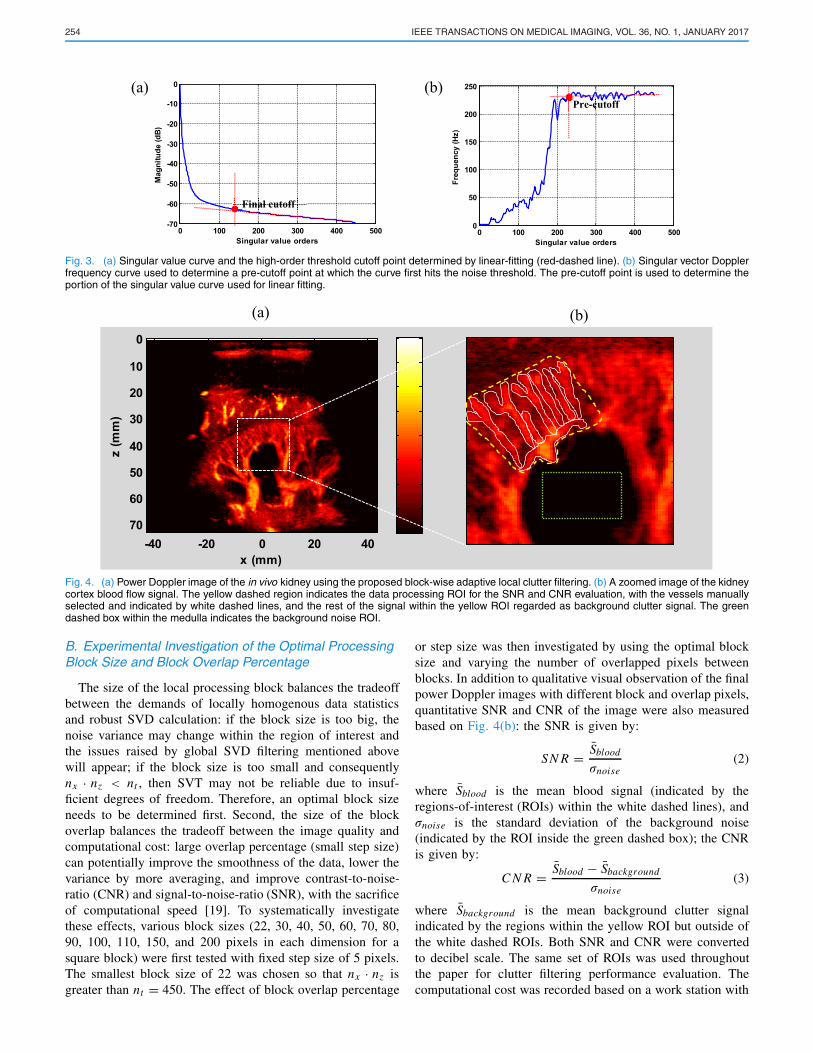
2) High-Order Singular Value Thresholding: Toremove singular values that are mainly corresponding to noise,a high-order singular value cutoff needs to be determined.Here we use the principles of the Marcenko-Pastur distrib-ution which describes the singular value distribution of ani.i.d. Gaussian random matrix [16], [17]. Assuming Gaussiannoise distribution within the local data block, the high-ordersingular values corresponding to noise should follow a lineardistribution under the logarithm scale (e.g. the part of the curvebeyond singular value order of 200 in Fig. 3(a)). By linear-fitting this part of the singular value curve, one can obtain aturning point from which the singular value curve begins todeviate from the fitted linear line, as shown in Fig. 3(a). Thisturning point can be used as a cutoff threshold differentiatingblood signal from noise. To determine the portion of thesingular value curve that can be used for linear fitting, apre-cutoff point is first determined from the singular vectorDoppler frequency curve shown in Fig. 3(b). The flat portionof the curve in Fig. 3(b) is used to calculate an averagednoise Doppler frequency threshold and a pre-cutoff pointis determined at which the frequency curve hits the noisethreshold for the first time. In this example, the pre-cutoff
point was 235. Therefore, the singular value curve from order235 and beyond was used for linear fitting, and a final cutoffvalue of 133 was determined as the high-order singular valuethreshold for this block.
3) Reconstruct Global Flow Signals From Blocks:Within each block, the clutter filtering was performed byforcing the singular values that are outside of the low- andhigh-order cutoff thresholds to zero, followed by an inverseSVD calculation [3], [7]. For each pixel, the final blood flowsignal S(x, z) is given by:
S(x, z) = 1
N
N∑
n=1
⎛
⎝ Sn∑High−cuto f f
Low−cutof f λ
⎞
⎠ (1)
where N is the total number of overlapped blocks containingthe target pixel (x, z), Sn is the clutter-filtered signal fromblock n, and λ are the remaining singular values correspondingto blood signal. Finally, the power Doppler signal is calculatedby cumulating the power of the blood signal along the tempo-ral direction and displayed on the decibel scale [18], as shownin Fig. 4(a).
254 IEEE TRANSACTIONS ON MEDICAL IMAGING, VOL. 36, NO. 1, JANUARY 2017
Fig. 3. (a) Singular value curve and the high-order threshold cutoff point determined by linear-fitting (red-dashed line). (b) Singular vector Dopplerfrequency curve used to determine a pre-cutoff point at which the curve first hits the noise threshold. The pre-cutoff point is used to determine theportion of the singular value curve used for linear fitting.
Fig. 4. (a) Power Doppler image of the in vivo kidney using the proposed block-wise adaptive local clutter filtering. (b) A zoomed image of the kidneycortex blood flow signal. The yellow dashed region indicates the data processing ROI for the SNR and CNR evaluation, with the vessels manuallyselected and indicated by white dashed lines, and the rest of the signal within the yellow ROI regarded as background clutter signal. The greendashed box within the medulla indicates the background noise ROI.
B. Experimental Investigation of the Optimal ProcessingBlock Size and Block Overlap Percentage
The size of the local processing block balances the tradeoffbetween the demands of locally homogenous data statisticsand robust SVD calculation: if the block size is too big, thenoise variance may change within the region of interest andthe issues raised by global SVD filtering mentioned abovewill appear; if the block size is too small and consequentlynx · nz < nt , then SVT may not be reliable due to insuf-ficient degrees of freedom. Therefore, an optimal block sizeneeds to be determined first. Second, the size of the blockoverlap balances the tradeoff between the image quality andcomputational cost: large overlap percentage (small step size)can potentially improve the smoothness of the data, lower thevariance by more averaging, and improve contrast-to-noise-ratio (CNR) and signal-to-noise-ratio (SNR), with the sacrificeof computational speed [19]. To systematically investigatethese effects, various block sizes (22, 30, 40, 50, 60, 70, 80,90, 100, 110, 150, and 200 pixels in each dimension for asquare block) were first tested with fixed step size of 5 pixels.The smallest block size of 22 was chosen so that nx · nz isgreater than nt = 450. The effect of block overlap percentage
or step size was then investigated by using the optimal blocksize and varying the number of overlapped pixels betweenblocks. In addition to qualitative visual observation of the finalpower Doppler images with different block and overlap pixels,quantitative SNR and CNR of the image were also measuredbased on Fig. 4(b): the SNR is given by:
SN R = Sblood
σnoise(2)
where Sblood is the mean blood signal (indicated by theregions-of-interest (ROIs) within the white dashed lines), andσnoise is the standard deviation of the background noise(indicated by the ROI inside the green dashed box); the CNRis given by:
C N R = Sblood − Sbackground
σnoise(3)
where Sbackground is the mean background clutter signalindicated by the regions within the yellow ROI but outside ofthe white dashed ROIs. Both SNR and CNR were convertedto decibel scale. The same set of ROIs was used throughoutthe paper for clutter filtering performance evaluation. Thecomputational cost was recorded based on a work station with

SONG et al.: ULTRASOUND SMALL VESSEL IMAGING WITH BLOCK-WISE ADAPTIVE LOCAL CLUTTER FILTERING 255
Fig. 5. (a) Power Doppler image of the noise field derived from thereference phantom data by applying the same global SVD filtering asin the in vivo data. (b) Noise-equalized global SVD-filtered result byapplying the noise field in (a) onto Fig. 1. Both power Doppler imagesare displayed under a 40 dB dynamic range.
two Intel® Xeon® E5-2650 processors (2.30 GHz) and runningMatlab (version R2013b, MathWorks, Natick, MA).
C. Background Noise Equalization for theGlobal SVD Approach
Due to the lack of transmit focusing of diverging waves, thelower-half of the power Doppler image processed by the globalSVD filter has significantly lower SNR than the upper-half ofthe image and appears to be noise-saturated (Fig. 1). To allevi-ate the background noise for the global SVD filtering, we useda reference phantom (CIRS Inc., Norfolk, VA. Attenuation= 0.5 dB/cm/MHz; sound speed = 1540 m/s) to collect oneset of reference data using the same ultrasound sequence as inthe in vivo data. The same global SVD processing was appliedto the reference data. Because no blood flow signal was presentin the phantom, the majority of the remaining signal afterglobal SVD filtering was noise, as shown in Fig. 5(a). We thenused the noise field derived from the reference phantom toequalize the power Doppler image (i.e. dividing by the noisefield), as shown in Fig. 5(b). The noise equalization providesa more uniform power Doppler image for the global SVDmethod, facilitating better comparisons between the globalSVD method and the proposed block-wise adaptive approach.
III. RESULTS
Low-Order and High-Order Singular Value Thresholding
Figure 6 shows the step-by-step filtering results of theproposed method. Before clutter filtering, the blood signal iscompletely overwhelmed by the strong tissue signal (Fig. 6(a))and no vessels can be observed. For local SVD adaptive clutterfiltering, a square block size of 80 pixels with 75 overlappingpixels was used. After low-order SVT (step 1a in the Methodssection), the tissue signal can be mostly removed (Fig. 6(b)).However, there is still significant noise in the image thatdeteriorates the contrast of the vessel signals. After high-orderSVT (step 1b), the image quality is substantially improvedwith significantly reduced noise. The contrast of the image isgreatly enhanced. Using the measurement ROIs defined above,the SNR and CNR are 28.3 dB and 14.5 dB respectivelyfor the vessel signals with only low-order SVT, as comparedto 33.5 dB and 25 dB for the vessel signals with both low-and high-order SVT. These results suggest that while it is
critical to use low-order SVT to reject tissue clutter, it is alsoimportant to use high-order SVT to reject noise for improvedSNR and CNR of small vessel imaging using high frame-rateplane waves with large ensembles.
Global SVD Clutter Filtering vs. Block-Wise AdaptiveLocal SVD Filtering
Figure 7 shows the comparison results between the globalSVD clutter filtering method [7], the noise-equalized globalSVD method, and the proposed block-wise adpative local SVDclutter filtering method on the same set of in vivo human nativekidney data. The local SVD method used a square block sizeof 80 pixels with 75 overlapping pixels. One can clearly seethat the spatially varying background noise can be effectivelysuppressed and the kidney vessels can be better visualizedby the proposed method, due to the fact that noise withineach block is approximately identically distributed and that theadaptive cutoff thresholds adjust locally for better extractionof the blood vessel signal. The actual thresholds chosen ateach block are as shown in Figs. 7(d) and (e). The SNR andCNR of the vessel signals for the global SVD clutter filteringare 15.5 dB and 7.59 dB, respectively, compared to an SNRof 33.5 dB and CNR of 25 dB with the block-wise local SVDapproach: a more than two-fold increase in SNR and more thanthree-fold increase in CNR in dB scale using the proposedmethod as compared to the global SVD filter without noiseequalization. Moreover, the near field subcutaneous tissue andthe strong noise in lower left and right corners of the imagecan be better suppressed using the proposed method comparedto the global SVD approach. The SNR and CNR for theglobal SVD clutter filter plus noise equalization are 28.6 dBand 19.9 dB, respectively, which are significantly higher thanglobal SVD without noise equalization, yet about 5 dB lowerthan the block-wise local SVD approach. As shown in Fig. 7(c)and Fig. 4(b), small kidney cortex vessels with diameters ofabout 0.5 - 1mm can be clearly resolved at a depth of about4 cm using the proposed clutter filtering method.
Various Block Size Experiments
Figure 8 shows the final power Doppler images processedwith different block sizes and a fixed step size of 5 (blockoverlap pixels = block size - 5). Figure 9 shows the SNR,CNR, and computational times for each of the block size resultin Fig. 8. From Fig. 8, one can see that the block-wise SVDclutter filtering does not offer benefit until a block size of 50,and provides similar results for block sizes from 80 to 150,until the background ramp-shaped noise and the near fieldtissue signal become prominent again at a block size of 200.The SNR and CNR plots in Fig. 9(a) match these observationsand indicate that the SNR and CNR both plateau at block sizesof 80 to 110 and gradually decrease with larger block sizes. Forcomputational cost, as shown in Fig. 9(b), the processing timegrows ∼ n3 with increased block size. This is not surprisingnoting the O(n3) complexity of the SVD [20]. From block sizeof 80 to block size of 110, the SNR and CNR are virtually thesame while the computational time almost doubled. Therefore,

256 IEEE TRANSACTIONS ON MEDICAL IMAGING, VOL. 36, NO. 1, JANUARY 2017
Fig. 6. (a) Power Doppler image of the original data set without clutter filtering. The image is displayed under 100 dB dynamic range. (b) PowerDoppler image obtained using local adaptive SVD filtering with only low-order SVT. (c) Power Doppler image obtained using local adaptive SVDfiltering with both low- and high-order SVT. Both (b) and (c) are displayed under 40 dB dynamic range.
Fig. 7. (a) Clutter filtering result using the global SVD filtering approach. (b) clutter filtering result after noise equalization of (a). (c) clutter filtering onthe same set of data using the proposed block-wise adaptive local SVD filtering approach. (d)and (e): adaptively selected low-order and high-ordersingular value thresholds by the proposed algorithms, respectively. Figures (d) and (e) were generated by placing the threshold values in the centerof each block and replacing the zero pixels between blocks (5 pixels in this case with a block size of 80 pixels and 75 pixels overlap) with the samethreshold values of the block center.
a block size of 80 was used for further testing of the optimalblock overlap percentage.
Various Block Overlap Percentage Experiments
Figure 10 shows the selected power Doppler image resultsusing different block overlap percentages for local SVDand computational cost plots versus the overlap percentages:0% - 90% with 10% increment (corresponding to overlappixels from 0 to 72 with a block size of 80) and 93.75%
(corresponding to overlap pixels of 75) which has been usedin previous experiments. From the results one can see severediscontinuities are observed in the power Doppler imagesfor overlap percentages smaller than 80%. The computationalcost dramatically increases for overlaps greater than 80%.From a closer look of the power Doppler image of 80%overlap, one can still appreciate subtle “tile” patterns ascompared to the 93.75% overlap image shown in Fig. 8 (blocksize 80). This suggests that further increasing the overlappercentage beyond 80% does improve the smoothness and

SONG et al.: ULTRASOUND SMALL VESSEL IMAGING WITH BLOCK-WISE ADAPTIVE LOCAL CLUTTER FILTERING 257
Fig. 8. Power Doppler results using different processing block sizes (in the unit of pixels), with fixed step size of 5 pixels. All images have 40 dBdisplay dynamic range.
Fig. 9. (a) SNR and CNR plots versus processing block size. (b) Computational cost versus block size.
visual appearance of the power Doppler images. However,80% overlap offers a good tradeoff between the image qualityand computational cost.
IV. DISCUSSION
This paper presents a block-wise local adaptive clutter filter-ing technique based on singular value decomposition (SVD)to address several key challenges of ultrasound small vesselimaging with high frame rate plane waves. A block-wiseprocessing algorithm, together with robust and adaptive sin-gular value thresholding (SVT) based on local data statis-tics, was proposed to facilitate better clutter rejection andimprove SNR and CNR of the power Doppler images bymulti-block averaging. The proposed method demonstratedsignificant improvement of background noise suppression forplane wave imaging, near-field subcutaneous tissue rejection,and a more than two-fold SNR increase and a more thanthree-fold CNR increase in dB scale over the existing SVD-based clutter filtering methods. Fine kidney cortex vessels withdiameters less than 1 mm could be clearly resolved at a depth
of 4 cm using the proposed technique, which is superior toexisting ultrasound Doppler imaging techniques.
Due to the complex tissue components and noise distributionin in vivo human imaging, the performance of a global SVD-based clutter filter will usually be compromised. This is clearlyrevealed in the results shown in Figs. 7(d) and (e), whereone can see that the SVT cutoff values can be significantlydifferent across the FOV. Block-wise processing provides theopportunity of fine tuning the cutoff thresholds based onthe distribution of tissue, blood, and noise within a localregion, to achieve much improved clutter rejection and bloodsignal extraction. In this study we also proposed to use ahigh-order singular value cutoff threshold to reject the high-order singular values that mainly correspond to noise, inaddition to the conventional low-order threshold to separateblood from tissue (Fig. 6). The high-order cutoff may notbe necessary for conventional Doppler imaging because largervessels are the common targets in conventional Doppler. Theblood signal strength of large vessels is typically superiorto noise, and therefore separating blood signal from tissue

258 IEEE TRANSACTIONS ON MEDICAL IMAGING, VOL. 36, NO. 1, JANUARY 2017
Fig. 10. (a) Selected power Doppler results with different block overlap percentages. All images were under 40 dB display dynamic range.(b) Computational cost versus the block overlap percentage.
is the primary goal for clutter filtering. For high frame ratesmall vessel imaging with large ensembles, however, the smallvessel signals are much weaker than large vessels and theirsingular values are not too much higher than noise (Fig. 2(c)).As shown in Fig. 6, a high-order SVT cutoff based on boththe singular value curve and singular vector Doppler frequencycurve can effectively reject the noise, preserve the small vesselsignal, and improve the SNR and CNR of the power Dopplerimage. Figure 8 shows that the local processing block sizeneeds to be greater than a certain value (50 in this study) forrobust clutter rejection. The optimal block size was found to be80×80 in this study, which provides 6400 spatial samples overthe 450 temporal samples. In practice one may need to adjustthe block size according to the ensemble length to achieveoptimal performance. A low block overlap percentage can beused for such a test to lower computational cost, and a highoverlap can be used later for better visual appearance.
Previous adaptive clutter filters have been proposed byYoo et al. [21] and Kargel et al. [22]. The adaptive clut-ter filter method by Yoo et al. utilized an adaptive fil-tering process but was Fourier transform-based and thusdid not use an adaptive signal transformation mechanism(e.g., SVD or eigen-decomposition). Conversely, the study byKargel et al. utilized an eigen-decomposition based signaltransform process– which, unlike Fourier transformation issignal adaptive– but the selection of cutoff singular valueswas not adaptive. In this work, we synergistically utilizeboth an adaptive signal transformation and filtering procedure,which overcomes the respective challenges faced in each ofthe previous methods. As shown in the paper, the higherperformance of the proposed method was mainly achievedby adaptively choosing the singular value cutoff thresholds
based on local data statistics. Note that in this study we didnot compare the proposed method with the adaptive Fouriertransform-based method of Yoo et al., which is a limitationof this study. A similar adaptive method based on a Fouriertransform instead of eigenvectors may significantly improvethe computational performance.
Figure 11 shows two more examples of kidney perfu-sion imaging and two examples of liver perfusion imag-ing processed with the global SVD filtering technique, theglobal SVD filtering plus noise equalization technique, andthe proposed local adaptive SVD filtering method. The sameprocessing parameters as used in Fig. 4(a), including block sizeand block overlap percentage (80 × 80 pixels with 75 pixelsoverlap), were used for all the examples. It can be seen thatthe proposed method achieved better performance than theglobal SVD filtering approach and the global SVD plus noiseequalization throughout all examples. More detailed vascula-ture could be resolved with the local adaptive SVD filter withsignificantly suppressed background noise and near field tissuesignal, and substantially improved contrast. The local adaptiveSVD filtering also showed robustness in processing differentorgans with different tissue and blood signal characteristics.No adjustments to the SVT cutoff values or noise equalizationas for global SVD were necessary thanks to the adaptive andblock-wise nature of the proposed method.
The proposed block-wise adaptive clutter filter method isa multi-ensemble-based clutter filtering technique. The spatialresolution of small vessel imaging, which is predominantlydetermined by the ultrasound imaging resolution (e.g. fre-quency, pulse length, etc.), should not be compromised by theproposed clutter filtering method because no spatial averagingwas performed within each processing block. As a comparison,

SONG et al.: ULTRASOUND SMALL VESSEL IMAGING WITH BLOCK-WISE ADAPTIVE LOCAL CLUTTER FILTERING 259
Fig. 11. In vivo kidney and liver perfusion power Doppler images processed with the global SVD clutter filtering approach (upper row), the globalSVD plus noise equalization approach (middle row), and the proposed local adaptive SVD clutter filtering method (lower row). Same processingparameters (80× 80 pixels with 75 pixels overlap) were used for all examples.
Fig. 12(a) shows the result of a single-ensemble-based eigen-processing clutter filter [5], [23] on the same set of kidneydata as in Fig. 4(a), together with the noise-equalized result.The single-ensemble-based clutter filtering provides morestatistically stationary ensembles (i.e. less spatially-varyingtissue motion) than the multi-ensemble approaches [5], [23].However, as compared to the proposed block-wise adaptiveSVD filter (Fig. 7(c)), the single-ensemble Hankel-SVD filtersuffers more from noise contamination and tissue clutters.This may be because the single-ensemble SVD filter does notdeploy a high-order singular value thresholding mechanismas proposed in this paper for noise suppression. The multi-ensemble SVD also provides more robust tissue and bloodsignal separation for the global and block-wise SVD methodsthan the single-ensemble Hankel-SVD. The SNR and CNRof the power Doppler image in Fig. 12(a) before noise-equalization are 12.56 dB and 3.54 dB, respectively, and26.8 dB and 16.7 dB after noise-equalization, which are bothlower than the global SVD (both before and after noise-equalization) and the block-wise local SVD methods (SNR of33.5 dB and CNR of 25 dB). Meanwhile, the results show thatthe spatial resolution was not compromised by the block-wiseadaptive SVD filter. The block-wise processing provides more
spatial information about adjacent tissue and blood signalsthan the single-ensemble SVD filter, enabling more robustseparation of tissue, blood and noise using singular valuethresholding and better perfusion images.
Figure 12(b) shows the clutter filtering result from a con-ventional Butterworth IIR highpass filter (5th order, cutofffrequency = 50 Hz, first 10 sampling points were discardedto suppress initialization effects). Figure 12(c) shows theclutter filter result from a regression clutter filter with quarticorder [24]. For each spatial pixel, the long temporal ensembleswere divided into small segments with 16 samples for quarticregression fitting. As compared to the proposed block-wiseadaptive SVD filter (Fig. 7(c)), both tissue clutter rejectionand noise suppression are inferior to the proposed method.The contrast of the small vessels is also worse than theproposed method. The SNR and CNR are 12.21 dB and2.49 dB before noise-equalization, and 26.8 dB and 15.9 dBafter noise-equalization for Butterworth IIR filter, respectively;11.71 dB and 1.81 dB before noise-equalization, and 26.1 dBand 15.0 dB after noise-equalization for the regression filter,which are all lower than the proposed block-wise local SVDmethods (SNR of 33.5 dB and CNR of 25 dB). The weakflow signal amplitude and slow flow speed of small vessels

260 IEEE TRANSACTIONS ON MEDICAL IMAGING, VOL. 36, NO. 1, JANUARY 2017
Fig. 12. Clutter filtering results using the single-ensemble Hankel-SVDfilter (a), the conventional IIR highpass filter (cutoff = 50 Hz) (b), andthe regression clutter filter (quartic-order regression) (c). The noise-equalized results of each method are also shown in the figure.
make it challenging to separate blood from noise purely in theFourier domain or by subtraction of tissue signal by regressionfitting. The proposed method uses block-wise local SVD anda Marcenko-Pastur distribution based high cutoff method toeffectively reject noise residing in the high singular valueorders. This has been shown to greatly improve the SNR andCNR of the image as compared to the global SVD, single-ensemble-based eigen-processing filter, and conventional IIRclutter filters.
In this study, only one set of in vivo data was used tocompare the performance of different clutter filters. Ideallya simulation study with ground truth would provide a morestable platform for evaluation studies. Most current blood flowsimulation methods use Gaussian white noise as the noisemodel [5], [9], [24], which may not be the best noise model tosimulate the spatially varying complicated noise we observein vivo for compounding plane wave imaging-based smallvessel perfusion imaging. In a future study, a better noisemodel needs to be developed to account for noise propagationfrom raw ultrasound RF channel data to the final beamformedimage. This includes investigating the noise behavior with
ultrasound gain settings (e.g., TGC) and the beamformingprocess (e.g., number of channels used in delay-and-sum,receive apodization, compounding, etc.), and integrating theseobservations into an analytical model to predict the noise ateach spatial location.
The lower part of the power Doppler images processed byglobal SVD and other conventional clutter filtering methodsare saturated by noise, while the proposed method is not thanksto the local power normalization and high-order singular-valuethresholding within each processing block. Because of the lackof transmit focusing of diverging waves used in this study,the lower half of the image has lower SNR and thereforehigher noise than the upper half of the image. Although ithas been shown that the noise-equalization approach couldsignificantly improve the appearance of the global SVD andother conventional clutter filter results, the SNR and CNR ofthe noise-equalized images are still significantly lower than theproposed block-wise adaptive SVD method. The small vesselsare better visualized with the proposed method with highercontrast. The proposed block-wise SVD approach calculateslocally adaptive low eigen-order cut-off values to robustlyseparate blood signal from tissue signal and high eigen-ordercut-off values to reject noise (Figs. 7(d) and (e)). The block-wise SVD also normalizes the power locally so that it auto-matically equalizes uneven noise distribution. These benefitscannot be obtained from conventional methods and globalSVD. The noise-equalization offers substantially improvedvisual appearance for the images processed by conventionalclutter filters with significantly less computational cost thanthe proposed method. The drawbacks of the noise-equalizationapproach include: 1) the noise-equalization method only equal-izes the intensity of the image across depths instead of actuallyrejecting any noise, as opposed to the proposed method; 2) thenoise-equalization approach needs the reference phantom datato be acquired with the exact same imaging setup (e.g., TGCgain, transmit voltage, etc.) as the targeted data. When theimaging setup is changed, a new set of reference phantomdata needs to be collected for equalization.
One limitation of the block-wise SVD clutter filtering,and for eigen-based clutter filtering techniques in general,is the relatively expensive computational cost compared toconventional high-pass-filter-based clutter filtering, especiallyfor large block sizes and block overlap percentages. This isdue to the O(n3) complexity of the SVD [20]. For the globalSVD clutter filtering performed in Fig. 1, the processing timewas 18.7 seconds. The fastest processing time with block-wise SVD was 25.7 seconds with block size of 80 and 0%overlap, while 472.3 seconds was needed to obtain the 80%overlap image with acceptable visual appearance. The majorityof the computational cost comes from the SVD calculationon large matrices, which are typical for high frame rateplane wave imaging with long ensembles to boost up Dopplersensitivity. Use of compiled– rather than interpreted– program-ming languages such as C/C++ or high-throughput parallelcomputing platforms (e.g., Intel Xeon Phi) may significantlyimprove the computational speed of the proposed method.Use of stochastic approximate SVD strategies [25] may alsolead to substantial computational acceleration, and will be

SONG et al.: ULTRASOUND SMALL VESSEL IMAGING WITH BLOCK-WISE ADAPTIVE LOCAL CLUTTER FILTERING 261
Fig. 13. Clutter filter results using different number of temporalensembles and data acquisition duration. All power Doppler images aredisplayed under 40 dB dynamic range. Bottom plot shows the SNR andCNR of the power Doppler maps versus different ensemble lengths.
studied in a separate, future work. In additional to the highcomputational cost, the long data acquisition time (typicallyseveral hundreds of milliseconds) also remains as a majorhurdle towards real-time implementation of the plane wavesmall vessel imaging technique. Figure 13 shows examples ofthe impact of data acquisition time on small vessel imaging.Given a PRF of 500 Hz used in this study, the original data wastruncated to 100 ensembles (200 ms acquisition), 50 ensembles(100 ms acquisition) and 25 ensembles (50 ms acquisition),to reflect a theoretical frame rate (i.e. ignoring computationalcost, etc.) of 5 Hz, 10 Hz, and 20 Hz, respectively (theoriginal 900 ms acquisition corresponds to about 1.1 Hz framerate). From Fig. 13, one can clearly see a degradation ofimage quality with decreased number of ensembles, whichis also quantitatively indicated in the SNR and CNR plot.With a potential frame rate of 5 Hz, the 100 ensemble lengthprovides a moderate to good visualization of the vasculatureof the kidney cortex, which suggests a balanced tradeoffbetween image quality and frame rate. However, ideally a longensemble length is preferred to increase Doppler sensitivityand achieve best visualization of the detailed vasculature. Onepotential solution to the low frame rate is to combine the small
vessel imaging with conventional real-time Doppler imaging,which can be used to first locate the targeted imaging region,and then followed by high frame rate plane wave transmissionsto provide a snapshot of the detailed vasculatures of the target.Moreover, although only power Doppler results were presentedin this paper, other Doppler measurements such as color flowand spectral Doppler can be retrospectively obtained for eachindividual vessel within the FOV based on the high framerate plane wave imaging data [26], [27]. These combinedmeasurements may be very useful in a wide range of clinicalapplications.
V. CONCLUSIONS
This paper presents an SVD-based block-wise adaptive localclutter filtering technique that can substantially improve clutterrejection and small vessel imaging quality for high frameplane wave ultrasound. The proposed technique successfullyovercame the challenges of small vessel clutter filtering forin vivo human imaging by facilitating robust singular valuethresholding on a local level and combining the results forimproved imaging quality. This method can facilitate thetranslation of high frame rate ultrasound small vessel imagingfrom laboratory to clinic, and has great potential for earlydetection, diagnosing, and follow up of many diseases.
REFERENCES
[1] E. Macé, G. Montaldo, I. Cohen, M. Baulac, M. Fink, and M. Tanter,“Functional ultrasound imaging of the brain,” Nature Methods, vol. 8,pp. 662–664, Jul. 2011.
[2] E. Macé, G. Montaldo, B. Osmanski, I. Cohen, M. Fink, andM. Tanter, “Functional ultrasound imaging of the brain: Theory andbasic principles,” IEEE Trans. Ultrason., Ferroelect., Freq. Control,vol. 60, no. 3, pp. 492–506, Mar. 2013.
[3] A. C. H. Yu and L. Lovstakken, “Eigen-based clutter filter designfor ultrasound color flow imaging: A review,” IEEE Trans. Ultrason.,Ferroelect., Freq. Control, vol. 57, no. 5, pp. 1096–1111, May 2010.
[4] A. C. H. Yu and R. S. C. Cobbold, “Single-ensemble-based eigen-processing methods for color flow imaging—Part II. The matrix pencilestimator,” IEEE Trans. Ultrason., Ferroelect., Freq. Control, vol. 55,no. 3, pp. 573–587, Mar. 2008.
[5] A. C. H. Yu and R. S. C. Cobbold, “Single-ensemble-based eigen-processing methods for color flow imaging—Part I. The Hankel-SVDfilter,” IEEE Trans. Ultrason., Ferroelect., Freq. Control, vol. 55, no. 3,pp. 559–572, Mar. 2008.
[6] L. Lovstakken, S. Bjaerum, K. Kristoffersen, R. Haaverstad, and H. Torp,“Real-time adaptive clutter rejection filtering in color flow imaging usingpower method iterations,” IEEE Trans. Ultrason., Ferroelect., Freq.Control, vol. 53, no. 9, pp. 1597–1608, Sep. 2006.
[7] C. Demené et al., “Spatiotemporal clutter filtering of ultrafast ultrasounddata highly increases Doppler and fUltrasound sensitivity,” IEEE Trans.Med. Imag., vol. 34, no. 11, pp. 2271–2285, Nov. 2015.
[8] D. E. Kruse and K. W. Ferrara, “A new high resolution color flowsystem using an eigendecomposition-based adaptive filter for clutterrejection,” IEEE Trans. Ultrason., Ferroelect., Freq. Control, vol. 49,no. 12, pp. 1384–1399, Dec. 2002.
[9] S. Bjaerum, H. Torp, and K. Kristoffersen, “Clutter filter design forultrasound color flow imaging,” IEEE Trans. Ultrason., Ferroelect.,Freq. Control, vol. 49, no. 2, pp. 204–216, Feb. 2002.
[10] T. L. Szabo, “Attenuation,” in Diagnostic Ultrasound Imaging: InsideOut, T. L. Szabo, Ed., 2nd ed. Boston, MA, USA: Academic, 2014,ch. 4, pp. 81–119.
[11] J. Shlens. (2014). “A tutorial on principal component analysis.” [Online].Available: http://arxiv.org/abs/1404.1100
[12] J.-F. Cai, E. J. Candes, and Z. Shen, “A singular value threshold-ing algorithm for matrix completion,” SIAM J. Opt., vol. 20, no. 4,pp. 1956–1982, 2010.

262 IEEE TRANSACTIONS ON MEDICAL IMAGING, VOL. 36, NO. 1, JANUARY 2017
[13] S. Ma, D. Goldfarb, and L. Chen, “Fixed point and Bregman iterativemethods for matrix rank minimization,” Math. Program., vol. 128,nos. 1–2, pp. 321–353, 2011.
[14] G. Montaldo, M. Tanter, J. Bercoff, N. Benech, and M. Fink, “Coherentplane-wave compounding for very high frame rate ultrasonographyand transient elastography,” IEEE Trans. Ultrason., Ferroelect., Freq.Control, vol. 56, no. 3, pp. 489–506, Mar. 2009.
[15] D. H. Evans, “Colour flow and motion imaging,” Proc. Inst. Mech. Eng.H, J. Eng. Med., vol. 224, no. 2, pp. 241–253, 2010.
[16] V. A. Marcenko and L. A. Pastur, “Distribution of eigenvalues forsome sets of random matrices,” Math. USSR-Sbornik, vol. 1, no. 4,pp. 457–483, 1967.
[17] M. Rudelson and R. Vershynin, “Non-asymptotic theory of ran-dom matrices: Extreme singular values,” in Proc. Int. Congr. Math.,vol. 3. 2010, pp. 1576–1602.
[18] D. H. Evans and W. N. McDicken, Doppler Ultrasound: Physics,Instrumentation and Signal Processing. Hoboken, NJ, USA: Wiley,2000.
[19] E. J. Candes, C. A. Sing-Long, and J. D. Trzasko, “Unbiased riskestimates for singular value thresholding and spectral estimators,” IEEETrans. Signal Process., vol. 61, no. 19, pp. 4643–4657, Oct. 2013.
[20] G. H. Golub and C. F. Van Loan, Matrix Computations. Baltimore, MD,USA: The Johns Hopkins Univ. Press, 1996.
[21] Y. M. Yoo, R. Managuli, and Y. Kim, “Adaptive clutter filtering forultrasound color flow imaging,” Ultrasound Med. Biol., vol. 29, no. 9,pp. 1311–1320, 2003.
[22] C. Kargel, G. Hoebenreich, G. Plevnik, B. Trummer, and M. Insana,“Velocity estimation and adaptive clutter filtering for color flowimaging,” Tech. Rep.
[23] W. You and Y. Wang, “A single-ensemble clutter rejection method basedon the analytic geometry for ultrasound color flow imaging,” UltrasoundMed. Biol., vol. 37, pp. 1909–1922, Nov. 2011.
[24] A. P. Kadi and T. Loupas, “On the performance of regression and step-initialized IIR clutter filters for color doppler systems in diagnosticmedical ultrasound,” IEEE Trans. Ultrason., Ferroelect., Freq. Control,vol. 42, no. 5, pp. 927–937, Sep. 1995.
[25] N. Halko, P. G. Martinsson, and J. A. Tropp, “Finding structurewith randomness: Probabilistic algorithms for constructing approximatematrix decompositions,” SIAM Rev., vol. 53, no. 2, pp. 217–288,2011.
[26] J. Bercoff et al., “Ultrafast compound Doppler imaging: Providing fullblood flow characterization,” IEEE Trans. Ultrason., Ferroelect., Freq.Control, vol. 58, no. 1, pp. 134–147, Jan. 2011.
[27] B. Y. Yiu and A. C. Yu, “High-frame-rate ultrasound color-encodedspeckle imaging of complex flow dynamics,” Ultrasound Med. Biol.,vol. 39, pp. 1015–1025, Jun. 2013.