ultrasound of the knee - semed femede · jumper’s knee jumper’s knee is a syndrome affecting...
TRANSCRIPT

Ultrasound of the KneeMarc J. Lee, M.D.
1and Kira Chow, M.D.
2
ABSTRACT
Ultrasound of the knee is best suited for the evaluation of tendons, fluidcollections and effusion, synovitis, periarticular soft tissue masses, muscles, and thecollateral ligaments. Advantages of sonographic evaluation include the lack of ionizingradiation, the noninvasive nature of the examination, focused evaluation correlated withthe specific site of pain, multiplanar capability, and utility in patients for whom magneticresonance imaging (MRI) is contraindicated. The ability to visualize the microanatomy oftendons, ligaments, nerves, and muscles is also an advantage over MRI. Moreover,ultrasound is generally readily accessible and of lower cost than MRI. Ultrasound is lesssuited for the evaluation of internal derangement. Certain structures that are exquisitelyevaluated by MRI, including the menisci, cruciate ligaments, bone marrow, and articularcartilage, are suboptimally evaluated by sonography. This article reviews the structures andpathological processes for which ultrasound has been shown useful, emphasizing appro-priate technique and normal anatomy, appearance of pathological processes, and advan-tages and limitations of sonographic evaluation.
KEYWORDS: Ultrasound, sonography, knee
Sonographic evaluation of the knee can be tail-ored to the history and clinical site of concern, correlat-ing the point of maximal symptoms with the imagingfindings. Comprehensive examination can be performed,and various protocols have been posited in the litera-ture.1 The examination should be performed with ahigh-resolution linear multifrequency transducer (7 to10 MHz). Patient positioning and transducer orienta-tion depend on the particular structure being imaged.Comparison with the asymptomatic contralateral knee isvery useful and increases diagnostic confidence.
We discuss the ultrasound evaluation of tendonabnormalities, fluid collections including bursae andeffusions, the menisci and parameniscal cysts, ligamenttears, arthritis and synovitis, as well as the utility ofultrasound in the setting of total knee arthroplasty.
EXTENSOR MECHANISM
The extensor mechanism of the knee is poised foroptimal sonographic evaluation given its superficiallocation. The patient can be positioned in a supine orsitting position with the knee flexed or semiflexed,thereby elongating the tendon. A support under theknee in the supine position can maximize patient com-fort. Both longitudinal and transverse images should beobtained of the quadriceps and patellar tendons, both ofwhich normally show a parallel fibrillar echogenicappearance (Fig. 1). The transducer should be parallelto the tendon to avoid creating artifactual hypoechoicareas that can simulate pathology. Extended field-of-view imaging is particularly useful to show the extensormechanism along its length, delineating anatomicalrelationships.
1Department of Radiology, Saint Joseph Medical Center, Burbank,California; 2Department of Radiology, Santa Monica–UCLAMedicalCenter and Orthopedic Hospital, Santa Monica, California.
Address for correspondence and reprint requests: Kira Chow,M.D., Department of Radiology, Santa Monica–UCLA MedicalCenter and Orthopedic Hospital, 1250 16th St., Ste. 275, 2nd Fl.,Tower, Santa Monica, CA 90404 (e-mail: [email protected]).
Musculoskeletal Ultrasound; Guest Editor, Kambiz Motamedi,M.D.
Semin Musculoskelet Radiol 2007;11:137–148. Copyright #2007 by Thieme Medical Publishers, Inc., 333 Seventh Avenue,New York, NY 10001 USA. Tel: + 1(212) 584-4662.DOI 10.1055/s-2007-1001879. ISSN 1089-7860.
137
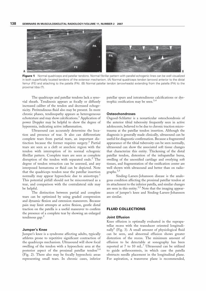
The quadriceps and patellar tendons lack a syno-vial sheath. Tendinosis appears as focally or diffuselyincreased caliber of the tendon and decreased echoge-nicity. Peritendinous fluid also may be present. In morechronic phases, tendinopathy appears as heterogeneousechotexture and may show calcifications.1 Application ofpower Doppler may be helpful to show the degree ofhyperemia, indicating active inflammation.
Ultrasound can accurately determine the loca-tion and presence of tear. It also can differentiatecomplete tears from partial tears, an important dis-tinction because the former requires surgery.2 Partialtears are seen as a cleft or anechoic region with thetendon with interruption of its normal echogenicfibrillar pattern. Complete tears are seen as completedisruption of the tendon with separated ends.2 Thedegree of tendon retraction can be assessed, and anyinterposed hematoma or fluid can be depicted. Notethat the quadriceps tendon near the patellar insertionnormally may appear hypoechoic due to anisotropy.1
This potential pitfall should not be misconstrued as atear, and comparison with the contralateral side maybe helpful.
The distinction between partial and completetears can be optimized by using graded compressionand dynamic flexion and extension maneuvers. Becausepain may limit attempts at active flexion, gentle distaltraction on the patella is a useful maneuver to confirmthe presence of a complete tear by showing an enlargedtendinous gap.2
Jumper’s Knee
Jumper’s knee is a syndrome affecting adults, typicallyathletes prone to repetitive significant contraction ofthe quadriceps mechanism. Ultrasound will show focalswelling of the tendon with a hypoechoic area at theposterior aspect of the proximal patellar tendon3,4
(Fig. 2). There also may be focally hypoechoic areasrepresenting small tears. In chronic cases, inferior
patellar spurs and intratendinous calcifications or dys-trophic ossification may be seen.3,4
Osteochondroses
Osgood-Schlatter is a nonarticular osteochondrosis ofthe anterior tibial tuberosity frequently seen in activeadolescents, believed to be due to chronic traction micro-trauma at the patellar tendon insertion. Although thediagnosis is generally made clinically, ultrasound can beuseful for diagnostic confirmation. Because a fragmentedappearance of the tibial tuberosity can be seen normally,ultrasound can show the associated soft tissue changesthat characterize this entity. Thickening of the distalpatellar tendon, distention of the infrapatellar bursa,swelling of the unossified cartilage and overlying softtissues, and fragmentation of the ossification center arewell shown with ultrasound and better than on radio-graphs.3,5
Sinding-Larsen-Johansson disease is the analo-gous condition affecting the proximal patellar tendon atits attachment to the inferior patella, and similar changesare seen in this entity.3,5 Note that the imaging appear-ances of jumper’s knee and Sinding-Larsen-Johanssonare similar.
FLUID COLLECTIONS
Joint Effusion
Knee effusion is optimally evaluated in the suprapa-tellar recess with the transducer oriented longitudi-nally6 (Fig. 3). A small amount of physiological fluidcan be seen, and abnormal effusion shows greaterdistention of the recess. The minimum amount ofeffusion to be detectable at sonography has beenreported at 7 to 10 mL.7 Ultrasound can be utilizedto guide arthrocentesis, in which case the patellaobstructs needle placement in the longitudinal plane.For aspiration, a transverse plane is recommended,
Figure 1 Normal quadriceps and patellar tendons. Normal fibrillar pattern with parallel echogenic lines can be well visualized
in both superficially located tendons of the extensor mechanism. (A) Normal quadriceps tendon (arrows) anterior to the distal
femur (FE) and attaching to the patella (PA). (B) Normal patellar tendon (arrowheads) extending from the patella (PA) to the
proximal tibia (T).
138 SEMINARS IN MUSCULOSKELETAL RADIOLOGY/VOLUME 11, NUMBER 2 2007

either anteromedially or anterolaterally depending onthe side of greater distention.6
In the setting of acute trauma, sonography canbe used to evaluate for lipohemarthrosis. Lipohe-marthrosis may appear as either a two-layered collec-tion with inferior anechoic blood and nondependenthyperechoic fat or as a trilaminar collection with asuperior hyperechoic fat layer, an intermediateanechoic serum layer, and a dependent hypoechoiclayer of blood cells.8,9 A prospective study comparingultrasound with radiographs and using computed to-mography (CT) as the gold standard showed thatalthough ultrasound could not directly depict intra-articular knee fractures, ultrasound was significantlymore sensitive (97%) than radiographs (55%) for de-piction of lipohemarthrosis in the suprapatellar bursa
Figure 3 Joint effusion. Longitudinal scan obtained ante-
riorly shows joint effusion in the suprapatellar recess (arrow-
heads) posterior to the quadriceps tendon (QT). Patella (PA)
and distal femur (FE) are also seen.
Figure 2 Jumper’s knee. (A) Longitudinal scans of the bilateral patellar tendons. On the right, the normal patellar tendon
demonstrates regular caliber and normal fibrillar pattern (arrowheads). On the left, the abnormal patellar tendon is thickened
(arrows) with a hypoechoic area at the posterior aspect of the proximal patellar tendon (asterisks). (B) Transverse image through
the proximal patellar tendon demonstrates heterogeneous disorganized appearance with enlargement of the tendon (arrow-
heads). (C) Longitudinal scan through the abnormal patellar tendon (PT) shows hyperemia along the posterior proximal aspect
on power Doppler image.
ULTRASOUND OF THE KNEE/LEE, CHOW 139

and could therefore potentially be used to evaluate foroccult knee fracture in acute trauma.8
Bursae
Bursae about the anterior knee include the prepatellarbursa and the superficial and deep infrapatellar bursae.The prepatellar bursa is located in the subcutaneoustissues anterior to the lower patella. This bursa canbecome distended after prolonged kneeling (Fig. 4),and the entity is called ‘‘housemaid’s knee.’’ Superficialinfrapatellar bursitis, ‘‘clergyman’s knee,’’ is seen ante-rior to the inferior patellar tendon. The deep infra-patellar bursa is located between the anterior tibia andthe inferior patellar tendon. This last bursa is the onlyone commonly seen in the normal knee as a flattened2- to 3-mm fluid structure.3 Bursitis is seen as a fluidcollection, which may be associated with wall thick-
ening. Power Doppler may show associated inflam-mation.
Pes anserine bursitis is seen as a cystic mass justdeep to the pes anserine tendon at the anteromedialaspect of the knee.
Bursae may contain echogenic material represent-ing inflammatory or infectious debris or hemorrhage.1
Baker Cyst
Evaluation for Baker cyst is best performed from aposterior approach with the patient supine and the kneefully extended. Ultrasound is highly sensitive for thedetection of Baker cysts, which are seen as thin-walledfluid collections (Fig. 5), sometimes containing debris.The fluid may be anechoic, hypoechoic, or of mixedechogenicity relative to skeletal muscle.10 A Baker cysttypically communicates with the knee joint, extending
Figure 4 Prepatellar bursitis. (A) Transverse image anterior to the patella (PA) demonstrates fluid in the prepatellar bursa
(arrowheads). (B) Longitudinal scan shows nodularity and thickening of the periphery of the prepatellar bursa (arrowheads),
secondary to inflammation. (C) Color Doppler longitudinal image shows hyperemia along the periphery of the prepatellar bursa.
(D) Lateral radiograph of the knee shows focal prominence of the prepatellar soft tissues (arrows).
140 SEMINARS IN MUSCULOSKELETAL RADIOLOGY/VOLUME 11, NUMBER 2 2007

between the semimembranosus and medial gastrocne-mius tendons. Demonstrating this connection is diag-nostic for a Baker cyst with 100% accuracy and caneliminate false-positive diagnoses and misinterpreta-tion of other cystic structures or masses as Baker cysts.10
A leaking Baker cyst shows fluid tracking between themuscles and along the subcutaneous tissues. Dopplercan be applied to differentiate a small Baker cyst from apopliteal aneurysm.
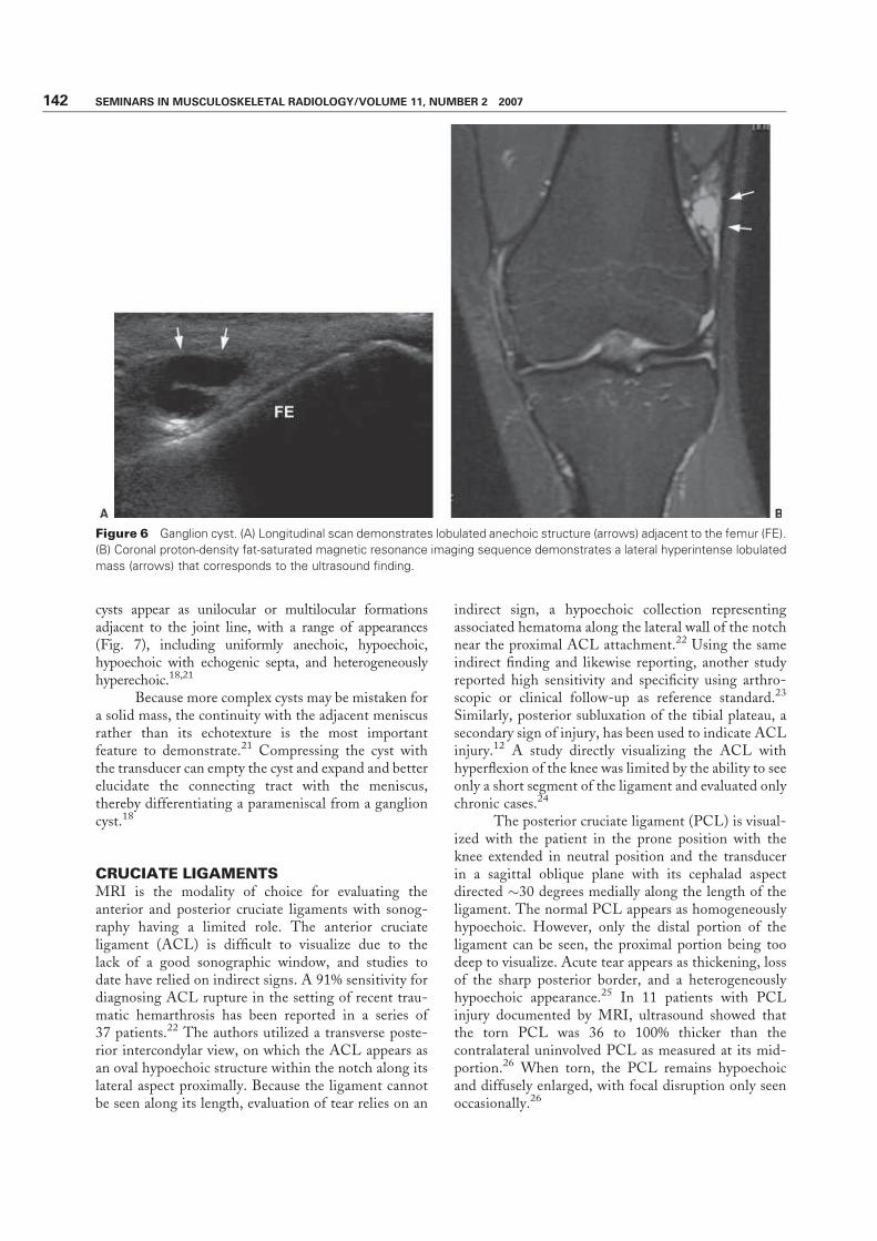
Ganglion Cysts
Ganglion cysts about the knee are frequent findingsbut may be incidental. They are mucin-filled cysticstructures that may be unilocular or septated andappear anechoic to hypoechoic (Fig. 6). Ultrasoundis useful for guidance of aspiration for symptomaticcysts.
MENISCI AND PARAMENISCAL CYSTS
Although magnetic resonance imaging (MRI) remainsthe gold-standard imaging modality for evaluation of themenisci, some tears can be demonstrated sonographicallywith meticulous technique. The utility of sonography formeniscal tear is controversial. Earlier studies reportedhigh sensitivities,11–13 but experienced sonologists in a2002 study comparing findings with MRI, CT, andarthroscopy results concluded that sonography is notaccurate enough to be reliable as the sole modality fordiagnosing or excluding tears.14
To examine the menisci, the transducer is placedlongitudinally spanning the joint line and always ori-ented perpendicular to the meniscus. The anterior
horns and midzones can be imaged with the patientsupine or in the lateral decubitus position with theknee extended; the posterior horns are imaged with thepatient prone. Dynamic flexion and rotation can helpopen the joint line to improve visualization. Visual-ization may be improved by applying mild valgus orvarus stress to image the medial and lateral meniscus,respectively. The entire meniscus cannot be routinelyvisualized due to its anatomical location. There maybe limited conspicuity of the free edge of the meni-scus; peripheral and posterior tears are the easiest todemonstrate.1,15,16
The normal meniscus appears as a triangular,uniformly hyperechoic structure sandwiched betweenthe hypoechoic articular cartilage surfaces of the fem-oral condyle and tibial plateau. Tears appear as eitherhypoechoic or anechoic focal linear defects and lessfrequently as hyperechoic abnormalities.1,17 In addi-tion, tears can be obscured by an overlying parameniscalcyst. Meniscal degeneration appears as decreased echo-genicity with a swollen appearance, sometimes withcystic regions or as heterogeneity.1,18 In advanceddegeneration, meniscal protrusion and fragmentationalso may be seen.18 The utility of ultrasound forevaluating meniscocapsular separation has not beenwell evaluated, with one study using arthroscopicallycreated lesions in cadavers concluding that sonographyis not accurate for the diagnosis.19
Sonography has an accuracy of > 90% for theevaluation of parameniscal cysts,18 which may present aspain or a palpablemass ormay be found incidentally. In 80to 90% of cases, parameniscal cysts are associated witha meniscal lesion, generally horizontal tears, althoughsome cysts may be due to degeneration.18,20 Parameniscal
Figure 5 Baker cyst. (A) Transverse scan obtained medially demonstrates anechoic fluid (arrowheads) in a Baker cyst. The
neck of the Baker cyst (arrow) is between the medial gastrocnemius head tendon (MG) and the semimembranosus tendon
(SM). (B) A second transverse image again demonstrates the Baker cyst (asterisks) with its neck (arrow) originating between
the tendons just cited. The tendons (arrowheads) are well-defined hyperechoic ovals.
ULTRASOUND OF THE KNEE/LEE, CHOW 141
cysts appear as unilocular or multilocular formationsadjacent to the joint line, with a range of appearances(Fig. 7), including uniformly anechoic, hypoechoic,hypoechoic with echogenic septa, and heterogeneouslyhyperechoic.18,21
Because more complex cysts may be mistaken fora solid mass, the continuity with the adjacent meniscusrather than its echotexture is the most importantfeature to demonstrate.21 Compressing the cyst withthe transducer can empty the cyst and expand and betterelucidate the connecting tract with the meniscus,thereby differentiating a parameniscal from a ganglioncyst.18
CRUCIATE LIGAMENTS
MRI is the modality of choice for evaluating theanterior and posterior cruciate ligaments with sonog-raphy having a limited role. The anterior cruciateligament (ACL) is difficult to visualize due to thelack of a good sonographic window, and studies todate have relied on indirect signs. A 91% sensitivity fordiagnosing ACL rupture in the setting of recent trau-matic hemarthrosis has been reported in a series of37 patients.22 The authors utilized a transverse poste-rior intercondylar view, on which the ACL appears asan oval hypoechoic structure within the notch along itslateral aspect proximally. Because the ligament cannotbe seen along its length, evaluation of tear relies on an
indirect sign, a hypoechoic collection representingassociated hematoma along the lateral wall of the notchnear the proximal ACL attachment.22 Using the sameindirect finding and likewise reporting, another studyreported high sensitivity and specificity using arthro-scopic or clinical follow-up as reference standard.23
Similarly, posterior subluxation of the tibial plateau, asecondary sign of injury, has been used to indicate ACLinjury.12 A study directly visualizing the ACL withhyperflexion of the knee was limited by the ability to seeonly a short segment of the ligament and evaluated onlychronic cases.24
The posterior cruciate ligament (PCL) is visual-ized with the patient in the prone position with theknee extended in neutral position and the transducerin a sagittal oblique plane with its cephalad aspectdirected �30 degrees medially along the length of theligament. The normal PCL appears as homogeneouslyhypoechoic. However, only the distal portion of theligament can be seen, the proximal portion being toodeep to visualize. Acute tear appears as thickening, lossof the sharp posterior border, and a heterogeneouslyhypoechoic appearance.25 In 11 patients with PCLinjury documented by MRI, ultrasound showed thatthe torn PCL was 36 to 100% thicker than thecontralateral uninvolved PCL as measured at its mid-portion.26 When torn, the PCL remains hypoechoicand diffusely enlarged, with focal disruption only seenoccasionally.26
Figure 6 Ganglion cyst. (A) Longitudinal scan demonstrates lobulated anechoic structure (arrows) adjacent to the femur (FE).
(B) Coronal proton-density fat-saturated magnetic resonance imaging sequence demonstrates a lateral hyperintense lobulated
mass (arrows) that corresponds to the ultrasound finding.
142 SEMINARS IN MUSCULOSKELETAL RADIOLOGY/VOLUME 11, NUMBER 2 2007

COLLATERAL LIGAMENTS
The collateral ligaments have an appearance similar totendons, comprised of echogenic parallel bands. Themedial collateral ligament (MCL) is visualized inthe coronal plane, and axial images also can be obtained.The MCL extends from the medial femoral condyle to
the medial tibial metaphysis, appearing as thin hyper-echoic bands comprised of superficial and deep com-ponents separated by hypoechoic fibroadipose tissue.Low-grade injury with sprain is seen as hypoechoic fluidparalleling the MCL possibly with ligament thicken-ing. In higher-grade injuries, there may be disruption
Figure 7 Meniscal cyst. (A) Longitudinal scan along the medial joint line demonstrates a heterogeneous mass (arrowheads)
with hypoechoic internal foci and echogenic periphery underneath the medial collateral ligament (large arrows). The
hyperechoic triangle (small arrows) represents the meniscus, and the location of the mass adjacent to the meniscus is a
helpful imaging characteristic in diagnosing meniscal cysts. The joint space is appreciated between the distal femur (F) and
proximal tibia (T). (B) Coronal proton-density magnetic resonance imaging (MRI) sequence demonstrates heterogeneous signal
in the meniscal cyst (arrows) underlying the medial collateral ligament (arrowheads). (C) Sagittal proton-density MRI sequence
shows the knee joint, with distal femur (F) and proximal tibia (T). In the posterior horn medial meniscus, there is a horizontal
meniscal tear (arrow), typically associated with meniscal cyst. Signal abnormality within the substance of the meniscus
represents underlying degeneration.
ULTRASOUND OF THE KNEE/LEE, CHOW 143

associated with bony avulsion at its attachments, or apartial- or full-thickness tear.19 With complete rupture,hypoechoic fluid or hematoma may occupy the site ofdisruption. In chronic injury, the MCL may have a well-defined thickened appearance or contain calcification.1
Gentle varus stress may help elucidate a tear.The lateral collateral ligament complex is imaged
with the patient supine and the leg in internal rotation orin the lateral decubitus position. The iliotibial (ITB)band is positioned somewhat anteriorly, and the fibular
collateral ligament and biceps femoris tendon are justposterior. The fibular collateral ligament is a thin bandextending from the lateral femoral epicondyle to thefibular head and appears slightly hypoechoic.1 A partialtear is seen as greater hypoechogenicity and ill-definedthickening with surrounding fluid, whereas a full-thick-ness tear is seen as a disruption of the fibers oftenassociated with hematoma.
The ITB friction syndrome, also called runner’sknee, is commonly seen in athletes and readily evaluated
Figure 8 Iliotibial bursitis. (A) Transverse scan along the lateral knee demonstrates fluid underlying the iliotibial band (ITB)
with thickened periphery and internal septation (arrows), representing an acquired bursa. Lateral aspect of the femur (F) is
appreciated deep to the bursa. (B) Axial T2-weighted fat-saturated magnetic resonance imaging (MRI) demonstrates fluid within
the iliotibial bursa (asterisk) deep to a thickened ITB (arrows) and superficial to the lateral patellar retinaculum. The linear
hypointense band within the bursa corresponds to the ultrasound finding. (C) Coronal proton-density fat-saturated MRI image
shows the septated fluid collection (asterisk) deep to the ITB (arrowheads), which is coursing distally to insert onto the lateral
tibial plateau.
144 SEMINARS IN MUSCULOSKELETAL RADIOLOGY/VOLUME 11, NUMBER 2 2007
with ultrasound. The normally hyperechoic ITB passesover the lateral femoral condyle, and repetitive motionmay cause inflammation in the ITB and adjacent softtissues. The ITB may be thickened and hypoechoicand may show surrounding fluid or an adventitialbursa deep to it over the condyle27 (Fig. 8). Thenormal ITB on ultrasound in asymptomatic subjectsmeasures �2 mm in thickness at the level of thefemoral condyle and 3 to 4 mm at the level of thetibial condyle.28
LATERAL PATELLAR DISLOCATION
The medial soft tissue restraints of the patella, themedial patellar retinaculum and vastus medialis obliquusmuscle, are important for patellofemoral instability.Several authors have evaluated the normal sonographicappearance of the retinaculum.29,30 The medial patellarretinaculum appears as a bilaminar hypoechoic structureat the level of the patella, appearing thinner and notbilaminar superior and inferior to the patella.30 It isnormally thicker toward the patellar attachment. Ultra-sound may show tear at either or both the patellarattachment, often associated with an avulsed bony frag-ment at the medial patella or at the femoral attachmentat the adductor tubercle.31,32
MUSCLES
Normal muscle appears hypoechoic with hyperechoiclines of fibroadipose septa interspersed throughout.Ultrasound is useful for the evaluation of muscle injury,including demonstration of partial- and full-thicknesstears of the muscle and tendon. A partial tear will appearas an anechoic cleft or hypoechoic collection in the bellyof the muscle.1 As previously discussed, the quadricepsmuscle and tendon are readily evaluated. Ultrasound can
be used to evaluate for herniation of muscle through thefascia, a finding that is sometimes transient and there-fore particularly amenable to real-time sonographicevaluation during which muscle flexion can evoke theabnormality.
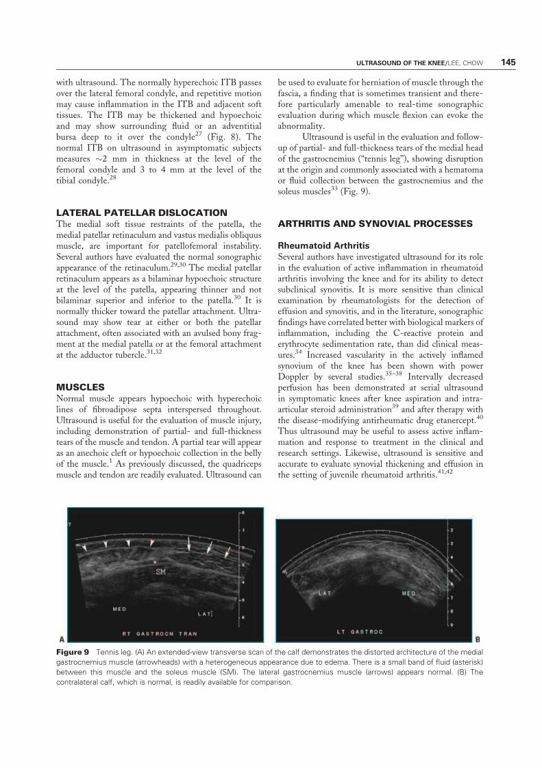
Ultrasound is useful in the evaluation and follow-up of partial- and full-thickness tears of the medial headof the gastrocnemius (‘‘tennis leg’’), showing disruptionat the origin and commonly associated with a hematomaor fluid collection between the gastrocnemius and thesoleus muscles33 (Fig. 9).
ARTHRITIS AND SYNOVIAL PROCESSES
Rheumatoid Arthritis
Several authors have investigated ultrasound for its rolein the evaluation of active inflammation in rheumatoidarthritis involving the knee and for its ability to detectsubclinical synovitis. It is more sensitive than clinicalexamination by rheumatologists for the detection ofeffusion and synovitis, and in the literature, sonographicfindings have correlated better with biological markers ofinflammation, including the C-reactive protein anderythrocyte sedimentation rate, than did clinical meas-ures.34 Increased vascularity in the actively inflamedsynovium of the knee has been shown with powerDoppler by several studies.35–38 Intervally decreasedperfusion has been demonstrated at serial ultrasoundin symptomatic knees after knee aspiration and intra-articular steroid administration39 and after therapy withthe disease-modifying antirheumatic drug etanercept.40
Thus ultrasound may be useful to assess active inflam-mation and response to treatment in the clinical andresearch settings. Likewise, ultrasound is sensitive andaccurate to evaluate synovial thickening and effusion inthe setting of juvenile rheumatoid arthritis.41,42
Figure 9 Tennis leg. (A) An extended-view transverse scan of the calf demonstrates the distorted architecture of the medial
gastrocnemius muscle (arrowheads) with a heterogeneous appearance due to edema. There is a small band of fluid (asterisk)
between this muscle and the soleus muscle (SM). The lateral gastrocnemius muscle (arrows) appears normal. (B) The
contralateral calf, which is normal, is readily available for comparison.
ULTRASOUND OF THE KNEE/LEE, CHOW 145

Osteoarthritis
The specific etiology of pain in the setting of osteo-arthritis has not been elucidated, and the severity ofradiographic findings does not always correlate withsymptoms. As such, ultrasound has been evaluated forits utility in painful episodes in osteoarthritis, with theauthors of one study reporting a higher risk of a painfulepisode when there is suprapatellar effusion and Bakercyst.43 However, ultrasound has a limited role in theevaluation of mechanical arthritis, with findings includ-ing meniscal pathology, osteophytes, and synovitis alsoevident.
Calcium Pyrophosphate Dihydrate Deposition
Calcium pyrophosphate dihydrate deposition (CPPD)can affect the hyaline articular cartilage surfaces of theknee or the meniscal fibrocartilage. Within the normallyuniformly hypoechoic band of articular cartilage alongthe articular surface, chondrocalcinosis appears as intra-substance linear hyperechoic foci or a line.44,45 Althoughthis finding is generally readily detected radiographically,the appearance at sonography should suggest the diag-nosis.
Pigmented Villonodular Synovitis and Synovial
Osteochondromatosis
Pigmented villonodular synovitis (PVNS) appears as afocal or diffuse mass or thickening within the joint or inthe bursae, sometimes extending beyond the joint cap-sule. Because there are no specific sonographic appear-ances, MRI may provide a more definitive diagnosis byshowing the characteristic hypointensity reflectinghemosiderin.46 Synovial chondromatosis, due to synovialmetaplasia, likewise has a nonspecific appearance. How-ever, if the cartilage nodules have undergone ossificationas frequently occurs, myriad ossific bodies with echo-genic surfaces and posterior acoustic shadowing will beevident.
EVALUATION AFTER TOTAL KNEE
ARTHROPLASTY
Sonography is a useful adjunctive examination to radio-graphs in evaluating the knee after total arthroplasty, inwhich case MRI may be limited by artifact. The evalua-tion of anterior knee pain after total knee arthroplastymay be due to abnormalities of the extensor mechanism,whose pathology is the same as in the virgin knee. Ararer form of painful extensor mechanism dysfunctionafter arthroplasty is termed ‘‘patellar clunk syndrome,’’ inwhich a fibrous nodule develops along the deep aspectof the distal quadriceps tendon. The nodule becomesentrapped in the anterior intercondylar notch, snappingfree with extension.47 Sonography shows an echogenic
area attached to the quadriceps tendon with vascularityby Doppler.48
Arthrofibrosis may cause chronic pain and stiff-ness after arthroplasty related to synovial hypertrophyand capsular thickening. In a study of patients withclinically established arthrofibrosis, ultrasound demon-strated a significantly greater thickness of the synovialmembrane and neovascularity compared with asympto-matic controls.49 Using a cutoff of 3.0 mm, sonographyhad a sensitivity of 84% and specificity of 82% forarthrofibrosis.49
The polyethylene liner of the arthroplasty is sub-ject to abrasion and thinning and can be a significantsource of debris predisposing to osteolysis. Although theliner is generally evaluated as a radiolucent band atradiography, ultrasound can demonstrate the liner as astrong linear echogenic interface with posterior acousticshadowing and is accurate for measuring its thickness.50
CONCLUSIONS
Ultrasound is a useful examination for evaluating theextensor mechanism, joint effusion and periarticularfluid collections, the collateral ligaments, muscle andtendon pathology, and synovitis. It may also be helpfulfor evaluation after total knee arthroplasty becauseMRI is limited by metallic artifact. Although sonog-raphy may demonstrate tears of the menisci and cru-ciate ligaments, MRI is generally preferred forevaluating internal derangement.
REFERENCES
1. Friedman L, Finlay K, Jurriaans E. Ultrasound of the knee.Skeletal Radiol 2001;30(7):361–377
2. Bianchi S, Zwass A, Abdelwahab IF, Banderali A.Diagnosis of tears of the quadriceps tendon of the knee:value of sonography. AJR Am J Roentgenol 1994;162(5):1137–1140
3. Carr JC, Hanly S, Griffin J, Gibney R. Sonography of thepatellar tendon and adjacent structures in pediatric and adultpatients. AJR Am J Roentgenol 2001;176(6):1535–1539
4. Khan KM, Bonar F, Desmond PM, et al. Patellar tendinosis(jumper’s knee): findings at histopathologic examination, US,and MR imaging. Victorian Institute of Sport Tendon StudyGroup. Radiology 1996;200(3):821–827
5. De Flaviis L, Scaglione P, Nessi R, Albisetti W. Ultrasoundin degenerative cystic meniscal disease of the knee. SkeletalRadiol 1990;19(6):441–445
6. Fessell DP, Jacobson JA, Craig J, et al. Using sonography toreveal and aspirate joint effusions. AJR Am J Roentgenol2000;174(5):1353–1362
7. Delaunoy I, Feipel V, Appelboom T, Hauzeur JP. Sonog-raphy detection threshold for knee effusion. Clin Rheumatol2003;22(6):391–392
8. Bonnefoy O, Diris B, Moinard M, Aunoble S, Diard F,Hauger O. Acute knee trauma: role of ultrasound. Eur Radiol2006;16(11):2542–2548
146 SEMINARS IN MUSCULOSKELETAL RADIOLOGY/VOLUME 11, NUMBER 2 2007

9. Costa DN, Cavalcanti CF, Sernik RA. Sonographic and CTfindings in lipohemarthrosis. AJR Am J Roentgenol 2007;188(4):W389
10. Ward EE, Jacobson JA, Fessell DP, Hayes CW, vanHolsbeeck M. Sonographic detection of Baker’s cysts:comparison with MR imaging. AJR Am J Roentgenol 2001;176(2):373–380
11. Casser HR, Sohn C, Kiekenbeck A. Current evaluation ofsonography of the meniscus. Results of a comparative studyof sonographic and arthroscopic findings. Arch OrthopTrauma Surg 1990;109(3):150–154
12. Friedl W, Glaser F. Dynamic sonography in the diagnosis ofligament and meniscal injuries of the knee. Arch OrthopTrauma Surg 1991;110(3):132–138
13. Gerngross H, Sohn C. Ultrasound scanning for the diagnosisof meniscal lesions of the knee joint. Arthroscopy 1992;8(1):105–110
14. Azzoni R, Cabitza P. Is there a role for sonography in thediagnosis of tears of the knee menisci? J Clin Ultrasound2002;30(8):472–476
15. Selby B, Richardson ML, Montana MA, Teitz CC, LarsonRV, Mack LA. High resolution sonography of the menisci ofthe knee. Invest Radiol 1986;21(4):332–335
16. Selby B, Richardson ML, Nelson BD, Graney DO, MackLA. Sonography in the detection of meniscal injuries of theknee: evaluation in cadavers. AJR Am J Roentgenol 1987;149(3):549–553
17. van Holsbeeck MIJ, Introcaso JH. Musculoskeletal Ultra-sound. St. Louis: Mosby-Year Book; 2001
18. Rutten MJ, Collins JM, van Kampen A, Jager GJ. Meniscalcysts: detection with high-resolution sonography. AJR Am JRoentgenol 1998;171(2):491–496
19. De Maeseneer M, Lenchik L, Starok M, Pedowitz R,Trudell D, Resnick D. Normal and abnormal medialmeniscocapsular structures: MR imaging and sonography incadavers. AJR Am J Roentgenol 1998;171(4):969–976
20. Court-Payen M. Sonography of the knee: intra-articularpathology. J Clin Ultrasound 2004;32(9):481–490
21. Seymour R, Lloyd DC. Sonographic appearances of meniscalcysts. J Clin Ultrasound 1998;26(1):15–20
22. Ptasznik R, Feller J, Bartlett J, Fitt G, Mitchell A,Hennessy O. The value of sonography in the diagnosis oftraumatic rupture of the anterior cruciate ligament of theknee. AJR Am J Roentgenol 1995;164(6):1461–1463
23. Skovgaard Larsen LP, Rasmussen OS. Diagnosis of acuterupture of the anterior cruciate ligament of the knee bysonography. Eur J Ultrasound 2000;12(2):163–167
24. Suzuki S, Kasahara K, Futami T, Iwasaki R, Ueo T,Yamamuro T. Ultrasound diagnosis of pathology of theanterior and posterior cruciate ligaments of the knee joint.Arch Orthop Trauma Surg 1991;110(4):200–203
25. Cho KH, Lee DC, Chhem RK, et al. Normal and acutelytorn posterior cruciate ligament of the knee at USevaluation: preliminary experience. Radiology 2001;219(2):375–380
26. Miller TT. Sonography of injury of the posterior cruciateligament of the knee. Skeletal Radiol 2002;31(3):149–154
27. Bonaldi VM, Chhem RK, Drolet R, Garcia P, Gallix B,Sarazin L. Iliotibial band friction syndrome: sonographicfindings. J Ultrasound Med 1998;17(4):257–260
28. Goh LA, Chhem RK, Wang SC, Chee T. Iliotibial bandthickness: sonographic measurements in asymptomatic vol-unteers. J Clin Ultrasound 2003;31(5):239–244
29. Phornphutkul C, Sekiya JK, Wojtys EM, Jacobson JA.Sonographic imaging of the patellofemoral medial jointstabilizing structures: findings in human cadavers. Orthope-dics 2007;30(6):472–478
30. Starok M, Lenchik L, Trudell D, Resnick D. Normal patellarretinaculum: MR and sonographic imaging with cadavericcorrelation. AJR Am J Roentgenol 1997;168(6):1493–1499
31. O’Reilly MA, O’Reilly PM, Bell J. Sonographic appearancesof medial retinacular complex injury in transient patellardislocation. Clin Radiol 2003;58(8):636–641
32. Trikha SP, Acton D, O’Reilly M, Curtis MJ, Bell J. Acutelateral dislocation of the patella: correlation of ultrasoundscanning with operative findings. Injury 2003;34(8):568–571
33. Kwak HS, Han YM, Lee SY, Kim KN, Chung GH.Diagnosis and follow-up US evaluation of ruptures of themedial head of the gastrocnemius (‘‘tennis leg’’). Korean JRadiol 2006;7(3):193–198
34. Naredo E, Moller I, Moragues C, et al. Interobserverreliability in musculoskeletal ultrasonography: results from a‘‘Teach the Teachers’’ rheumatologist course. Ann RheumDis 2006;65(1):14–19
35. Carotti M, Salaffi F, Manganelli P, Salera D, Simonetti B,Grassi W. Power Doppler sonography in the assessment ofsynovial tissue of the knee joint in rheumatoid arthritis: apreliminary experience. Ann Rheum Dis 2002;61(10):877–882
36. Kasukawa R, Yamadera Y, Takahashi A, Takeda I, Kanno T.Power Doppler sonography for detection of intraarticularvascularization in knee joints of patients with rheumatoidarthritis. Mod Rheumatol 2004;14(2):149–153
37. Qvistgaard E, Kristoffersen H, Terslev L, Danneskiold-Samsoe B, Torp-Pedersen S, Bliddal H. Guidance byultrasound of intra-articular injections in the knee and hipjoints. Osteoarthritis Cartilage 2001;9(6):512–517
38. Walther M, Harms H, Krenn V, Radke S, Faehndrich TP,Gohlke F. Correlation of power Doppler sonography withvascularity of the synovial tissue of the knee joint in patientswith osteoarthritis and rheumatoid arthritis. Arthritis Rheum2001;44(2):331–338
39. Newman JS, Laing TJ, McCarthy CJ, Adler RS. PowerDoppler sonography of synovitis: assessment of therapeuticresponse—preliminary observations. Radiology 1996;198(2):582–584
40. Fiocco U, Cozzi L, Rubaltelli L, et al. Long-termsonographic follow-up of rheumatoid and psoriatic pro-liferative knee joint synovitis. Br J Rheumatol 1996;35(2):155–163
41. Cellerini M, Salti S, Trapani S, D’Elia G, Falcini F, Villari N.Correlation between clinical and ultrasound assessment ofthe knee in children with mono-articular or pauci-articularjuvenile rheumatoid arthritis. Pediatr Radiol 1999;29(2):117–123
42. Sureda D, Quiroga S, Arnal C, Boronat M, Andreu J, CasasL. Juvenile rheumatoid arthritis of the knee: evaluation withUS. Radiology 1994;190(2):403–406
43. de Miguel Mendieta E, Cobo Ibanez T, Uson Jaeger J,Bonilla Hernan G, Martin Mola E. Clinical and ultrasono-graphic findings related to knee pain in osteoarthritis.Osteoarthritis Cartilage 2006;14(6):540–544
44. Coari G, Iagnocco A, Zoppini A. Chondrocalcinosis:sonographic study of the knee. Clin Rheumatol 1995;14(5):511–514
ULTRASOUND OF THE KNEE/LEE, CHOW 147

45. Sofka CM, Adler RS, Cordasco FA. Ultrasound diagnosis ofchondrocalcinosis in the knee. Skeletal Radiol 2002;31(1):43–45
46. Wakefield RJ, Brown A, O’Connor P, et al. Rheumatologicalultrasound. Rheumatology (Oxford) 2003;42(8):1001
47. Miller TT. Sonography of joint replacements. SeminMusculoskelet Radiol 2006;10(1):79–85
48. Okamoto T, Futani H, Atsui K, Fukunishi S, Koezuka A,Maruo S. Sonographic appearance of fibrous nodules in
patellar clunk syndrome: a case report. J Orthop Sci 2002;7(5):590–593
49. Boldt JG, Munzinger UK, Zanetti M, Hodler J. Arthrofib-rosis associated with total knee arthroplasty: gray-scale andpower Doppler sonographic findings. AJR Am J Roentgenol2004;182(2):337–340
50. Sofka CM, Adler RS, Laskin R. Sonography of polyethyleneliners used in total knee arthroplasty. AJR Am J Roentgenol2003;180(5):1437–1441
148 SEMINARS IN MUSCULOSKELETAL RADIOLOGY/VOLUME 11, NUMBER 2 2007