ultrasound of the acute abdomen - bmus.org€¦ · ultrasound of the acute abdomen – a survival...
TRANSCRIPT

Ultrasound Of The Acute Abdomen ndash a survival guide
Alison McGuinness
Consultant sonographer
Mid Yorks Hospitals NHS Trust
Definition
bull Rapid onset of severe abdominal pain usually requiring admission to hospital
Speed of diagnosis
bull Stability of patient site and severity of pain
bull Trauma non-traumatic
bull Large proportion of patients will not have a diagnosis for their pain when they are discharged
bull Ultrasound v CT v laparotomy
Patient Care
bull Avoid delay bull Assess patient condition take a good history and
above all LISTEN to what is not being said bull Use all information ndash biochemical and previous
imaging bull Reassurance bull Ensure report delivered efficiently to appropriate
point of referral for early patient management
History taking
bull Demographic details occupation recent travel history of recent abdominal trauma
bull Pain ndash Onset (including whether new pain or previously
experienced) ndash Site (ask the patient to point) localised or diffuse ndash Nature (constantintermittentcolicky) ndash Radiation ndash Severity ndash Relievingaggravating factors (eg if worsened by
movementcoughing suspect active peritonitis pancreatitis is relieved by sitting forward)
Associated symptoms
bull Vomiting and the nature of vomitus (undigested food or bile suggests upper GI pathology or obstruction faeculent vomiting suggests lower GI obstruction)
bull Haematemesis or melaena bull Stoolurine colour bull New lumps in the abdominal regiongroins bull Eating and drinking - including when the patients last meal occurred bull Bowels - including presence of diarrhoea constipation and ability to pass
flatus bull Fainting dizziness or palpitations bull Feverrigors bull Rash or itching bull Urinary symptoms bull Recent weight loss
Trauma to the abdomen
Who provides the service
Core and out of hours
bull Paramedic
bull Emergency medicine physician
bull Sonographer
bull Radiologist
bull Trauma radiographer
Principles of Ultrasound in Emergency Medicine
bull Limited examination
bull Focused
bull Specific question
bull lsquoRule inrsquo
bull Record images
bull cf radiology departmental examination
Free fluid Fast(Focused Assessment with Sonography for Trauma)
bull Employed to answer a single rule-in question rather than specifically evaluate a system
bull Takes less than 5 minutes
bull Can be performed during resuscitation
bull Has replaced diagnostic peritoneal lavage (DPL) as the primary assessment of blunt abdominal trauma
FAST
bull Employs a limited 4-6 view of the abdomen to rule in free fluid ndash Hepatorenal space
(Morrisonrsquos pouch)
ndash pericardial space
ndash perisplenic space
ndash suprapubic region
ndash bilateral paracolic views
bull 250ml of fluid
Liver Trauma
Renal trauma
Splenic collection
Non-traumatic pain
Ultrasound Of The Acute Abdomen
Answer SIX basic questions
bull Is there blood (free fluid) in the peritoneal cavity
bull Is an abdominal aortic aneurysm (AAA) present
bull Is there evidence of obstructive uropathy
bull Is there evidence of biliary disease (cholelithiasis)
bull Is there evidence of a living intrauterine pregnancy
bull Is there a pericardial effusion raquo Brenchley J Sloan J 2000
Aetiology
bull 34 no cause for pain identified bull 28 appendicitis bull 10 acute cholecystitis bull 4 small bowel obstruction bull 4 acute gynae bull 3 acute pancreatitis bull 3 ureteric colic bull 3 perforated ulcer bull 11 others ndash vascular origin aortic aneurysm
cardiac problems
AAA Abdominal Pain And Hypotension
bull Aortic dimensions can be easily measured and although leakage cannot be assessed the presence of a dilated aorta in patients with circulatory instability significantly speeds up diagnosis of a leaking abdominal aortic aneurysm and vascular referral
raquo BALLARD et al 1998
Kidneys
Renal colic
bull Ultrasound ndash hydronephrosis pyelonephritis ndash management pathway
bull CTKUB ndash stones (exception ndash females of child bearing age)
Hydronephrosis - pregnancy
Is it normal for gestation
Hydronephrosis ndash chronic retention
Cortical
thickness
Renal calculi
bull Is it in the collecting system
bull Is it causing obstruction
bull Is it solitary
Renal and ureteric stones Hydronephrosis
Hydronephrosis ndashpelvic mass
Rhabdomyosarcoma
Pyelonephritis
bull Often unilateral
bull Discrepancy in size
bull Focal echogenic area
bull Hypervascularity
Increased cortico-medullary
differentiation
Pancreas
Acute Pancreatitis
bull Oedematous inflammation of the pancreas with severe upper abdominal pain
bull Diagnosis made on clinical grounds with a serum amylase gt1000 iul
bull Predisposing factors include alcoholism gallstones and mild blunt abdominal trauma
Acute pancreatitis
Chronic Pancreatitis
bull Chronic changes due to recurrent bouts of inflammation with resultant fibrosis stone formation and permanent damage
bull Patient presents with a similar pain to acute pancreatitis only more persistent and not as severe
bull Frequently occurs in alcoholics following multiple episodes of acute pancreatitis
Chronic pancreatitis
Traumatic Pancreatitis
bull Severe trauma may disrupt the duct spilling pancreatic juices into the surrounding tissues resulting in the development of large pseudocysts
bull Neck of pancreas is most vulnerable the duct rupturing when compressed between the aorta and the spine
bull Distal pancreatectomy is often necessary
Traumatic dissection pancreatic body
Biliary Tree
Abnormal LFTs
bull Serum albumin and bilirubin levels used to provide a true measure of hepatic function
bull Alanine aminotransferase (ALT) or asparate transaminase (AST) sensitive markers of hepatocyte injury ndash ndash raised in acute hepatitis but also in chronic liver disease ndash Very high levels of ALT (gt 1000iUl) can due to hepatic
ischaemia or cholangitis due to biliary stones
bull Gammo-glutamyl transpeptidase (GGT) is a marker for biliary tract disease
bull GGT along with ALP used as a specific marker of alcohol misuse
bull History and exam should include possible non hepatic causes of raised LFTs eg right sided heart failure endocrine disorders diabetes and thyroid dysfunction
Abnormal LFTs
Liver Enzymes Normal Moderate Severe
AST under 40 IUl 40-200 Over 200
ALP under 40 IUl 40-200 Over 200
GGT under 60 IUl 60-200 Over 200
Alk Phos under 112 IUl 112-300 Over 300
LFTs Normal Moderate Severe
Bilirubin under 12 mgdl 12- 25 Over 25
Albumin 35-45 gdl 30 -35 Under 30
Prothrombin time Under 14 secs 14-17 Over 17
Chronic cholelithiasis
Acute cholecystitis
Acute cholecystitis
CBD dilatation
Biliary obstruction ndash IHD dilatation
Biliary obstruction - stones
Biliary obstruction ndash intra duct mass
Cholangiocarcinoma
Liver
Liver Mets- previous Ho Ca
Acute left loin pain ndashno prev history
Acute RUQ pain and hepatomegaly
Portal vein patency
Check for thrombus in PV varicosities and
recannulised umbilical vein
recannulised umbilical vein
Collections
Collections
RUQ pain pyrexial recent travel to Asia
Ho Cholecystectomy 10 years ago RUQ
pain
Biloma
Rectus sheath haematoma
Psoas muscle abscess
Psoas muscle abscess may present with fever flank pain or abdominal pain
Primary abscess occurs probably as a result of spread of an infectious process from an occult source in the body and can occur in patients with bull diabetes mellitus
bull intravenous drug abuse
bull AIDS
bull renal failure
bull immuno suppression
Secondary spread of infection from gastrointestinal disease (eg appendicitis Crohnrsquos disease diverticulitis)
bull Renal disease is the second most common source
The rest
Acute Appendicitis
bull Non-compressible sausage shaped structure demonstrating no peristalsis
bull 75 lie behind the caecum and colon making ultrasound access difficult
bull In cross section it gives a target appearance gt6mm in diameter usually between 7 and 10 mm The wall thickness can be 3mm or more
Right Sided Colonic Diverticulitis
bull Self limiting benign condition
bull All ages
bull Congenital
bull Not so rare
bull Clinically appendicitis
bull In 40 unnecessary hemicolectomy performed
Diverticulitis
Crohnrsquos
Lymphoma
General Medical Ultrasound Leeds
University
Conclusion
bull Abdominal ultrasound is first line investigation in the assessment of suspected acute biliary colic renal colic acute colonic diverticulitis appendicitis and abdominal masses Requested for acute pancreatitis with limited success
bull Patients with localised abdominal pain and tenderness are more likely to have a positive diagnosis on ultrasound
bull Raised WCC or an abnormal LFT in patients with acute
abdominal pain is predictive of a higher yield of positive findings on ultrasound
bull Only one third of children with appendicitis will present with classic symptoms Pain initially in midline and extending to RIF with guarding favourable for appendicitis NB High or ectopic appendix may be seen
Conclusion
bull Use all the clinical history to make a diagnosis
bull Avoid over diagnosis gallstones and dilated CBD may be incidental findings
bull Cholecystitis has thickened GB wall and intermittent pain whereas pancreatitis continual pain
bull Check biochemical results to aid diagnosis
bull Use colour doppler to check patency and hypervascularity
bull Check all previous imaging before suggesting follow up
References
bull Ballard RB 1998 THE SURGEONS USE OF ULTRASOUND IN THE ACUTE SETTING Surgical Clinics of North America 78 (2) 337ndash364 ltimg alt=Cover image src=httparsels-cdncomcontentimageS00396109gif class=toprightlogo gt
bull Brenchley J1 Sloan JP Thompson PK 2000 Echoes of things to come Ultrasound in UK emergency medicine practice 17(3)170-5
bull Brenchley J Walker A Sloan J et al 2006 Evaluation of focused assessment J Accid Emerg Med with sonography in trauma by emergency physicians Emerg Med J 23446ndash8
bull El Sayed MJ and Zaghrini E 2013 Prehospital Emergency Ultrasound A Review of Current Clinical Applications Challenges and Future Implications Emergency Medicine International 2013 ID 531674 httpdxdoiorg1011552013531674
bull Puylaert J 2007 Acute Abdomen - Role of Ultrasound Radiology Assistant httpwwwradiologyassistantnlenp4613dde72e42cacute-abdomen-role-of-ultrasoundhtml
bull Raman S 2004 Are we overusing ultrasound in non-traumatic acute abdominal pain Postgrad Med J 200480177-179 doi101136pgmj20030138
bull Snaith B M Hardy M and Walker A 2011 ldquoEmergency ultrasound in the prehospital setting the impact of environment on examination outcomesrdquo Emergency Medicine Journal 28 (12) 1063ndash1065
bull White EK et al 2014 Seeing past the appendix the role of ultrasound in right iliac fossa pain Ultrasound 22 (2) 104-112
Thank You
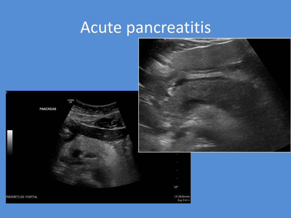

Abnormal LFTs
Liver Enzymes Normal Moderate Severe
AST under 40 IUl 40-200 Over 200
ALP under 40 IUl 40-200 Over 200
GGT under 60 IUl 60-200 Over 200
Alk Phos under 112 IUl 112-300 Over 300
LFTs Normal Moderate Severe
Bilirubin under 12 mgdl 12- 25 Over 25
Albumin 35-45 gdl 30 -35 Under 30
Prothrombin time Under 14 secs 14-17 Over 17
Chronic cholelithiasis
Acute cholecystitis
Acute cholecystitis
CBD dilatation
Biliary obstruction ndash IHD dilatation
Biliary obstruction - stones
Biliary obstruction ndash intra duct mass
Cholangiocarcinoma
Liver
Liver Mets- previous Ho Ca
Acute left loin pain ndashno prev history
Acute RUQ pain and hepatomegaly
Portal vein patency
Check for thrombus in PV varicosities and
recannulised umbilical vein
recannulised umbilical vein
Collections
Collections
RUQ pain pyrexial recent travel to Asia
Ho Cholecystectomy 10 years ago RUQ
pain
Biloma
Rectus sheath haematoma
Psoas muscle abscess
Psoas muscle abscess may present with fever flank pain or abdominal pain
Primary abscess occurs probably as a result of spread of an infectious process from an occult source in the body and can occur in patients with bull diabetes mellitus
bull intravenous drug abuse
bull AIDS
bull renal failure
bull immuno suppression
Secondary spread of infection from gastrointestinal disease (eg appendicitis Crohnrsquos disease diverticulitis)
bull Renal disease is the second most common source
The rest
Acute Appendicitis
bull Non-compressible sausage shaped structure demonstrating no peristalsis
bull 75 lie behind the caecum and colon making ultrasound access difficult
bull In cross section it gives a target appearance gt6mm in diameter usually between 7 and 10 mm The wall thickness can be 3mm or more
Right Sided Colonic Diverticulitis
bull Self limiting benign condition
bull All ages
bull Congenital
bull Not so rare
bull Clinically appendicitis
bull In 40 unnecessary hemicolectomy performed
Diverticulitis
Crohnrsquos
Lymphoma
General Medical Ultrasound Leeds
University
Conclusion
bull Abdominal ultrasound is first line investigation in the assessment of suspected acute biliary colic renal colic acute colonic diverticulitis appendicitis and abdominal masses Requested for acute pancreatitis with limited success
bull Patients with localised abdominal pain and tenderness are more likely to have a positive diagnosis on ultrasound
bull Raised WCC or an abnormal LFT in patients with acute
abdominal pain is predictive of a higher yield of positive findings on ultrasound
bull Only one third of children with appendicitis will present with classic symptoms Pain initially in midline and extending to RIF with guarding favourable for appendicitis NB High or ectopic appendix may be seen
Conclusion
bull Use all the clinical history to make a diagnosis
bull Avoid over diagnosis gallstones and dilated CBD may be incidental findings
bull Cholecystitis has thickened GB wall and intermittent pain whereas pancreatitis continual pain
bull Check biochemical results to aid diagnosis
bull Use colour doppler to check patency and hypervascularity
bull Check all previous imaging before suggesting follow up
References
bull Ballard RB 1998 THE SURGEONS USE OF ULTRASOUND IN THE ACUTE SETTING Surgical Clinics of North America 78 (2) 337ndash364 ltimg alt=Cover image src=httparsels-cdncomcontentimageS00396109gif class=toprightlogo gt
bull Brenchley J1 Sloan JP Thompson PK 2000 Echoes of things to come Ultrasound in UK emergency medicine practice 17(3)170-5
bull Brenchley J Walker A Sloan J et al 2006 Evaluation of focused assessment J Accid Emerg Med with sonography in trauma by emergency physicians Emerg Med J 23446ndash8
bull El Sayed MJ and Zaghrini E 2013 Prehospital Emergency Ultrasound A Review of Current Clinical Applications Challenges and Future Implications Emergency Medicine International 2013 ID 531674 httpdxdoiorg1011552013531674
bull Puylaert J 2007 Acute Abdomen - Role of Ultrasound Radiology Assistant httpwwwradiologyassistantnlenp4613dde72e42cacute-abdomen-role-of-ultrasoundhtml
bull Raman S 2004 Are we overusing ultrasound in non-traumatic acute abdominal pain Postgrad Med J 200480177-179 doi101136pgmj20030138
bull Snaith B M Hardy M and Walker A 2011 ldquoEmergency ultrasound in the prehospital setting the impact of environment on examination outcomesrdquo Emergency Medicine Journal 28 (12) 1063ndash1065
bull White EK et al 2014 Seeing past the appendix the role of ultrasound in right iliac fossa pain Ultrasound 22 (2) 104-112
Thank You

Acute cholecystitis
Acute cholecystitis
CBD dilatation
Biliary obstruction ndash IHD dilatation
Biliary obstruction - stones
Biliary obstruction ndash intra duct mass
Cholangiocarcinoma
Liver
Liver Mets- previous Ho Ca
Acute left loin pain ndashno prev history
Acute RUQ pain and hepatomegaly
Portal vein patency
Check for thrombus in PV varicosities and
recannulised umbilical vein
recannulised umbilical vein
Collections
Collections
RUQ pain pyrexial recent travel to Asia
Ho Cholecystectomy 10 years ago RUQ
pain
Biloma
Rectus sheath haematoma
Psoas muscle abscess
Psoas muscle abscess may present with fever flank pain or abdominal pain
Primary abscess occurs probably as a result of spread of an infectious process from an occult source in the body and can occur in patients with bull diabetes mellitus
bull intravenous drug abuse
bull AIDS
bull renal failure
bull immuno suppression
Secondary spread of infection from gastrointestinal disease (eg appendicitis Crohnrsquos disease diverticulitis)
bull Renal disease is the second most common source
The rest
Acute Appendicitis
bull Non-compressible sausage shaped structure demonstrating no peristalsis
bull 75 lie behind the caecum and colon making ultrasound access difficult
bull In cross section it gives a target appearance gt6mm in diameter usually between 7 and 10 mm The wall thickness can be 3mm or more
Right Sided Colonic Diverticulitis
bull Self limiting benign condition
bull All ages
bull Congenital
bull Not so rare
bull Clinically appendicitis
bull In 40 unnecessary hemicolectomy performed
Diverticulitis
Crohnrsquos
Lymphoma
General Medical Ultrasound Leeds
University
Conclusion
bull Abdominal ultrasound is first line investigation in the assessment of suspected acute biliary colic renal colic acute colonic diverticulitis appendicitis and abdominal masses Requested for acute pancreatitis with limited success
bull Patients with localised abdominal pain and tenderness are more likely to have a positive diagnosis on ultrasound
bull Raised WCC or an abnormal LFT in patients with acute
abdominal pain is predictive of a higher yield of positive findings on ultrasound
bull Only one third of children with appendicitis will present with classic symptoms Pain initially in midline and extending to RIF with guarding favourable for appendicitis NB High or ectopic appendix may be seen
Conclusion
bull Use all the clinical history to make a diagnosis
bull Avoid over diagnosis gallstones and dilated CBD may be incidental findings
bull Cholecystitis has thickened GB wall and intermittent pain whereas pancreatitis continual pain
bull Check biochemical results to aid diagnosis
bull Use colour doppler to check patency and hypervascularity
bull Check all previous imaging before suggesting follow up
References
bull Ballard RB 1998 THE SURGEONS USE OF ULTRASOUND IN THE ACUTE SETTING Surgical Clinics of North America 78 (2) 337ndash364 ltimg alt=Cover image src=httparsels-cdncomcontentimageS00396109gif class=toprightlogo gt
bull Brenchley J1 Sloan JP Thompson PK 2000 Echoes of things to come Ultrasound in UK emergency medicine practice 17(3)170-5
bull Brenchley J Walker A Sloan J et al 2006 Evaluation of focused assessment J Accid Emerg Med with sonography in trauma by emergency physicians Emerg Med J 23446ndash8
bull El Sayed MJ and Zaghrini E 2013 Prehospital Emergency Ultrasound A Review of Current Clinical Applications Challenges and Future Implications Emergency Medicine International 2013 ID 531674 httpdxdoiorg1011552013531674
bull Puylaert J 2007 Acute Abdomen - Role of Ultrasound Radiology Assistant httpwwwradiologyassistantnlenp4613dde72e42cacute-abdomen-role-of-ultrasoundhtml
bull Raman S 2004 Are we overusing ultrasound in non-traumatic acute abdominal pain Postgrad Med J 200480177-179 doi101136pgmj20030138
bull Snaith B M Hardy M and Walker A 2011 ldquoEmergency ultrasound in the prehospital setting the impact of environment on examination outcomesrdquo Emergency Medicine Journal 28 (12) 1063ndash1065
bull White EK et al 2014 Seeing past the appendix the role of ultrasound in right iliac fossa pain Ultrasound 22 (2) 104-112
Thank You

Acute cholecystitis
CBD dilatation
Biliary obstruction ndash IHD dilatation
Biliary obstruction - stones
Biliary obstruction ndash intra duct mass
Cholangiocarcinoma
Liver
Liver Mets- previous Ho Ca
Acute left loin pain ndashno prev history
Acute RUQ pain and hepatomegaly
Portal vein patency
Check for thrombus in PV varicosities and
recannulised umbilical vein
recannulised umbilical vein
Collections
Collections
RUQ pain pyrexial recent travel to Asia
Ho Cholecystectomy 10 years ago RUQ
pain
Biloma
Rectus sheath haematoma
Psoas muscle abscess
Psoas muscle abscess may present with fever flank pain or abdominal pain
Primary abscess occurs probably as a result of spread of an infectious process from an occult source in the body and can occur in patients with bull diabetes mellitus
bull intravenous drug abuse
bull AIDS
bull renal failure
bull immuno suppression
Secondary spread of infection from gastrointestinal disease (eg appendicitis Crohnrsquos disease diverticulitis)
bull Renal disease is the second most common source
The rest
Acute Appendicitis
bull Non-compressible sausage shaped structure demonstrating no peristalsis
bull 75 lie behind the caecum and colon making ultrasound access difficult
bull In cross section it gives a target appearance gt6mm in diameter usually between 7 and 10 mm The wall thickness can be 3mm or more
Right Sided Colonic Diverticulitis
bull Self limiting benign condition
bull All ages
bull Congenital
bull Not so rare
bull Clinically appendicitis
bull In 40 unnecessary hemicolectomy performed
Diverticulitis
Crohnrsquos
Lymphoma
General Medical Ultrasound Leeds
University
Conclusion
bull Abdominal ultrasound is first line investigation in the assessment of suspected acute biliary colic renal colic acute colonic diverticulitis appendicitis and abdominal masses Requested for acute pancreatitis with limited success
bull Patients with localised abdominal pain and tenderness are more likely to have a positive diagnosis on ultrasound
bull Raised WCC or an abnormal LFT in patients with acute
abdominal pain is predictive of a higher yield of positive findings on ultrasound
bull Only one third of children with appendicitis will present with classic symptoms Pain initially in midline and extending to RIF with guarding favourable for appendicitis NB High or ectopic appendix may be seen
Conclusion
bull Use all the clinical history to make a diagnosis
bull Avoid over diagnosis gallstones and dilated CBD may be incidental findings
bull Cholecystitis has thickened GB wall and intermittent pain whereas pancreatitis continual pain
bull Check biochemical results to aid diagnosis
bull Use colour doppler to check patency and hypervascularity
bull Check all previous imaging before suggesting follow up
References
bull Ballard RB 1998 THE SURGEONS USE OF ULTRASOUND IN THE ACUTE SETTING Surgical Clinics of North America 78 (2) 337ndash364 ltimg alt=Cover image src=httparsels-cdncomcontentimageS00396109gif class=toprightlogo gt
bull Brenchley J1 Sloan JP Thompson PK 2000 Echoes of things to come Ultrasound in UK emergency medicine practice 17(3)170-5
bull Brenchley J Walker A Sloan J et al 2006 Evaluation of focused assessment J Accid Emerg Med with sonography in trauma by emergency physicians Emerg Med J 23446ndash8
bull El Sayed MJ and Zaghrini E 2013 Prehospital Emergency Ultrasound A Review of Current Clinical Applications Challenges and Future Implications Emergency Medicine International 2013 ID 531674 httpdxdoiorg1011552013531674
bull Puylaert J 2007 Acute Abdomen - Role of Ultrasound Radiology Assistant httpwwwradiologyassistantnlenp4613dde72e42cacute-abdomen-role-of-ultrasoundhtml
bull Raman S 2004 Are we overusing ultrasound in non-traumatic acute abdominal pain Postgrad Med J 200480177-179 doi101136pgmj20030138
bull Snaith B M Hardy M and Walker A 2011 ldquoEmergency ultrasound in the prehospital setting the impact of environment on examination outcomesrdquo Emergency Medicine Journal 28 (12) 1063ndash1065
bull White EK et al 2014 Seeing past the appendix the role of ultrasound in right iliac fossa pain Ultrasound 22 (2) 104-112
Thank You

Collections
Collections
RUQ pain pyrexial recent travel to Asia
Ho Cholecystectomy 10 years ago RUQ
pain
Biloma
Rectus sheath haematoma
Psoas muscle abscess
Psoas muscle abscess may present with fever flank pain or abdominal pain
Primary abscess occurs probably as a result of spread of an infectious process from an occult source in the body and can occur in patients with bull diabetes mellitus
bull intravenous drug abuse
bull AIDS
bull renal failure
bull immuno suppression
Secondary spread of infection from gastrointestinal disease (eg appendicitis Crohnrsquos disease diverticulitis)
bull Renal disease is the second most common source
The rest
Acute Appendicitis
bull Non-compressible sausage shaped structure demonstrating no peristalsis
bull 75 lie behind the caecum and colon making ultrasound access difficult
bull In cross section it gives a target appearance gt6mm in diameter usually between 7 and 10 mm The wall thickness can be 3mm or more
Right Sided Colonic Diverticulitis
bull Self limiting benign condition
bull All ages
bull Congenital
bull Not so rare
bull Clinically appendicitis
bull In 40 unnecessary hemicolectomy performed
Diverticulitis
Crohnrsquos
Lymphoma
General Medical Ultrasound Leeds
University
Conclusion
bull Abdominal ultrasound is first line investigation in the assessment of suspected acute biliary colic renal colic acute colonic diverticulitis appendicitis and abdominal masses Requested for acute pancreatitis with limited success
bull Patients with localised abdominal pain and tenderness are more likely to have a positive diagnosis on ultrasound
bull Raised WCC or an abnormal LFT in patients with acute
abdominal pain is predictive of a higher yield of positive findings on ultrasound
bull Only one third of children with appendicitis will present with classic symptoms Pain initially in midline and extending to RIF with guarding favourable for appendicitis NB High or ectopic appendix may be seen
Conclusion
bull Use all the clinical history to make a diagnosis
bull Avoid over diagnosis gallstones and dilated CBD may be incidental findings
bull Cholecystitis has thickened GB wall and intermittent pain whereas pancreatitis continual pain
bull Check biochemical results to aid diagnosis
bull Use colour doppler to check patency and hypervascularity
bull Check all previous imaging before suggesting follow up
References
bull Ballard RB 1998 THE SURGEONS USE OF ULTRASOUND IN THE ACUTE SETTING Surgical Clinics of North America 78 (2) 337ndash364 ltimg alt=Cover image src=httparsels-cdncomcontentimageS00396109gif class=toprightlogo gt
bull Brenchley J1 Sloan JP Thompson PK 2000 Echoes of things to come Ultrasound in UK emergency medicine practice 17(3)170-5
bull Brenchley J Walker A Sloan J et al 2006 Evaluation of focused assessment J Accid Emerg Med with sonography in trauma by emergency physicians Emerg Med J 23446ndash8
bull El Sayed MJ and Zaghrini E 2013 Prehospital Emergency Ultrasound A Review of Current Clinical Applications Challenges and Future Implications Emergency Medicine International 2013 ID 531674 httpdxdoiorg1011552013531674
bull Puylaert J 2007 Acute Abdomen - Role of Ultrasound Radiology Assistant httpwwwradiologyassistantnlenp4613dde72e42cacute-abdomen-role-of-ultrasoundhtml
bull Raman S 2004 Are we overusing ultrasound in non-traumatic acute abdominal pain Postgrad Med J 200480177-179 doi101136pgmj20030138
bull Snaith B M Hardy M and Walker A 2011 ldquoEmergency ultrasound in the prehospital setting the impact of environment on examination outcomesrdquo Emergency Medicine Journal 28 (12) 1063ndash1065
bull White EK et al 2014 Seeing past the appendix the role of ultrasound in right iliac fossa pain Ultrasound 22 (2) 104-112
Thank You

Collections
RUQ pain pyrexial recent travel to Asia
Ho Cholecystectomy 10 years ago RUQ
pain
Biloma
Rectus sheath haematoma
Psoas muscle abscess
Psoas muscle abscess may present with fever flank pain or abdominal pain
Primary abscess occurs probably as a result of spread of an infectious process from an occult source in the body and can occur in patients with bull diabetes mellitus
bull intravenous drug abuse
bull AIDS
bull renal failure
bull immuno suppression
Secondary spread of infection from gastrointestinal disease (eg appendicitis Crohnrsquos disease diverticulitis)
bull Renal disease is the second most common source
The rest
Acute Appendicitis
bull Non-compressible sausage shaped structure demonstrating no peristalsis
bull 75 lie behind the caecum and colon making ultrasound access difficult
bull In cross section it gives a target appearance gt6mm in diameter usually between 7 and 10 mm The wall thickness can be 3mm or more
Right Sided Colonic Diverticulitis
bull Self limiting benign condition
bull All ages
bull Congenital
bull Not so rare
bull Clinically appendicitis
bull In 40 unnecessary hemicolectomy performed
Diverticulitis
Crohnrsquos
Lymphoma
General Medical Ultrasound Leeds
University
Conclusion
bull Abdominal ultrasound is first line investigation in the assessment of suspected acute biliary colic renal colic acute colonic diverticulitis appendicitis and abdominal masses Requested for acute pancreatitis with limited success
bull Patients with localised abdominal pain and tenderness are more likely to have a positive diagnosis on ultrasound
bull Raised WCC or an abnormal LFT in patients with acute
abdominal pain is predictive of a higher yield of positive findings on ultrasound
bull Only one third of children with appendicitis will present with classic symptoms Pain initially in midline and extending to RIF with guarding favourable for appendicitis NB High or ectopic appendix may be seen
Conclusion
bull Use all the clinical history to make a diagnosis
bull Avoid over diagnosis gallstones and dilated CBD may be incidental findings
bull Cholecystitis has thickened GB wall and intermittent pain whereas pancreatitis continual pain
bull Check biochemical results to aid diagnosis
bull Use colour doppler to check patency and hypervascularity
bull Check all previous imaging before suggesting follow up
References
bull Ballard RB 1998 THE SURGEONS USE OF ULTRASOUND IN THE ACUTE SETTING Surgical Clinics of North America 78 (2) 337ndash364 ltimg alt=Cover image src=httparsels-cdncomcontentimageS00396109gif class=toprightlogo gt
bull Brenchley J1 Sloan JP Thompson PK 2000 Echoes of things to come Ultrasound in UK emergency medicine practice 17(3)170-5
bull Brenchley J Walker A Sloan J et al 2006 Evaluation of focused assessment J Accid Emerg Med with sonography in trauma by emergency physicians Emerg Med J 23446ndash8
bull El Sayed MJ and Zaghrini E 2013 Prehospital Emergency Ultrasound A Review of Current Clinical Applications Challenges and Future Implications Emergency Medicine International 2013 ID 531674 httpdxdoiorg1011552013531674
bull Puylaert J 2007 Acute Abdomen - Role of Ultrasound Radiology Assistant httpwwwradiologyassistantnlenp4613dde72e42cacute-abdomen-role-of-ultrasoundhtml
bull Raman S 2004 Are we overusing ultrasound in non-traumatic acute abdominal pain Postgrad Med J 200480177-179 doi101136pgmj20030138
bull Snaith B M Hardy M and Walker A 2011 ldquoEmergency ultrasound in the prehospital setting the impact of environment on examination outcomesrdquo Emergency Medicine Journal 28 (12) 1063ndash1065
bull White EK et al 2014 Seeing past the appendix the role of ultrasound in right iliac fossa pain Ultrasound 22 (2) 104-112
Thank You
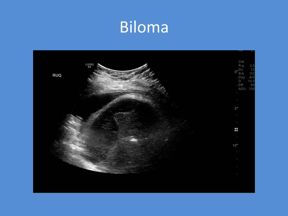
Conclusion
bull Use all the clinical history to make a diagnosis
bull Avoid over diagnosis gallstones and dilated CBD may be incidental findings
bull Cholecystitis has thickened GB wall and intermittent pain whereas pancreatitis continual pain
bull Check biochemical results to aid diagnosis
bull Use colour doppler to check patency and hypervascularity
bull Check all previous imaging before suggesting follow up
References
bull Ballard RB 1998 THE SURGEONS USE OF ULTRASOUND IN THE ACUTE SETTING Surgical Clinics of North America 78 (2) 337ndash364 ltimg alt=Cover image src=httparsels-cdncomcontentimageS00396109gif class=toprightlogo gt
bull Brenchley J1 Sloan JP Thompson PK 2000 Echoes of things to come Ultrasound in UK emergency medicine practice 17(3)170-5
bull Brenchley J Walker A Sloan J et al 2006 Evaluation of focused assessment J Accid Emerg Med with sonography in trauma by emergency physicians Emerg Med J 23446ndash8
bull El Sayed MJ and Zaghrini E 2013 Prehospital Emergency Ultrasound A Review of Current Clinical Applications Challenges and Future Implications Emergency Medicine International 2013 ID 531674 httpdxdoiorg1011552013531674
bull Puylaert J 2007 Acute Abdomen - Role of Ultrasound Radiology Assistant httpwwwradiologyassistantnlenp4613dde72e42cacute-abdomen-role-of-ultrasoundhtml
bull Raman S 2004 Are we overusing ultrasound in non-traumatic acute abdominal pain Postgrad Med J 200480177-179 doi101136pgmj20030138
bull Snaith B M Hardy M and Walker A 2011 ldquoEmergency ultrasound in the prehospital setting the impact of environment on examination outcomesrdquo Emergency Medicine Journal 28 (12) 1063ndash1065
bull White EK et al 2014 Seeing past the appendix the role of ultrasound in right iliac fossa pain Ultrasound 22 (2) 104-112
Thank You

Thank You