ultrasound in coronary artery disease - springer978-94-009-0611-2/1.pdf · ultrasound in coronary...
TRANSCRIPT
ULTRASOUND IN CORONARY ARTERY DISEASE
Developments in Cardiovascular Medicine
VOLUME 113
The titZes published in this series are listed at the end ofthis voZume.

Ultrasound in Coronary Artery Disease Present Role and Future Perspectives
edited by
Sabino Iliceto Department 01 Cardiology, University 01 Bari, Bari, Italy
Paolo Rizzon Department 01 Cardiology, University 01 Bari, Bari, Italy
and
J os R. T. c. Roelandt Thorax Center, Erasmus University, ROtlerdam, The Netherlands
Kluwer Academic Publi hers Dordrecht I Bo ton / London
Library of Congress Cataloging-in-Publication Data
Ultrasound in coronary artery disease : present role and future perspectives / edited by Sabino Iliceto, Paolo Rizzon, and Jos R. T. C. Roelandt.
p. cm. - (Developments in cardiovascular medicine: v. 113) ISBN-13:978-94-010·6762-1 e-ISBN-13:978-94-009-0611-2 DOI: 10.1007/978-94-009-0611-2
1. Coronary heart disease - Ultrasonic imaging. 2. Echocardiography. I. Iliceto, Sabino. H. Rizzon, Paolo. III. Roelandt, Jos. IV Series.
[DNLM: 1. Coronary Diseases - diagnosis. 2. Echocardiography. 3. Myocardial Diseases - diagnosis. Wl DE997VME v. 113/ WG 141.5.E2 U47] RC685.C6U46 1990 616.1'2307543 - dc20 DLC for Library of Congress
ISBN-13:978-94-010·6762·1
Published by Kluwer Academic Publishers, P.O. Box 17, 3300 AA Dordrecht, The Netherlands
Kluwer Academic Publishers incorporates the publishing programmes of D. Reidel, Martinus Nijhoff, Dr W. Junk and MTP Press.
Sold and distributed in the U.S.A. and Canada by Kluwer Academic Publishers, 101 Philip Drive, Norwell, MA 02061, U.S.A.
In all other countries, sold and distributed by Kluwer Academic Publishers Group, P.O. Box 322, 3300 AH Dordrecht, The Netherlands.
All rights reserved
© 1991 Kluwer Academic Publishers Softcover reprint of the hardcover 1st edition 1991
90-4496
No part of the material protected by this copyright notice may be reproduced or utilized in any form or by anY means, electronic or mechanical, including photocopying, recording or by any information storage and retrieval system, without written permission from the copyright owner.

This book is dedicated to my father, Nicola Iliceto
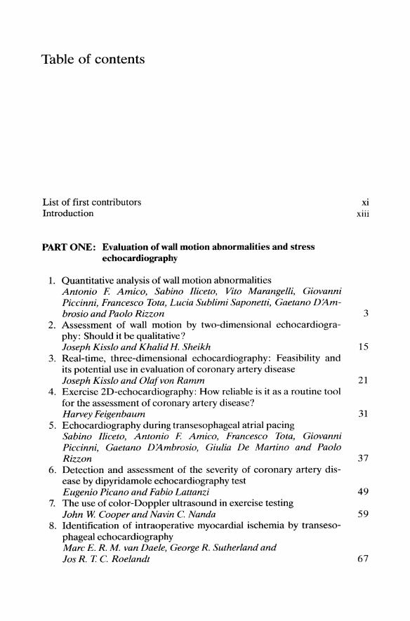
Table of contents
List of first contributors Introduction
PART ONE: Evaluation ofwall motion abnormalities and stress echocardiography
1. Quantitative analysis of wall motion abnormalities Antonio F. Amieo, Sabino llieeto, Vito Marangelli, Giovanni Piecinni, Franeeseo Tota, Lucia Sublimi Saponetti, Gaetano D'Am-
Xl
xiii
brosio and Paolo Rizzon 3 2. Assessment of wall motion by two-dimensional echocardiogra
phy: Should it be qualitative? loseph Kisslo and Khalid H. Sheikh 15
3. Real-time, three-dimensional echocardiography: Feasibility and its potential use in evaluation of coronary artery disease loseph Kisslo and Olajvon Ramm 21
4. Exercise 2D-echocardiography: How reliable is it as a routine tool for the assessment of coronary artery disease? Harvey Feigenbaum 31
5. Echocardiography during transesophageal atrial pacing Sabino llieeto, Antonio F. Amieo, Franeeseo Tota, Giovanni Piecinni, Gaetano D'Ambrosio, Giulia De Martino and Paolo Rizzon 37
6. Detection and assessment of the severity of coronary artery disease by dipyridamole echocardiography test Eugenio Pieano and Fabio Lattanzi 49
7. The use of color-Doppler ultrasound in exercise testing lohn W. Cooper and Navin C. Nanda 59
8. Identification of intraoperative myocardial ischernia by transesophagealechocardiography Mare E. R. M. van Daele, George R. Sutherland and los R. T. C. Roelandt 67

viii
9. Silent myoeardial isehemia: Clinical implieations, deteetion and management Giuseppe Specchia, Colomba Falcone, Cristina Opas ich, Maria Teresa La Rovere 83
10. Digital eine loop teehnology: A new tool for the evaluation of wall motion abnormalities Vito Marangelli, Gaetano D/lmbrosio, Luigi Carella, Sabino lliceto and Paolo Rizzon 95
PART TWO: Evaluation ofmyocardial infarction
11. Two-dimensional eehoeardiography in the early management of aeute myoeardial infarction Luciano Agati, Maria Penco, Carmine D. Vizza, Marco Renzi and Armando Dagianti 107
12. Left ventrieular shape changes and modelling during aeute myocardial infaretion Paolo Marino, Giorgio Golia and Piero Zardini 121
13. Two-dimensional eehoeardiographie quantification of myoeardial infarct size Natesa G. Pandian and Brenda S. Kusay 133
14. Deteetion and evaluation of left ventrieular thrombosis in myocardial infaretion: Role of eehocardiography Antonio F Amico, Sabino lliceto, Cataldo Memmola, Giovanni Piccinni, Carlo Caiati, Cesare Pellegrini and Paolo Rizzon 143
15. Two-dimensional echocardiography and Doppler findings in right ventrieular infarction M. A. Garcia-Fernandez, 1. Lopez-Sendon and M. Moreno Yan-güela 151
16. The role of cardiae ultrasound in the diagnosis of the 'surgieal' complications of aeute myocardial infarction lohn H. Smyllie, Patricia E. Assmann, George R. Sutherland, Alan G. Fraser and los R. T. C. Roelandt 183
17. Prognostie information obtained by 2D-eeho-Doppler evaluation in aeute myoeardial infarction Cees A. Visser, Ben l. Delemarre and Arend l. Dunning 197
18. Analysis of left ventriucular function in patients with myocardial infarction: Methodological problems and possible solutions Alan G. Fraser, lohn H. Smyllie, Patricia E. Assmann, George R. Sutherland and los R. T. C. Roelandt 213
19. Stress eehoeardiography for identifying patients at risk after myoeardial infaretion Sabino Iliceto, Antonio F Amico, Carlo Caiati, Giovanni Piccinni, Francesco Tota, Vito Marangelli, Cataldo Memmola and Paolo Rizzon 231
20. Two-dimensional echocardiography for the assessment of therapeutic interventions and long-term follow-up of patients with acute myocardial infarction
ix
Raimund Erbei, V. NixdorjJ, G. Görge, R. Brennecke and J. Meyer 241 21. Possibilities of ultrasonic tissue identification in the heart
N. Bom, J. Rijsterborgh, C. T. Lancee and Jos R. T. C. Roelandt 255
PART THREE: Evaluation of coronary anatomy and flow
22. Visualization of the coronary arteries by precordial echocardiography H. Feigenbaum 265
23. Evaluation of proximal left coronary artery anatomy and blood flow using digital transesophageal echocardiography Sabino Iliceto, Cataldo Memmola, Giulia De Marlino, Vito Maran-gelli, Ca rio Caiati and Paolo Rizzon 269
24. Intra-arterial ultrasonic imaging N. Bom, W J. Gussenhoven, C. T. Lancee, C. J. Slager, Pa trick W Serruys, H. ten Hoff and Jos R. T. C. Roelandt 277
25. Intracoronary blood flow velocity, reactive hyperemia and coronary blood flow reserve during and following PTCA Patrick W Serruys, Felix Zijlstra, Hans H. C. Reiber, Kevin Beatt, G. J. Laarman, Jos R. T. C. Roelandt and P. J. de Feyter 285
26. Long term result of revascularization after angioplasty: Should we randomize? Patrick W Serruys, B. J. Meesterand P. J. Feyter 311
PART FOUR: Myocardial perfusion
27. Pathophysiology of the coronary circulation: Role of myocardial contrast echocardiography and relation with other techniques David D. McPherson 325
28. Contrast agents for myocardial perfusion studies: Mechanisms, state of the art, and future prospects Richard S. Meltzer, Antonio F. Amico, Shimon A. Reisner and Janine R. Shapiro 351
29. Coronary anatomy and myocardial perfusion: Role of contrast echocardiography Antonio F. Amico, Sabino lliceto, Richard S. Meltzer, Gaetano D'Ambrosio, Vito Marangelli, Cataldo Memmola, Giulia De Mar-lino, Lucia Sublimi Saponetti and Paolo Rizzon 367

x
30. Physiological heterogeneity of coronary blood flow in space and time by contrast echocardiography Daniele Rovai, Massimo Lombardi, Steven E. Nissen, Mario Marzilli, Alessandro Distante, Luigi Taddei, Anthony N. DeMaria and Antonio L~bbate 377
31. Myocardial contrast echocardiography for the evaluation of coronary flow reserve: Potential and limitations Antonio F Amico, Sabino lliceto, Lucia Sublimi Saponetti, Catal-do Memmola and Paolo Rizzon 389
Subject index 397

List of first contributors
Luciano Agati, 1 Cattedra di Cardiologia, Univ. La Sapienza, Policlinico Umberto 1,1-00161, Roma,Italy
Antonio F. Amico, Division of Cardiology, University of Bari, 1-70124 Bari, Italy N. Born, Thorax Center, Erasmus University, P.O. Box 1738, 3000 DR Rotterdam, The Nether
lands Raimund Erbel, II Medical Clinic, Johannes Gutenberg University, Langenbeckstrasse 1,
D-6500 Mainz, FRG Harvey Feigenbaum, Hemodynamic Lab., Indianapolis University, School of Medicine, 926
West Michigan Street, Indianapolis, IN 46223, USA Alan G. Fraser. Department of Cardiology. University of Wales, College of Medicine, Heath
Park, Cardiff CF4 4XN, Wales, UK M. Garcia-Fernandez, Lab. Cardiologia No Invasiva, Escuela Nacional deI Thorax, Ciudad
Universitaria, 28040 Madrid, Spain Sabino lliceto, Division of Cardiology, University of Bari, 1-70124 Bari, Italy Joseph Kisslo, Cardiac Diagnostic Unit, Duke University Medical Center, Box 3818, Durham,
NC 27710, USA Vito Marangelli, Division of Cardiology, University of Bari, 1-70124 Bari, Italy Paolo Marino, Division of Cardiology, University of Verona, Centro Ospedaliero di Borgo
Trento, P.le A. Stefani 1,1-37126 Verona, Italy David D. McPherson, Dir. Echocardiography Lab., Northwestern Memorial Hospital, Suite
586, Wesley Pazalian-Superior and Farbacks Court, Chicago, IL 60611, USA Richard S. Meltzer, University of Rochester Medical Center, Cardiology Box 679, 601 Elm
wood Ave., Rochester, NY 14642, USA Navin C. Nanda, Heart Station SWB-W001, University of Alabama School of Medicine, Spain
Wallace Building, Birrningham, AL 35294, USA Natesa G. Pandian, Tufts University School of Medicine, Non Invasive Cardiac Lab., New Eng-
land Med. Center Hospitals, 750 Washington Street, Box 32, Boston, MA 02111, USA Eugenio Picano, Ist. di Fisiologia Clinica deI C.N.R., Via Savi 8, 1-56100 Pisa, Italy Shimon A. Reisner, Department of Cardiology, Rambam Medical Center, Haifa 31096, Israel Paolo Rizzon, Division of Cardiology, University of Bari, 1-70124 Bari, Italy Jos R. T. C. Roelandt, Thorax Center, Erasmus University, P.O. Box 1738, 3000 DR Rotterdam,
The N etherlands Daniele Rovai, CNR Clinical Physiology Inst., V. Savi 8,1-56100 Pisa, Italy Patrick W. Serruys, Thorax Center, Erasmus University, P.O. Box 1738,3000 DR Rotterdam,
The Netherlands Janine R. Shapiro, Box 604, University of Rochester Medical Center, Rochester, NY 14642,
USA

xn
lohn H. Smyllie, Thorax Center, Erasmus University, P.O. Box 1738,3000 DR, The Netherlands
Giuseppe Specchia, Div. di Cardiologia, Univ. degii Studi, Policlinico S. Matteo, 27100 Pavia, Italy
Mare van Daele, Thorax Center, Erasmus University, P.O. Box 1738, 3000 DR, The Netherlands
Cees A. Visser, Dept. of Cardiology, Academisch Medisch Centrum, Meibergdreef 9, 1105 AZ Amsterdam. The Netherlands

Introduction
Today, coronary artery disease is one of the major causes of mortality and morbidity in the Western World. In the last decade many major diagnostic and therapeutic advances have been made, considerably furthering our potential in the management of coronary artery disease. At the same time, a new generation of cardiac tools has appeared. The field which has, perhaps, undergone the most important technological innovations is echocardiography. Nowadays, in fact, the world of ultrasounds ofters the cardiologist a wide range of technical applicatons: two-dimensional real-time imaging, intra- and extra-cardiac Doppler flow measurements, real-time imaging of cardiac structure and flow by 2D color Doppler, high resolution cardiac imaging by transesophageal echocardiography, tissue characterization by analysis of ultrasound wave characteristics, information on myocardial perfusion by contrast echocardiography, etc. Thanks to these technical improvements and to its consequent increased potentiality, echocardiography now plays an important and irreplaceable role in the management of all cardiac diseases. In the field of coronary artery disease, echocardiography can reliably be used not only in the acute phases of the disease to derive useful functional and prognostic information but also as a stress diagnostic procedure (thanks to new stress modalities and the continuing improvement of reviewing digital systems) for the diagnosis of coronary artery disease and for the evaluation of various therapeutic interventions. Furthermore, other promising applications of ultrasounds in this disease are currently being investigated: tissue characterization, myocardial contrast echocardiography, coronary artery anatomy and flow evaluation by specially-designed ultrasound catheters.
This book evaluates the current situation and future potential of echocardiography in the management of patients with coronary artery disease.