ultrasound imaging in vascular diseases
TRANSCRIPT
10
Ultrasound Imaging in Vascular Diseases Mariantina Fragou1, Andreas Karabinis2, Eugene Daphnis3,
Nicolaos Labropoulos4 and Dimitrios Karakitsos1 1Department of Intensive Care Unit, General State Hospital of Athens
2Department of Intensive Care Unit, Onassis Cardiac Surgery Center, Athens 3Nephrology Department, Heraklion University Hospital, Heraklion, Crete
4Department of Surgery, Stony Brook University Medical Center, Stony Brook NY 1,2,3Greece
4USA
1. Introduction
Ultrasound scanning has become one of the most important noninvasive investigations used in the assessment of vascular diseases. Duplex ultrasound is currently the main diagnostic modality used in deep venous thrombosis and carotid disease. It enables monitoring of bypasses for patency and neointimal hyperplasia as well as transcranial examination of brain vessels in subarachnoid hemorrhage. Nowdays, the role of the vascular laboratory is continuously changing with the introduction of new technology. New interventions such as carotid stenting and endovascular aneurysm repair have necessitated the use of duplex ultrasound for detecting in-stent restenosis or endoleaks. Ultrasound is also used intra-operatively in evaluating endovenous procedures such as laser and radiofrequency ablation and the patency of in situ vein bypasses. Arterial mapping of the lower extremity by duplex ultrasound has replaced contrast angiography (CA) and magnetic resonance angiography (MRA) for lower extremity bypass procedures. Most recently, a variety of new applications for vascular ultrasound has arised. Contrast-
enhanced ultrasound imaging allows the assessment of microcirculation and can be used for
delivery of drugs and genetic therapies. Physicians use three-dimensional ultrasound to
assess carotid plaque volume and monitor its evolution with time and its response to
various treatments. In the aera of atherosclerosis, ultrasound has provided a non-invasive
way of assessing the elastic properties of an artery and to evaluate the patient’s risks of
adverse cardiovascular events. Finally, intravascular ultrasound has opened new frontiers in
the measuring of intimal thickness and assessing vulnerable plaques.
2. Ultrasound in the era of atherosclerotic disease
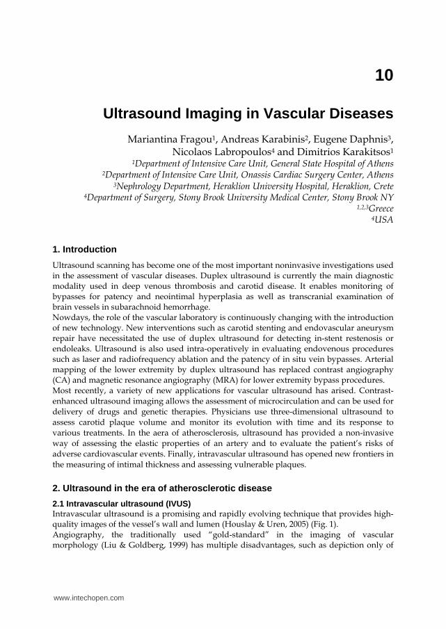
2.1 Intravascular ultrasound (IVUS) Intravascular ultrasound is a promising and rapidly evolving technique that provides high-quality images of the vessel’s wall and lumen (Houslay & Uren, 2005) (Fig. 1). Angiography, the traditionally used “gold-standard” in the imaging of vascular morphology (Liu & Goldberg, 1999) has multiple disadvantages, such as depiction only of
www.intechopen.com
Ultrasound Imaging – Medical Applications
196
contrast agent filled lumen and not the vessel wall, as well as the risk of contrast-induced nenal failure. Moreover, it underestimates the plaque burden, especially for the detection of concentrical lumen narrowing (Liu & Goldberg, 1999).
Fig. 1. Intravascular ultrasound (IVUS) advancing via the inferior vena cava (IVC) at various levels depicting the right atrium (A), the hepatic veins (B), the renal veins (C), and the infrarenal IVC (D)
With the use of high-frequency catheter based transducers, all basic components of the vessel are identified: the cross sectional luminal size, shape and vessel wall, as well as the various layers of the wall such as the intima, media, adventitia and perivascular structures (Liu & Goldberg, 1999). In conventional gray-scale intravascular sonography arteries can be classified as elastic (lying centrally in the arterial tree) or muscular (peripherally distributed). Calcifications are demonstrated as hyperechoic areas, whereas hemorrage or fat deposition inside an atheromatic plaque is hypoechoic. Subsequently, the plaque is subclassified as lipid, calcified and fibrous, according to its differential acoustic properties (Liu & Goldberg, 1999). Furthermore, the concommitant use of colour-flow intravascular sonography allows for a real-time dynamic assessment of plaque morphology and provides a better understanding of the blood flow, the lumen size and the success of endovascular treatment (Zacharatos et al., 2010). Hence, the physician can obtain a realistic virtual histology map of the patient’s arteries, visualize the atheroma without the need of true histologic sections of the diseased coronary arteries and tailor his treatments (Fayad & Fuster, 2001; Martin et al., 1997). The correlation of IVUS plaque characteristics and histopathologic speciments is well validated in literature (Potkin et al., 1990).The CAPITAL study showed a strong correlation of IVUS findings with histologic speciments from endarterectomy (Diethrich et al., 2007) while Potkin et al compared IVUS with coronary histopathologic speciments, to show that fibrous and calcified plaques were identified with good precision (Potkin et al., 1990).
www.intechopen.com
Ultrasound Imaging in Vascular Diseases
197
In patients with coronal artery stenosis IVUS can act as a prognostication tool, as vulnerable plaques, heavy in fat deposition are considered at higher risk for cardiovascular events and might need more aggressive treatment. In addition, plaque regression in respond to lipid lower therapies with statins has been successfully monitored by the use of serial IVUS (Crouse
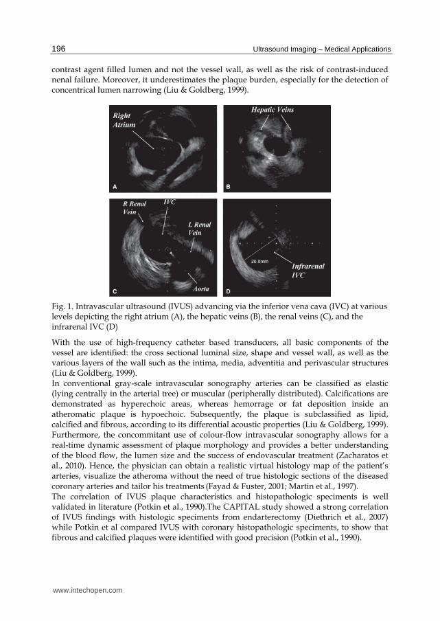
et al., 2007; Nissen, 2005; Nissen et al., 2008). Many dynamic phenomena in the setting of an atheroma, such as positive and negative remodeling or restenosis after angioplasty and stenting are timely demonstrated and treated accordingly (Liu & Goldberg, 1999). The concommitant use of intravascular elastography(Fayad & Fuster, 2001) (images obtained from cardiac IVUS images associated with intraluminar pressures during the cardiac circle) expands the frontiers of the IVUS technique, as it illustrates the mechanical properties of the vessel wall and provides strain information. IVUS allows a direct visualization of the processes associated with lumen expansion by ballon dilation. It is believed that about 80% of the post-dilation increase in the lumen area is due to an increase in vessel size, and 20% is due to a reduction in plaque area, due to axial plaque redistribution (Liu & Goldberg, 1999). IVUS can be utilized as a means of planning and evaluating the effects of vascular brachytherapy in the treatment of post-stent restenosis (Carlier et al., 2000). Apart from coronal artery disease, it is a safe method of choosing the appropriate intervention procedure in peripheral vascular disease (Fig. 2) and carotid stenting (Liu & Goldberg, 1999). It’s only risk, the disruption of a plaque and release of embolic material, seems minor compared to it’s numerous advantages.
Fig. 2. Obstruction of the iliac vein: pre-intervention imaging (A) and post-intervention result (B) by intravascular ultrasound (IVUS)
2.2 3D ultrasound
Three-dimentional ultrasound is a recent development that came as an answer to the
multiple problems inherent to 2D ultrasound images, leading to incorrect quantification of
plaque morphology, difficult localization and significant operator dependence (Fenster et
al., 2004) (Fig. 3).
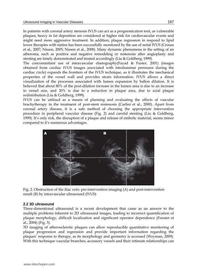
3D imaging of atherosclerotic plaques can allow reproducible quantitative monitoring of plaque progression and regression and provide important information regarding the plaques’ response to therapy, as its morphology and geometry is accessed (Weyman, 2009). With this technique vascular branches, accessory vessels and their intimate relationships can
www.intechopen.com
Ultrasound Imaging – Medical Applications
198
be easily demonstrated (Liu & Goldberg, 1999). Arterial dissections, aneurysms and aortic dissections and flaps are depicted with 3D IVUS much better than conventional angiography.
Fig. 3. 3D IVUS of an iliac artery
Although the importance of 3D imaging has been demonstrated in several studies, the widespread use of this technique has been prevented by inherent disadvantages, such as the slow frame rate, the lengthy analysis time and the limited spacial resolutions (Weyman, 2009). The combination of contrast with 3D ultrasound will likely compensate for the latter and should enhance its value in areas such as stress echocardiography (Liu & Goldberg, 1999).
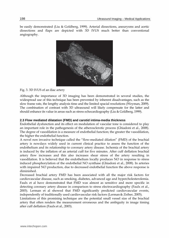
2.3 Flow mediated dilatation (FMD) and carotid intima-media thickness Endothelial dysfunction and its effect on modulation of vascular tone is considered to play an important role in the pathogenesis of the atherosclerotic process (Ghiadoni et al., 2008). The degree of vasodilation is a measure of endothelial function; the greater the vasodilation, the higher the endothelial function. A novel non invasive technique called the “flow-mediated dilation” (FMD) of the brachial artery is nowdays widely used in current clinical practice to assess the function of the endothelium and its relationship to coronary artery disease. Ischemia of the brachial artery is induced by the inflation of an arterial cuff for five minutes. After cuff deflation brachial artery flow increases and this also increases shear stress of the artery resulting in vasodilation. It is believed that the endothelium locally produces NO in response to stress induced phosphorylation of the endothelial NO synthase (Ghiadoni et al., 2008). In arteries with impaired NO production, due to decreased endothelial function the above response is diminished. Decreased brachial artery FMD has been associated with all the major risk factors for cardiovascular disease, such as smoking, diabetes, advanced age and hypercholesterolemia. Faulx et al have demonstrated that FMD was almost as sensitive and more specific in detecting coronary artery disease in comparison to stress electrocardiography (Faulx et al., 2003). Lerman et al showed that FMD significantly predicted cardiovascular events, independently of traditionally used cardiovascular risk factors (Lerman & Zeiher, 2005). Limitations of this promising technique are the potential small vessel size of the brachial artery that often renders the measurement erroneous and the ambiguity in image timing after cuff deflation (Faulx et al., 2003).
www.intechopen.com
Ultrasound Imaging in Vascular Diseases
199
Another marker used to quantify atherosclerosis by B-mode ultrasound is the measurement of carotid intima-media thickness (CIMT) (de Groot et al., 2008; Labropoulos et al., Dec 2005). A simple distance measurement between the leading edges of the lumen-intima and media-adventitia ultrasound interfaces is performed and this measurement can be used to measure the degree of existing atherosclerosis and future cardiovascular disease risk (de Groot et al., 2008; Bots et al., 1997; Chambless et al., 1997). Futhermore, several studies in literature have proved the efficacy of CIMT in monitoring the patient’s response to lipid lowering therapies (Blankenhorn et al., 1993; de Groot et al., 1998; Furberg et al., 1994; Smilde et al., 2001).
3. Pre-operative ultrasound use
In patients with lower extremity ischemia, angiography has traditionally been considered the “gold standard” of arterial mapping. However, in recent years, duplex ultrasonography (DA) has gained a major role in the pre-operative evaluation of patients undergoing lower extremity bypass (Ascher et al., 1999; Grassbaugh et al., 2003; Ligush et al., 1998; Pemberton et al., 1996; Proia et al., 2001). Mazzariol et al reported that the use of dupplex ultrasonography can provide enough information for surgery in more than 83% of patients with arterial stenosis and that an abnornal femoral artery waveform was 100% predictive for detecting stenosis greater than 80% (Mazzariol et al., 2000) Proia et al have also confirmed these results (Proia et al., 2001). In comparison with conventional angiography or MRA, DA has a lower sensitivity for detecting arterial disease of the lower extremity. Moreover, it is often cumbersome and time-consuming to scan the entire arterial tree from the aorta to the pedal vessels. Imaging can also be obscured in obese patients or heavily calcified vessels. Nevertheless, the simplicity and safety of this technique underlines its importance as a first-line examination before planning a bypass surgery (Pearce & Astleford, 2004).
4. Intra-operative and post-operative ultrasound
Completion angiography has been traditionally used as an adjunctive technique after bypass procedures to scan the graft for anatomic and flow abnormalities. Nowdays, duplex scanning is used as a safer alternative to visualize these abnormalities (Johnson et al., 2000). Defective grafts are identified by direct measurement of PSV and ratio at the lesion devided by proximal velocity (Johnson et al., 2000). Moreover, surveillance Duplex ultrasound has acquired an important role in the post-operative assessment of patients following lower extremity revascularization. It enables prompt detection of hemodynamically significant lesions and early intervention (Bandyk, 2002, Mills et al., 2001). Patients undergoing EVAR (endovascular aneurysm repair) are also monitored by serial ultrasound examinations (Bendick et al., 2003; Carter et al., 2000; McWilliams et al., 1999; Pearce & Astleford, 2004). The value of duplex ultrasound scanning in this group of patients is that repeated studies can be performed frequently to detect an endoleak or a restenosis, without the risk of contrast-induced nephropathy or the presence of adjuscent metal artifacts. Carotid artery stenting (CAS) is currently being clinically evaluated as an alternative to conventional carotid endarterectomy (CEA). Dupplex ultrasound is commonly used to monitor restenosis in patients following CEA. Several authors in literature begin to report
www.intechopen.com

Ultrasound Imaging – Medical Applications
200
their experience in the use of ultrasonography as a follow-up tool in CAS too (Lal et al., 2004).
5. Ultrasound in venous disease
In the field of deep venous thrombosis ultrasound is nowdays the sole diagnostic modality in most centers and has contibuted extensively in our understanding of the natural history and the pathophysiology of the disease (Labropoulos et al., Feb 2005; Labropoulos et al., Jan 2010; Apr 2010) (Figures 4, 5).
Fig. 4. Acute thrombosis in the common femoral vein. There is absence of color, the vein is dilated (twice the size of the adjacent common femoral artery in red) with homogenous echolucent texture
Fig. 5. Chronic inferior vana cava (IVC) obstruction with partial recanalization. The lumen of IVC is smaller compared to the adjacent aorta. The azygos vein is dilated and larger than the aorta.
www.intechopen.com
Ultrasound Imaging in Vascular Diseases
201
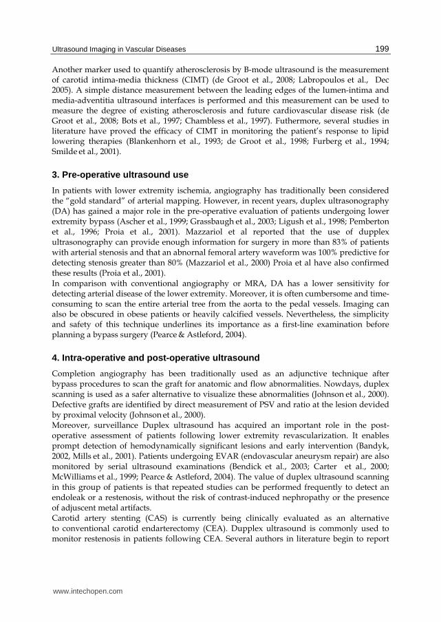
Duplex ultrasound is also used as a follow-up tool to detect venous reflux up to six months after treatment (Labropoulos et al., Feb 2005) (Fig. 6) and to investigate conditions such as thrombus neovascularization (Labropoulos et al., Sep 2005).
Fig. 6. Chronic thrombus with partial recanalization in great saphenous vein. Flow channels with reflux are seen over the old thrombus that is seen as an echogenic band in the lumen
Patients undergoing varicose vein stripping and venous ablative surgery routinely undergo preoperative ultrasound scanning to assure that the deep venous system is clot-free and to identify the location of perforators and accessory veins as well as the presence of lesser saphenous reflux. Intraoperatively, the use of ultrasound allows for placement of a catheter under direct guidance close to the sapheno-femoral junction, and then the anesthesia agent is injected (Min et al., 2001; Pichot et al., 2004). Successful ablation is then established by monitoring venous occlusion by means of ultrasonography.
6. Contrast-enhanced ultrasound
Gramiak was the first to describe contrast enhanced ultrasound in 1969, using air bubbles in the aorta (Gramiak et al., 1969). In modern clinical practice contrast agents for ultrasound use are microbubbles of different gases, 1-7μm in diameter (Cosgrove, 2006). The gas can be air, which unfortunately dissolves quickly in water or blood, or newer agents such as sulphur hexafloride, perfluocarbons or heavy gases, which minimally dissolve in water (Cosgrove, 2006). In order to reach and persist in systemic circulation the microbubbles are stabilized with albumin, simple phospholipids micelles, bilayered membranes and biocompatible polymers (Cosgrove, 2006). Inside the vessels microbubbles expand and contract in the alternating pressure waves of the ultrasound beam, while tissues are almost incompressible. Special software using multiple pulse sequences enhances this blood-tisue border detection (Weyman, 2009). Adverse effects of contrast agents are headache, bruising, injection site pain, paresthesias and burning. Microbubbles are currently used to improve Doppler studies especially when flow information makes conventional imaging difficult, a technique called “Doppler rescue” (Cosgrove, 2006). Common applications include imaging of the basilar and vertebral arteries, transcranial ultrasound examinations in patients with severe hyperostosis of the
www.intechopen.com
Ultrasound Imaging – Medical Applications
202
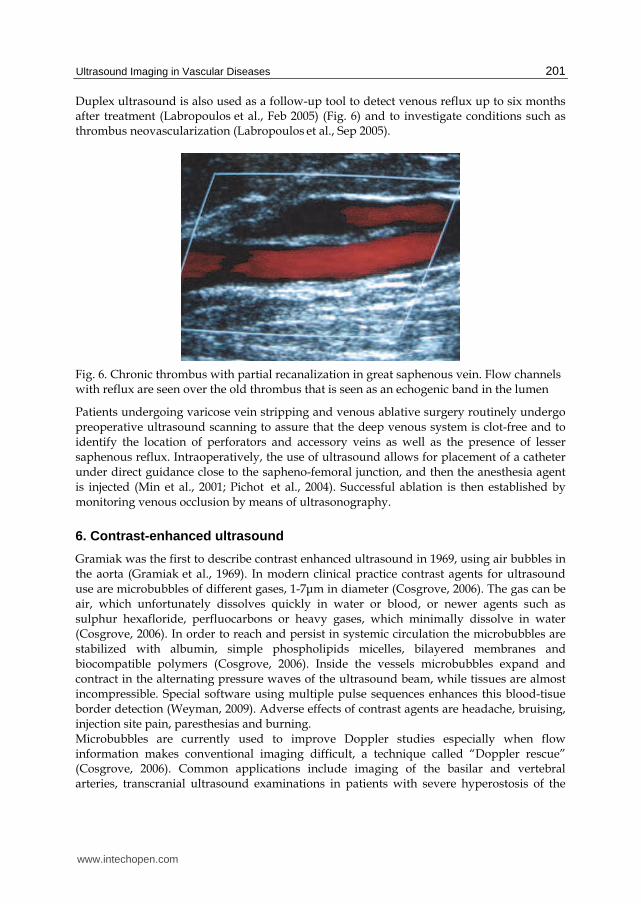
skull, measurement of internal carotid stenosis in calcified arteries (Cosgrove, 2006) and differentiation of subtotal stenosis from occlusion in acute stroke (Seidel & Meairs, 2009). Moreover, contrast enhanced ultrasound has been used in renal artery stenosis, portal and hepatic artery examinations (e.g. in cirrhosis, tips shunts or after liver transplantation) (Furlow, 2009) and in liver oncology (Wilson & Burns, 2006) (Fig. 7).
Fig. 7. Contrast-enhanced ultrasound of an hepatic hemangioma
Neurosurgeons can intra-operatively assess vascular pathologies such as middle cerebral artery aneurysms and arteriovenous malformations in real time and make appropriate surgical planning (Hölscher et al., 2007). The microbubbles used in contrast-enhanced ultrasound techniques after attachment of antibodies or other ligants to their shell can bind to specific cell receptors in areas of disease (Voigt, 2009), thus provide information about the stage of the disease (Lindner, 2009). Several promising studies have targeted pathologic processes with contrast-enhanced ultrasound (Lindner, 2009; Voigt, 2009), such as angiogenesis (by targeting endothelial integrins) (Lerman & Zeiher, 2005), thrombus (by targeting IIbIIIa receptors or fibrinogen) (Alonso et al., 2007), inflammation (by targeting ICAM-1, VCAM-1 P- and L- selectin) and plaque (by targeting ICAM-1, VCAM-1 and several angiogenesis markers) (Voigt, 2009). In fact, it is believed that by targeting plaque inflammation and neovascularization we could identify patients at high risk for cardiovascular events well before angiography (Lindner, 2009; Voigt, 2009) or even recognise “vulnerable plaques” and begin early treatment (Vicenzini et al., 2009). Several future promising therapeutic applications of microbubbles are under research. Therapeutic agents, genes, siRNAs, drugs and molecules can be attached to or dissolved within the surface shell or deposited within the bubbles themselves (Hernot & Klibanov, 2008; Hynynen, 2008; Weyman, 2009). Local cavitation due to microbubble destruction can produce clot fragmentation and enhance thrombolysis (Medel et al., 2009; Trübestein et al., 1976). Continuous transcranial Doppler has been shown to augment the t-PA induced arterial recanalization in stroke victims (Alexandrov et al., 2004). Future studies need to be performed in order to improve the safety profile and therapeutic indexes of these challenging techniques.
www.intechopen.com
Ultrasound Imaging in Vascular Diseases
203
7. Conclusion
Ultrasound imaging in vascular diseases has evolved during the past years due to new challenging technologies. Virtually all peripheral arterial and venous structures can be visualized with color duplex ultrasound. In the era of atherosclerosis, the use of intravascular ultrasound, contrast-enhanced ultrasound, 3D ultrasound, flow mediated dilatation and carotid intima media thickness techniques allow for more precise and localized diagnosis, treatment and follow-up. Ultrasound techniques routinely performed before, during or after surgical procedures are nowdays commonplace. The low cost and noninvasive aspect of most ultrasound methods make it the technique of choice in studying the human vasculature.
8. References
Alexandrov AV, Molina CA, Grotta JC, Garami Z, Ford SR, Alvarez-Sabin J, Montaner J, Saqqur M, Demchuk AM, Moyé LA, Hill MD & Wojner AW. Ultrasound-enhanced systemic thrombolysis for acute ischemic stroke. N Engl J Med, Vol. 351, No. 21, (Nov 2004), pp. 2170-2178
Alonso A, Della Martina A, Stroick M, Fatar M, Griebe M, Pochon S, Schneider M, Hennerici M, Allémann E, Meairs S. Molecular imaging of human thrombus with novel abciximab immunobubbles and ultrasound. Stroke, Vol. 38, No. 5, (May 2007), pp. 1508-1514
Ascher E, Mazzariol F, Hingorani A, Salles-Cunha S & Gade P. The use of Duplex ultrasound arterial mapping as an alternative to conventional arteriography for primary and secondary infrapopliteal bypasses. Am J Surg, Vol. 178, No. 2, (Aug 1999), pp. 162–165
Bandyk DF. Infrainguinal vein bypass graft surveillance: how to do it, when to intervene, and is it cost effective? J Am Coll Surg, Vol. 194, No. 1 suppl, (Jan 2002), pp. 540-552 Bendick PJ, Bove PG, Long GW, Zelenock GB, Brown OW & Shanley CJ. Efficacy of ultrasound scan contrast agents in the noninvasive follow-up of aortic stent grafts. J Vasc Surg, Vol. 37, No. 2, (Feb 2003), pp. 381–385
Blankenhorn DH, Selzer RH, Crawford DW, Barth JD, Liu CR, Liu CH, Mack WJ & Alaupovic P. Beneficial effects of colestipol niacin therapy on the common carotid artery. Two- and four-year reduction of intima-media thickness measured by ultrasound. Circulation, Vol. 88, No. 1, (Jul 1993), pp. 20-28
Bots ML, Hoes AW, Koudstaal PJ, Hofman A & Grobbee DE. Common carotid ntima-media thickness and risk of stroke and myocardial infarction: the Rotterdam Study. Circulation, Vol. 96, No. 5, (Sep 1997), pp. 1432-1437
Carlier SG, Coen VL, Sabaté M, Kay IP, Ligthart JM, Van Der Giessen WJ, Levendag PC, Bom K & Serruys PW. The role of intravascular ultrasound imaging in vascular brachytherapy. Int J Cardiovasc Intervent, Vol. 3, No. 1, (Mar 2000), pp. 3-12
Carter KA, Nelms CR, Bloch PHS, Gregory RT, Parent EN & DeMasi RJ. Doppler waveform assessment of endoleak following endovascular repair of abdominal aortic aneurysm: predictors of endoleak thrombosis. J Vasc Technol, Vol. 24, No. 2, (2000), pp. 119 122
Chambless LE, Heiss G, Folsom AR, Rosamond W, Szklo M, Sharrett AR & Clegg LX. Association of coronary heart disease incidence with carotid arterial wall thickness and major risk factors: the Atherosclerosis Risk in Communities (ARIC) Study, 1987-1993. Am J Epidemiol, Vol. 146, No. 6, (Sep 1997), pp. 483-494
www.intechopen.com
Ultrasound Imaging – Medical Applications
204
Cosgrove D. Ultrasound contrast agents: an overview. Eur J Radiol, Vol. 60, No. 3, (Dec 2006), pp. 324-330
Crouse JRr, Raichlen JS, Riley WA, Evans GW, Palmer MK, O'Leary DH Grobbee DE & Bots ML. Effect of rosuvastatin on progression of carotid Intima media thickness in low-risk individuals with
subclinical atherosclerosis: the METEOR Trial. JAMA, Vol. 297, No. 12, (Mar 2007), pp. 1344-1353
de Groot E, Jukema JW, Montauban van Swijndregt AD, Zwinderman AH, Ackerstaff RG, van der Steen AF, Bom N, Lie KI & Bruschke AV. B-mode ultrasound assessment of pravastatin treatment effect on carotid and femoral artery walls and its correlations with coronary arteriographic findings: a report of the Regression Growth Evaluation Statin Study (REGRESS). J Am Coll Cardiol, Vol. 31, No. 7, (Jun 1998), pp. 1561-1567
de Groot E, van Leuven SI, Duivenvoorden R, Meuwese MC, Akdim F, Bots ML & Kastelein JJ. Measurement of carotid intima-media thickness to assess progression and regression of atherosclerosis. Nat Clin Pract Cardiovasc Med, Vol. 5, No. 5, (May 2008), pp. 280 288
Diethrich EB, Pauliina Margolis M, Reid DB, Burke A, Ramaiah V, Rodriguez - Lopez JA, Wheatley G, Olsen D & Virmani R. Virtual histology intravascular ultrasound assessment of carotid artery disease: the Carotid Artery Plaque Virtual Histology Evaluation (CAPITAL) study. J Endovasc Ther, Vol. 14, No. 5, (Oct 2007), pp. 676-86
Faulx MD, Wright AT & Hoit BD. Detection of endothelial dysfunction with brachial artery ultrasound scanning. Am Heart J, Vol. 145, No. 6, (Jun 2003), pp. 943-951
Fayad ZA & Fuster V. Clinical imaging of the high-risk or vulnerable atherosclerotic plaque. Circ Res, Vol. 89, No. 4, (Aug 2001), pp. 305-316
Fenster A, Landry A, Downey DB, Hegele RA & Spence JD. 3D ultrasound imaging of the carotid arteries. Curr Drug Targets Cardiovasc Haematol Disord, Vol. 4, No. 2, (Jun 2004), pp. 161-175
Furberg CD, Adams HPJ, Applegate WB, Byington RP, Espeland MA, Hartwell T, Hunninghake DB, Lefkowitz DS, Probstfield J & Riley WA. Effect of lovastatin on early carotid atherosclerosis and cardiovascular events. Asymptomatic Carotid Artery Progression Study (ACAPS) Research Group. Circulation, Vol. 90, No. 4, (Oct 1994), pp. 1679-1687
Furlow B. Contrast-enhanced ultrasound. Radiol Technol, Vol. 80, No. 6, (Jul Aug 2009), pp. 547S-561S
Ghiadoni L, Versari D, Giannarelli C, Faita F & Taddei S. Non-invasive diagnostic tools for investigating endothelial dysfunction. Curr Pharm, Vol. 14, No. 35, (Dec 2008), pp. 3715-3722
Gramiak R, Shah PM & Kramer DH: Ultrasound cardiography: contrast studies in anatomy and function. Radiology, Vol. 92, No. 5, (Apr 1969), pp. 939-948
Grassbaugh JA, Nelson PR, Rzucidlo EM, Schermerhorn ML, Fillinger MF, Powell RJ, Zwolak RM, Cronenwett JL & Walsh DB. Blinded comparison of preoperative duplex ultrasound scanning and contrast arteriography for planning revascularization at the level of the tibia. J Vasc Surg, Vol. 37, No 6, (Jun 2003), pp. 1186-1190
Hernot S & Klibanov AL. Microbubbles in ultrasound-triggered drug and gene delivery. Adv Drug Deliv Rev, Vol. 60, No. 10, (Jun 2008), pp. 1153-1166
Hölscher T, Ozgur B, Singel S, Wilkening WG, Mattrey RF & Sang H. Intraoperative ultrasound using phase inversion harmonic imaging: first experiences.
www.intechopen.com
Ultrasound Imaging in Vascular Diseases
205
Neurosurgery, Vol. 60, No. 4, Suppl. 2, (Apr 2007), pp. 382-386, discussion 386-387 Houslay ES & Uren NG. Intravascular ultrasound: defining plaque regression. Hosp Med, Vol. 66, No. 1, (Jan 2005), pp. 27-31
Hynynen K. Ultrasound for drug and gene delivery to the brain. Adv Drug Deliv Rev, Vol. 60, No. 10, (Jun 2008), pp. 1209-1217
Johnson BL, Bandyk DF, Back MR, Avino AJ & Roth SM. Intraoperative duplex monitoring of infrainguinal vein bypass procedures. J Vasc Surg, Vol. 31, No. 4, (Apr 2000), pp. 678-690
Labropoulos N, Bhatti AF, Amaral S, Leon L, Borge M, Rodriguez H & Kalman P. Neovascularization in acute venous thrombosis. J Vasc Surg, Vol. 42, No. 3, (Sep 2005), pp. 515-518
Labropoulos N, Kokkosis AA, Spentzouris G, Gasparis AP & Tassiopoulos AK. The distribution and significance of varicosities in the saphenous trunks. J Vasc Surg, Vol. 51, No 1, (Jan 2010), pp. 96
Labropoulos N, Leon L, Kwon S, Tassiopoulos A, Gonzalez-Fajardo JA, Kang SS, Mansour MA & Littooy FN. Study of the venous reflux progression. J Vasc Surg, Vol. 41, No. 2, (Feb 2005), pp. 291-295
Labropoulos N, Leon LRJ, Brewster LP, Pryor L, Tiongson J, Kang SS, Mansour MA & Kalman P. Are your arteries older than your age?. Eur J Vasc Endovasc Surg, Vol. 30, No. 6, (Dec 2005), pp. 588-596
Labropoulos N, Jen J, Jen H, Gasparis AP & Tassiopoulos AK: Recurrent deep vein thrombosis: long-term incidence and natural history. Ann Surg, Vol. 251, No. 4, (Apr 2010), pp. 749-753
Lal BK, Hobson RW 2nd, Goldstein J, Chakhtoura EY & Duran WN. Carotid artery stenting: is there a need to revise ultrasound velocity criteria? J Vasc Surg, Vol. 39, No.1, (Jan 2004), pp. 58–66
Lerman A & Zeiher AM. Endothelial function: cardiac events. Circulation, Vol. 111, No. 3, (Jan 2005), pp. 363-368
Ligush J Jr, Reavis SW, Preisser JS & Hansen KJ. Duplex ultrasound scanning Defines operative strategies for patients with limb-threatening ischemia. J Vasc Surg, Vol. 28, No. 3, (Sep 1998), pp. 482–490
Lindner JR. Contrast ultrasound molecular imaging of inflammation in cardiovascular disease. Cardiovasc Res, Vol. 84, No. 2, (Nov 2009), pp. 182-189
Liu JB & Goldberg BB. 2-D and 3-D endoluminal ultrasound: vascular and nonvascular applications. Ultrasound Med Biol, Vol. 5, No. 2, (Feb 1999), pp. 159-173
Martin AJ, Ryan LK, Gotlieb AI, Henkelman RM & Foster FS . Arterial imaging: comparison of high-resolution US and MR imaging with histologic correlation. Radiographics, Vol. 17, No. 1, (Jan- Feb 1997), pp. 189-202
Mazzariol F, Ascher E, Hingorani A, Gunduz Y, Yorkovich W & Salles-Cunha S. Lower-extremity revascularisation without preoperative contrast arteriography in 185 cases: lessons learned with duplex ultrasound arterial mapping. Eur J Vasc Endovasc Surg, Vol. 19, No. 5, (May 2000), pp. 509-515
McWilliams RG, Martin J, White D, Gould DA, Harris PL, Fear SC, Brennan J, Gilling-Smith GL, Bakran A & Rowlands PC. Use of contrast-enhanced ultrasound in follow-up after endovascular aortic aneurysm repair. J Vasc Interven Radiol , Vol. 10, No. 8, (Sep 1999), pp. 1107–1114
Medel R, Crowley RW, McKisic MS, Dumont AS & Kassell NF. Sonothrombolysis: an emerging modality for the management of stroke. Neurosurgery, Vol. 65, No. 5, (Nov 2009), pp. 979-993, discussion 993
www.intechopen.com
Ultrasound Imaging – Medical Applications
206
Mills JL Sr, Wixon CL, James DC, Devine J, Westerband A & Hughes JD. The natural history of intermediate and critical vein graft stenosis: recommendations for continued surveillance or repair. J Vasc Surg, Vol. 33, No. 2, (Feb 2001), pp. 273–278 [discussion 278–280]
Min RJ, Zimmet SE, Isaacs MN & Forrestal MD. Endovenous laser treatment of the incompetent greater saphenous vein. J Vasc Interven Radiol, Vol. 12, No. 10, (Oct 2001), pp. 1167–1171
Nissen SE. Effect of intensive lipid lowering on progression of coronary atherosclerosis: evidence for an early benefit from the Reversal of Atherosclerosis with Aggressive Lipid Lowering (REVERSAL) trial. Am J Cardiol, Vol. 96, No. 5A, (Sep 2005), pp. 61F-68F
Nissen SE, Nicholls SJ, Wolski K, Rodés-Cabau J, Cannon CP, Deanfield JE, Després JP, Kastelein JJ, Steinhubl SR, Kapadia S, Yasin M, Ruzyllo W, Gaudin C, Job B, Hu B, Bhatt DL, Lincoff AM & Tuzcu EM. Effect of rimonabant on progression of atherosclerosis in patients with abdominal obesity and coronary artery disease: the STRADIVARIUS randomized controlled trial. JAMA, Vol. 299, No. 13, (Apr 2008), pp. 1547-1560
Pearce WH & Astleford P: What's new in vascular ultrasound. Surg Clin North Am, Vol. 84, No. 4, (Aug 2004), pp. 1113-1126
Pemberton M, Nydahl S, Hartshorne T, Naylor AR, Bell PR & London NJ. Colour-coded duplex imaging can safely replace diagnostic arteriography in patients with lower-limb arterial disease. Br J Surg, Vol. 83, No. 12, (Dec 1996), pp. 1725–1728
Pichot O, Kabnick LS, Creton D, Merchant RF, Schuller-Petroviae S & Chandler JG. Duplex ultrasound scan findings two years after great saphenous vein radiofrequency endovenous obliteration. J Vasc Surg, Vol. 39, No. 1, (Jan 2004), pp. 189–195
Potkin BN, Bartorelli AL, Gessert JM, Neville RF, Almagor Y, Roberts WC, & Leon MB. Coronary artery imaging with intravascular high frequency ultrasound. Circulation, Vol. 81, No. 5, (May 1990), pp. 1575-1585
Proia RR, Walsh DB, Nelson PR, Powell RJ, Zwolak RM, Fillinger MF & Cronenwett JL. Early results of infragenicular revascularization based solely on duplex arteriography. J Vasc Surg, Vol. 33, No. 6, (Jun 2001), pp. 1165–1170
Seidel G & Meairs S. Ultrasound contrast agents in ischemic stroke. Cerebrovasc Dis, Vol. 27, No. Suppl 2 (2009), pp. 25-39
Smilde TJ, van Wissen S, Wollersheim H, Trip MD, Kastelein JJ & Stalenhoef AF. Effect of aggressive versus conventional lipid lowering on atherosclerosis progression in familial hypercholesterolaemia (ASAP): a prospective, randomised, double-blind trial. Lancet, Vol. 357, No. 9256, (Feb 2001), pp. 577-581
Trübestein G, Engel C, Etzel F, Sobbe A, Cremer H & Stumpff U. Thrombolysis by ultrasound. Clin Sci Mol Med, Vol. Suppl 3, (Dec 1976), pp. 697s-698s
Vicenzini E, Giannoni MF, Benedetti-Valentini F & Lenzi GL. Imaging of carotid plaque angiogenesis. Cerebrovasc Dis, Vol. 27, No. Suppl 2, (2009), pp.48-54
Voigt JU. Ultrasound molecular imaging. Methods, Vol. 48, No. 2, (Jun 2009), pp. 92-97 Weyman AE. Future directions in echocardiography. Rev Cardiovasc Med, Vol. 10, No. 1,
(Winter 2009), pp. 4-13 Wilson SR & Burns PN. Microbubble contrast for radiological imaging: 2. Applications.
Ultrasound Q, Vol. 22, No. 1, (Mar 2006), pp: 15-18 Zacharatos H, Hassan AE & Qureshi A. (2010). Intravascular ultrasound: principles and
cerebrovascular applications. AJNR Am J Neuroradiol, Vol. 31, No. 4, (Apr 2010), pp. 586-597
www.intechopen.com
Ultrasound Imaging - Medical ApplicationsEdited by Prof. Oleg Minin
ISBN 978-953-307-279-1Hard cover, 330 pagesPublisher InTechPublished online 23, August, 2011Published in print edition August, 2011
InTech EuropeUniversity Campus STeP Ri Slavka Krautzeka 83/A 51000 Rijeka, Croatia Phone: +385 (51) 770 447 Fax: +385 (51) 686 166www.intechopen.com
InTech ChinaUnit 405, Office Block, Hotel Equatorial Shanghai No.65, Yan An Road (West), Shanghai, 200040, China
Phone: +86-21-62489820 Fax: +86-21-62489821
This book provides an overview of ultrafast ultrasound imaging, 3D high-quality ultrasonic imaging, correctionof phase aberrations in medical ultrasound images, etc. Several interesting medical and clinical applicationsareas are also discussed in the book, like the use of three dimensional ultrasound imaging in evaluation ofAsherman’s syndrome, the role of 3D ultrasound in assessment of endometrial receptivity and follicularvascularity to predict the quality oocyte, ultrasound imaging in vascular diseases and the fetal palate, clinicalapplication of ultrasound molecular imaging, Doppler abdominal ultrasound in small animals and so on.
How to referenceIn order to correctly reference this scholarly work, feel free to copy and paste the following:
Mariantina Fragou, Andreas Karabinis, Eugene Daphnis, Nicolaos Labropoulos and Dimitrios Karakitsos(2011). Ultrasound Imaging in Vascular Diseases, Ultrasound Imaging - Medical Applications, Prof. Oleg Minin(Ed.), ISBN: 978-953-307-279-1, InTech, Available from: http://www.intechopen.com/books/ultrasound-imaging-medical-applications/ultrasound-imaging-in-vascular-diseases

© 2011 The Author(s). Licensee IntechOpen. This chapter is distributedunder the terms of the Creative Commons Attribution-NonCommercial-ShareAlike-3.0 License, which permits use, distribution and reproduction fornon-commercial purposes, provided the original is properly cited andderivative works building on this content are distributed under the samelicense.