ultrasonograph yin gastrointestinal disease in cattle
TRANSCRIPT
7/29/2019 Ultrasonograph Yin Gastrointestinal Disease in Cattle
http://slidepdf.com/reader/full/ultrasonograph-yin-gastrointestinal-disease-in-cattle 1/13
Review
Ultrasonography in gastrointestinal disease in cattle
U. Braun
Department of Farm Animals, University of Zurich, Winterthurerstrasse 260, CH-8057 Zurich, Switzerland
Accepted 28 October 2002
Abstract
Ultrasonography is an ideal diagnostic tool for investigating gastrointestinal disorders in cattle. It is performed on standing non-
sedated cattle using a 3.5 MHz linear transducer. In animals with traumatic reticuloperitonitis, inflammatory fibrinous changes, and
abscesses can be imaged; however, magnets and foreign bodies are difficult to visualize because of the gas content of the reticulum.
Ultrasonography can be used to assess the size, position and contents of the abomasum. Percutaneous ultrasound-guided abom-
asocentesis can be performed to evaluate the nature and chemical composition of its contents. In left displacement of the abomasum,
the abomasum is seen between the left abdominal wall and the rumen. It contains fluid ingesta ventrally and a gas cap of varying size
dorsally. Occasionally, the abomasal folds are seen in the ingesta. In cattle with right displacement of the abomasum, the liver is
displaced medially from the right abdominal wall by the abomasum, which has an ultrasonographic appearance similar to that
described for left displacement. Motility and diameter of the intestine are the most important criteria for ultrasonographic as-
sessment of ileus. However, the cause of the ileus is rarely determined using ultrasonography. In cases with ileus of the small in-
testine, there is at least one region of dilatation of the intestine and motility is reduced or absent. In cattle with caecal dilatation, the
caecum can always be imaged from the right lateral abdominal wall. The wall of the caecum closest to the transducer appears as a
thick, echogenic, semi-circular line.
Ó 2003 Elsevier Science Ltd. All rights reserved.
Keywords: Ultrasonography; Cattle; Reticulum; Abomasum; Small intestine; Caecum
1. Introduction
Ultrasonography is an ideal diagnostic tool for the
investigation of bovine gastrointestinal disorders, the
most common of which include traumatic reticuloperi-
tonitis, left and right displacement of the abomasum,
ileus of the small intestine and dilatation and displace-
ment of the caecum. An ultrasonographic examination
is performed on non-sedated, standing cattle using a 3.5
MHz linear transducer1 after the application of trans-
mission gel. The hair is clipped from the area where the
transducer is to be applied; for optimal transmission of
ultrasound waves, remaining hair may be removed using
a razor or depilatory cream.
2. Reticulum/rumen
2.1. Ultrasonographic examination of the normal
reticulum
For ultrasonographic examination of the reticulum,
the transducer is applied to the ventral aspect of the
thorax on the left and right of the sternum as well as to
the left and right lateral thorax up to the level of the
elbow (Braun, 1997; Braun and G€ootz, 1994; G€ootz, 1992;
Kaske et al., 1994). The reticulum is first examined from
the left side and then from the right (Fig. 1). The normal
reticulum appears as a half-moon-shaped structure with
an even contour (Fig. 2). It contracts at regular intervals
and, when relaxed, is situated immediately adjacent to
the diaphragm and ventral portion of the abdominal
wall. The different layers of the reticular wall usually
cannot be imaged, and the honeycomb-like structure of
the mucosa is not often seen clearly. Contents of the
reticulum cannot be normally imaged because of their
The Veterinary Journal 166 (2003) 112–124
The Veterinary Journal
www.elsevier.com/locate/tvjl
E-mail address: [email protected] The figures in the manuscript were obtained using ultrasound
scanners LSC7000 (Picker International), EUB-515A (Hitachi) and
EUB-405 (Hitachi).
1090-0233/$ - see front matter Ó 2003 Elsevier Science Ltd. All rights reserved.
doi:10.1016/S1090-0233(02)00301-5

7/29/2019 Ultrasonograph Yin Gastrointestinal Disease in Cattle
http://slidepdf.com/reader/full/ultrasonograph-yin-gastrointestinal-disease-in-cattle 2/13
partly gaseous composition. Foreign bodies and mag-
nets also cannot usually be seen in the reticulum because
of the gas content of the reticulum. Radiography is the
method of choice for identifying radiodense foreign
bodies and magnets (Braun et al., 1993a). On the left
side of the animal, the wall of the craniodorsal blind sac
of the rumen, the ruminoreticular groove and the wall of
the ventral sac of the rumen are seen as echogenic lines
caudal to the reticulum (Fig. 3). From the right ventral
thorax, part of the omasum, abomasum, and sometimes
the liver are imaged. The abomasum is frequently seen
between the craniodorsal blind sac of the rumen or ru-
men and ventral abdominal wall, immediately caudal to
the reticulum.
For an assessment of reticular motility, the trans-
ducer is placed over the left ventral thoracic region. The
reticulum is located and observed for 3 min without
moving the transducer. The number, amplitude, dura-
tion and speed of reticular contractions and the duration
of the interval of relaxation between two biphasic
reticular contractions are assessed. The reticulum
normally contracts once per minute. Thus, in the 3-min-
observation period, the reticulum has three biphasic
contractions, the first of which is incomplete. Contrac-
tion of the craniodorsal blind sac of the rumen is fre-
quently seen immediately after the second reticular
contraction. Rumination is associated with an addi-tional ruminal contraction, which occurs immediately
prior to the biphasic contraction. The duration of the
first reticular contraction ranges from 2.0 to 3.5 s and
the second reticular contraction lasts from 3.0 to 5.5 s
(G€ootz, 1992). The velocity of the first reticular contrac-
tion varies from 3.3 to 10.0 cm/s. The interval of relax-
ation between two biphasic contractions ranges from 25
to 75 s.
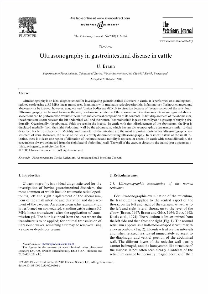
The abdominal wall, spleen and wall of the rumen
or reticulum can usually be imaged from the sixth
and seventh intercostal spaces of the left ventral thorax
(Fig. 4). Ultrasonographically, the spleen appears in
cross-section as a 2–3 cm thick structure situated be-
tween the abdominal wall and the rumen or the reticu-
lum. It tapers off ventrally, where the lateral and the
medial surface merge. The capsule of the spleen appears
as an echogenic line, and the splenic pulp appears as
many small, evenly distributed echoes. The vasculature
of the spleen is seen in cross-section as oval hypoecho-
genic structures in the pulp.
The reticulum, omasum and liver can be imaged from
the intercostal spaces of the right thorax. The omasal
wall is seen as a circular, distinctly echogenic line, im-
mediately adjacent to the right thoracic wall. The wall of
Fig. 1. Ultrasonographic examination of the left paramedian view of
the reticulum. (1) Reticulum, (2) craniodorsal blind sac of the rumen,
(3) ventral sac of rumen, (4) diaphragm (reproduced from Braun and
G€ootz, 1994).
Fig. 2. Ultrasonogram of the normal reticulum viewed from the left
ventral thorax. (1) Ventral abdominal wall, (2) musculophrenic vein,
(3) reticulum. Cr: cranial and Cd: caudal.
Fig. 3. Ultrasonogram of the normal reticulum, craniodorsal blind sac
of the rumen, ventral sac of the rumen, and abomasum viewed from
the left ventral thorax. (1) Ventral abdominal wall, (2) musculophrenic
vein, (3) abomasum, (4) reticulum, (5) craniodorsal blind sac of the
rumen, (6) cranial part of the ventral sac of the rumen. Cr: cranial andCd: caudal.
U. Braun / The Veterinary Journal 166 (2003) 112–124 113
7/29/2019 Ultrasonograph Yin Gastrointestinal Disease in Cattle
http://slidepdf.com/reader/full/ultrasonograph-yin-gastrointestinal-disease-in-cattle 3/13
the omasum is thicker than that of the reticulum. The
contents of the omasum cannot usually be imaged.
The left wall of the rumen and the longitudinal
groove, which divides the rumen into the dorsal and
ventral sacs of the rumen, can be seen from the left
thoracic and abdominal walls.
2.2. Traumatic reticuloperitonitis
In cattle with traumatic reticuloperitonitis, ultraso-
nography can be used to identify morphological changes
in the region of the cranial, ventral, or caudal reticularwall (Braun et al., 1993b). The caudoventral reticular
wall is the most frequently affected, often in association
with the craniodorsal blind sac of the rumen. The
changes in the contour of the reticulum depend on the
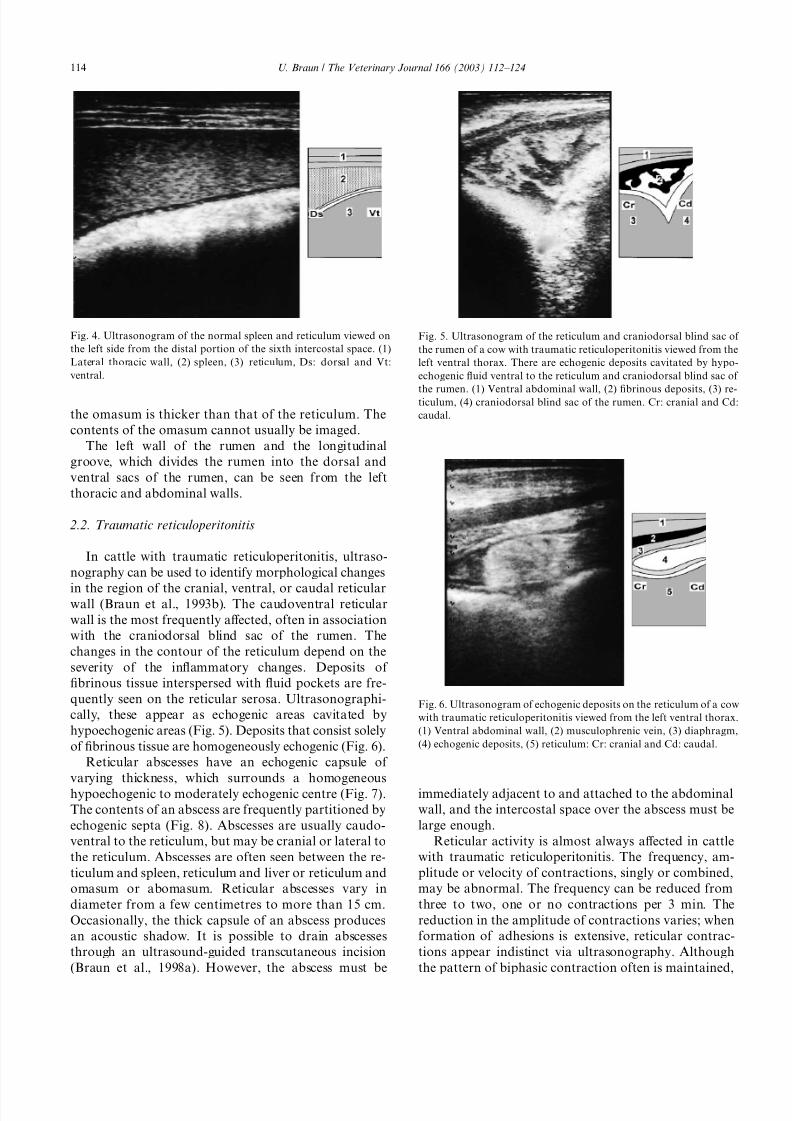
severity of the inflammatory changes. Deposits of
fibrinous tissue interspersed with fluid pockets are fre-
quently seen on the reticular serosa. Ultrasonographi-
cally, these appear as echogenic areas cavitated by
hypoechogenic areas (Fig. 5). Deposits that consist solely
of fibrinous tissue are homogeneously echogenic (Fig. 6).
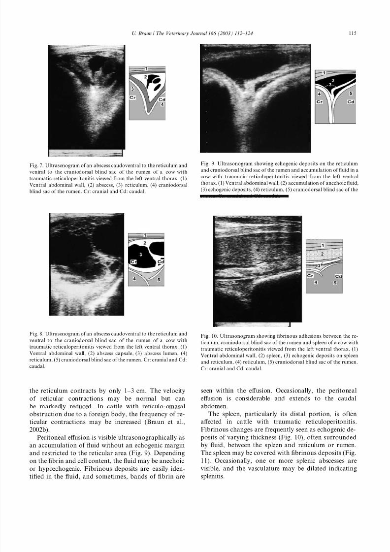
Reticular abscesses have an echogenic capsule of
varying thickness, which surrounds a homogeneous
hypoechogenic to moderately echogenic centre (Fig. 7).
The contents of an abscess are frequently partitioned by
echogenic septa (Fig. 8). Abscesses are usually caudo-
ventral to the reticulum, but may be cranial or lateral to
the reticulum. Abscesses are often seen between the re-
ticulum and spleen, reticulum and liver or reticulum and
omasum or abomasum. Reticular abscesses vary in
diameter from a few centimetres to more than 15 cm.
Occasionally, the thick capsule of an abscess produces
an acoustic shadow. It is possible to drain abscesses
through an ultrasound-guided transcutaneous incision
(Braun et al., 1998a). However, the abscess must be
immediately adjacent to and attached to the abdominal
wall, and the intercostal space over the abscess must be
large enough.
Reticular activity is almost always affected in cattle
with traumatic reticuloperitonitis. The frequency, am-
plitude or velocity of contractions, singly or combined,
may be abnormal. The frequency can be reduced from
three to two, one or no contractions per 3 min. The
reduction in the amplitude of contractions varies; when
formation of adhesions is extensive, reticular contrac-
tions appear indistinct via ultrasonography. Although
the pattern of biphasic contraction often is maintained,
Fig. 5. Ultrasonogram of the reticulum and craniodorsal blind sac of
the rumen of a cow with traumatic reticuloperitonitis viewed from the
left ventral thorax. There are echogenic deposits cavitated by hypo-
echogenic fluid ventral to the reticulum and craniodorsal blind sac of
the rumen. (1) Ventral abdominal wall, (2) fibrinous deposits, (3) re-ticulum, (4) craniodorsal blind sac of the rumen. Cr: cranial and Cd:
caudal.
Fig. 6. Ultrasonogram of echogenic deposits on the reticulum of a cow
with traumatic reticuloperitonitis viewed from the left ventral thorax.
(1) Ventral abdominal wall, (2) musculophrenic vein, (3) diaphragm,
(4) echogenic deposits, (5) reticulum: Cr: cranial and Cd: caudal.
Fig. 4. Ultrasonogram of the normal spleen and reticulum viewed on
the left side from the distal portion of the sixth intercostal space. (1)
Lateral thoracic wall, (2) spleen, (3) reticulum, Ds: dorsal and Vt:
ventral.
114 U. Braun / The Veterinary Journal 166 (2003) 112–124
7/29/2019 Ultrasonograph Yin Gastrointestinal Disease in Cattle
http://slidepdf.com/reader/full/ultrasonograph-yin-gastrointestinal-disease-in-cattle 4/13
the reticulum contracts by only 1–3 cm. The velocity
of reticular contractions may be normal but can
be markedly reduced. In cattle with reticulo-omasal
obstruction due to a foreign body, the frequency of re-
ticular contractions may be increased (Braun et al.,
2002b).
Peritoneal effusion is visible ultrasonographically as
an accumulation of fluid without an echogenic margin
and restricted to the reticular area (Fig. 9). Depending
on the fibrin and cell content, the fluid may be anechoic
or hypoechogenic. Fibrinous deposits are easily iden-
tified in the fluid, and sometimes, bands of fibrin are
seen within the effusion. Occasionally, the peritoneal
effusion is considerable and extends to the caudal
abdomen.
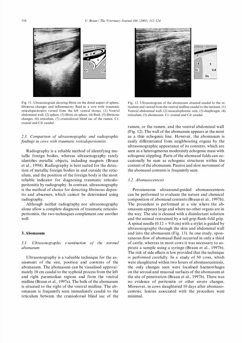
The spleen, particularly its distal portion, is often
affected in cattle with traumatic reticuloperitonitis.
Fibrinous changes are frequently seen as echogenic de-
posits of varying thickness (Fig. 10), often surrounded
by fluid, between the spleen and reticulum or rumen.
The spleen may be covered with fibrinous deposits (Fig.
11). Occasionally, one or more splenic abscesses are
visible, and the vasculature may be dilated indicating
splenitis.
Fig. 8. Ultrasonogram of an abscess caudoventral to the reticulum and
ventral to the craniodorsal blind sac of the rumen of a cow with
traumatic reticuloperitonitis viewed from the left ventral thorax. (1)
Ventral abdominal wall, (2) abscess capsule, (3) abscess lumen, (4)
reticulum, (5) craniodorsal blind sac of the rumen. Cr: cranial and Cd:
caudal.
Fig. 7. Ultrasonogram of an abscess caudoventral to the reticulum and
ventral to the craniodorsal blind sac of the rumen of a cow with
traumatic reticuloperitonitis viewed from the left ventral thorax. (1)
Ventral abdominal wall, (2) abscess, (3) reticulum, (4) craniodorsal
blind sac of the rumen. Cr: cranial and Cd: caudal.
Fig. 10. Ultrasonogram showing fibrinous adhesions between the re-
ticulum, craniodorsal blind sac of the rumen and spleen of a cow with
traumatic reticuloperitonitis viewed from the left ventral thorax. (1)
Ventral abdominal wall, (2) spleen, (3) echogenic deposits on spleen
and reticulum, (4) reticulum, (5) craniodorsal blind sac of the rumen.
Cr: cranial and Cd: caudal.
Fig. 9. Ultrasonogram showing echogenic deposits on the reticulum
and craniodorsal blind sac of the rumen and accumulation of fluid in a
cow with traumatic reticuloperitonitis viewed from the left ventral
thorax. (1) Ventral abdominal wall, (2) accumulation of anechoic fluid,
(3) echogenic deposits, (4) reticulum, (5) craniodorsal blind sac of the
rumen. Cr: cranial and Cd: caudal.
U. Braun / The Veterinary Journal 166 (2003) 112–124 115
7/29/2019 Ultrasonograph Yin Gastrointestinal Disease in Cattle
http://slidepdf.com/reader/full/ultrasonograph-yin-gastrointestinal-disease-in-cattle 5/13
2.3. Comparison of ultrasonographic and radiographic
findings in cows with traumatic reticuloperitonitis
Radiography is a reliable method of identifying me-
tallic foreign bodies, whereas ultrasonography rarely
identifies metallic objects, including magnets (Braun
et al., 1994). Radiography is best suited for the detec-
tion of metallic foreign bodies in and outside the retic-
ulum, and the position of the foreign body is the most
reliable indicator for diagnosing traumatic reticulo-peritonitis by radiography. In contrast, ultrasonography
is the method of choice for detecting fibrinous depos-
its and abscesses, which cannot be determined using
radiography.
Although neither radiography nor ultrasonography
alone allow a complete diagnosis of traumatic reticulo-
peritonitis, the two techniques complement one another
well.
3. Abomasum
3.1. Ultrasonographic examination of the normal
abomasum
Ultrasonography is a valuable technique for the as-
sessment of the size, position and contents of the
abomasum. The abomasum can be visualised approxi-
mately 10 cm caudal to the xyphoid process from the left
and right paramedian regions and from the ventral
midline (Braun et al., 1997a). The bulk of the abomasum
is situated to the right of the ventral midline. The ab-
omasum is frequently seen immediately caudal to the
reticulum between the craniodorsal blind sac of the
rumen, or the rumen, and the ventral abdominal wall
(Fig. 12). The wall of the abomasum appears at the most
as a thin echogenic line. However, the abomasum is
easily differentiated from neighbouring organs by the
ultrasonographic appearance of its contents, which are
seen as a heterogeneous moderately echogenic mass with
echogenic stippling. Parts of the abomasal folds can oc-
casionally be seen as echogenic structures within the
content of the abomasum. Passive and slow movement of
the abomasal contents is frequently seen.
3.2. Abomasocentesis
Percutaneous ultrasound-guided abomasocentesis
can be performed to evaluate the nature and chemical
composition of abomasal contents (Braun et al., 1997b).
The procedure is performed at a site where the ab-
omasum appears large and where no other organs are in
the way. The site is cleaned with a disinfectant solution
and the animal restrained by a tail grip flank-fold grip.
A spinal needle (0:12Â 9:0 cm) with a stylet is guided by
ultrasonography through the skin and abdominal wall
and into the abomasum (Fig. 13). In one study, spon-
taneous flow of abomasal fluid occurred in only a third
of cattle, whereas in most cows it was necessary to as-
pirate a sample using a syringe (Braun et al., 1997b).
The risk of side effects is low provided that the technique
is performed carefully. In a study of 50 cows, which
were slaughtered within two hours of abomasocentesis,
the only changes seen were localised haemorrhages
on the serosal and mucosal surfaces of the abomasum at
the site of penetration (Braun et al., 1997b). There was
no evidence of peritonitis or other severe changes.
Moreover, in cows slaughtered 10 days after abomaso-
centesis, lesions associated with the procedure were
minimal.
Fig. 11. Ultrasonogram showing fibrin on the distal aspect of spleen,
fibrinous changes and inflammatory fluid in a cow with traumatic
reticuloperitonitis viewed from the left ventral thorax. (1) Ventral
abdominal wall, (2) spleen, (3) fibrin on spleen, (4) fluid, (5) fibrinous
changes, (6) reticulum, (7) craniodorsal blind sac of the rumen. Cr:cranial and Cd: caudal.
Fig. 12. Ultrasonogram of the abomasum situated caudal to the re-
ticulum and viewed from the ventral midline caudal to the sternum. (1)
Ventral abdominal wall, (2) musculophrenic vein, (3) diaphragm, (4)
reticulum, (5) abomasum. Cr: cranial and Cd: caudal.
116 U. Braun / The Veterinary Journal 166 (2003) 112–124

7/29/2019 Ultrasonograph Yin Gastrointestinal Disease in Cattle
http://slidepdf.com/reader/full/ultrasonograph-yin-gastrointestinal-disease-in-cattle 6/13
Abomasal fluid is assessed for colour, smell and thepresence of blood, and the pH is determined. Additional
analyses include determination of the concentrations of
sodium, potassium, volatile fatty acids, bile acids and
pepsin. Normally, the colour ranges from olive green to
grey, and the fluid has a sour smell. The pH of abomasal
fluid varies from 1.38 to 4.50 (2:5Æ 0:87) (Braun et al.,
1997a). Higher values occur with abomasal haemor-
rhage, addition of bile or chronic abomasitis due to
ostertagiosis. Blood or haemoglobin, which is deter-
mined using a commercial test kit, such as hemoFEC
(Boehringer), does not normally occur in abomasal fluid
and its presence indicates disease. In the majority of cases, blood originates from an abomasal ulcer (Braun
et al., 1997b) and rarely from the rumen or small
intestine.
3.3. Left displacement of the abomasum
An ultrasonographic examination is useful to confirm
the diagnosis of left displacement of the abomasum in
unclear cases (Braun et al., 1997c). The last three in-
tercostal spaces on the left side are examined ventrally to
dorsally with the transducer held parallel to the ribs
(Fig. 14). Normally, the rumen is immediately adjacent
to the left abdominal wall (Fig. 15). Thus, on ultrason-
ograms, the ruminal wall is medial to the abdominal
wall and ventrodorsally appears as a smooth, thick,
echogenic line, which is indented at the left longitudinal
sulcus. The ruminal contents cannot be visualised be-
cause of their gaseous nature. With left displacement of
the abomasum, the wall of the rumen often remains
immediately adjacent to the abdominal wall in the
ventral region (Fig. 16). When the transducer is moved
dorsally, it becomes apparent that the wall of the rumen
is pushed medially and can no longer be imaged ultra-
sonographically. Instead the abomasum is seen, located
between the abdominal wall and rumen. Moving the
transducer further dorsally, the abomasum disappears
and the rumen reappears on the screen. The abomasal
contents do not appear uniform because, ventrally, there
are fluid ingesta, and dorsally, there is a gas cap that
varies in extent. The ingesta visible ventrally in the ab-
omasum appear hypoechogenic (Fig. 17). Occasionally,
the abomasal folds are visible among the ingesta and
appear as elongated, echogenic, sickle-shaped structures
(Fig. 18). The ruminal wall can often be seen medial to
the ingesta. The abomasal gas cap, seen further dorsally,
is characterised by reverberation artifacts (Fig. 19)
similar to those observed during the ultrasonographic
examination of lung. The artifacts are caused by the
reflection of the ultrasound waves by abomasal gas and
Fig. 14. Ultrasonographic examination of the abdominal cavity of a
cow from the last intercostal space of the left abdominal wall. In an
animal with a normal abomasum, the rumen is situated immediately
adjacent to the abdominal wall. (1) Abdominal wall, (2) rumen
(reproduced from Braun et al., 1997c).
Fig. 15. Ultrasonogram showing the rumen adjacent to the abdominal
wall in a healthy cow; viewed from the 12th intercostal space on the left
side. (1) Abdominal wall, (2) wall of rumen, (3) rumen. Ds: dorsal and
Vt: ventral. (reproduced from Braun et al., 1997c).
Fig. 13. Schematic representation of ultrasound-guided abomasocen-
tesis as viewed from the right side. (1) Abomasum, (2) omasum, (3)
reticulum, (4) rumen, (5) diaphragm, (6) udder (reproduced from
Braun et al., 1997b).
U. Braun / The Veterinary Journal 166 (2003) 112–124 117
7/29/2019 Ultrasonograph Yin Gastrointestinal Disease in Cattle
http://slidepdf.com/reader/full/ultrasonograph-yin-gastrointestinal-disease-in-cattle 7/13
reverberation between the transducer and the abomasal
surface. They appear as lines of varying echogenicityrunning parallel to the abomasal surface and become
weaker as the distance from the transducer increases. At
a depth of 7–8 cm, they are no longer visible (Braun
et al., 1997c). Reverberation artifacts prevent the visu-
alisation of the ruminal wall medial to the abomasal gas
cap.
3.4. Right displacement of the abomasum
Ultrasonography is a useful diagnostic tool in
doubtful cases of right displacement of the abomasum.
The area immediately caudal to the last rib and the
caudal two to three intercostal spaces on the right side
are examined ventrodorsally with the transducer held
parallel to the ribs (Braun, 1997). Normally, loops of
small intestine are imaged in cross-section and, less
commonly, longitudinally in the ventral abdomen; fur-
ther dorsally, the liver is seen immediately adjacent to
the right abdominal wall. In animals with right dis-
placement of the abomasum, the liver is displaced from
the abdominal wall and cannot be imaged. The ab-
omasum is seen where the liver would normally be,
immediately adjacent to the right abdominal wall.
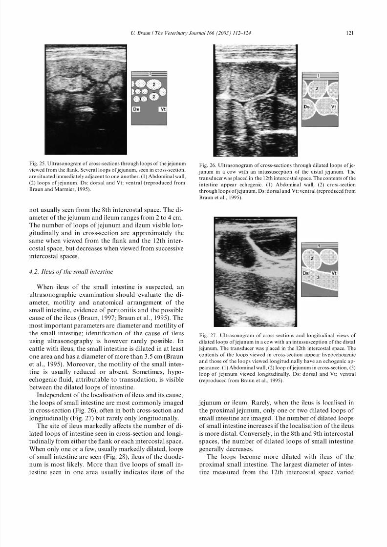
Fig. 17. Ultrasonogram of left displacement of the abomasum viewed
from the ventral region of the 12th intercostal space. (1) Abdominal
wall, (2) abomasum with hypoechogenic ingesta, (3) rumen displaced
medially. Ds: dorsal and Vt: ventral. (reproduced from Braun et al.,
1997c).
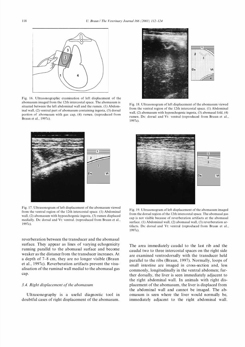
Fig. 18. Ultrasonogram of left displacement of the abomasum viewed
from the ventral region of the 12th intercostal space. (1) Abdominal
wall, (2) abomasum with hypoechogenic ingesta, (3) abomasal fold, (4)
rumen. Ds: dorsal and Vt: ventral (reproduced from Braun et al.,
1997c).
Fig. 19. Ultrasonogram of left displacement of the abomasum imaged
from the dorsal region of the 12th intercostal space. The abomasal gas
cap is not visible because of reverberation artifacts at the abomasal
surface. (1) Abdominal wall, (2) abomasal wall, (3) reverberation ar-
tifacts. Ds: dorsal and Vt: ventral (reproduced from Braun et al.,
1997c).
Fig. 16. Ultrasonographic examination of left displacement of the
abomasum imaged from the 12th intercostal space. The abomasum is
situated between the left abdominal wall and the rumen. (1) Abdom-
inal wall, (2) ventral part of abomasum containing ingesta, (3) dorsal
portion of abomasum with gas cap, (4) rumen. (reproduced from
Braun et al., 1997c).
118 U. Braun / The Veterinary Journal 166 (2003) 112–124

7/29/2019 Ultrasonograph Yin Gastrointestinal Disease in Cattle
http://slidepdf.com/reader/full/ultrasonograph-yin-gastrointestinal-disease-in-cattle 8/13
Its ultrasonographic appearance is the same as that
described previously for left displacement.
3.5. Defects in abomasal emptying
Defects in abomasal emptying occur with functional
or mechanical pyloric stenosis. Abomasal emptying canalso be impaired secondary to intestinal ileus. With all
these disorders, the abomasum is dilated but not dis-
placed dorsally or torsed. Depending on its degree of
fill, the dilated abomasum can be imaged on the right
side from the ventral region of intercostal spaces 8 to
12, or even from the ventral abdomen caudal to the
ribs. In contrast to left or right displacement, there is
no accumulation of gas in the non-displaced, dilated
abomasum. The abomasal contents appear predomi-
nantly hypoechogenic and homogeneous, and because
of sequestration of hydrochloric acid (HCl), are fre-
quently fluid in nature. This often allows for good vi-
sualisation of the abomasal folds, which appear as thin,
echogenic, wavy structures (Fig. 20). In animals with
ileus, the loops of intestine are dilated and clearly
visible. With pyloric stenosis, the small intestine is
empty.
Discrete lesions of the abomasal mucosa such as
chronic abomasitis, parasitic nodules, erosions and type-
I ulcers cannot be imaged ultrasonographically using a
3.5 MHz linear transducer. In a study involving 50 cows,
chronic abomasitis identified on postmortem examina-
tion in 48 cows could not be visualised by ultrasono-
graphy before slaughter (Wild, 1995; Braun et al.,
1997a). The same was true for mucosal erosions ortype-I ulcers found in the fundic and pyloric regions,
respectively, in 16 cows.
4. Intestine
4.1. Normal small intestine
For ultrasonography of the small intestine in cattle
(Fig. 21), the area from the tuber coxae to the eighth
intercostal space and from the transverse processes of the vertebrae to the linea alba on the right side is ex-
amined (Braun and Marmier, 1995; Marmier, 1993).
The appearance of loops of small intestine and their
diameter, contents and motility are assessed. The wall of
the normal small intestine is 2–3 mm thick and its lu-
minal diameter is 2–4 cm (Braun and Marmier, 1995).
Evaluation of the contents of the small intestine in cattle
is usually straightforward because there is generally no
gas. As, unlike in small animals and humans, ruminants
digest carbohydrates principally in the forestomachs,
from which the gas is eructated (G€uurtler, 1980).
The ultrasonographic appearance of the contents of
the small intestine varies. Most commonly, the intestine
contains mucus or feed, which appears hyperechogenic.
In these cases, not only the intestinal wall closest to the
transducer, but also the intestinal contents and the wall
furthest from the transducer can be visualised. This is
also true for intestine filled with fluid, which is hypoe-
chogenic. In rare cases with gaseous intestinal contents,
the intestinal wall closest to the transducer appears as a
hyperechogenic line adjacent to an acoustic shadow.
Because of reflection of the ultrasound waves at the soft
tissue–air interface, the intestinal contents and the in-
testinal wall furthest from the transducer cannot be
Fig. 20. Ultrasonogram showing dilatation of the abomasum second-
ary to intussusception of the jejunum of a cow, viewed from the right
paramedian region. (1) Abdominal wall, (2) abomasum, (3) abomasal
fold. Cr: cranial, Cd: caudal, and Ds: dorsal (reproduced from Braun,
1997).
Fig. 21. Ultrasonographic examination of the intestine viewed from the
level of the first lumbar vertebra on the right side. (1) First lumbar
vertebra, (2) right kidney, (3) aorta, (4) rumen, (5) large intestine, (6)
small intestine (reproduced from Braun, 1997).
U. Braun / The Veterinary Journal 166 (2003) 112–124 119

7/29/2019 Ultrasonograph Yin Gastrointestinal Disease in Cattle
http://slidepdf.com/reader/full/ultrasonograph-yin-gastrointestinal-disease-in-cattle 9/13
visualised. In contrast with results in dogs (Penninck
et al., 1989), there is no difference between the diameter
of most of the small intestine before and after feeding in
cattle. This uniformity is probably because the fore-
stomachs constitute a feed reservoir and ingesta passes
along the intestine continuously, independent of feed
intake, and without changes in the diameter of the in-
testinal lumen, which would be measurable by ultraso-
nography.
The cranial part of the duodenum is relatively easy to
identify because it originates from the abomasum and is
in close proximity with the liver and gallbladder (Fig.
22). It is seen in cross section or longitudinally, medial
or ventral to the gallbladder and almost always from the
10th or 11th intercostal space (Fig. 23). The diameter of
the cranial part of the duodenum ranges from 0.9 to 5.5
cm (Braun and Marmier, 1995). The descending duo-
denum is also quite easily imaged in most cattle. From
the 10th, 11th or 12th intercostal space and from
the dorsal region of the right flank, it is observed in
cross-section or longitudinally (Fig. 24). The following
characteristics allow accurate identification of the de-
scending duodenum: it is situated immediately adjacent
to the abdominal wall and runs horizontally and cau-dally between the serosal lamellae of the omentum,
which appears as an echogenic envelope around the
descending duodenum. It then courses caudally to form
the caudal duodenal flexure at the level of the tuber
coxae. After this, it runs medially and cranially to form
the ascending duodenum. The diameter of the de-
scending duodenum varies from 1.5 to 3.5 cm. The as-
cending duodenum is situated more than 20.0 cm from
the right abdominal wall and as a result cannot be im-
aged using ultrasonography.
The jejunum and ileum form the longest part of the
small intestine and cannot be differentiated from one
another ultrasonographically. It is typical to see more
than ten loops of jejunum and ileum immediately adja-
cent to one another from the flank and lateral abdomi-
nal wall and from intercostal spaces 9 to 12 (Braun and
Marmier, 1995). The loops of small intestine are usually
seen in cross-section and occasionally longitudinally.
They can be differentiated from the descending duode-
num because they are not surrounded by omentum and
because they are constantly in motion (Fig. 25). Loops
of jejunum and ileum are seen from the 10th intercostal
space in approximately 70% of cows and from the 9th
intercostal space in approximately 10% cows. They are
Fig. 22. Schematic representation of ultrasonographic examination of
the duodenum in cows. L: liver, G: gallbladder, A: abomasum. (1)
Position of the transducer for the longitudinal examination of the
descending part of the duodenum. (2) Position of the transducer for the
examination of the descending duodenum in cross-section. (3) Position
of the transducer for examination of the cranial part of the duodenum
(reproduced from Braun and Marmier, 1995).
Fig. 23. Ultrasonogram of a cross-section through the cranial part of
the duodenum, viewed from the 10th intercostal space. The cranial
part of the duodenum lies medially to the gallbladder, which serves as
an acoustic window. (1) Abdominal wall, (2) liver, (3) gallbladder, (4)
cranial part of the duodenum, (5) omasum. Ds: dorsal and Vt: ventral
(reproduced from Braun and Marmier, 1995).
Fig. 24. Ultrasonogram of a cross-section through the descending part
of the duodenum, viewed from the flank. The descending part of the
duodenum is between the abdominal wall and the large intestine and is
surrounded by the greater omentum. (1) Abdominal wall, (2) de-
scending duodenum, (3) greater omentum, (4) colon. Ds: dorsal andVt: ventral (reproduced from Braun and Marmier, 1995).
120 U. Braun / The Veterinary Journal 166 (2003) 112–124
7/29/2019 Ultrasonograph Yin Gastrointestinal Disease in Cattle
http://slidepdf.com/reader/full/ultrasonograph-yin-gastrointestinal-disease-in-cattle 10/13
7/29/2019 Ultrasonograph Yin Gastrointestinal Disease in Cattle
http://slidepdf.com/reader/full/ultrasonograph-yin-gastrointestinal-disease-in-cattle 11/13
from 6.5 to 9.9 cm (7:7Æ 1:9) in animals with ileus of
the duodenum, from 3.5 to 9.8 cm (5:5Æ 1:7) in animals
with ileus of the jejunum and from 4.4 to 5.5 cm
(5:0Æ 0:4 cm) in animals with ileus of the ileum (Braun
et al., 1995). When interpreting the diameter of the in-
testine, it is important to remember that in healthy
cows, in which the intestine is full of ingesta, all parts of
the intestine will have a similar diameter. By contrast, in
animals with ileus, in addition to the extremely dilatedloops of intestine proximal to the ileus, there are usually
empty loops of intestine distal to the ileus (Fig. 29).
Furthermore, the intestinal lumen of a healthy cow is
constantly changing, whereas the increased intestinal
diameter of a cow with ileus remains unchanged be-
cause the intestinal motility is markedly reduced or
absent.
The contents of the small intestine appear predomi-
nantly echogenic and rarely anechoic. Different parts of the intestine of the same animal may be echogenic or
anechogenic. Intraluminal gas, which is associated with
reverberation artifacts, is rarely observed.
In the majority of cows with ileus of the small intes-
tine, intestinal motility is markedly reduced or absent.
However, movement of intestinal contents often is ap-
parent although no intestinal contractions are visible.
This flowing movement is presumably due to the passive
movement of the intestine by respiratory activity and
possibly by the movement of adjacent organs such as the
rumen or abomasum.
The cause of ileus can seldom be determined ultra-
sonographically. Often, this is because the cause of
ileus is further from the abdominal wall than the
penetration capacity of the transducer. A common
cause of ileus is intussusception, the ultrasonographic
appearance of which in cross-section has been de-
scribed as bowel within bowel, bullÕs eye lesion, target
pattern or as multiple layered, onion ring-type mass
with varing echogenicities (Fig. 30). Typically, the in-
vaginated intestinal wall is swollen. Depending on the
severity of oedema and the imaging plane, the affected
area of intestine may appear hyperechogenic or hyp-
oechogenic. Viewed longitudinally, the typical lumen-
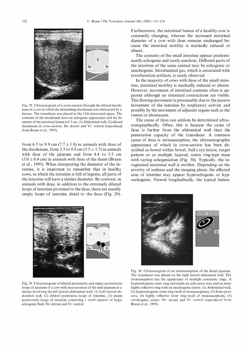
Fig. 28. Ultrasonogram of a cross-section through the dilated duode-
num of a cow in which the descending duodenum was obstructed by a
bezoar. The transducer was placed in the 11th intercostal space. The
contents of the duodenum have an echogenic appearance and the di-
ameter of the intestinal lumen is 8.5 cm. (1) Abdominal wall, (2) dilatedduodenum in cross-section. Ds: dorsal and Vt: ventral (reproduced
from Braun et al., 1995).
Fig. 29. Ultrasonogram of dilated prestenotic and empty poststenotic
loops of jejunum of a cow with incarceration of the mid-jejunum in a
hernia involving the left lateral abdominal wall. (1) Left lateral ab-
dominal wall, (2) dilated prestenotic loops of intestine, (3) empty
poststenotic loops of intestine containing a small amount of hypo-
echogenic fluid. Ds: dorsal and Vt: ventral.
Fig. 30. Ultrasonogram of an intussusception of the distal jejunum.
The transducer was placed on the right lateral abdominal wall. The
intussusception has the appearance of multiple concentric rings. A
hyperechogenic outer ring surrounds an echo-poor area and an inner
highly reflective ring with an anechogenic centre. (1) Abdominal wall,
(2) hyperechogenic outer ring (wall of intussuscipiens), (3) Echo-poor
area, (4) highly reflective inner ring (wall of intussusceptum), (5)
anechogenic centre. Ds: dorsal and Vt: ventral (reproduced from
Braun et al., 1995).
122 U. Braun / The Veterinary Journal 166 (2003) 112–124

7/29/2019 Ultrasonograph Yin Gastrointestinal Disease in Cattle
http://slidepdf.com/reader/full/ultrasonograph-yin-gastrointestinal-disease-in-cattle 12/13
within-a-lumen can be clearly identified and has been
described as a ‘‘sandwich’’ configuration. In rare cases,
compression of the small intestine by abscesses in the
region of the liver or compression of the duodenum
between the liver and gallbladder can be identified.
Ileus can also be caused by generalised peritonitis with
fibrinous adhesions involving the small intestine. Insuch cases, thickening of the intestinal wall, fibrinous
deposits and accumulation of intraabdominal fluid are
usually seen.
Failure of ingesta to pass through the small intestine
results in delayed passage of ingesta through the
abomasum, omasum and rumen, which consequently
become dilated (see Section 3.5).
Uncomplete perforation of the intestine usually leads
to chronic peritonitis. Ultrasonographically, this is seen
as intraabdominal fluid with bands of fibrin, which may
have a spiderweb-type of appearance among the loops
of intestine and organs. Free intra-abdominal gas with
its associated reverberation artifacts may be seen if in-
testinal gas has leaked through the perforation.
4.3. Normal large intestine
Carbohydrates remaining in the ingesta after their
passage through the forestomachs are fermented in the
large intestine, and the gas produced makes it more
difficult to image this section of the bowel (Marmier,
1993). The large intestine is always visible from the flank
and is situated medial to the descending duodenum,
whereby the colon is more dorsal and the proximal loop
of the colon and caecum are more ventral. The largeintestine is usually easy to differentiate from the small
intestine based on its marked gas content (Amrein, 1999;
Braun and Amrein, 2001; Marmier, 1993). Because of
the gas, only the wall of the large intestine closest to the
transducer can usually be imaged and appears as a thick
echogenic line. However, reverberation artifacts origi-
nating from the tissue-gas interphase may become su-
perimposed on the image of the wall and obscure it. The
wall of the large intestine furthest from the transducer
cannot be imaged. Usually, the proximal loop of the
large colon, the caecum and the colon can be visualised.
The proximal loop of the large colon and the caecum
appear as thick, echogenic, continuous and slightly
curved lines (Fig. 31). The spiral loop of the colon has
the appearance of a garland with a number of echogenic
arched lines next to each other (Fig. 32). In contrast to
the small intestine, which has vigorous peristaltic ac-
tivity and segmental contractions, very few contractions
are observed in the large intestine.
4.4. Caecal dilatation
Diagnosis of caecal dilatation usually is straightfor-
ward but may be difficult when the dilatation is com-
plicated by retroflexion of the caecum. In that case,
either no abnormal findings can be palpated, or a dis-
tended viscus can be palpated only with the tips of the
fingers. The differential diagnoses must include ileus of
the small intestine and right displacement of the ab-
omasum, respectively. A diagnosis by clinical examina-
tion alone may not be possible, but ultrasonography
allows the differentiation between right displacement of
the abomasum, ileus of the small intestine and caecal
dilatation. A dilated caecum can always be imaged from
the lateral abdominal wall (Amrein, 1999; Braun et al.,
Fig. 31. Ultrasonogram of a cross-section of the caecum viewed from
the flank. The caecal wall closest to the transducer is echogenic and the
wall furthest from the transducer cannot be visualised because of the
gas content of the caecum. Several loops of small intestine, viewed in
cross-section, are seen between the abdominal wall and caecum. (1)
Abdominal wall of right flank, (2) cross-sections of loops of small
intestine, (3) caecum. Ds: dorsal and Vt: ventral (reproduced from
Braun, 1997).
Fig. 32. Ultrasonogram of a cross-section of the spiral colon viewed
from the flank. The portion of colon has a garland-like appearance.
The wall of the colon closest to the transducer appears echogenic. (1)
Abdominal wall of right flank, (2) Colon. Ds: dorsal and Vt: ventral
(reproduced from Braun, 1997).
U. Braun / The Veterinary Journal 166 (2003) 112–124 123

7/29/2019 Ultrasonograph Yin Gastrointestinal Disease in Cattle
http://slidepdf.com/reader/full/ultrasonograph-yin-gastrointestinal-disease-in-cattle 13/13
2002a) and in some cases, may be seen from the 12th,
11th and 10th intercostal spaces. The dilated caecum
and the proximal loop of the colon are almost always
immediately adjacent to the abdominal wall. Because of
the gaseous contents, only the wall of the dilated caecum
(Fig. 33) and proximal loop of the colon closest to the
transducer are seen ultrasonographically and appear asthick, echogenic, semi-circular lines (Fig. 34). In cases
with fluid ingesta instead of gaseous contents in the
caecum and proximal loop of the colon, the lumen ap-
pears moderately echogenic. Differentiation of caecum
and proximal loop of the colon may be difficult ultra-
sonographically unless the ileocaecal fold of the perito-
neum between the two can be identified.
References
Amrein, E.M., 1999. Ultraschalluntersuchungen bei K€
uuhen mitBlinddarmdilatation. DrMedVet Thesis, Faculty of Veterinary
Medicine, University of Zurich.
Braun, U., 1997. Atlas und Lehrbuch der Ultraschalldiagnostik beim
Rind. Parey Buchverlag, Berlin.
Braun, U., Amrein, E., 2001. Ultrasonographic examination of the
caecum and proximal and spiral loop of the colon of cattle. The
Veterinary Record 149, 45–48.
Braun, U., Amrein, E., Koller, U., Lischer, C., 2002a. Ultrasono-
graphic findings in cows with dilatation, torsion, and retroflexion
of the caecum. The Veterinary Record 150, 75–79.
Braun, U., Fl€uuckiger, M., G€ootz, M., 1994. Comparison of ultrasono-
graphic and radiographic findings in cows with traumatic reticu-
loperitonitis. The Veterinary Record 135, 470–478.
Braun, U., Fl€uuckiger, M., N€aageli, F., 1993a. Radiography as an aid in
the diagnosis of traumatic reticuloperitonitis in cattle. The Veter-inary Record 132, 103–109.
Braun, U., G€ootz, M., 1994. Ultrasonography of the reticulum in cows.
American Journal of Veterinary Research 55, 325–332.
Braun, U., G€ootz, M., Marmier, O., 1993b. Ultrasonographic findings
in cows with traumatic reticuloperitonitis. The Veterinary Record
133, 416–422.
Braun, U., Iselin, U., Lischer, C., Fluri, E., 1998a. Ultrasonographic
findings in five cows before and after treatment of reticular
abscesses. The Veterinary Record. 142, 184–189.
Braun, U., Marmier, O., 1995. Ultrasonographic examination of
the small intestine of cows. The Veterinary Record 136, 239–244.
Braun, U., Marmier, O., Pusterla, N., 1995. Ultrasonographic exam-
ination of the small intestine of cows with ileus of the duodenum,
jejunum, or ileum. The Veterinary Record 137, 209–215.
Braun, U., Pusterla, N., Sch€oonmann, M., 1997c. Ultrasonographicfindings in cows with left displacement of the abomasum. The
Veterinary Record 141, 331–335.
Braun, U., Schweizer, G., Fl€uuckiger, M., 2002b. Radiographic and
ultrasonographic findings in three cows with reticulo-omasal ob-
struction dueto a foreignbody.The VeterinaryRecord 150,580–581.
Braun, U.,Wild, K.,Guscetti,F., 1997a.Ultrasonographicexamination
of the abomasum of 50 cows. The Veterinary Record 140, 93–98.
Braun, U., Wild, K., Merz, M., Hertzberg, H., 1997b. Percutaneous
ultrasound-guided abomasocentesis in cows. The Veterinary
Record 140, 599–602.
G€ootz, M., 1992. Sonographische Untersuchungen an der Haube des
Rindes. DrMedVet Thesis, Faculty of Veterinary Medicine, Uni-
versity of Zurich.
G€uurtler, H., 1980. Die Physiologie der Verdauung und Absorption. In:
Kolb, E. (Ed.), Lehrbuch der Physiologie der Haustiere. VebGustav Fischer, Stuttgart, pp. 177–339.
Kaske, M., Midasch, A., Rehage, J., 1994. Sonographic investigation
of reticular contractions in healthy sheep, cows, and goats and in
cows with traumatic reticulo-peritonitis. Journal of Veterinary
Medicine, Series A 41, 748–756.
Marmier, O., 1993. Sonographische Untersuchungen am Darm des
Rindes. DrMedVet Thesis, Faculty of Veterinary Medicine, Uni-
versity of Zurich.
Penninck, D.G., Nyland, T.G., Fisher, P.E., Kerr, L.Y., 1989.
Ultrasonography of the normal canine gastrointestinal tract.
Veterinary Radiology 30, 272–276.
Wild, K., 1995. Sonographische Untersuchungen am Labmagen des
Rindes. DrMedVet Thesis, Faculty of Veterinary Medicine, Uni-
versity of Zurich.
Fig. 33. Ultrasonogram of a dilated caecum of a cow with caecal di-
latation and torsion viewed from the right flank. The caecal wall
closest to the transducer appears as a curved echogenic line. The caecalcontents and wall furthest from the transducer are not visible. (1)
Lateral abdominal wall, (2) caecal wall. Ds: dorsal and Vt: ventral
(reproduced from Braun, 1997).
Fig. 34. Ultrasonogram of part of the spiral colon of a cow with caecal
dilatation and torsion viewed from the right flank. The intestinal wall
closest to the transducer appears echogenic. (1) Lateral abdominal
wall, (2) part of the colon. Ds: dorsal and Vt: ventral (reproduced from
Braun, 1997).
124 U. Braun / The Veterinary Journal 166 (2003) 112–124