ultra-sensitive detection of cysteine by gold nanorod … detection of cysteine by gold nanorod...
TRANSCRIPT

Biosensors and Bioelectronics 25 (2010) 2078–2083
Contents lists available at ScienceDirect
Biosensors and Bioelectronics
journa l homepage: www.e lsev ier .com/ locate /b ios
Ultra-sensitive detection of cysteine by gold nanorod assembly
Haowen Huanga,c,∗, Xuanyong Liub,c, Tao Huc, Paul K. Chuc,∗∗
a Key Laboratory of Theoretical Chemistry and Molecular Simulation of Ministry of Education, School of Chemistry and Chemical Engineering,Hunan University of Science and Technology, Xiangtan 411201, Chinab Shanghai Institute of Ceramics, Chinese Academy of Science, Shanghai 200050, Chinac Department of Physics & Materials Science, City University of Hong Kong, Tat Chee Avenue, Kowloon, Hong Kong, China
a r t i c l e i n f o
Article history:Received 11 January 2010Received in revised form 5 February 2010Accepted 5 February 2010Available online 12 February 2010
Keywords:CysteineGold nanorodsBiosensor
a b s t r a c t
In this paper, we have developed a simple, rapid, selective, and ultra-sensitive assay to detect cysteineby means of gold nanorods assembly. This methodology is based on the end-to-end assembly of goldnanorods and it is very sensitive due to the useful formation of the gold nanorods chain in the presenceof cysteine under acidic aqueous solution. The sharp absorption peak changes observed from the assem-bly formed by the gold nanorods allow one to monitor very subtle differences induced by cysteine in anacidic medium and therefore, it can provide a quantitative measure of the cysteine concentration. Onthe other hand, the high extinction coefficients of gold nanorods allow one to monitor the changes atlower concentrations compared to that accomplished by conventional absorbance-based chromophores.Therefore, one of the significant features is the ability to detect cysteine in the presence of other essen-tial �-amino acids at concentrations as low as 10 pM, which to the best of our knowledge is the lowestreported for a colorimetric cysteine detection system. More importantly, this assay is particularly attrac-tive because it does not rely on organic cosolvents, enzymatic reactions, light-sensitive dye molecules,lengthy protocols, and sophisticated instrumentation.
© 2010 Elsevier B.V. All rights reserved.
1. Introduction
As an essential amino acid in natural proteins which is biolog-ically active as well as thiol-containing, cysteine plays a crucialbiological role in the human body including protein synthesis,detoxification, and metabolism. It is also a potential neurotoxin(Wang and Cynader, 2001) biomarker for various medical con-ditions (Liu et al., 2000), and disease-associated physiologicalregulator (Droge and Holm, 1997) that is widely used in the medi-cal and food industry. As knowledge of cysteine provides criticalinsight into the proper physiological functions and diagnosis ofdisease, accurate detection of cysteine is very important.
Several methods such as fluorometry based on fluorescent dyes(Shao et al., 2006; Rusin et al., 2004; Wang et al., 2005; Shang andDong, 2009), electrochemical analysis (Zhou et al., 2007; Spãtaruet al., 2001; Bai et al., 2009) and chromatography separation (Luet al., 2007; Amarnath et al., 2003) have been developed to detectcysteine. Recently, advances have been made in the development
∗ Corresponding author at: Key Laboratory of Theoretical Chemistry and Molec-ular Simulation of Ministry of Education, School of Chemistry and ChemicalEngineering, Hunan University of Science and Technology, Xiangtan 411201, China.Tel.: +86 731 58290045; fax: +86 731 58290509.∗∗ Corresponding author. Tel.: +86 731 58290045; fax: +86 731 58290509.
E-mail addresses: [email protected] (H. Huang), [email protected](P.K. Chu).
of chromophoric colorimetric sensors to detect cysteine by usinggold nanoparticles (Lee et al., 2008; Li and Li, 2009). However, oneof the major difficulties associated with the selective detection of�-amino acids using chemosensors is their structural similarity dueto the presence of both carboxylic and amine groups. �-Amino acidscan be detected by chemosensors (Ros-Lis et al., 2004) and sphericalnanoparticles (Shang et al., 2007). Utilization of gold nanorods asprobes for cysteine detection has several advantages compared tospherical gold nanoparticles (Sudeep et al., 2005; Daniel and Astruc,2004). Analyte-induced aggregation of spherical gold nanoparti-cles results in a decrease in the plasmon absorption at around520 nm and formation of a long wavelength band (Kelly et al., 2003;Hao et al., 2004; Hallock et al., 2005). However, various functionalgroups such as amines, thiols, and carboxylic acids present in aminoacids and other organic molecules also influence their plasmonabsorption. In contrast, the presence of molecules containing suchfunctional groups does not influence plasmon absorption by goldnanorods thereby yielding excellent selectivity for cysteine (Sethiet al., 2009; Pramod and Thomas, 2008; Chang et al., 2005).
Gold nanorods exhibit strong optical extinction at the longitu-dinal plasmon band. The extinction coefficient of gold nanorodsis around 6 × 10⁹ M−1 cm−1 which is larger than the value of2.7 × 108 M−1 cm−1 for gold nanoparticles and several orders ofmagnitude more than those of traditional organic chromophores(El-sayed, 2001). More importantly, slight variation of the refractiveindex in the vicinity of the gold nanorods will induce a significant
0956-5663/$ – see front matter © 2010 Elsevier B.V. All rights reserved.doi:10.1016/j.bios.2010.02.003
H. Huang et al. / Biosensors and Bioelectronics 25 (2010) 2078–2083 2079
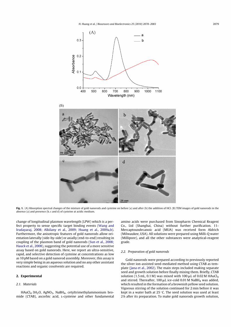
Fig. 1. (A) Absorption spectral changes of the mixture of gold nanorods and cysteine on before (a) and after (b) the addition of HCl. (B) TEM images of gold nanorods in theabsence (a) and presence (b, c and d) of cysteine at acidic medium.
change of longitudinal plasmon wavelength (LPW) which is a per-fect property to sense specific target binding events (Wang andIrudayaraj, 2008; Alkilany et al., 2009; Huang et al., 2009a,b).Furthermore, the anisotropic features of gold nanorods allow ori-entation laterally (side-by-side) or axially (end-to-end) resulting incoupling of the plasmon band of gold nanorods (Sun et al., 2008;Hauck et al., 2008), suggesting the potential use of a more sensitiveassay based on gold nanorods. Here, we report an ultra-sensitive,rapid, and selective detection of cysteine at concentrations as lowas 10 pM based on a gold nanorod assembly. Moreover, this assay isvery simple being in an aqueous solution and no any other assistantreactions and organic cosolvents are required.
2. Experimental
2.1. Materials
HAuCl4·3H2O, AgNO3, NaBH4, cetyltrimethylammonium bro-mide (CTAB), ascorbic acid, l-cysteine and other fundamental
amino acids were purchased from Sinopharm Chemical ReagentCo., Ltd (Shanghai, China) without further purification. 11-Mercaptoundecanoic acid (MUA) was received form Aldrich(Milwaukee, USA). All solutions were prepared using Milli-Q water(Millipore), and all the other substances were analytical-reagentgrade.
2.2. Preparation of gold nanorods
Gold nanorods were prepared according to previously reportedthe silver ion-assisted seed-mediated method using CTAB as tem-plate (Jana et al., 2002). The main steps included making separateseed and growth solution before finally mixing them. Briefly, CTABsolution (1.5 mL, 0.1 M) was mixed with 100 �L of 0.02 M HAuCl4and stirred. Thereafter, 100 �L ice-cold 0.01 M NaBH4 was added,which resulted in the formation of a brownish yellow seed solution.Vigorous stirring of the solution continued for 2 min before it waskept in a water bath at 25 ◦C. The seed solution was used at least2 h after its preparation. To make gold nanorods growth solution,

2080 H. Huang et al. / Biosensors and Bioelectronics 25 (2010) 2078–2083
1.5 mL of 0.02 M HAuCl4 and 1.0 mL of 0.01 M AgNO3 were addedinto 30 mL of 0.1 M CTAB, and 0.8 mL of 0.08 M ascorbic acid solu-tion was added. Ascorbic acid as a mild reducing agent changes thegrowth solution from dark yellow to colorless. After both solutionswere ready, 70 �L seed was taken out and added to the growthsolution at 25 ◦C. The color of the solution gradually changed in thefirst 15 min until finally stabilized.
2.3. Detection of cysteine
Prior to the detection of cysteine, excess CTAB was removedfrom the suspension of gold nanorods by careful centrifugation at12,000 rpm for 10 min. The nanorods, then, were redispersed inwater via sonication. Cysteine solution was prepared by dissolv-ing some cysteine with water as the stock solution (10 mM); theworking solution of cysteine was prepared by serial dilution of thestock solution. A typical analysis was realized by following steps.First, cysteine solution was mixed with gold nanorods. This solu-tion was allowed to react for 10 min at room temperature. Then, theHCl solution was added into the mixture solution. Absorption spec-tra of the reacted solution were recorded with 1 cm path-lengthcell.
3. Results and discussion
3.1. Spectral and TEM characterization of gold nanorods
Detection of cysteine based on the absorption spectra of goldnanorods is investigated in aqueous solution. Gold nanorods canbe suspended in the cysteine solution for a long time albeit at lowcetyltrimethylammonium bromide (CTAB) concentration. Additionof HCl into the mixture of gold nanorods and cysteine brings fortha significant change in the optical spectra, red-shift of longitudi-nal plasmon peak, as well as dramatic decrease in the intensity ofthe longitudinal plasmon band as shown in Fig. 1A. A new red-shifted peak at 950 nm emerges after HCl addition resulting fromcoupling of the plasmon absorption of gold nanorods assisted byself-assembly. TEM studies show that the gold nanorods beforeHCl addition are randomly distributed whereas cysteine-boundnanorods are preferentially self-assembled in the end-to-end con-figuration in the acidic medium as shown in Fig. 1B.
It is interesting that no noticeable spectral changes are observedfrom the mixture of gold nanorods and cysteine. Akin to regular sur-face plasmon resonance based on thin gold films (Huang and Chen,2006; Homola, 2008; Yao et al., 2008), localized surface plasmonresonance based on gold nanoparticles is suitable for the detection
Fig. 2. Influence of the addition sequence of reagents on the absorption spectra:(A) addition of AuNR into mixture of cysteine and HCl, (B) addition of HCl into themixture of cysteine and AuNR.
of macromolecules. A significant change occurs after the additionof HCl to the mixture of gold nanorods and cysteine. It resultsfrom the coupling plasmon band shown in Fig. 1B arising from theinteraction among the gold nanorods bound cysteine. As previousreports, gold nanorods can be assembled in either end-to-end orside-by-side orientation due to their shape anisotropy (Sun et al.,2008; Hauck et al., 2008; Sethi et al., 2009). The CTAB micelle cap-sulize gold nanorods and enable them stabilized in water. Most of
Scheme 1. Detection of cysteine using self-assembly of gold nanorods in the acidic medium.

H. Huang et al. / Biosensors and Bioelectronics 25 (2010) 2078–2083 2081
the CTAB surround the nanorods and few CTAB gather the end ofnanorods, which favors to make the cysteine functionalized goldnanorod form end-to-end linkage. In the first step, the thiol groupsin the cysteine molecules are functionalized on the surface of thegold nanorods. The isoelectric point of cysteine is 5.02, leaving mostof the amine and carboxylic moieties in the nearly neutral solu-tion with a pH of about 7 (Scheme 1). In this procedure, the smallmolecular cysteine will not induce evident changes in the LPW. Inthe second step, with the addition of HCl, cysteine leaves the proto-
nated amine moieties exposed on the surface of the gold nanorods.At the same time, the appended protonated amine moieties are ableto interact with oxygen atoms present in the carboxylic groups toform hydrogen bonds immediately adjacent to them on the surfaceof the neighboring gold nanorods. As a result, significant spectralchanges appear as shown in Fig. 1A. Accordingly, the acidic mediummakes the anisotropic gold nanorods self-assembled in end-to-end,which plays a very important role in the sensitive detection of cys-teine in the aqueous solution.
Fig. 3. Effect of AuNR and Cysteine ratio on absorption spectra, the molar ratio of cysteine and gold nanorods of (A), (B), (C), (D) and (E) are 2 × 10⁵:1, 1.5 × 10⁵:1, 3 × 10⁴:1,2 × 10⁴:1, 1.2 × 10⁴:1, respectively.

2082 H. Huang et al. / Biosensors and Bioelectronics 25 (2010) 2078–2083
3.2. Optimization of detection conditions of cysteine
To further verify the role of pH in this system, media with variouspH values are investigated. Different ionic forms of cysteine dependon the pH of the solution and cysteine molecules are present inionic forms at higher pH. Carboxylic groups are used to deprotonateamino acids larger than pH of 6. Therefore, the electrostatic inter-action among the negatively charged cysteine molecules preventsthe coupling of cysteine molecules. No noticeable LPW changes areobserved from the gold nanorods with the addition of cysteine inthe basic medium, indicating that end-to-end assembly does notoccur. A lower pH value accelerates end-to-end assembly of thegold nanorods and a higher degree of protonated amine moietiesfavors the formation of hydrogen bonds. As a result, rapid opticalspectra response can be accomplished when the pH value of themedium is less than 1.
We have investigated the effects of reagents added in differ-ent sequences. Firstly, the HCl solution and cysteine are mixedand then gold nanorods are added to the solution 10 min later.The intensity of absorption peak slowly decreases and a tail occurssimultaneously. A shoulder peak appears after 30 min and a signif-icant red-shifted peak appears as shown in Fig. 2A. In contrast, adrastic change occurs as shown in Fig. 2B if the gold nanorods, cys-teine and HCl are mixed simultaneously. In the former case, besidesformation of hydrogen bond among cysteine molecules, the strongacidic medium passivates the thiol group present in cysteine toreact with gold nanorods. The results indicate that the end-to-endassembly of gold nanorods arises from the ionic forms of cysteine. Inthis system, detection of cysteine must be carried out in the acidicmedium. Surprisingly, subsequent addition of NaOH to make thesolution basic solution does not damage the end-to-end assemblyand no significant influence is observed in the further formation ofassembly, as shown in Fig. S1. A plausible explanation is that thesteric hindrance of the nanorods chain and CTAB bilayer around thenanorods prevent damage of the hydrogen bond from the addedbase.
The sensing performance for cysteine is strongly influenced bythe assay conditions such as gold nanorods concentration and dif-ferent assay conditions are investigated. A fixed 1 �M cysteine anda series of concentrations of gold nanorods are first exploited asshown in Fig. 3. The different concentrations of gold nanorods leadto different response time and optical spectral changes. Generally, ahigher concentration of gold nanorods accelerates the assay for cys-teine detection. When the concentration of gold nanorods is below2 pM, the detection sensitivity for cysteine diminishes and responsetime increase significantly. Our results suggest that the appropri-ate molar ratio of cysteine and gold nanorods is round 104 to 105:1.Under this condition we investigated optimum reaction time andobserved that the detection signal reach the maximum within 8 minand no longer changes could be found after 10 min, thus, the opticalspectra were recorded 10 min later.
A previous study of the surface chemistry of Au colloids showsthat the amine (–NH2) group of some amino acids can bind to thegold nanoparticles at low pH values (Zhong et al., 2004). To inves-tigate the influence of other amino acids on selective detection ofcysteine, we study its optical spectral response to other amino acidsat a concentration of 100 �M. It is clear that only cysteine showsa significant change of optical spectra and the longitudinal plas-mon peak in the presence of other amino acids remain unchanged.This means that the amine group of the amino acids cannot bind tothe gold nanorods stabilized by CTAB in the acidic medium. There-fore, only cysteine of the essential amino acids can be specificallydetected by this assay. In addition, we investigated the effects ofMUA on the self-assembled mode of gold nanorods. MUA can alsoinduce end-to-end assembly of gold nanorods at pH above 8, asshown in Fig. S2. However, the response time is longer and a grad-
Fig. 4. The characteristic absorption spectra of 50 nM (A) and 10 nM (B) cysteine.
ually red-shift is still observed after 1 h reaction. On contrary, thenanorods stay isolated without showing end-to-end assembly inthe presence of MUA in the acidic medium (pH < 4). These resultsdemonstrate that the method mentioned above can specificallydetect cysteine.
The cysteine concentration changes the spectral characteristicsin the assay, as shown in Fig. 4, and 10 nM is a critical concentration.When the cysteine concentration is larger than 20 nM, a significantred-shift is observed from the longitudinal plasmon peak whereasonly a tail peak appears if the cysteine concentration is less than10 nM. The assay for cysteine detection relies on the end-to-endassembly of gold nanorods and therefore, it is very sensitive dueto the useful formation of the gold nanorods chain in the acidicaqueous solution. However, when the concentration of cysteine istoo low, the low proportion of assembled nanorods induce only atail peak to appear in the absorption spectra. The optical spectrashown in Supplementary information (Fig. S3A) reveal detectionof 10 pM cysteine. Compared to the control experiment shown inFig. S3B (Supplementary information), cysteine at a low concentra-tion of 10 pM can be detected readily.
Two modes of optical spectra changes occur in this assay asillustrated in Fig. 4A and B. Fig. 4A is the preferred mode to detectcysteine. In principle, the ideal state is that when all the cysteinemolecules are bound to the surface of the gold nanorods and thereare no unbounded gold nanorods in the solution. Thus, with theaddition of HCl, a significant red-shift in the longitudinal plas-mon peak can occur. A lower concentration of cysteine requireslower concentration of gold nanorods. Theoretically, a lower con-centration of gold nanorods is suitable for the detection of a lowerconcentration of cysteine. However, in practice, no red-shift inthe longitudinal plasmon peak occurs with the exception of the

H. Huang et al. / Biosensors and Bioelectronics 25 (2010) 2078–2083 2083
Fig. 5. The relationship between the concentration of cysteine (larger than 10 nM)and the variation of absorbance, and the relative standard deviations (RSD) are1.2–5.4% (n = 3).
appearance of a tail when the gold nanorod concentration is low.In addition, two other factors limit the use of a low concentrationof gold nanorods in the cysteine assay. A low concentration of goldnanorods leads to a long response time in this assay and a low con-centration of gold nanorods yields instability in detecting cysteine.Therefore, the concentration of gold nanorods is chosen to be largerthan 0.01 nM in this study.
The response LPW change is slightly variation when differ-ent gold nanorods or different concentration were employed todetect cysteine. A detectable LPW change can be observed with agood linear range within the cysteine concentration of 10 nm–1 �Mbased on around 0.1 nM gold nanorods with LPW 650 nm illus-trated in Fig. 5. With the concentration decrease, there is no clearlyobserved LPW change, however, during the concentration rangeof 10 pM–10 nm, a significant change of the optical spectra occurswith decreasing absorption intensity and the appearance of a tail,as shown in Fig. S3.
As aforementioned, three factors are responsible for the highsensitivity and selectivity of this assay. Firstly, the selectivity andtight binding of cysteine to the gold nanorods by the Au–S bondcompared to other essential amino acids lead to an assay withspecificity. Secondly, the sharp absorption peak changes observedfrom the assembly formed by the gold nanorods allow one to mon-itor very subtle differences in an acidic medium and therefore, itmay provide a quantitative measure of the cysteine concentration.Finally, the high extinction coefficients of gold nanorods allow oneto monitor the changes at lower concentrations compared to thataccomplished by conventional absorbance-based chromophores(extinction coefficients of about 104–105 cm−1 M−1).
4. Conclusions
In conclusion, this paper describes a simple, rapid, selective, andultra-sensitive assay to detect cysteine be means of gold nanorods.This assay is based on the thiophilicity of Au and the uniqueoptical properties. One of the significant features is the ability todetect cysteine in the presence of other essential �-amino acids atconcentrations as low as 10 pM, which to the best of our knowl-edge is the lowest reported for a colorimetric cysteine detection
system. This assay is particularly attractive because it does notrely on organic cosolvents, enzymatic reactions, light-sensitive dyemolecules, lengthy protocols, and sophisticated instrumentation.Considering the physiological link between cysteine and diseases,this assay has large potential in medicine.
Acknowledgments
This work was jointly supported by Scientific Research Fund ofHunan Provincial Education Department (06B028), National BasicResearch Fund (2005CB623901), Shanghai Science and TechnologyR&D Fund (0952nm04400, 0852nm03300, 07JC14057), and HongKong Research Grants Council (RGC) General Research Funds (GRF)No. CityU 112307.
Appendix A. Supplementary data
Supplementary data associated with this article can be found, inthe online version, at doi:10.1016/j.bios.2010.02.003.
References
Amarnath, K., Amarnath, V., Amarnath, K., Valentine, H.L., Valentine, W.M., 2003.Talanta 60, 1229–1238.
Alkilany, A.M., Nagaria, P.K., Hexel, C.R., Shaw, T.J., Murphy, C.J., Wyatt, M.D., 2009.Small 5, 701–708.
Bai, Y.H., Xu, J.J., Chen, H.Y., 2009. Biosens. Bioelectron. 24, 2895–2990.Chang, J.Y., Wu, H., Chen, H., Ling, Y.C., Tan, W., 2005. Chem. Commun., 1092–1094.Daniel, M.C., Astruc, D., 2004. Chem. Rev. 104, 293–346.Droge, W., Holm, E., 1997. FASEB J. 11, 1077–1089.El-sayed, M.A., 2001. Acc. Chem. Res. 34, 257–264.Hallock, A.J., Redmond, P.L., Brus, L.E., 2005. Proc. Natl. Acad. Sci. 102, 1280–1284.Hao, E., Schatz, G.C., Hupp, J.T., 2004. J. Fluoresc. 14, 331–341.Hauck, T.S., Ghazani, A.A., Chan, W.C.W., 2008. Small 4, 153–159.Homola, J., 2008. Chem. Rev. 108, 462–493.Huang, H., Chen, Y., 2006. Biosens. Bioelectron. 22, 644–648.Huang, H., He, C., Zeng, Y., Xia, X., Yu, X., Yi, P., Chen, Z., 2009a. Biosens. Bioelectron.
24, 2255–2259.Huang, H., Huang, S., Liu, X., Zeng, Y., Yu, X., Liao, B., Chen, Y., 2009b. Biosens. Bio-
electron. 24, 3025–3029.Jana, N.R., Gearheart, L., Obare, S.O., Murphy, C.J., 2002. Langmuir 18, 922–927.Kelly, K.L., Coronado, E., Zhao, L.L., Schatz, G.C., 2003. J. Phys. Chem. B 107, 668–677.Lee, J.S., Ulmann, P.A., Han, M.S., Mirkin, C.A., 2008. Nano Lett. 8, 529–533.Li, L., Li, B., 2009. Analyst 134, 1361–1365.Liu, J., Yeo, H.C., Overvik-Douki, E., Hagen, T., Doniger, S.J., Chu, D.W., Brooks, G.A.,
Ames, B.N., 2000. J. Appl. Physiol. 89, 21–28.Lu, C., Zu, Y., Yam, V.W.W., 2007. J. Chromatogr. A 1163, 328–332.Pramod, P., Thomas, K.G., 2008. Adv. Mater. 20, 4300–4305.Ros-Lis, J.V., Garcia, B., Jimenez, D., Martinez-Manez, R., Sancenon, F., Soto, J., Gon-
zalvo, F., Valldecabres, M.C., 2004. J. Am. Chem. Soc. 126, 4064–4065.Rusin, O., Luce, N.N.S., Agbaria, R.A., Escobedo, J.O., Jiang, S., Warner, I.M., Dawan,
F.B., Lian, K., Strongin, R.M., 2004. J. Am. Chem. Soc. 126, 438–439.Sethi, M., Joung, G., Knecht, M.R., 2009. Langmuir 25, 1572–1581.Shang, L., Dong, S., 2009. Biosens. Bioelectron. 24, 1569–1573.Shang, L., Qin, C., Wang, T., Wang, M., Wang, L., Dong, S., 2007. J. Phys. Chem. C 111,
13414–13417.Shao, N., Jin, J.Y., Cheung, S.M., Yang, R.H., Chan, W.H., Mo, T., 2006. Angew. Chem.
Int. Ed. 24, 4944–4948.Spãtaru, N., Sarada, B.V., Popa, E., Tryk, D.A., Fujishima, A., 2001. Anal. Chem. 73,
514–519.Sudeep, P.K., Joseph, S.T.S., Thomas, K.G., 2005. J. Am. Chem. Soc. 127, 6516–6517.Sun, Z., Ni, W., Yang, Z., Kou, X., Li, L., Wang, J., 2008. Small 4, 1287–1292.Wang, C., Irudayaraj, J., 2008. Small 4, 2204–2208.Wang, W., Rusin, O., Xu, X., Kim, K.K., Escobedo, J.O., Fakayode, S.O., Fletcher, K.A.,
Lowry, M., Schowalter, C.M., Lawrence, C.M., Fronczek, F.R., Warner, I.M., Stron-gin, R.M., 2005. J. Am. Chem. Soc. 127, 15949–15958.
Wang, X.F., Cynader, M.S., 2001. J. Neurosci. 21, 3322–3331.Yao, J., Stewart, M.E., Maria, J., Lee, T.W., Gray, S.K., Rogers, J.A., Nuzzo, R.G., 2008.
Angew. Chem. Int. Ed. 47, 5013–5017.Zhong, Z., Patskovskyy, S., Bouvrette, P., Luong, J.H.T., Gedanken, A., 2004. J. Phys.
Chem. B 108, 4046–4052.Zhou, M., Ding, J., Guo, L., Shang, Q.K., 2007. Anal. Chem. 79, 5328–5335.