ucma, a novel secreted cartilage-specific protein with implications
TRANSCRIPT
Ucma, a Novel Secreted Cartilage-specific Protein withImplications in Osteogenesis*□S
Received for publication, April 2, 2007, and in revised form, December 4, 2007 Published, JBC Papers in Press, December 21, 2007, DOI 10.1074/jbc.M702792200
Cordula Surmann-Schmitt‡, Uwe Dietz‡, Trayana Kireva‡, Nadia Adam‡, Jung Park‡, Andreas Tagariello§,Patrik Onnerfjord¶, Dick Heinegård¶, Ursula Schlotzer-Schrehardt�, Rainer Deutzmann**, Klaus von der Mark‡,and Michael Stock‡1
From the ‡Department of Experimental Medicine I, Nikolaus-Fiebiger Center of Molecular Medicine, University ofErlangen-Nuremberg, 91054 Erlangen, Germany, the §Institute of Human Genetics, University Hospital Erlangen,91054 Erlangen, Germany, the ¶Department of Experimental Medical Science, Lund University, 221 84 Lund, Sweden,the �Eye Hospital of the University Hospital Erlangen, 91054 Erlangen, Germany, and the **Institute of Biochemistry,University of Regensburg, 93053 Regensburg, Germany
Here we report on the structure, expression, and function of anovel cartilage-specific gene coding for a 17-kDa small, highlycharged, and secreted protein that we termedUcma (unique carti-lage matrix-associated protein). The protein is processed by afurin-like protease into an N-terminal peptide of 37 amino acidsand a C-terminal fragment (Ucma-C) of 74 amino acids. Ucma ishighly conserved between mouse, rat, human, dog, clawed frog,and zebrafish, but has no homology to other known proteins.Remarkable are 1–2 tyrosine sulfate residues/molecule and denseclusters of acidic and basic residues in the C-terminal part. In thedeveloping mouse skeleton Ucma mRNA is expressed in restingchondrocytes in the distal and peripheral zones of epiphyseal andvertebralcartilage.Ucmaissecreted intotheextracellularmatrixasanuncleavedprecursor and shows the same restricteddistributionpattern in cartilage as Ucma mRNA. In contrast, antibodies pre-pared against the processed C-terminal fragment located Ucma-Cin the entire cartilagematrix, indicating that it either diffuses or isretained until chondrocytes reach hypertrophy. During differenti-ation of an MC615 chondrocyte subclone in vitro, Ucma expres-sion parallels largely the expression of collagen II and decreaseswithmaturation toward hypertrophic cells. RecombinantUcma-Cdoes not affect expression of chondrocyte-specific genes or prolif-eration of chondrocytes, but interferes with osteogenic differenti-ation of primary osteoblasts, mesenchymal stem cells, andMC3T3-E1pre-osteoblasts.These findings suggest thatUcmamaybe involved in the negative control of osteogenic differentiation ofosteochondrogenic precursor cells in peripheral zones of fetal car-tilage and at the cartilage-bone interface.
Elucidation of molecular mechanisms underlying chondro-cyte differentiation is not only important for our understandingof skeletal development, but also of particular interest for ourknowledge on the behavior of chondrocytes following articularcartilage damage during cartilage repair and treatment ofdegenerative cartilage diseases. Initial steps of chondrogenesis,i.e. the formation of a cartilage blastema from limb budmesen-chymal cells, include cell condensation and onset of chondro-cyte differentiation marked by the expression of cartilage-spe-cific matrix proteins such as aggrecan, collagen II, IX, and XIand others (1, 2). These events are regulated by the orchestratedaction of several growth factors including BMPs, Wnt factors,FGFs, and the transcription factors Sox5, 6, and 9 (3, 4). Furthersteps of chondrocyte growth, maturation, and replacement bybone in the growth plate of long bones, ribs, and vertebrae dur-ing endochondral ossification can be defined by the stepwiseonset or decline of differentially expressed genes: collagen IIand Sox9 for resting and proliferating, FGFR3 for proliferatingand prehypertrophic, Ihh and PTHrP receptor for prehypertro-phic, collagen X for hypertrophic, and Runx2, osteocalcin, andMMP13 for late hypertrophic chondrocytes (2, 5–7). Similar tochondrogenesis, the steps of chondrocyte differentiation dur-ing endochondral ossification are regulated in a complex man-ner by BMPs, Wnt factors, and FGFs. In addition, Ihh/PTHrP,HIF-1�, thyroxine, and others are important regulators of thisprocess (2, 7).Little is known, however, on the early differentiation events
occurring in the distal, so-called “resting” zone of epiphysealcartilage. In a search for further chondrocyte specific genes, weidentified a novel, highly charged extracellular protein that isabundantly expressed in the upper immature zone of fetal andjuvenile epiphyseal cartilage. The gene, termedUcma, was dis-covered in a screen for differentially expressed genes in retinoicacid-treated mouse chondrocytes. Independently, Ucma wasalso identified in a human chondrocyte EST screen for candi-date genes of skeletal dysplasias (8). Here we report on the tran-sient expression of Ucma in the developing mouse skeletonbetween day E13.5 of embryonic development and 5 months ofpostnatal development. Ucma is predominantly expressed indistal peripheral, non-articulating zones of fetal epiphyses andvertebrae, but not by chondrocytes of the hypertrophic zone.Ucma contains tyrosine sulfate and is cleaved by a furin-like
* This work was supported in part by grants from the Deutsche Forschungs-gemeinschaft (to M. S., STO 824/1-1, and K. v. d. M., MA 534/18-2), and fromthe Swedish Research Council (to P. O.). The costs of publication of thisarticle were defrayed in part by the payment of page charges. This articlemust therefore be hereby marked “advertisement” in accordance with 18U.S.C. Section 1734 solely to indicate this fact.
□S The on-line version of this article (available at http://www.jbc.org) containssupplemental Figs. S1 and S2.
The nucleotide sequence(s) reported in this paper has been submitted to the Gen-BankTM/EBI Data Bank with accession number(s) EU368184.
1 To whom correspondence should be addressed: Dept. of ExperimentalMedicine I, Nikolaus-Fiebiger Center of Molecular Medicine, University ofErlangen-Nuremberg, Glueckstr. 6, 91054 Erlangen, Germany. Fax: 49-0-9131-8526341; E-mail: [email protected].
THE JOURNAL OF BIOLOGICAL CHEMISTRY VOL. 283, NO. 11, pp. 7082–7093, March 14, 2008© 2008 by The American Society for Biochemistry and Molecular Biology, Inc. Printed in the U.S.A.
7082 JOURNAL OF BIOLOGICAL CHEMISTRY VOLUME 283 • NUMBER 11 • MARCH 14, 2008
by guest on April 11, 2019
http://ww
w.jbc.org/
Dow
nloaded from
protease into N- and C-terminal fragments of 37 and 74 aminoacid residues, respectively. With specific antibodies we showthat the distribution of the unprocessed form of Ucma isrestricted to sites of UcmamRNA expression, while the C-ter-minal fragment Ucma-C is found in almost the entire cartilagematrix. During differentiation of MC615 chondrocytes in vitroUcma expression largely parallels the expression of collagen II,and declines with onset of hypertrophy. Furthermore, we pro-vide evidence that recombinant Ucma-C protein suppressesosteogenic differentiation of preosteoblasts in vitro. Therefore,we postulate that this protein may have a transient function inthe formation and stabilization of the juvenile cartilage matrixof epiphyseal, vertebral, and rib cartilage by suppressing osteo-genic differentiation of precursor cells present in peripheralzones and perichondrium of fetal cartilage.
EXPERIMENTAL PROCEDURES
Cell Lines and Primary Skeletal Cells—MC615 cells were cul-tured inDulbecco’smodified Eagle’smedium (DMEM)2/Ham’sF12 supplemented with 10% fetal calf serum (FCS) and pas-saged before reaching confluency as outlined before (9, 10).C3H10T1/2, C2C12, NIH3T3, and ATDC5 cells were culturedas previously reported inDMEMwith 10%FCS and 2mM L-glu-tamine (11). MC3T3-E1 cells were maintained in DMEM/Ham’s F12 with 10% FCS and subcultured when reaching con-fluency. To obtain clones ofMC615with unique differentiationstates individual subclones of this cell line were isolated by lim-iting dilution. Chondrogenic differentiation of one of these sub-clones (4C6) in vitro was achieved within 3–5 weeks in normalgrowth medium.Osteogenic differentiation of MC3T3-E1 preosteoblasts was
achieved by prolonged confluent culture in differentiationmedium (DMEM/Ham’s F12, 10% FCS, 50�g/ml ascorbic acid,10 mM �-glycerophosphate) as previously described (12). Pri-mary mouse calvarial cells were isolated from 3–6-day-oldmice as outlined earlier (11).Humanmesenchymal stem cells (hMSCs)were isolated from
fresh umbilical cord blood from healthy donors. After expan-sion osteogenic differentiation of hMSCs was achieved by pro-longed culture in differentiation medium (60% DMEM-LG(Invitrogen), 40% MCDB-201, 1� insulin-transferrin-sele-nium, 1� linoleic-acid-bovine-serum-albumin, 10�9 M dexa-methasone, 10�4 M ascorbic acid 2-phosphate, 10 ng/ml EGF(all from Sigma-Aldrich), 10 ng/ml PDGF-BB (R&D Systems),100 units of penicillin, 1000 units of streptomycin (Invitrogen)and 2% fetal calf serum) as reported earlier (13). For isolation ofprimary murine chondrocytes rib cages from newborn micewere treatedwith 1mg/ml trypsin in serum-freeDMEM/Ham’sF12 medium (PAA) at 37 °C for 45 min followed by digestionwith collagenase P (Roche Applied Science, Mannheim, Ger-many) (2 mg/ml in DMEM/Ham’s F12, 10% FCS, 2 h at 37 °C)(10). Rib chondrocytes were seeded in DMEM/Ham’s F12 con-
taining 10% FCS at densities of 1 � 104 to 2.5 � 104 cells/cm2.Bovine epiphyseal chondrocytes were isolated from the growthplates of fetal calves (about 4–6 months of gestation) and sep-arated into distinct fractions by centrifugation through a linearPercoll gradient as previously described (10, 14). For BMP stim-ulation of cells normal growth medium was replaced byDMEM/Ham’s F12 medium without or with 1% FCS supple-mented with recombinant BMP-2 at the indicated concentra-tions for 24 h or 48 h.Detection of the Transcription Start Site of the Ucma Gene—
The transcription start site of Ucma was detected by rapidamplification of 5�-cDNA ends (5�-RACE) using the Gene-RacerTM kit (Invitrogen) according to the manufacturer’sinstructions. For this purpose poly(A)� RNA was obtainedfrom primary murine rib chondrocytes using the Oligotex�mRNA Mini kit (Qiagen, Hilden, Germany). 150 ng poly(A)�RNAwas used in a 5�GeneRacer assay with gene specific prim-ers 5�-GGC GGT TGT AGA GGT AGG AAG GA-3� and5�-TCATCTCTCTGTTCCTCCACGAAGT-3� for primaryand nested PCR reactions, respectively. The product of thenested PCR reaction was cloned into pCR4-TOPO (Invitrogen,Karlsruhe, Germany) by TOPO-TA cloning and 12 individualclones were sequenced to reveal the 5� cDNA end representingthe transcription start site of Ucma.Analysis of mRNA Expression—Total RNA for expression
analyses and cloning was isolated using the RNeasy Kit (Qia-gen) according to themanufacturer’s instructions. To avoid anycontamination with genomic DNA the optional DNase stepwas included. Reverse transcription for (real-time) RT-PCRwas performed using SuperScriptTM II reverse transcriptase(Invitrogen) according to themanufacturer’s instructions. PCRreactions were carried out as previously reported using theTaqPCR Core Kit (Qiagen) and gene-specific primers (11).Real-time RT-PCR was performed as described previously
(10). Cyclophilin A and/or�-actin were used to standardize thetotal amount of cDNA in real-time PCR approaches. The fol-lowing primers were used for detection of respective mRNAlevels by (real-time) RT-PCR: murine Ucma: 5�-GTT TCTGGG GGT GCT CTG TA-3� (sense primer); 5�-GAG GAAATTGGAGGCATCAG-3� (antisense primer); bovineUcma:5�-CTC AGC CTT TCC AAA GAT GG-3� (sense primer);5�-GGG TAC AGG CCG TCG TAA T-3� (antisense primer);murine Runx2: 5�- ATA CCC CCT CGC TCT CTG TT-3�(sense primer); 5�-AGG TTG GAG GCA CAC ATA GG-3�(antisense primer); murine Col10a1: 5�-CAT AAA GGG CCCACTTGCTA-3� (sense primer); 5�-CAGGAATGCCTTGTTCTC CT-3� (antisense primer); murine �-actin: 5�-AGA GGGAAA TCG TGC GTG AC-3� (sense primer) and 5�-CAA TAGTGA TGA CCT GGC CGT-3� (antisense primer); murinecyclophilin A: 5�-CCA CCG TGT TCT TCG ACA T-3� (senseprimer) and 5�-CAG TGC TCA GAG CTC GAA AG-3� (anti-sense primer).For RT-PCR control reactions sequences of murine and
bovine glyceraldehyde-3-phosphate dehydrogenase (Gapdh)primers were adopted from PCR-Select cDNA Subtraction Kit(BD Clontech Germany, Heidelberg, Germany) and Schmidl etal. (10), respectively.
2 The abbreviations used are: DMEM, Dulbecco’s modified Eagle’s medium;FCS, fetal calf serum; RACE, rapid amplification of cDNA ends; MALDI-TOF,matrix-assisted laser desorption/ionization-time of flight; hMSCs, humanmesenchymal stem cells; RT, reverse transcription; MS, mass spectrometry;AA, ascorbic acid; �-GP, �-glycerophosphate; AP, alkaline phosphatase.
Ucma, a Novel Cartilage-specific Secreted Protein
MARCH 14, 2008 • VOLUME 283 • NUMBER 11 JOURNAL OF BIOLOGICAL CHEMISTRY 7083
by guest on April 11, 2019
http://ww
w.jbc.org/
Dow
nloaded from

Northern blot analyses were carried out as described before(12). A cDNA probe forUcmawas generated by RT-PCR usingthe following cDNA-specific primers: 5�-CCAGTGGCAT-TATGATGGC-3� (sense primer); 5�-TGAAAGTGTCTAT-CAGATGGGTG-3� (antisense primer). For detection of colla-gen type II transcripts a cDNA probe encompassingnucleotides 4464 to 4858 of the procollagen, type II, alpha 1(Col2a1), mRNA sequence (GenbankTM: NM_031163.2) wasemployed andGapdh transcripts were hybridized with a probepreviously described (12).RNA in situ hybridization on paraffin sections of mouse tis-
sues with digoxigenin-labeled antisense riboprobes for Ucmaand collagens �1(II) and �1(X) was carried out as reportedbefore (10). Specific cDNA fragments forUcma antisense ribo-probes including nucleotides 451–784 or 131–544 of themurine UcmamRNA sequence were obtained by RT-PCR andcloned into the pCRII-TOPO vector.Generation of Polyclonal Rabbit Antibodies (TAGL-1 and
UCMA-1) Directed against Ucma—For preparation of antibodiesagainst unprocessedUcma, a 17-amino acidpeptideoptimized forrabbit immune response (MSNFLKRRGKR�SPKSRD) and over-lapping the furin cleavage site (arrowhead) was conjugatedto KLH. A rabbit was injected subcutaneously with 0.4 mg ofUcma-KLH peptide in complete Freund’s adjuvant followedby four booster injections in incomplete adjuvant. The anti-sera were purified by affinity chromatography with a HiTrapNHS-activated HP column (GE Healthcare, Sweden) withUcma peptides and the resulting purified antibody wastermed TAGL-1. Rabbit antisera against cleaved anduncleaved forms of Ucma were generated by David Biotech-nologie GmbH (Regensburg, Germany) using purifiedrecombinant KLH-conjugated Ucma-C for immunization.Antibodies were purified by affinity chromatography withrecombinant Ucma-C as described above and termedUCMA-1.Analysis of Protein Expression—For Western blot analysis,
protein samples were resolved by SDS-polyacrylamide gel elec-trophoresis through denaturing 15% polyacrylamide gels asreported before (12, 15). After transfer onto nitrocellulosemembranes protein immunodetection was carried out asdescribed previously (10). Recombinant Ucma protein wasdetected with a mouse monoclonal anti-FLAG antibody(1:1000; Sigma). Endogenous Ucma protein was visualizedby incubating blots over night at 4 °C with TAGL-1 antibodydiluted 1:1000 in 5% skim milk powder in TBS-T and a poly-clonal goat anti rabbit IgG peroxidase conjugate as secondaryantibody. As loading control �-actin levels were determined onthe same blot as Ucma using an anti-�-actin antibody (Sigma).
Immunohistochemistry was performed as described before(10). Briefly, paraffin sections were rehydrated and pretreatedwith testicular hyaluronidase (2 mg/ml) for 1 h at 37 °C. Afterwashing with TBS and blocking with 5% bovine serum albuminsections were incubated with TAGL-1 antibody (1:1000) over-night at 4 °C or for 2 h at room temperature. Bound antibodywas visualized using the Link-Label IHC Detection System(Biogenex, San Ramon, CA) with biotinylated anti rabbit Ig,avidin-coupled alkaline phosphatase, and Fast Red tablets con-
taining levamisole (Sigma-Aldrich) according to the manufac-turer’s instructions.For postembedding immunogold labeling, tissue specimens
were fixed in 4% paraformaldehyde and 0.1% glutaraldehyde in0.1 M cacodylate buffer (pH 7.4) for 2 to 5 h at 4 °C. Specimenswere dehydrated serially to 70% ethanol at �20 °C and embed-ded in resin (LR White; Electron Microscopy Sciences). Ultra-thin sections were successively incubated in Tris-bufferedsaline (TBS), 1 mg/ml testicular hyaluronidase (pH 7.0), 0.05 Mglycine in TBS, 0.5% ovalbumin, and 0.5% fish gelatin in TBS,TAGL-1 antibody diluted 1:50 in TBS-ovalbumin overnight at4 °C, and finally in 10 nm gold-conjugated secondary antibody(BioCell, Cardiff,Wales,UK) diluted 1:30 inTBS-ovalbumin for1 h. After rinsing, the sections were stained with uranyl acetateand examined with a transmission electron microscope (906E,Leo, Oberkochen, Germany). In negative control samples, theprimary antibodywas replaced by PBS or equimolar concentra-tions of nonimmune rabbit IgG.Preparation of Recombinant Ucma—For episomal expres-
sion of recombinant Ucma the coding sequence of murineUcma including the signal peptide and C-terminally fused Hisand FLAG tags was cloned into the pCEP-Pu vector. This con-struct was stably transfected into HEK293EBNA cells. Episo-mally expressed Ucma(His)6FLAG was collected from serum-free culturemedium, concentrated by ultrafiltration on aYM10Amiconmembrane and purified by affinity chromatography onnickel-nitriloacetic acid Sepharose (Qiagen). Purity, integrity,and size of purified protein were tested by SDS-PAGE, Coo-massie Blue staining, andWestern blotting using an anti-FLAGantibody. The peptide sequence was determined byN-terminalsequencing on an automated amino acid sequencer (AppliedBiosystems).Mass Spectrometry—Ucma protein (250 �g/ml) was diluted
1:10 using either 0.2% formic acid for intact protein analysis or25 mM ammonium bicarbonate pH 7.8 for trypsin (Promega)digestion. 10 ng of trypsin was used (1:125), and the digestionwas performed overnight at 37 °C. Intact protein samples weremixed in 1:1 ratio with matrix (10 mg/ml sinapinic acid in 50%acetonitrile, 0.1% trifluoroacetic acid) and then applied (1–2�l)onto the sample target plate. Digested samples were purifiedand concentrated using reversed phase tips, homemade Stopand Go extraction tips (16). The peptides retained from sam-ples for MALDI-TOF MS were eluted onto dried matrix (2,5-dihydroxybenzoic acid) spots on AnchorchipTM target plates(Bruker) using 1–2 �l 50% acetonitrile, 0.1% formic acid. Sam-ples for electrospray ionization (ESI)-MS were eluted using 10�l 50% acetonitrile, 0.1% formic acid into autosampler glassvials (Qsert, Waters). The organic solvent was evaporated, andreplaced with 0.1% formic acid.The MALDI-TOF MS analyses were done with a Bruker
Reflex III instrument run in linear mode with delayed extrac-tion and an acceleration voltage of 20 kV. The polarity wasswitched between positive and negative ion mode and 50–100spectra were summed. Trypsin autolysis peaks at 842.5100m/zand 2211.1046m/zwere used for internal calibration in positiveionmode. The electrosprayMS analysis used a Bruker Esquire-HCT ion trap instrument equipped with an Ultimate HPLCsystem (LC Packings) with a PepmapTM nano-precolumn (LC
Ucma, a Novel Cartilage-specific Secreted Protein
7084 JOURNAL OF BIOLOGICAL CHEMISTRY VOLUME 283 • NUMBER 11 • MARCH 14, 2008
by guest on April 11, 2019
http://ww
w.jbc.org/
Dow
nloaded from
Packings, C18, 300 �m i.d. and 5 mm long) and an AtlantisTManalytical column (Waters, C18, 3 �m particles, 150 mm � 150�m). Samples (1–10 �l) were loaded onto the precolumn andrinsed with 0.1% formic acid for 5 min. Bound peptides wereeluted using a gradient consisting of solutionA (3% acetonitrile,0.1% formic acid) and solution B (80% acetonitrile, 0.1% formicacid). The elution gradient was 5–50% B solution in 30 min.The analytical column was coupled to the MS instrument
using a microflow nebuliser. The equipment was controlled byHyStar software (Bruker); generated spectra were processedusing DataAnalysis (Bruker) and data base searches were per-formed via MASCOT MS/MS Ions Search (17). The experi-ment was performed in separate runs using either positive ornegative ionmode. Ions selected in the positive mode (multiplycharged ions) were run with MS/MS for identification. Todetect tyrosine sulfations, which aremore stable in negative ionmode, the polarity of the instrument was switched (18). Tolocate the positions of the sulfation sites the protein wasdigested with trypsin to generate peptides which give moredetailed structural information. The peptide map identifiedUcma protein with 10 peptides matching the theoreticalsequence covering amino acids 68–135 of totalUcma, thus rep-resenting nearly the complete secreted form of Ucma (aminoacids 65–138). Themass shifts corresponding to a sulfate group(80 Da) upon polarity switching were found by manual inspec-tion of the data. Quantitative comparisons were made by com-paring the relative peak intensities of different species in thenegative ion mode.
RESULTS
Identification of Ucma as a Gene Repressed in Retinoic Acid-treated Dedifferentiated Chondrocytes—Ucma was discoveredas one of the genes most strongly repressed by retinoic acid-induced dedifferentiation of murine chondrocytes. Chondro-cytes were obtained from 18.5 dpcmurine embryos and treatedwith retinoic acid (3 � 10�6 M) to induce dedifferentiation.Control cells were incubated without retinoic acid. Subsequentmicroarray-based gene expression profiling identified the so farnon-described cDNAcloneXM_129985 (AW121955) as one ofthe most strongly retinoic acid-repressed transcripts in thisstudy.3 The most complete corresponding cDNA that could beuncovered by BLAST search was RIKEN cDNA 1110017I16(NM_026754) which is orthologous to the predicted humangene C10orf49 (AAH18068).Genomic Organization, mRNA, and Deduced Amino Acid
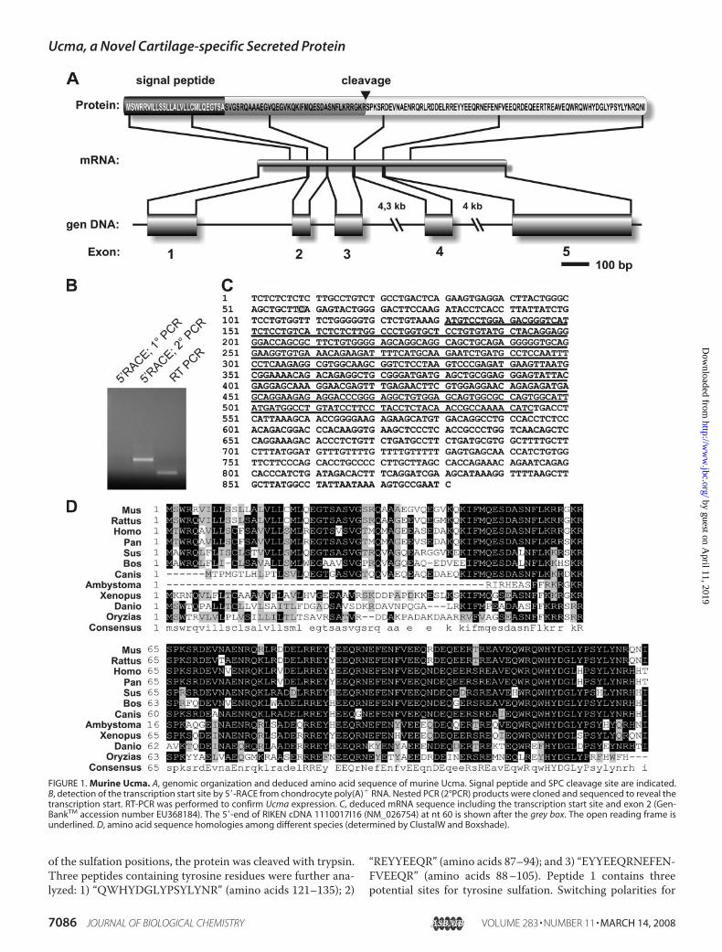
Sequence of Ucma—ThemurineUcma gene is located on chro-mosome 2. It is composed of five exons spanning �9 kb (Fig.1A). 5�-RACE determined a transcription start site �60-bpupstream of the 5� terminus of RIKEN cDNA clone 1110017I16(Fig. 1, B and C). cDNA sequencing and annotated genomicdata revealed the presence of an additional exon (exon 2) ascompared with the 1110017I16 cDNA clone. The mRNAsequence thus comprises 881 nt including an open readingframe of 414 nt (Fig. 1C; GenBankTM accession numberEU368184). The open reading frame encodes a polypeptide of138 amino acids with a predicted molecular mass of 16.5 kDa.
The deduced amino acid sequence predicts a 27-amino acidsignal peptide as calculated by the web-based signal peptideprediction program SignalP 3.0 (Fig. 1A). Ucma contains aRGKR/S consensus sequence predicting processing by a furin-like protease (see below).Recently, the same gene has been identified independently in
an EST project as a transcript from a human fetal cartilagelibrary (8).Interestingly, the C-terminal fragment is exceptionally rich
in acidic and basic amino acids: the 111 C-terminal amino acidscontain 27 acidic residues and 21 basic residues. Further con-served functional domains, however, could not be identified bymeans of bioinformatic analyses. Murine and human mRNAand amino acid sequences exhibit a homology of �65 and 80%,respectively. Highly conserved orthologues of Ucma can befound in various other species including rat, dog, clawed frog,and zebra fish, among others (Fig. 1D). The putative furin cleav-age site is conserved in most species analyzed. In contrast,paralogous genes or pseudogenes were not revealed afterscreening the published murine genome.Ucma Is Proteolytically Processed and Contains Tyrosine
Sulfates—For in vitro assays on the physiologic function ofUcma, and for analysis of potential posttranslational modifica-tions, recombinant (His)6- and FLAG-tagged Ucma proteinwas produced in HEK293EBNA cells. In cell lysates the anti-FLAG antibody detected a major band at about 18 kDa, corre-sponding to the full-length Ucma protein including His andFLAG tags (Fig. 2A, Lys) and a minor band migrating at �13kDa. Ucma purified from cell culture supernatant showed onlythe 13-kDa band, indicating processing of Ucma during or aftersecretion (Fig. 2A, SN). Fig. 2B displays the purified Ucma pro-tein in a Coomassie-stained PAA gel. TheN-terminal sequenceof recombinant Ucma (SPKSRDE . . . ) as determined by aminoacid sequencing indicated that Ucma was proteolytically pro-cessed by a furin-like protease into a C-terminal fragment of 74amino acid residues (Ucma-C), containing the His-FLAG-Tagat the carboxyl end, and an N-terminal fragment of 37 aminoacids (Ucma-N) (Fig. 1A) (19). The actualmolecularmass of thesecretedC-terminalUcma fragment (Ucma-C)was determinedto 11510Da usingMALDI-TOFMS,matching the theoreticallypredicted molecular weight of its amino acid sequence includ-ing His and FLAG tags.Polyclonal rabbit antibodies were raised 1) against a Ucma
peptide containing the SPC cleavage site (TAGL-1) (8), and 2)against recombinant Ucma-C (UCMA-1). Western blot analy-sis showed that the TAGL-1 antibodies only detected theuncleaved form of Ucma, while UCMA-1 antibodies detectedboth Ucma-C and the uncleaved Ucma precursor in lysates ofHEK293-EBNA cells transfected with Ucma (Fig. 2C).The presence of several tyrosine residues in an environment
rich in acidic amino acids raised the question, whether Ucma ispost-translationally modified by tyrosine sulfation which is fre-quently found in secreted and membrane-associated proteins(20, 21). To address this question, tyrosine sulfation of thesecreted 13 kDa Ucma-C was analyzed byMALDI-TOFMS. Intwo independent preparations protein species were detectedwith a molecular mass higher than predicted, indicating sulfa-tion at 1 or 2 positions (Fig. 3A, arrows). For further localization3 U. Dietz et al., manuscript in preparation.
Ucma, a Novel Cartilage-specific Secreted Protein
MARCH 14, 2008 • VOLUME 283 • NUMBER 11 JOURNAL OF BIOLOGICAL CHEMISTRY 7085
by guest on April 11, 2019
http://ww
w.jbc.org/
Dow
nloaded from
of the sulfation positions, the protein was cleaved with trypsin.Three peptides containing tyrosine residues were further ana-lyzed: 1) “QWHYDGLYPSYLYNR” (amino acids 121–135); 2)
“REYYEEQR” (amino acids 87–94); and 3) “EYYEEQRNEFEN-FVEEQR” (amino acids 88–105). Peptide 1 contains threepotential sites for tyrosine sulfation. Switching polarities for
FIGURE 1. Murine Ucma. A, genomic organization and deduced amino acid sequence of murine Ucma. Signal peptide and SPC cleavage site are indicated.B, detection of the transcription start site by 5�-RACE from chondrocyte poly(A)� RNA. Nested PCR (2°PCR) products were cloned and sequenced to reveal thetranscription start. RT-PCR was performed to confirm Ucma expression. C, deduced mRNA sequence including the transcription start site and exon 2 (Gen-BankTM accession number EU368184). The 5�-end of RIKEN cDNA 1110017I16 (NM_026754) at nt 60 is shown after the grey box. The open reading frame isunderlined. D, amino acid sequence homologies among different species (determined by ClustalW and Boxshade).
Ucma, a Novel Cartilage-specific Secreted Protein
7086 JOURNAL OF BIOLOGICAL CHEMISTRY VOLUME 283 • NUMBER 11 • MARCH 14, 2008
by guest on April 11, 2019
http://ww
w.jbc.org/
Dow
nloaded from
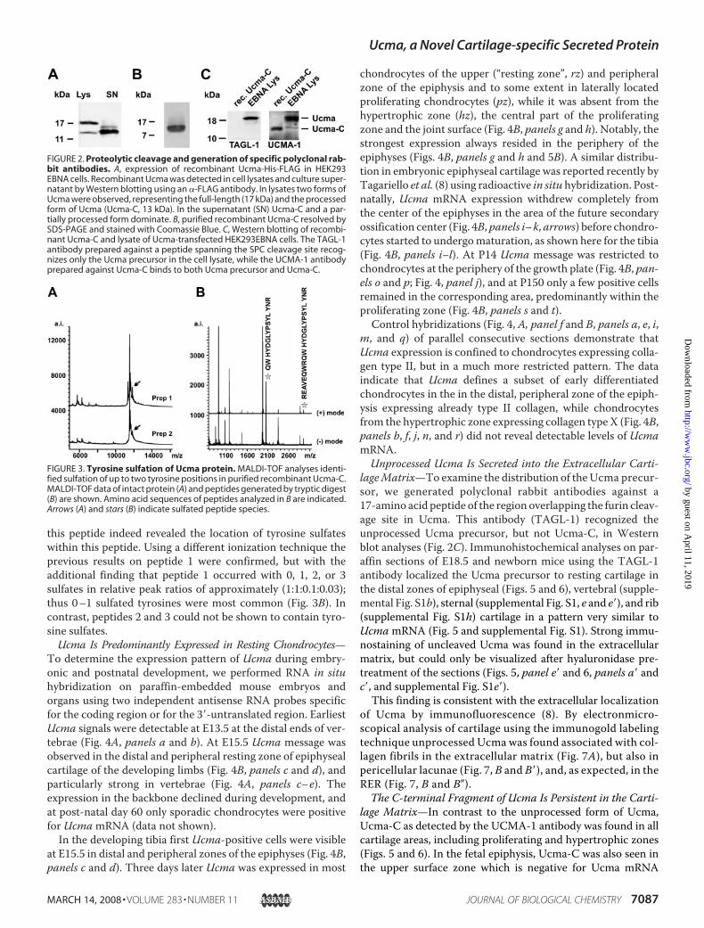
this peptide indeed revealed the location of tyrosine sulfateswithin this peptide. Using a different ionization technique theprevious results on peptide 1 were confirmed, but with theadditional finding that peptide 1 occurred with 0, 1, 2, or 3sulfates in relative peak ratios of approximately (1:1:0.1:0.03);thus 0–1 sulfated tyrosines were most common (Fig. 3B). Incontrast, peptides 2 and 3 could not be shown to contain tyro-sine sulfates.Ucma Is Predominantly Expressed in Resting Chondrocytes—
To determine the expression pattern of Ucma during embry-onic and postnatal development, we performed RNA in situhybridization on paraffin-embedded mouse embryos andorgans using two independent antisense RNA probes specificfor the coding region or for the 3�-untranslated region. EarliestUcma signals were detectable at E13.5 at the distal ends of ver-tebrae (Fig. 4A, panels a and b). At E15.5 Ucma message wasobserved in the distal and peripheral resting zone of epiphysealcartilage of the developing limbs (Fig. 4B, panels c and d), andparticularly strong in vertebrae (Fig. 4A, panels c–e). Theexpression in the backbone declined during development, andat post-natal day 60 only sporadic chondrocytes were positivefor UcmamRNA (data not shown).In the developing tibia first Ucma-positive cells were visible
at E15.5 in distal and peripheral zones of the epiphyses (Fig. 4B,panels c and d). Three days later Ucma was expressed in most
chondrocytes of the upper (“resting zone”, rz) and peripheralzone of the epiphysis and to some extent in laterally locatedproliferating chondrocytes (pz), while it was absent from thehypertrophic zone (hz), the central part of the proliferatingzone and the joint surface (Fig. 4B, panels g and h). Notably, thestrongest expression always resided in the periphery of theepiphyses (Figs. 4B, panels g and h and 5B). A similar distribu-tion in embryonic epiphyseal cartilage was reported recently byTagariello et al. (8) using radioactive in situ hybridization. Post-natally, Ucma mRNA expression withdrew completely fromthe center of the epiphyses in the area of the future secondaryossification center (Fig. 4B, panels i–k, arrows) before chondro-cytes started to undergomaturation, as shown here for the tibia(Fig. 4B, panels i–l). At P14 Ucma message was restricted tochondrocytes at the periphery of the growth plate (Fig. 4B, pan-els o and p; Fig. 4, panel j), and at P150 only a few positive cellsremained in the corresponding area, predominantly within theproliferating zone (Fig. 4B, panels s and t).
Control hybridizations (Fig. 4, A, panel f and B, panels a, e, i,m, and q) of parallel consecutive sections demonstrate thatUcma expression is confined to chondrocytes expressing colla-gen type II, but in a much more restricted pattern. The dataindicate that Ucma defines a subset of early differentiatedchondrocytes in the in the distal, peripheral zone of the epiph-ysis expressing already type II collagen, while chondrocytesfrom the hypertrophic zone expressing collagen typeX (Fig. 4B,panels b, f, j, n, and r) did not reveal detectable levels of UcmamRNA.Unprocessed Ucma Is Secreted into the Extracellular Carti-
lageMatrix—To examine the distribution of theUcma precur-sor, we generated polyclonal rabbit antibodies against a17-amino acid peptide of the region overlapping the furin cleav-age site in Ucma. This antibody (TAGL-1) recognized theunprocessed Ucma precursor, but not Ucma-C, in Westernblot analyses (Fig. 2C). Immunohistochemical analyses on par-affin sections of E18.5 and newborn mice using the TAGL-1antibody localized the Ucma precursor to resting cartilage inthe distal zones of epiphyseal (Figs. 5 and 6), vertebral (supple-mental Fig. S1b), sternal (supplemental Fig. S1, e and e�), and rib(supplemental Fig. S1h) cartilage in a pattern very similar toUcmamRNA (Fig. 5 and supplemental Fig. S1). Strong immu-nostaining of uncleaved Ucma was found in the extracellularmatrix, but could only be visualized after hyaluronidase pre-treatment of the sections (Figs. 5, panel e� and 6, panels a� andc�, and supplemental Fig. S1e�).This finding is consistent with the extracellular localization
of Ucma by immunofluorescence (8). By electronmicro-scopical analysis of cartilage using the immunogold labelingtechnique unprocessed Ucma was found associated with col-lagen fibrils in the extracellular matrix (Fig. 7A), but also inpericellular lacunae (Fig. 7, B and B�), and, as expected, in theRER (Fig. 7, B and B�).The C-terminal Fragment of Ucma Is Persistent in the Carti-
lage Matrix—In contrast to the unprocessed form of Ucma,Ucma-C as detected by the UCMA-1 antibody was found in allcartilage areas, including proliferating and hypertrophic zones(Figs. 5 and 6). In the fetal epiphysis, Ucma-C was also seen inthe upper surface zone which is negative for Ucma mRNA
FIGURE 2. Proteolytic cleavage and generation of specific polyclonal rab-bit antibodies. A, expression of recombinant Ucma-His-FLAG in HEK293EBNA cells. Recombinant Ucma was detected in cell lysates and culture super-natant by Western blotting using an �-FLAG antibody. In lysates two forms ofUcma were observed, representing the full-length (17 kDa) and the processedform of Ucma (Ucma-C, 13 kDa). In the supernatant (SN) Ucma-C and a par-tially processed form dominate. B, purified recombinant Ucma-C resolved bySDS-PAGE and stained with Coomassie Blue. C, Western blotting of recombi-nant Ucma-C and lysate of Ucma-transfected HEK293EBNA cells. The TAGL-1antibody prepared against a peptide spanning the SPC cleavage site recog-nizes only the Ucma precursor in the cell lysate, while the UCMA-1 antibodyprepared against Ucma-C binds to both Ucma precursor and Ucma-C.
FIGURE 3. Tyrosine sulfation of Ucma protein. MALDI-TOF analyses identi-fied sulfation of up to two tyrosine positions in purified recombinant Ucma-C.MALDI-TOF data of intact protein (A) and peptides generated by tryptic digest(B) are shown. Amino acid sequences of peptides analyzed in B are indicated.Arrows (A) and stars (B) indicate sulfated peptide species.
Ucma, a Novel Cartilage-specific Secreted Protein
MARCH 14, 2008 • VOLUME 283 • NUMBER 11 JOURNAL OF BIOLOGICAL CHEMISTRY 7087
by guest on April 11, 2019
http://ww
w.jbc.org/
Dow
nloaded from

expression and Ucma precursor (Fig. 5, panels d–f). This maybe a result of enhanced diffusion of the small C-terminal frag-ment within the cartilagematrix. A strong immunoreaction forUcma-C was also seen in the proliferating and upper hyper-trophic zone, both in the fetal and post-natal growth plate (Figs.5, panel l and 6, panels b, d, f, and supplemental Fig. S1, panelsc and f�), where the antibody TAGL-1 for unprocessed Ucmawas negative. This indicates complete processing of Ucma inthese zones and persistence of Ucma-C in the cartilage matrixat the site of initial deposition for several days or even weeks
after secretion and processing,while chondrocytes continue to dif-ferentiate and mature to hypertro-phy (see also “Discussion”).In Vitro, Ucma Is Expressed dur-
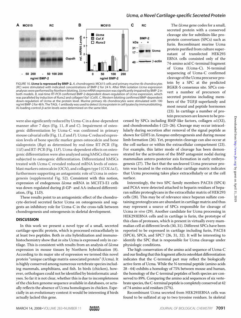
ing Early Stages of ChondrocyteDifferentiation—To investigateUcma expression during chondro-cyte differentiation in vitro, a varietyof murine mesenchymal cell linesand primary skeletal cells weretested for Ucma expression by RT-PCR. Among cell lines only chon-drogenic ATDC5 and MC615 cellsrevealed substantial Ucma expres-sion, whereas Ucma expressionwas absent from the mesenchymalprogenitor cell lines C3H10T1/2and C2C12. Only minor levels ofUcma mRNA were detectable inMC3T3-E1 pre-osteoblasts andNIH3T3 fibroblasts (Fig. 8A). RT-PCR analysis furthermore revealedhigh levels of Ucma mRNA expres-sion in primary murine rib chon-drocytes (RC) as comparedwith pri-mary osteoblasts from murinecalvariae (CC) (Fig. 8B). In bovineepiphyseal chondrocytes Ucmaexpression was confined to the rest-ing zone fraction (RZ), whereashypertrophic chondrocytes (HZ)merely expressed marginal levels ofUcma (Fig. 8C). These analyses con-firmed the predominant expressionof Ucma in resting chondrocytesobserved by in situ hybridization(Fig. 4).Changes in Ucma expression
during chondrocyte differentiationwere investigated using a subclone(4C6)4 of MC615 chondrocytes (9).Clone 4C6 exhibited a strong induc-tion of type II collagen (Col II)expression after 1 week in culture,reaching nearly the same level asthat of primary rib chondrocytes. Inlater stages (2 to 5 weeks after seed-
ing) Col II expression decreased with time, while type X colla-gen (Col X) expression started to increase strongly after 3weeks in culture (Fig. 9). In the 4C6 chondrocyte line Ucmaexpression largely followed the temporal kinetics of Col IIexpression although its peak expression lagged approximately 1week behind. Expression of Ucma in 4C6 cells reached similarlevels as in primary chondrocytes (Fig. 9A). These results are in
4 C. Surmann-Schmitt, N. Widmann, K. von der Mark, and M. Stock, manuscriptin preparation.
FIGURE 4. Ucma is predominantly expressed in juvenile cartilage of murine embryos and newborn mice,following expression of type II collagen. RNA in situ hybridization (ISH) on paraffin sections of murine embry-onic (E13.5, E15.5) vertebral column and whole E15.5 embryo (A) and of tibiae from embryonic, newborn,young, and adult mice (E15.5, E18.5, P6-P150) (B). A, Ucma expression was first observed in the vertebral columnat E13.5 (A, panels a and b); at E15.5 it is expressed mostly in the surface of the vertebrae (A, panels c– e). B, in thetibia expression of Ucma was first detectable in the epiphyses of tibiae from E15.5 murine embryos in earlydifferentiated chondrocytes in the epiphyseal surface zone (B, panels c and d). During further developmentUcma expression expanded into the entire distal and peripheral resting zone of the epiphysis (B, panels g andh). After birth Ucma expression was more and more restricted to the peripheral zone of epiphyseal cartilageand disappeared from the secondary ossification center (so) of the epiphysis (B, panels k, l, o, p: arrows). In adultmice (P150) only sporadic Ucma-positive cells were present in the epiphyseal growth zone (B, panels o and q).bo, bone; hz, hypertrophic zone; pz, proliferating zone; rz, resting zone; so, secondary ossification center; t,trachea; v, vertebral column.
Ucma, a Novel Cartilage-specific Secreted Protein
7088 JOURNAL OF BIOLOGICAL CHEMISTRY VOLUME 283 • NUMBER 11 • MARCH 14, 2008
by guest on April 11, 2019
http://ww
w.jbc.org/
Dow
nloaded from

agreement with a similar study on the time course of Ucmaexpression in differentiating ATDC5 cells (8) and indicate thatUcma expression in chondrocytes peaks in an early maturationstage following expression of Col II, while it declines with theonset of Col X expression.BMP-2 Represses Ucma Transcription in Chondrogenic Cells—
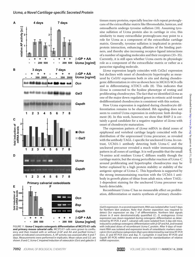
The finding thatUcma expression started in early chondrocytesin the distal zones of epiphyseal cartilage after type II collagen,but disappeared with chondrocyte maturation and hypertro-phy raised the question, how Ucma expression may be reg-ulated. Tagariello et al. (8) have shown thatUcma expressionin the developing limb is Indian hedgehog independent. Toinvestigate a potential regulatory effect on Ucma expressionby BMP factors, which have been shown to promote chon-drocyte maturation toward the hypertrophic phenotype invitro and in vivo we treated chondrogenic MC615 cells andprimary murine rib chondrocytes with increasing amountsof BMP-2 for 24 h (22). Northern blot analysis revealed adose-dependent repression of Ucma mRNA expression inresponse to BMP-2 in both MC615 cells and primary chon-
drocytes (Fig. 10A). While down-regulating Ucma expres-sion, BMP-2 enhanced the expression of the hypertrophicchondrocyte markers Col X and Runx2 in MC615 cells asshown by real-time RT-PCR (Fig. 10B). BMP-dependentdown-regulation of Ucma expression could also be con-firmed at protein level using cell lysates of primary rib chon-drocytes stimulated with 100 ng/ml BMP-2 in aWestern blotanalysis (Fig. 10C). These results implicate that BMP-2 mayhave a role in the suppression of Ucma during chondrocytematuration and hypertrophy.
FIGURE 5. Ucma mRNA and protein expression in cartilage of newbornand adult mice. The uncleaved Ucma precursor (detected with TAGL-1, pan-els b, e, e�, h, and k) largely colocalized with Ucma mRNA (panels a, d, d�, g, andj) in distal epiphyseal regions, predominantly in resting chondrocytes. In con-trast, the UCMA-1 antibody detecting both Ucma-C and Ucma precursorstained virtually the entire cartilage matrix of the epiphysis including resting,proliferating and hypertrophic zones (panels c, f, f�, and i), and the postnatalgrowth plate (panel l), indicating that Ucma-C is present in all epiphysealcartilage regions. Panels a– c, scapula and humerus, newborn; panels d–f, dis-tal femur epiphysis, newborn; panels d�–f�, enlarged region of panels d–f; pan-els g–i, tibia, newborn; panels j–l, tibia, adult (P150).
FIGURE 6. Ucma-C exhibits a wider protein distribution than Ucma pre-cursor in epiphyseal cartilage. Uncleaved Ucma precursor, detected withTAGL-1 is located predominantly in the resting, distal and peripheral zones ofepiphyseal cartilage (panels a, a�, c, c�, e), while Ucma-C, detected with theUCMA-1 antibody is distributed throughout the epiphysis including prolifer-ating and hypertrophic zones (panels b, b�, d, d�, f). Panels a and b, digits,newborn; panels a� and b�, enlarged region of panels a and b; panels c and d,hip, newborn; panels c� and d�, enlarged region of panels c and d; panels e andf, tibia, adult (P60).
Ucma, a Novel Cartilage-specific Secreted Protein
MARCH 14, 2008 • VOLUME 283 • NUMBER 11 JOURNAL OF BIOLOGICAL CHEMISTRY 7089
by guest on April 11, 2019
http://ww
w.jbc.org/
Dow
nloaded from
Ucma-C Suppresses Osteogenic, but Not Chondrogenic,Differentiation—For the analysis of possible Ucma-depend-ent effects on cell growth and differentiation, chondrocytesand a number of chondrocytic and osteoblastic cell lines
were treated with purified recombinant Ucma-C protein invitro. After treatment of primary murine rib chondrocytes orC3H10T1/2 cells with Ucma-C doses ranging from 125ng/ml to 1000 ng/ml for up to 11 days, no significant alter-ations on cell growth were observed (data not shown). Also,treatment of MC615 cells or primary murine chondrocyteswith variable amounts of purified recombinant Ucma-C didnot significantly change expression of chondrogenesis-re-lated genes as determined in a gene expression array (GE-Array Mouse Osteogenesis Microarray, Superarray) and byreal-time RT-PCR (data not shown).Interestingly however, Ucma impaired osteogenesis in vitro.
Confluent cultures of MC3T3-E1 cells, a cell line derived frommurine calvarial cells known to undergo osteogenic differenti-ation, were induced to differentiate with ascorbic acid (AA) and�-glycerophosphate (�-GP). Alkaline phosphatase (AP) activ-ity increased �4- and 4.5-fold after 4 and 7 days, respectively.After 7 days AA/�-GP-dependent induction of AP activity wassuppressed by Ucma-C approximately by 45%. Higher doses(500 ng/ml) of recombinant Ucma suppressed AP inductionalready after 4 days (by �24%). Osteoblast markers osteocalcin(Ocn) and galectin-3 (Gal-3) measured by Northern blotting
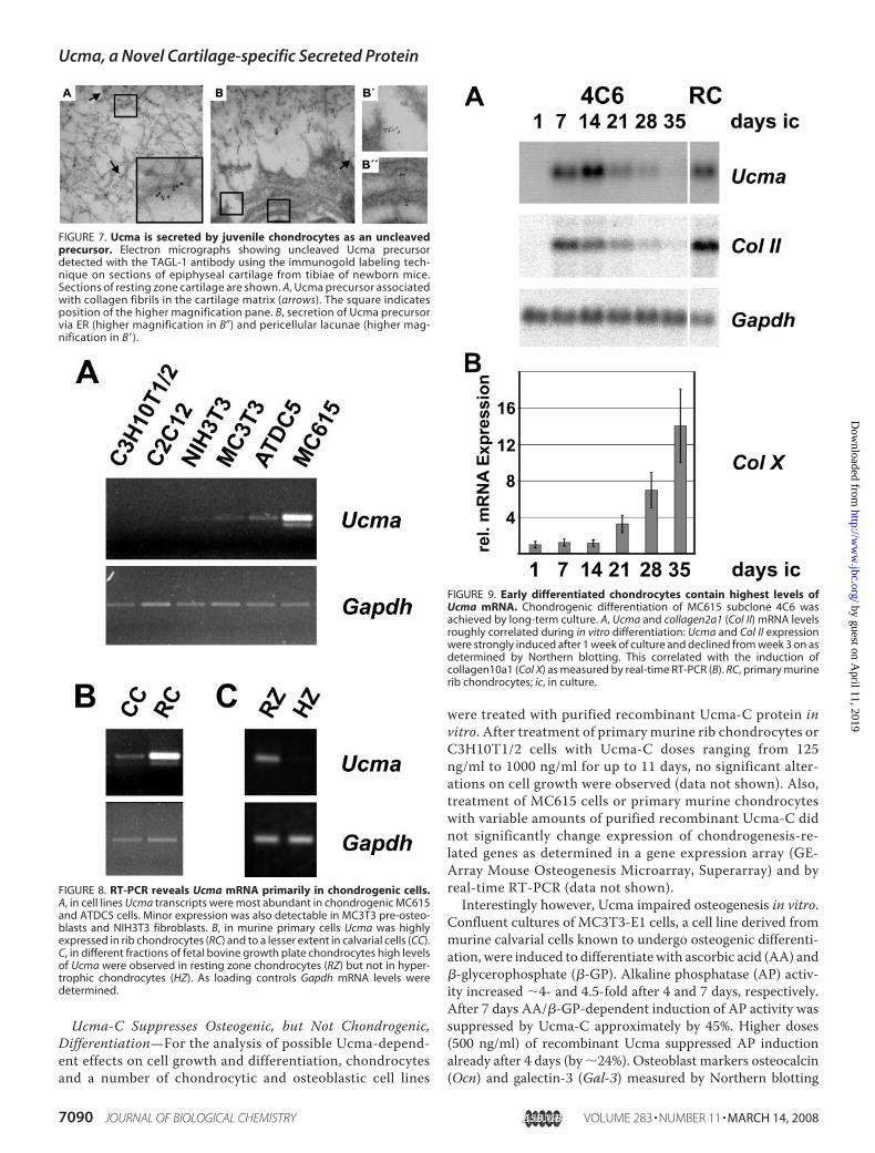
FIGURE 7. Ucma is secreted by juvenile chondrocytes as an uncleavedprecursor. Electron micrographs showing uncleaved Ucma precursordetected with the TAGL-1 antibody using the immunogold labeling tech-nique on sections of epiphyseal cartilage from tibiae of newborn mice.Sections of resting zone cartilage are shown. A, Ucma precursor associatedwith collagen fibrils in the cartilage matrix (arrows). The square indicatesposition of the higher magnification pane. B, secretion of Ucma precursorvia ER (higher magnification in B�) and pericellular lacunae (higher mag-nification in B�).
FIGURE 8. RT-PCR reveals Ucma mRNA primarily in chondrogenic cells.A, in cell lines Ucma transcripts were most abundant in chondrogenic MC615and ATDC5 cells. Minor expression was also detectable in MC3T3 pre-osteo-blasts and NIH3T3 fibroblasts. B, in murine primary cells Ucma was highlyexpressed in rib chondrocytes (RC) and to a lesser extent in calvarial cells (CC).C, in different fractions of fetal bovine growth plate chondrocytes high levelsof Ucma were observed in resting zone chondrocytes (RZ) but not in hyper-trophic chondrocytes (HZ). As loading controls Gapdh mRNA levels weredetermined.
FIGURE 9. Early differentiated chondrocytes contain highest levels ofUcma mRNA. Chondrogenic differentiation of MC615 subclone 4C6 wasachieved by long-term culture. A, Ucma and collagen2a1 (Col II) mRNA levelsroughly correlated during in vitro differentiation: Ucma and Col II expressionwere strongly induced after 1 week of culture and declined from week 3 on asdetermined by Northern blotting. This correlated with the induction ofcollagen10a1 (Col X) as measured by real-time RT-PCR (B). RC, primary murinerib chondrocytes; ic, in culture.
Ucma, a Novel Cartilage-specific Secreted Protein
7090 JOURNAL OF BIOLOGICAL CHEMISTRY VOLUME 283 • NUMBER 11 • MARCH 14, 2008
by guest on April 11, 2019
http://ww
w.jbc.org/
Dow
nloaded from
were also significantly reduced byUcma-C in a dose-dependentmanner after 7 days (Fig. 11, B and C). Impairment of osteo-genic differentiation by Ucma-C was confirmed in primarymouse calvarial cells (Fig. 11,E andF). Ucma-C reduced expres-sion levels of bone specific marker genes osteocalcin and bonesialoprotein (Bsp) as determined by real-time RT-PCR (Fig.11E) andRT-PCR (Fig. 11F). Ucma-dependent effects on osteo-genic differentiationwere also analyzed using hMSCs that weresubjected to osteogenic differentiation. Differentiated hMSCstreated with Ucma-C revealed reduced mRNA levels of osteo-blastmarkers osteocalcin (OCN), and collagen type I (COL1a1),furthermore supporting an antagonistic role of Ucma in osteo-genesis (supplemental Fig. S2). Consistent with this notion,expression of endogenous Ucma mRNA in MC3T3-E1 cellswas down-regulated during �-GP- and AA-induced differenti-ation, (Fig. 11D).These results point to an antagonistic effect of the chondro-
cyte-derived secreted factor Ucma on osteogenesis and sug-gests an inhibitory role for Ucma-C in the cross-talk betweenchondrogenesis and osteogenesis in skeletal development.
DISCUSSION
In this work we present a novel type of a small, secretedcartilage-specific protein, which is processed extracellularly inat least two peptides. Both in situ hybridization and immuno-histochemistry show that in situUcma is expressed only in car-tilage. This is consistent with results from an analysis of Ucmaexpression in mouse tissues by Northern hybridization (8).According to its major site of expression we termed this novelprotein “unique cartilagematrix-associated protein” (Ucma). Itis highly conserved among numerous vertebrate species includ-ing mammals, amphibians, and fish. In birds (chicken), how-ever, orthologues could not be identified by bioinformatic anal-yses. So far it is not clear, whether this is due to incompletenessof the chicken genome sequence available in databases, or actu-ally reflects the absence of Ucma homologues in chicken. Espe-cially in an evolutionary context it would be interesting if birdsactually lacked this gene.
TheUcma gene codes for a small,secreted protein with a conservedcleavage site for subtilisin-like pro-protein convertases (SPCs) such asfurin. Recombinant murine Ucmaprotein purified from culture super-natant of transfected HEK293EBNA cells consisted only of the74-amino acid C-terminal fragmentof Ucma (Ucma-C). N-terminalsequencing of Ucma-C confirmedcleavage of theUcmaprecursor pro-tein by a SPC at the predictedRGKR-S consensus site. SPCs con-vert a number of precursors ofsecreted proteins including mem-bers of the TGF� superfamily andmost neural and peptide hormones(23). In cartilage a number of pro-tein precursors are known to be pro-
cessed by SPCs including BMP-like factors, collagen �1(XI),and chondromodulin-I (23–26). Cleavage may occur intracel-lularly during secretion after removal of the signal peptide asshown for GDF5 in Xenopus embryogenesis and during mouselimb formation (26). Yet, proprotein cleavage can also occur atthe cell surface or within the extracellular compartment (23).For example, this latter mode of cleavage has been demon-strated for the activation of the BMP antagonist nodal duringmammalian antero-posterior axis formation in early embryo-genesis (27). The fact that the uncleaved Ucma precursor pro-tein was located in the extracellular cartilage matrix indicatesthat Ucma processing takes place extracellularly or at the cellsurface.Interestingly, the two SPC family members PACE4 (SPC4)
and PC6A were detected attached to heparin residues of hepa-ran sulfate proteoglycans in the extracellularmatrix of HEK293cells (28). This may be of relevance since heparan sulfate con-taining proteoglycans are abundant in cartilagematrix and thusmay represent a source of SPCs responsible for cleavage ofUcma in vivo (29). Another candidate for Ucma processing inHEK293EBNA cells and in cartilage is furin, the prototype ofthis class of proteases, which is present in virtually every mam-malian cell at different levels (30, 31). Different SPCs have beenreported to be expressed in cartilage including furin, PACE4(SPC4), SPC6, and SPC7 (26, 31, 32). It will be interesting toidentify the SPC that is responsible for Ucma cleavage underphysiologic conditions.The high conservation of the amino acid sequence of Ucma-C
andour finding that this fragmentaffectsosteoblastdifferentiationindicates that the C-terminal part may reflect the biologicallyactive form of Ucma. While the N-terminal peptide (amino acids28–64) exhibits a homology of 75% between mouse and human,the homology of the C-terminal peptides of both species are con-served to 89%. Comparing the amino acid sequences of six verte-brate species, theC-terminalpeptide is completely conservedat42of 74 amino acid residues (57%).Recombinant Ucma secreted from HEK293EBNA cells was
found to be sulfated at up to two tyrosine residues. In skeletal
FIGURE 10. Ucma is repressed by BMP-2. A, chondrogenic MC615 cells and primary murine rib chondrocytes(RC) were stimulated with indicated concentrations of BMP-2 for 24 h. After RNA isolation Ucma expressionanalyses were performed by Northern blotting. Ucma mRNA expression was significantly impaired by BMP-2 inboth models. B, real-time RT-PCR confirmed BMP-2-dependent down-regulation of Ucma expression, whichwas paralleled by induction of Runx2 and collagen10a1 (ColX). C, Western blotting confirmed BMP-dependentdown-regulation of Ucma at the protein level. Murine primary rib chondrocytes were stimulated with 100ng/ml BMP-2 for 48 h. The TAGL-1 antibody was used to detect Ucma protein in cell lysates by immunoblotting.As loading control �-actin levels were determined on the same blot.
Ucma, a Novel Cartilage-specific Secreted Protein
MARCH 14, 2008 • VOLUME 283 • NUMBER 11 JOURNAL OF BIOLOGICAL CHEMISTRY 7091
by guest on April 11, 2019
http://ww
w.jbc.org/
Dow
nloaded from
tissues many proteins, especially leucine-rich repeat proteogly-cans of the extracellular matrix like fibromodulin, lumican, andosteoadherin undergo tyrosine sulfation (18). Assuming tyro-sine sulfation of Ucma protein also in cartilage in vivo, thissimilarity to many extracellular proteoglycans may point to arole for Ucma as a component of the extracellular cartilagematrix. Generally, tyrosine sulfation is implicated in protein-protein interaction, enhancing affinities of the binding part-ners, and thereby also increasing receptor-ligand interactionsof a number of signalingmolecules and their receptors (33–35).Currently, it is still open whether Ucma exerts its physiologicrole as a component of the extracellular matrix or rather as aparacrine signaling molecule.Ucma expression largely coincides with Col2a1 expression
but declines with onset of chondrocyte hypertrophy as meas-ured by Col101 expression both in situ and during chondro-genic differentiation in vitro as shown here inMC615/4C6 cellsand in differentiating ATDC5 cells (8). This indicates thatUcma is connected to the hyaline phenotype of resting andproliferating chondrocytes. The fact that we identifiedUcma asone of the major down-regulated genes in retinoic acid-treateddedifferentiated chondrocytes is consistent with this notion.How Ucma expression is regulated during chondrocyte dif-
ferentiation remains to be elucidated. Ihh signaling does notseem to control Ucma expression in embryonic limb develop-ment (8). In this work, however, we show that BMP-2 is cer-tainly a good candidate for a negative regulator of Ucma withonset of chondrocyte maturation.The expression pattern of Ucma mRNA in distal zones of
epiphyseal and vertebral cartilage largely coincided with thedistribution of the unprocessed Ucma precursor, as revealedwith the antibodyTAGL-1 specific for uncleavedUcma. In con-trast, UCMA-1 antibody detecting both Ucma-C and theuncleaved precursor revealed a much wider immunostainingpattern in all zones of cartilage. It is well possible that the small74-amino acid residues Ucma-C diffuses readily though thecartilagematrix, but the strong pericellular reaction of Ucma-Caround proliferating and hypertrophic chondrocytes may bebetter explained by a high protein stability or stability of theantigenic epitope of Ucma-C. This hypothesis is supported bythe strong immunostaining reaction with the UCMA-1 anti-body in growth plates of tibiae from adult mice, where TAGL-1-dependent staining for the uncleaved Ucma precursor wasbarely detectable.Recombinant Ucma-C has no measurable effect on prolifer-
ation, differentiation or matrix synthesis of primary chondro-
FIGURE 11. Ucma-C impairs osteogenic differentiation of MC3T3-E1 cellsand primary mouse calvarial cells. MC3T3-E1 cells were grown to conflu-ency and then treated with or without �-GP and AA and purified Ucma-Cprotein at indicated concentrations. A, AP activity was assessed after 4 and 7days. Measurements were performed in triplicates. Mean values and S.D. areshown. B and C, Ucma-C impaired induction of osteocalcin (Ocn) and galectin-3
(Gal3) expression. In a second experiment, RNA was isolated after 3 and 7 daysfor Northern blot analyses. Note that shorter exposition was required todetect Ocn expression on day 7 of differentiation. Northern blot signalsshown in B were densitometrically quantified (C). D, endogenous Ucmaexpression was down-regulated during osteogenic differentiation as deter-mined by RT-PCR. E and F, calvarial cells were isolated from 3-day-old mice,induced to undergo osteogenic differentiation with �-GP and AA, and treatedwith indicated doses of recombinant Ucma-C protein. After 9 days of treat-ment RNA was isolated and expression levels of osteoblastic markers osteo-calcin (Ocn) and bone sialoprotein (Bsp) were determined by real-time RT-PCR(Ocn, E) and RT-PCR (Ocn and Bsp, F). Cyclophilin A (real-time RT-PCR) andGapdh (RT-PCR) mRNA levels were assessed for standardization of relativemRNA expression.
Ucma, a Novel Cartilage-specific Secreted Protein
7092 JOURNAL OF BIOLOGICAL CHEMISTRY VOLUME 283 • NUMBER 11 • MARCH 14, 2008
by guest on April 11, 2019
http://ww
w.jbc.org/
Dow
nloaded from
cytes or chondrocyte lines. However, the expression of osteo-genic markers in MC3T3-E1 cells that had been induced todifferentiate to osteoblasts was significantly inhibited byUcma-C, suggesting a paracrine action of chondrocyte-derivedUcma on bone cell development. Similar findings in primarymouse calvarial cells and human mesenchymal stem cells sup-port this hypothesis.There is numerous evidence formutual interactions between
chondrocytes and bone cells in the regulation of chondrogenicversus osteogenic differentiation (36, 37). Thus, Ucmamay rep-resent a paracrine factor supporting the balance between chon-drogenic versus osteogenic differentiation at the cartilage inter-face with perichondrium or periosteum in the developingskeleton. So far, reports on paracrine factors mediating signalsbetween different cell types involved in bone and cartilagedevelopment focused mainly on the Ihh/PTHrP system, andthe control of vascularization by VEGF produced by hyper-trophic chondrocytes. Several other paracrine factors includingFGF18 derived from the perichondrium have been reported tonegatively influence osteoblast differentiation (2, 7). In con-trast, Ucma may act as a direct link between epiphyseal chon-drocytes and pre-osteoblasts in the control of a well concertedand balanced formation of cartilage and bone.Themechanismof such regulatory functions is, however, still
unclear. Ucma-specific cellular receptors have not been identi-fied, yet. Because of its highly polar character and the potentialfor multiple protein interactions, matrix-bound Ucma-C mayexert indirect effects by binding growth factors or their inhibi-tors and controlling their release.The same features, however, propagate Ucma-C also as an
integrative constituent of the cartilage matrix, where it maycontrol the assembly of collagens, proteoglycans and glycopro-teins. The generation of Ucma-deficient and Ucma-overex-pressing mice is in progress to answer these questions.
Acknowledgments—We thank Britta Schlund and Eva Bauer forexcellent technical assistance.
REFERENCES1. Cancedda, R., Descalci-Cancedda, F., and Castagnola, P. (1995) Int. Rev.
Cytology 159, 265–3582. Goldring, M. B., Tsuchimochi, K., and Ijiri, K. (2006) J. Cell Biochem. 97,
33–443. de Crombrugghe, B., Lefebvre, V., and Nakashima, K. (2001) Curr. Opin.
Cell Biol. 13, 721–7274. DeLise, A. M., Fischer, L., and Tuan, R. S. (2000) Osteoarth. Cartilage 8,
309–3345. Vortkamp, A., Lee, K., Lanske, B., Segre, G. V., Kronenberg, H. M., and
Tabin, C. J. (1996) Science 273, 613–6226. Koziel, L., Wuelling, M., Schneider, S., and Vortkamp, A. (2005) Develop-
ment 132, 5249–52607. Wagner, E. F., and Karsenty, G. (2001) Curr. Opin. Genet. Dev. 11,
527–532
8. Tagariello, A., Luther, J., Streiter, M., Didt-Koziel, L., Wuelling, M., Sur-mann-Schmitt, C., Stock, M., Adam, N., Vortkamp, A., andWinterpacht,A. (2008)Matrix Biol. 27, 3–11
9. Mallein-Gerin, F., andOlsen, B. R. (1993) Proc. Natl. Acad. Sci. U. S. A. 90,3289–3293
10. Schmidl, M., Adam, N., Surmann-Schmitt, C., Hattori, T., Stock, M., Di-etz, U., de Crombrugghe, B., Poschl, E., and von derMark, K. (2006) J. Biol.Chem. 281, 31790–31800
11. Stock, M., Schafer, H., Fliegauf, M., and Otto, F. (2004) J. Bone Miner Res.19, 959–972
12. Stock, M., Schafer, H., Stricker, S., Gross, G., Mundlos, S., and Otto, F.(2003) J. Biol. Chem. 278, 17360–17367
13. Wixler, V., Hirner, S.,Muller, J.M., Gullotti, L.,Will, C., Kirfel, J., Gunther,T., Schneider,H., Bosserhoff, A., Schorle, H., Park, J., Schule, R., andBuett-ner, R. (2007) J. Cell Biol. 177, 163–172
14. Weisser, J., Riemer, S., Schmidl, M., Suva, L. J., Poschl, E., Brauer, R., andvon der Mark, K. (2002) Exp. Cell Res. 279, 1–13
15. Laemmli, U. K. (1970) Nature 227, 680–68516. Rappsilber, J., Ishihama, Y., andMann,M. (2003)AnalChem.75, 663–67017. Perkins, D. N., Pappin, D. J., Creasy, D. M., and Cottrell, J. S. (1999) Elec-
trophoresis 20, 3551–356718. Onnerfjord, P., Heathfield, T. F., and Heinegard, D. (2004) J. Biol. Chem.
279, 26–3319. Duckert, P., Brunak, S., and Blom, N. (2004) Protein Eng. Des. Sel. 17,
107–11220. Bundgaard, J. R., Vuust, J., and Rehfeld, J. F. (1997) J. Biol. Chem. 272,
21700–2170521. Moore, K. L. (2003) J. Biol. Chem. 278, 24243–2424622. Grimsrud, C. D., Romano, P. R., D’Souza, M., Puzas, J. E., Schwarz, E. M.,
Reynolds, P. R., Roiser, R. N., and O’Keefe, R. J. (2001) J. Orthop. Res. 19,18–25
23. Seidah, N. G., and Chretien, M. (1999) Brain Res. 848, 45–6224. Cui, Y., Jean, F., Thomas, G., and Christian, J. L. (1998) EMBO J. 17,
4735–474325. Azizan, A., Holaday, N., and Neame, P. J. (2001) J. Biol. Chem. 276,
23632–2363826. Thomas, J. T., Prakash, D.,Weih, K., andMoos,M., Jr. (2006) J. Biol. Chem.
281, 26725–2673327. Beck, S., Le Good, J. A., Guzman,M., BenHaim, N., Roy, K., Beermann, F.,
and Constam, D. B. (2002) Nat. Cell Biol. 4, 981–98528. Tsuji, A., Sakurai, K., Kiyokage, E., Yamazaki, T., Koide, S., Toida, K.,
Ishimura, K., andMatsuda, Y. (2003)Biochim. Biophys. Acta1645, 95–10429. Farach-Carson, M. C., Hecht, J. T., and Carson, D. D. (2005) Crit. Rev.
Eukaryot. Gene Expr. 15, 29–4830. Seidah, N. G., Chretien, M., and Day, R. (1994) Biochimie. (Paris) 76,
197–20931. Zheng, M., Streck, R. D., Scott, R. E., Seidah, N. G., and Pintar, J. E. (1994)
J. Neurosci. 14, 4656–467332. Constam, D. B., Calfon, M., and Robertson, E. J. (1996) J. Cell Biol. 134,
181–19133. Leyte, A., van Schijndel, H. B., Niehrs, C., Huttner, W. B., Verbeet, M. P.,
Mertens, K., and van Mourik, J. A. (1991) J. Biol. Chem. 266, 740–74634. Huang, S. C., Yu, D. H.,Wank, S. A.,Mantey, S., Gardner, J. D., and Jensen,
R. T. (1989) Peptides 10, 785–78935. Seibert, C., Cadene, M., Sanfiz, A., Chait, B. T., and Sakmar, T. P. (2002)
Proc. Natl. Acad. Sci. U. S. A. 99, 11031–1103636. Bianco, P., Cancedda, F. D., Riminucci, M., and Cancedda, R. (1998)Ma-
trix Biol. 17, 185–19237. Karsenty, G., and Wagner, E. F. (2002) Dev. Cell 2, 389–406
Ucma, a Novel Cartilage-specific Secreted Protein
MARCH 14, 2008 • VOLUME 283 • NUMBER 11 JOURNAL OF BIOLOGICAL CHEMISTRY 7093
by guest on April 11, 2019
http://ww
w.jbc.org/
Dow
nloaded from
Rainer Deutzmann, Klaus von der Mark and Michael StockAndreas Tagariello, Patrik Önnerfjord, Dick Heinegård, Ursula Schlötzer-Schrehardt,
Cordula Surmann-Schmitt, Uwe Dietz, Trayana Kireva, Nadia Adam, Jung Park,Osteogenesis
Ucma, a Novel Secreted Cartilage-specific Protein with Implications in
doi: 10.1074/jbc.M702792200 originally published online December 21, 20072008, 283:7082-7093.J. Biol. Chem.
10.1074/jbc.M702792200Access the most updated version of this article at doi:
Alerts:
When a correction for this article is posted•
When this article is cited•
to choose from all of JBC's e-mail alertsClick here
Supplemental material:
http://www.jbc.org/content/suppl/2007/12/26/M702792200.DC1
http://www.jbc.org/content/283/11/7082.full.html#ref-list-1
This article cites 37 references, 16 of which can be accessed free at
by guest on April 11, 2019
http://ww
w.jbc.org/
Dow
nloaded from