uc san francisco - pdfs.semanticscholar.org · this rat anti-cd44 was routinely used for ha-related...
TRANSCRIPT

UC San FranciscoUC San Francisco Previously Published Works
TitleHyaluronan-CD44 interaction stimulates Rac1 signaling and PKN gamma kinase activation leading to cytoskeleton function and cell migration in astrocytes
Permalinkhttps://escholarship.org/uc/item/5mg6z3vx
AuthorsSwanson, Raymond A.Brightman, AmyBourguignon, Lilly Y. W.et al.
Publication Date2007-05-01 Peer reviewed
eScholarship.org Powered by the California Digital LibraryUniversity of California
Hyaluronan-CD44 Interaction Stimulates Rac1 Signaling and PKNγ Kinase Activation Leading to Cytoskeleton Function and Cell Migration in Astrocytes Lilly Y.W. Bourguignon1#, Eli Gilad1, Karine Peyrollier1, Amy Brightman1 and Raymond Swanson2 Departments of Medicine1 and Neurology2, University of California San Francisco, Endocrine Unit (111N), San Francisco VA Medical Center, 4150 Clement Street, San Francisco, CA 94121. #Reprint requests should be addressed to:
Dr. Lilly Y.W. Bourguignon Endocrine Unit (111N), Department of Medicine University of California-San Francisco and VA Medical Center 4150 Clement Street San Francisco, CA 94121 TEL: (415) 221-4810 x 3321 FAX: (415) 383-1638 e-mail:[email protected]
Running Title: HA/CD44-mediated Rac1-PKNγ signaling in astrocytes. Key Words: Hyaluronan, CD44, Rac1, PKNγ, Cytoskeleton, Astrocyte, Migration. ABBREVIATIONS: HA, Hyaluronan; PKNγ, protein kinase N-γ; ACC, the antiparallel coiled-
coil; F-actin, filamentous actin; CNS, central nervous system; PLCγ, phospholipase C-γ; PKC,
protein kinase C.
1

ABSTRACT
Both hyaluronan [HA, the major glycosaminoglycans in the extracellular matrix (ECM)]
and CD44 (a primary HA receptor) are associated with astrocyte activation and tissue repair
following central nervous system (CNS) injury. In this study we investigated the question of
whether HA-CD44 interaction influences astrocyte signaling and migration. Our data indicated
that HA binding to the cultured astrocytes stimulated Rac1 signaling and cytoskeleton-mediated
migration. To determine the cellular and molecular basis of these events, we focused on PKNγ, a
Rac1-activated serine/threonine kinase in astrocytes. We determined that HA binding to
astrocytes stimulated Rac1-dependent PKNγ kinase activity which, in turn, upregulated the
phosphorylation of the cytoskeletal protein, cortactin, and attenuated the ability of cortactin to
cross-link F-actin. Further analyses indicated that the N-terminal antiparallel coiled-coil (ACC)
domains of PKNγ interacted with Rac1, and transfection of astrocytes with PKNγ-ACCcDNA
inhibited PKNγ activity. Overexpression of the PKNγ-ACC domain also functions as a dominant-
negative mutant to block HA/CD44-mediated PKNγ activation of cortactin and astrocyte
migration. Taken together, these findings strongly suggest that hyaluronan/CD44 interaction with
Rac1-PKNγ plays a pivotal role in cytoskeleton activation and astrocyte migration. These newly
discovered HA/CD44-induced astrocyte function may provide important insight into novel
therapeutic treatments for tissue repair following CNS injury.
INTRODUCTION
Astrocytes are the most abundant cells in the central nervous system (CNS) and
traditionally are thought to be primarily neuron-supporting cells (Fitch and Silver, 2001; Barres
and Barde, 2000). Evidence is now accumulating that astrocytes not only play an essential role in
maintaining the homeostatic neuronal environment, but also participate in CNS pathogenesis due
to trauma, viral/bacterial infections or chronic neurodegeneration (Haydon, 2000; Song, et al.,
2002; Ullian, et al., 2001; Chen and Swanson, 2003). During CNS injury, a breakdown of the
blood-brain barrier generally leads to reactive astrogliosis or glial scarring which involves
2
3
increased cell division, protein synthesis and cell migration of the astrocytes (Eddleston and
Mucke, 1993; Ridet et al., 1997). Formation of a glial scar is considered to be one of the major
obstacles to neuronal regeneration (Stichel and Muller, 1998; Nieto-Sampedro, 1999; David and
Lacroix, 2003). Other cellular behaviors, including abnormal astrocyte migration to the site of
injury to provide support for neuron regeneration and axonal regrowth (Rhodes et al., 2003;
Silver and Miller, 2004), could also contribute to the poor ability of CNS to repair itself (Shearer
and Fawcett, 2001; Sivron and Schwartz, 1995). Thus, understanding the cellular and molecular
mechanisms involved in regulating astrocyte functions (in particular, cell migration) following
CNS wounding/damage may be very useful for designing new therapeutic treatments for CNS
repair.
Hyaluronan (HA) is one of the major glycosaminoglycans in the extracellular matrix
(ECM) and plays an important role in morphogenesis, remodeling and integrity of the CNS
(Asher et al., 1991; Bignami et al., 1992; Bignami and Asher, 1992; Eggli et al., 1992). It is
synthesized by several HA synthases (Lee and Spicer, 2000; Toole, 2001) and modified by
hyaluronidases (Tona and Bignami, 1993). Under physiological conditions, HA in these tissues
exists as a large polymer in excess of 1 x 10⁶ Da (“high molecular weight HA”). However,
following CNS and other injuries, large HA becomes degraded (via hyaluronidases) into smaller
HA units (1 x 10⁴-1 x 10³ Da; “low molecular weight HA”) as reactive astrogliosis proceeds
(Struve et al., 2005). In fact, injection of hyaluronidases into normal spinal cord tissue induces
HA degradation and astrocyte proliferation (Struve et al., 2005). Both high and low molecular
weight HAs are capable of binding to CD44 which is a ubiquitous, abundant and functionally
important receptor expressed on the surface of many cells, including astrocytes (Underhill, 1992;
Haegel et al., 1993). In uninjured CNS, the expression of CD44 is restricted to astrocytes in the
white matter (Quackenbush, et al., 1985). However, upon CNS injury CD44 expression is
upregulated at the injured sites (Jones et al., 2000). The question of whether HA-CD44
interaction actually plays an important role in regulating astrocyte function (e.g. migration)
4
following injury has not been carefully investigated; and thus is the focus of this study.
Several lines of evidence indicate that the actin cytoskeleton is directly involved in
regulating astrocyte morphological changes and migration (Baorto et al., 1992; Ramakers and
Moolenaar, 1998; Etienne-Manneville and Hall 2002; Holtje et al., 2005). Members of the Rho
subclass of the Ras superfamily (small molecular weight GTPases, e.g. RhoA, Rac1 and Cdc42)
are known to transduce incoming signals which result in the formation of cytoskeleton-mediated
membrane protrusions and cell migration in many cell types including astrocytes (Hall, 1998).
Overexpression of certain RhoGTPases in astrocytes occurs at the site of the CNS injury
(Erschbamer et al., 2005). RhoGTPases have also been found to be involved in astrocytic wound
closure following in vitro wounding using a scratch-wound assay (Etienne-Manneville and Hall
2002; Holtje et al., 2005). Specifically, RhoA has been shown to negatively regulate astrocyte
morphology and migratory response after injury (Holtje et al., 2005). Other GTPases, such as
Rac1, are also known to be key components in regulating actin cytoskeleton and cell migration
(Hall, 1998). The question of whether Rac1 positively regulates cytoskeleton function and cell
migration in astrocytes following wounding has not been addressed previously.
HA has been shown to activate CD44-mediated Rac1 signaling and cell migration in a
variety of non-neuronal cell types (Bourguignon et al., 2000; Bourguignon et al., 2001a;
Bourguignon et al., 2004). Furthermore, several enzymes have been identified as possible
downstream targets for Rac1 signaling (Burbelo et al., 1995). One such enzyme is protein kinase
N-γ (PKNγ) (also called PRK2 or PKN-2) which belongs to a family of serine-threonine kinases
known to interact with Rac1 in a GTP-dependent manner and to share a great deal of sequence
homology with protein kinase C (PKC) in the C-terminal region (Quilliam et al., 1996; Vincent
and Settleman, 1997; Mukai, 2003). The N-terminal region of PKN contains three homologous
stretches of approximately 70 amino acids (relatively rich in charged residues) that form the
antiparallel coiled-coil fold (ACC domain) (Mukai, 2003; Vincent and Settleman, 1997; Quilliam
et al., 1996; Yoshinaga et al., 1999). This ACC domain is known to interact with RhoGTPases

5
such as Rac1 (and to a lesser extent with RhoA or Cdc42) (Vincent and Settleman, 1997;
Quilliam et al., 1996). The C-terminal region contains the C2-like region which functions as an
auto-inhibitory domain (Yoshinaga et al., 1999). The ACC and the C2-like domains, together
with the catalytic domain, are conserved among the PKN family members (Mukai, 2003). Most
importantly, the ACC domain has been shown to interact with GTP-bound RhoGTPases such as
Rac1 (Quilliam et al., 1996; Vincent and Settleman, 1997; Bourguignon, et al., 2004). Our recent
work demonstrates that HA-CD44 interaction with Rac1-activated PKNγ promotes PLCγ1-
regulated Ca2+ signaling and cortactin-cytoskeleton interaction required for a variety of cellular
functions (Bourguignon et al., 2004). Although Rac1 has been closely associated with the
remodeling of cortical actin, the Rac1-specific effector and its roles in regulating HA/CD44-
mediated astrocyte functions have not been identified or characterized. In this study we have
examined the role of HA/CD44-mediated Rac1-PKNγ kinase signaling and its downstream
effectors in the regulation of astrocyte functions involving cytoskeleton activation and cell
migration.
MATERIALS AND METHODS
Cell Cultures: Astrocyte cultures were prepared from cortices of one-day-old Swiss-Webster
mice (Simonsen, Gilroy, CA) as described previously (Swanson et al., 1997) and maintained in
Eagle’s MEM containing 10% fetal bovine serum (FBS) and glutamine (2mM) in a humidified,
5% CO2 incubator at 37oC. The medium were exchanged with fresh medium at day 5. At
confluence (days 12-15), 10µM cytosine arabinoside were added to prevent proliferation of other
cell types. This medium were removed after 48h, replaced with medium containing 3% FBS and
used for experiments at 20 days in vitro. Cells were routinely serum starved for 24h (and
therefore deprived of serum HA) before adding HA.
Antibodies and Reagents: Monoclonal rat anti-CD44 antibody (Clone: 020; Isotype: IgG2b;
obtained from CMB-TECH, Inc., San Francisco, CA.) interacts with a common determinant of
6
the CD44 class of glycoproteins including CD44s (the standard form), and CD44 variant
isoforms. This anti-CD44 antibody also recognizes a determinant of the HA-binding region
common to CD44 and its principal variant isoforms (Bourguignon, et al, 2000; Bourguignon, et
al, 2001a; Bourguignon, et al, 2001b; Bourguignon, et al, 2004). This rat anti-CD44 was routinely
used for HA-related blocking experiments and immunoprecipitation. Rabbit anti-phospho-
threonine antibody and rabbit anti-phospho-serine antibody were obtained from Zymed
Laboratories Inc. Rabbit anti-PKNγ and mouse anti-Rac1 were obtained from Santa Cruz
Biotechnology. Mouse anti-His and mouse anti-cortactin antibody (clone 4F11) were purchased
from Invitrogene and Upstate Biotechnology (Lake Placid, NY), respectively. GST-tagged Rac1
was obtained from Calbiochem. Cytochalasin D was purchased from Sigma. High molecular
weight HA polymers (~106 daltons) (HealonTM) were purchased from Amersham Pharmacia
Biotech.
Method for Preparing His-tagged Dominant-Negative Form [containing ACC domains] of
PKNγ Kinase: The cDNA fragment encoding the ACC domains of PKNγ (aa98-aa256) was
generated by reverse transcription-polymerase chain reaction (RT/PCR) using two specific
primers (5'-GGTGCAGCAGAAATTGGATGA-3' and 5'-
CTTTGACGTGGACTTAGTGTTGGTG-3'). PCR product digested with EcoR I and Hind III
was purified with QIAquick PCR purification Kit (Qiagen). The PKNγ-ACC fragment cDNA
was subsequently cloned into pcDNA3.1/HisC vector that contains Xpress-epitope to create His-
tagged PKNγ-ACCcDNA. The inserted ACC domain sequence was confirmed by nucleotide
sequencing analyses. This His-tagged PKNγ-ACC cDNA was then used for transient expression
in astrocytes as described below.
Cell Transfection: To establish a transient expression system, cultured astrocytes were
transfected with various plasmid DNAs (e.g. His-tagged PKNγ-ACC domain cDNA or vector
alone) using lipofectamin 2000 methods (Invitrogen, Carsbad, CA). Briefly, cells were plated at a
density of 2 x 10⁶ cells per 100 mm dish and transfected with 25µg plasmid cDNA/dish using

7
lipofectamin 2000. Transfected cells were grown in the culture medium for at least 24-48 h.
Various transfectants were then analyzed for their protein expression and functional properties as
described below.
Measurement of Rac1 Activation: Cultured astrocytes (~5.0 x 10⁵ cells) were incubated in a
buffer containing 118 mM KCl, 5 mM NaCl, 0.4 mM CaCl2, 1 mM EGTA, 1.2 mM Mg-acetate,
1.2 mM KH2PO4, 25 mM Tris-HCl (pH 7.4), 20 mg/ml BSA followed by adding [35S]GTPγS
(12.5 µCi). Subsequently, cells were electroporated at 25 microfarads and 2.0 kV/cm followed by
incubating with 50µg/ml HA [in the presence or absence of rat anti-CD44 antibody (50µg/ml)] or
without any HA treatment at 37oC for 10 min. [35S]GTPγS labeled cells were then solubilized by
NP-40 followed by incubating with mouse anti-Rac1 IgG (5µg/ml) plus goat anti-mouse
conjugated beads. The amount of [35S]GTPγS-Rac1 associated with anti-Rac1-conjugated
immuno-beads was measured by a gamma-counter.
Immunoblotting and Immunoprecipitation Techniques: Cultured astrocytes (untransfected
cells, PKNγ-ACCcDNA-transfected cells or vector-transfected cells) grown in 0.03mM Ca2+
were treated with no HA or with HA (50µg/ml) or pretreated with anti-CD44 antibody followed
by adding HA (50µg/ml) at 37oC for various time intervals (5min, 10min, 15min, 30min, 2h, 24h,
36h and 48h). These cells were then solubilized in 50mM HEPES (pH 7.5), 150mM NaCl, 20mM
MgCl2, 1.0% Nonidet P-40 (NP-40), 0.2mM Na3VO4, 0.2mM phenylmethylsulfonyl fluoride,
10µg/ml leupeptin, and 5µg/ml aprotinin. The sample was then analyzed by SDS-PAGE in a 5%
or 7.5% polyacrylamide gel. Separated polypeptides were then transferred onto nitrocellulose
filters. After blocking non-specific sites with 2% bovine serum albumin, the nitrocellulose filters
were incubated with each of the specific immuno-reagents [e.g. rat anti-CD44 IgG (5µg/ml), or
rabbit anti-PKNγ (5µg/ml), or rabbit anti-PLCγ1 (5µg/ml), or mouse anti-cortactin (5µg/ml) or
mouse anti-His antibody (5µg/ml)] followed by incubating with horseradish peroxidase (HRP)-
labeled goat anti-rat IgG, or HRP-labeled goat anti-rabbit IgG or HRP-labeled goat anti-mouse
8
IgG. The blots were then developed by the ECL™ system (Amersham Co.). For analyzing the
recruitment of endogenous PKNγ into CD44 complex, cells [either treated with HA (50µg/ml) or
without any HA treatment] were solubilized by 1.0% NP-40 and immunoprecipitated with rat
anti-CD44 antibody followed by anti-PKNγ-mediated immunoblot.
Untransfected astrocytes [untreated or pretreated with cytochalasin D (20µg/ml) for 1h at
37oC] or astrocytes transfected with PKNγ-ACCcDNA (or vector alone) were incubated with HA
(50µg/ml) at 37oC for 10 min [or pre-treated with anti-CD44 antibody followed by adding HA
(50µg/ml) or incubated with no HA]. These cells were then immunoprecipitated with anti-PLCγ1
(or anti-cortactin) antibody followed by immunoblotting with anti-phospho-serine/anti-phospho-
threonine or anti-Rac1/anti-His, respectively. Subsequently, these blots were then developed
using ECL chemiluminescence reagent according to the manufacturer’s instructions. During these
immunological analyses, an equal amount of cellular protein (50µg/ml) immunoprecipitated with
the antibody was applied to SDS-PAGE followed by immunoblot analyses.
PKNγ-Mediated Protein Phosphorylation Assay In Vitro: The PKNγ kinase reaction was
carried out in 50 µl of the reaction mixture containing 40 mM Tris-HCl (pH7.5), 2 mM EDTA, 1
mM DTT, 7 mM MgCl2, 0.1% CHAPS, 0.1µM calyculin A, 100µM ATP, 100ng PKNγ [isolated
from astrocytes (untransfected or transfected with His-tagged PKNγ-ACCcDNA or vector only)
using anti-PKNγ-conjugated Sepharose beads] and 1µg astrocyte cortactin (isolated from
astrocytes using anti-cortactin-conjugated Sepharose beads) in the presence of Rac1 (incubated
with 200µM GDP or 200µM GTP). After incubation for 30 min at 30oC, the reaction mixtures
were immunoprecipitated with anti-cortactin-beads, boiled in SDS-sample buffer and subjected to
SDS-PAGE. The protein bands associated with anti-cortactin-beads were revealed by anti-
phospho-serine/anti-phospho-threonine-mediated immunoblot or anti-cortactin-mediated
immunoblot, respectively. In some cases, PKNγ [isolated from astrocytes (untransfected or
transfected with His-tagged PKNγ-ACCcDNA or vector treated with HA or no HA) using anti-

9
PKNγ-conjugated Sepharose beads] was incubated with the reaction mixture containing 40mM
Tris-HCl (pH 7.5), 2mM EDTA, 1mM DTT, 7mM MgCl2, 0.1% CHAPS, 0.1µM calyculin A,
10µCi of [γ-32P]ATP (5000Ci/mmol), and 1µg astrocyte cortactin (isolated from astrocytes using
anti-cortactin-conjugated Sepharose beads). After 30min at 30oC, reactions were then terminated
by adding 20% cold trichloroacetic acid (TCA); and 2mg/ml BSA was then added as a carrier.
TCA precipitated proteins were spotted on 3MM filter papers followed by extensive wash with
10% TCA. The radioactivity associated with TCA-precipitated materials was analyzed by liquid
scintillation counting.
In vitro Scratch Wound Model: A scratch wound was made by scraping the cell monolayer
(untransfected, transfected with PKNγ-ACCcDNA or vector alone) across the cover glass with a
sterile cell lifter according to procedures described previously (Yu et al., 1993; Mukhin et al.,
1998). We changed the culture medium immediately after wounding to prevent the medium from
being conditioned with cell debris and factors released from deteched cells. Wounded cultured
were incubated with 50µg/ml HA (or no HA or with anti-CD44 antibody pretreatment followed
by HA addition) for various time intervals (e.g. 0, 4h, 12h or 24h) at 37oC. At different time
intervals after wounding, astrocytes were then fixed with 4% paraformaldehyde in 0.1M
phosphphate-buffered saline (PBS, pH 7.4) at room temperature for 1h. Both morphological
changes and cell migration were analyzed by an inverted phase-contrast microscope.
Double immunofluorescence staining of astrocytes using in vitro scratch wound models: To
test the effects of HA on cultured astrocytes (untransfected, transfected with PKNγ-ACCcDNA or
vector alone) under in vitro wounding conditions, a scratch wound were made by scraping the cell
monolayer across the cover glass with sterile cell lifter as described above. After wounding, the
culture medium was incubated with HA (50µg/ml) (in the presence or absence of anti-CD44) or
incubated with no HA for various time intervals (0, 18h or 24h). Subsequently, astrocytes were
fixed with 2% paraformaldehyde and stained with fluorescein (FITC)-labeled rat anti-CD44 IgG.
These FITC-labeled cells were then rendered permeable by ethanol treatment followed by

10
incubating with Texas Red-conjugated anti-Rac1 IgG or pholloidin. To detect non-specific
antibody binding, FITC-conjugated anti-CD44 or Texas Red-conjugated Rac1 IgG or pholloidin
labeled cells were incubated with FITC-conjugated normal rat IgG or Texas Red-conjugated
normal rabbit/normal mouse, respectively. No labeling was observed in such control samples.
These fluorescein- and Texas red-labeled samples were examined with a confocal laser scanning
microscope.
F-Actin Cross-Linking Assay: The procedures for conducting F-actin cross-linking experiments
were the same as those described previously (Huang et al., 1997; Huang et al., 1998;
Bourguignon et al., 2004) with some modifications. Unphosphorylated cortactin or PKNγ
phosphorylated cortactin (as described above)(50-100nM) in 50µl of TKM buffer [50mM Tris-
HCl (pH7.4), 134mM KCl and 1mM MgCl2] was mixed with an equal volume of 8µM of 125I-
labeled F-actin followed by a 30 min incubation at room temperature. Subsequently, the mixture
was centrifuged at 25,000 x g for 10 min at room temperature. The supernatant was then collected
and the radioactivity in this fraction was counted. The decrease (or loss) of radioactivity in the
supernatant fraction reflects F-actin precipitation due to the cross-linking reaction (Bourguignon
et al., 2004). The F-actin cross-linking reaction in the presence of unphosphorylated cortactin
(control) is designated as 100%. In some cases, both phosphorylated cortactin (from vector-
transfected cells treated with HA) or unphosphorylated cortactin (isolated from vector-transfected
cells treated with no HA or from PKNγ-ADDcDNA-transfected cells treated with no HA or with
HA) were isolated using anti-cortactin-conjugated Sepharose beads. These cortactin samples were
then used for the F-actin cross-linking reaction as described above. Unphosphorylated cortactin
isolated from vector-transfected cells treated with no HA (control) was designated as 100%.
Transwell cell migration assays: Twenty-four transwell units were used for monitoring in vitro
cell migration as described previously (Bourguignon et al., 2000; 2001a; 2001b). Specifically, the 5
µm porosity polycarbonate filters (CoStar Corp., Cambridge, MA) were used for the cell migration
assay. Cultured astrocytes (untransfected, transfected with PKNγ-ACCcDNA or vector alone) [in
11
the presence or absence of rat anti-CD44 antibody (5µg/ml) or 20µg/ml cytochalasin D] (1 x 10⁴
cells/well) were placed in the upper chamber of the transwell unit. The medium containing 50µg/ml
HA or no HA was placed in the lower chamber of the transwell unit. After 18h incubation at 37oC
in a humidified 95% air/5% CO2 atmosphere, vital stain MTT [3-(4,5-dimethyl thiazol-2-yl)-2,5-
diphenyl tetrazolium bromide] (Sigma Co.) was added at a final concentration of 0.2mg/ml to both
the upper and the lower chambers and incubated for additional 4h at 37oC. The CD44-specific cell
migration was determined by subtracting non-specific cell migration (i.e. cell migrate to the lower
chamber in the presence of anti-CD44 antibody treatment). Cell migration in untreated cultured
astrocytes (control) or in astrocytes treated with no HA (or vector-transfected cells treated with no
HA) is designated as 100%.
RESULTS
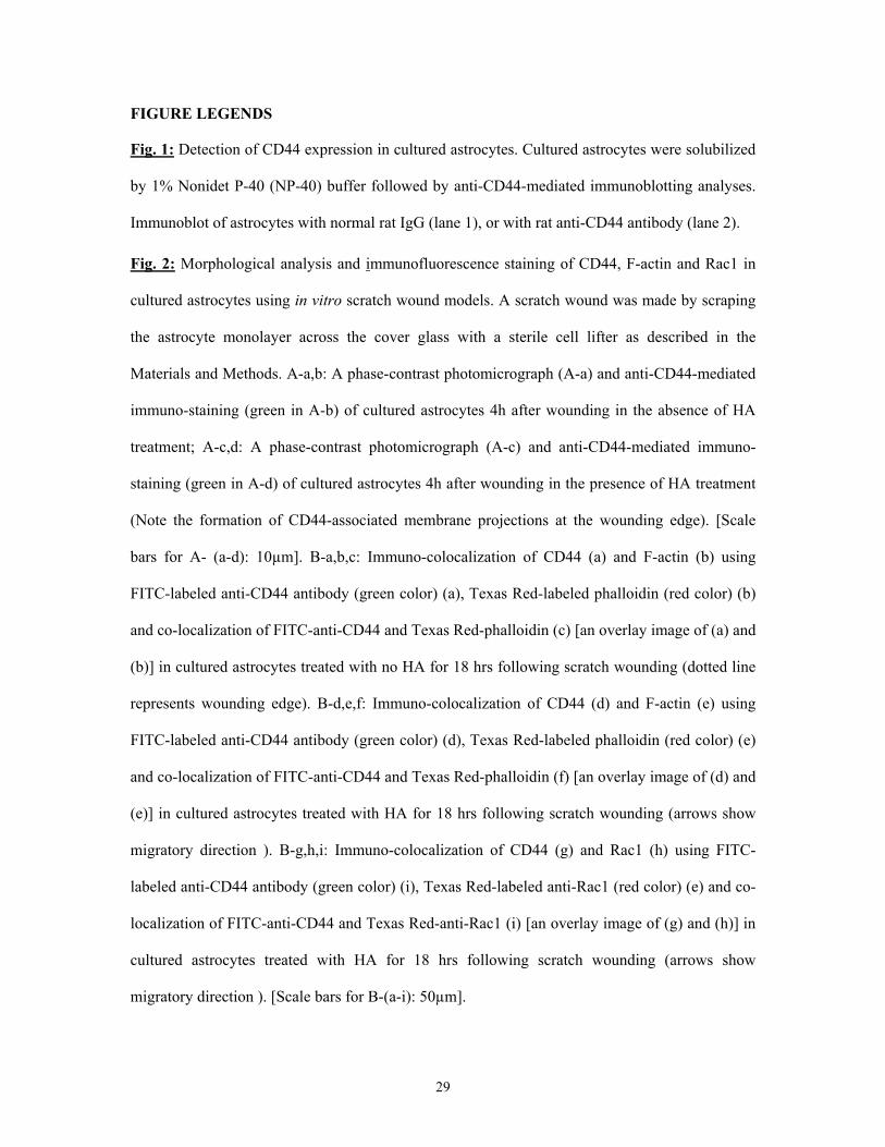
HA-CD44 Interaction in Cultured Astrocytes: CD44 is one of the major hyaluronan (HA) cell
surface receptors (Underhill 1992) in many different cell types, including astrocytes (Haegel et
al., 1993). Immunoblotting with monoclonal rat anti-CD44 antibody showed that a single band of
CD44 protein (molecular mass ~85kDa) was detected in cultured astrocytes isolated from mouse
brain (Fig. 1, lane 2). We conclude that the expression of CD44 in cultured astrocytes, as shown
by anti-CD44-mediated immunoblot, is specific since no protein was detected in these cells using
normal rat IgG (Fig. 1, lane 1). By employing a “scratch wound” assay with cultured astrocytes,
we found that HA induced CD44-associated membrane projections (Figs. 2A-c and 2A-d) and
CD44-F-actin colocalization in membrane protrusions at the wounding edges (Fig. 2B-d, e, f). In
contrast, very few CD44-F-actin-associated membrane projections were detected in astrocytes not
treated with HA (Figs. 2A-a, b and 2B-a, b, c). Following the formation of a scratch wound,
astrocytes were noted to polarize perpendicularly to the scratch and then migrate to close the
wound in the presence of HA (Figs. 3A-c, 3A-d and 3B-b). In contrast, pretreatment of cultured
astrocytes with anti-CD44 antibody followed by HA addition (Figs. 3A-e, 3A-f and 3B-c) or
12
without any HA treatment (Fig. 3A-a, 3A-b and 3B-a) resulted in a great reduction in migration
and wound closure. Using a Transwell cell migration assay, we also observed that a higher level
of cell migration occurred with HA-treated astrocytes as compared to untreated cells (Table 1).
Cultured astrocytes pretreated with anti-CD44 antibody or cytochalasin D (an inhibitor known to
impair filamentous actin function), showed significantly less HA-mediated migration (Table 1).
These findings clearly indicate that HA promotes actin cytoskeleton-mediated astrocyte migration
in a CD44-dependent manner.
HA-Stimulated Rac1 Signaling and CD44-PKNγ Kinase Association in Cultured
Astrocytes: The binding of HA to CD44 is known to induce important changes in certain
RhoGTPases such as Rac1 (Bourguignon et al., 2000; 2001a). Using an in vitro [35S]GTPγS
binding assay, we determined that Rac1 isolated from mouse cultured astrocytes displayed
specific guanine nucleotide binding activity (Table 2). In particular, we demonstrated that the
addition of HA to CD44-expressing astrocytes caused almost a three-fold increase in the binding
of [35S]GTPγS to Rac1 as compared to the amount of binding present in untreated astrocytes
(Table 2), or in astrocytes pretreated with anti-CD44 antibody followed by HA treatment (Table
2). Double immunofluorescence staining data indicated that HA also promoted Rac1 association
with CD44 in the membrane projections of astrocytes at the wounding edges (Fig. 2B-g, h, i).
These findings suggest that HA and CD44 are directly involved in the activation of Rac1 in
cultured astrocytes.
Several lines of evidence indicate that PKNγ binds to RhoGTPases (e.g. Rac1), and that
PKNγ’s activity is upregulated in the presence of the GTP-bound form of Rac1 (Quilliam et al.,
1996; Vincent and Settleman, 1997). In order to identify possible downstream targets for the
activated Rac1 in astrocytes, we focused our investigation on PKNγ kinase (also called PRK2
kinase). First, using a specific anti-PKNγ immunoblot technique, we determined that PKNγ
(molecular mass ~120kD) was expressed in astrocytes (data not shown). Next, we addressed the
question of whether there is an interaction between CD44 and PKNγ in cultured astrocytes. To

13
this end we carried out anti-CD44-mediated immunoprecipitation followed by anti-PKNγ
immunoblot (Fig. 4A-a) or anti-CD44 immunoblot (Fig. 4A-b), respectively, using untreated
astrocytes. Our results showed that a low level of PKNγ (Fig. 4A-a, lane 1) was present in the
anti-CD44-immunoprecipitated materials (Fig. 4A-b, lane 1). Subsequently, we determined that
HA treatment induced the recruitment of a significant amount of PKNγ (Fig. 4A-a, lane 2) into
the CD44-PKNγ complex (Fig. 4A-b, lane 2). These findings clearly establish that CD44 and
PKNγ are closely associated with each other, particularly following HA treatment of cultured
astrocytes.
Cortactin Serves as a Cellular Substrate for Rac1-Dependent PKNγ Kinase and Interacts
with Filamentous Actin (F-Actin): Although GTPase-activated PKNγ has been shown to
regulate actin cytoskeleton organization (Bourguignon et al., 2004), the specific cytoskeletal
components regulated by PKNγ in HA-mediated CD44 signaling in astrocytes have not been
clearly defined. In this study we observed that PKNγ kinase isolated from astrocytes was capable
of phosphorylating cortactin at both serine (Fig. 4B-a) and threonine residues (Fig. 4B-b) [as
detected by anti-phospho-serine and anti-phospho-threonine-mediated immunoblot, respectively
followed by reblotting with anti-cortactin antibody (Fig. 4B-c)] in the presence of activated Rac1
(GTP-bound Rac1) (Figs. 4B-a, lane 3 and 4B-b, lane 3) [and to a much lesser extent with GDP-
Rac1 treatment (Figs. 4B-a, lane 2 and 4B-b, lane 2)]. The level of serine/threonine
phosphorylation of cortactin was relatively low when PKNγ was not incubated with any Rac1
(Figs. 4B-a, lane 1 and 4B-b, lane 1). These results suggest that cortactin phosphorylation by
PKNγ involves Rac1 in a GTP-dependent fashion.
Further analyses indicated that the level of serine and threonine phosphorylation of
cortactin in cultured astrocytes [as detected by anti-cortactin-mediated immunoprecipitation
followed by immunoblotting with anti-serine (Fig. 4C-a) or anti-threonine (Fig. 4C-b) or anti-
cortactin (Fig. 4C-c), respectively] was significantly enhanced in astrocytes treated with HA (Fig.
4C-a, lane 2; Fig. 4C-b, lane 2). In contrast, cortactin serine and threonine phosphorylation was
14
relatively low in astrocytes without any HA treatment (Fig. 4C-a, lane 1; Fig. 4C-b, lane 1).
These observations strongly support the conclusion that cortactin phosphorylation is induced by
HA.
It is well known that cortactin is an important actin binding protein (Huang et al., 1997;
Huang et al., 1998). Our data indicated that cortactin with a low level of phosphorylation (in the
absence of PKNγ) (Fig. 4B-lane 1) or in the presence of PKNγ and unactivated Rac1 (GDP-
bound Rac1) (Fig. 4B-lane 2), was capable of cross-linking the actin filaments into bundles in
vitro (Table 3). However, serine/threonine phosphorylation of cortactin by Rac1 (GTP-bound
form)-activated PKNγ (Fig. 4B-lane 3) significantly decreased its ability to bind to filamentous
actin (Table 3). These results are consistent with previous findings suggesting cortactin
(phosphorylated vs. unphosphorylated forms) plays a critical role as a filamentous actin (F-actin)
modulator required for cytoskeleton reorganization.
Effects of PKNγ-ACC domains on Cortactin Phosphorylation, Actin Binding and Astrocyte
Migration: Several studies have determined that the N-terminal antiparallel coiled-coil (ACC)
domains of PKNγ (Fig. 5A-a) bind to RhoGTPases (e.g. Rac1) and upregulate PKNγ’s activity in
the presence of the GTP-bound form of Rac1 (Quilliam et al., 1996; Vincent and Settleman,
1997; Bourguignon et al., 2004). These findings suggest that the PKNγ-ACC domains play an
important role in regulating the activation of Rac1-dependent PKNγ kinase. In order to confirm
that the PKNγ-ACC domain regulate astrocyte signaling, PKNγ-ACC domain cDNA was cloned
into a His tagged expression vector pcDNA 3.1 (Fig. 5A-b) and then introduced into cultured
astrocytes by a transient transfection. To test the interaction between the PKNγ-ACC domain and
Rac1, we incubated purified His-tagged PKNγ-ACC domains with either GDP- or GTP-loaded
forms of Rac1-GST conjugated beads. Bound proteins were detected by immunoblotting with
anti-His antibody (Fig. 5B). Our results indicated that PKNγ-ACC domains appeared to
preferentially bind to GTP-bound Rac1 (Fig. 5B, lane 2), and to a much lesser extent to GDP-
bound Rac1 (Fig. 5B, line 1). These results agree with previous findings that PKNγ-ACC
15
domains are capable of binding to Rac1 in a GTP-dependent manner.
In order to examine the effects of PKNγ-ACC fragment overexpression on Rac1
interaction with endogenous PKNγ, we transfected cultured astrocytes with His-tagged PKNγ-
ACCcDNA (or vector alone). Our data showed that endogenous PKNγ was expressed at
comparable levels in vector-transfected (Fig. 5C-b, lane 1 and lane 2) and PKNγ-ACCcDNA-
transfected astrocytes (Fig. 5C-b, lane 3 and lane 4). Furthermore, we observed that a low levels
of endogenous Rac1 and PKNγ were co-precipitated in untreated vector-transfected astrocytes
(Fig. 5C-a, lane 1; Fig. 5C-b, lane 1). Moreover, we demonstrated that HA induced an additional
recruitment of endogenous Rac1 (Fig. 5C-a, lane 2) into a complex with PKNγ (Fig. 5C-b, lane 2)
in vector-transfected astrocytes. These results suggest that the recruitment of endogenous Rac1
into PKNγ in cultured astrocytes is HA-dependent. In addition, our observations indicated that
transfection of cultured astrocytes with PKNγ-ACCcDNA not only caused a significant inhibition
of HA-mediated recruitment of endogenous Rac1 (Fig. 5C-a, lane 4) to PKNγ (Fig. 5C-b, lane 4),
but also decreased the basal level of endogenous Rac1 (Fig. 5C-a, lane 3) association with PKNγ
(Fig. 5C-b, lane 3). These findings establish the fact that the PKNγ fragment containing ACC
domains acts as a potent competitive inhibitor of endogenous Rac1 binding to PKNγ during HA-
mediated signaling in cultured astrocytes.
In addition, we determined that phosphorylation of cortactin was stimulated using PKNγ
isolated from vector-transfected astrocytes treated with HA (Fig. 6A-b). In contrast, very little
cortactin phosphorylation was observed using PKNγ isolated from vector-transfected astrocytes
treated with no HA (Figs. 6A-a). We also found that cortactin phosphorylation was greatly
blocked using PKNγ isolated from PKNγ-ACCcDNA-transfected astrocytes treated with HA
(Fig. 6A-d) or without HA (Fig. 6A-c). Further analyses indicated that unphosphorylated
cortactin (treated by PKNγ kinase isolated from vector-transfected cells in the absence of HA)
promoted F-actin cross-linking activity in vitro (Fig. 6B-a). In contrast, PKNγ kinase-

16
phosphorylated cortactin (treated by PKNγ kinase isolated from vector-transfected cells in the
presence of HA) significantly reduced its ability to cross-link F-actin (Fig. 6B-b). Moreover, we
found that unphosphorylated cortactin (treated by PKNγ kinase isolated from PKNγ-ACCcDNA-
transfected cells with or without HA addition) retained its F-actin cross-linking properties (Fig.
6B-c and Fig. 6B-d). These results agree with a previous finding suggesting that inhibition of
Rac1 binding to PKNγ (Fig. 5C, lane 3 and 4) can effectively impair HA-mediated PKNγ activity
and cortactin-F-actin-based cytoskeleton function (Bourguignon et al., 2004).
Furthermore, we demonstrated that cortactin phosphorylation (Table 3B) was greatly
enhanced in vector-transfected astrocytes treated with HA as compared with those observed in
untreated vector-transfected astrocytes (Table 3B). Cortactin phosphorylation (isolated from
vector-transfected astrocytes treated with HA) significantly down-regulated its ability to cross-
link filamentous actin (Table 3B). In contrast, the reduction of cortactin phosphorylation (isolated
from vector-transfected astrocytes with no HA treatment or from PKNγ-ACCcDNA-transfected
astrocytes in the presence or absence of HA) was sufficient to promote cross-linking of actin
filaments into bundles in vitro (Table 3B). These results demonstrate that the ACC fragment of
PKNγ acts as a dominant-negative mutant that downregulates HA-induced Rac1-PKNγ activation
and cortactin phosphorylation required for cytoskeleton function.
Furthermore, using Transwell cell migration assays, we observed that cell migration of
vector-transfected astrocytes were stimulated during HA treatment (Fig. 7A-a, b). However,
transfection of cultured astrocytes with PKNγ-ACCcDNA, resulted in a significant inhibition of
HA-mediated astrocyte migration (Fig. 7A-c, d). Similarly, using a scratch wound-induced
migration assay of vector-transfected astrocytes, we found a faster rate of migration occurring
with HA-treated cells following wounding (Fig. 7B-(I)-c, d; 7B-(II)-b). In contrast, very little cell
migration was detected in vector-transfected astrocytes that were not treated with HA (Fig. 7B-
(I)-a, b; 7B-(II)-a). We also noted that overexpression of PKNγ-ACC in astrocytes transfected
with PKNγ-ACCcDNA significantly reduced cell migration under wounding conditions either in

17
the presence (Fig. 7B-(I)-g,h; 7B-(II)-d) or absence of HA (Fig. 7B-(I)-e,f; 7B-(II)-c). Thus, HA-
mediated Rac1-PKNγ signaling and cytoskeleton function are directly involved in astrocyte
migration following mechanical injury in vitro.
DISCUSSION
Astrocytes are the major glial cell type in the central nervous system (CNS), and play an
essential role in maintaining the physiological function of neurons (Fitch and Silver, 2001; Barres
and Barde, 2000). In the CNS, astrocytes often display drastic morphological changes when they
interact with neurons and the actin cytoskeleton is known to play an important role in the
regulation of astrocyte shape changes (Baorto et al., 1992; Ramakers and Moolenaar, 1998;
Etienne-Manneville and Hall 2002; Holtje et al., 2005). After CNS injury, astrocytes also undergo
morphological changes. For example, the retraction of astrocyte processes has been described as
one of the major ultrastructural changes following CNS injury (Sperry et al., 1993). As early as
one hour after CNS injury, there are alterations in the astrocyte cytoskeleton and increased glial
fibrillary acidic protein (GFAP) immunoreactivity (Goshgarian et al., 1989; Hadley, 1997).
Astrocytes that participate in these early activation processes also proliferate and/or migrate to
form the glial scar tissue that encases the damaged area (Hadley, 1997). This process, known as
“reactive astrogliosis”, is considered to be an accurate indicator of neuronal damage and a major
hindrance to axonal regeneration following CNS injury (Stichel and Muller, 1998; Nieto-
Sampedro, 1999). The abnormal astrocyte migration to the injured sites also correlates with the
failure of regrowing axons to transverse this site (Rhodes et al., 2003; Silver and Miller, 2004;
Shearer and Fawcett, 2001; Sivron and Schwartz, 1995). Presently, very little is known about the
signaling mechanisms that control astrocyte cytoskeletal changes and cellular function (e.g.
migration) following injury.
Hyaluronan (HA) is a very high molecular weight glycosaminoglycan consisting of
repeating disaccharide units of D-glucuronic acid and N-acetylglucosamine (Tammi et al., 2002).
HA production is closely associated with embryogenesis, wound healing and tumorigenesis
18
(Toole, 2001; Toole, 2004), and has been implicated in the regulation of astrocyte functions
during CNS repair and glial scar formation (Asher et al., 1991; Bignami et al., 1992; Bignami and
Asher, 1992; Eggli et al., 1992; Struve et al., 2005). However, the cellular and molecular
mechanisms by which astrocytes respond to HA are not well understood. The predominant
receptor for HA on the cell surface is CD44 (Haegel et al., 1993) CD44 is encoded by a single
gene which contains 19 exons (Screaton et al., 1992). The most common form, CD44s (CD44
standard form), contains exon 1-5 (N-terminal 150 amino acids), exon 15 and 16 (membrane
proximal 85 amino acids), exon 17 (transmembrane domain), and a portion of exon 17 and 19
(cytoplasmic tail, 70 amino acids) (Screaton et al., 1992). Out of the 19 exons, 12 exons can be
alternatively spliced (Screaton et al., 1992). In this study we have determined that the CD44
detected in astrocytes isolated from normal brain tissues is a ~85kDa polypeptide which is the
standard form of CD44 (CD44s) (Fig. 1). A number of astrocyte-type gliomas have also been
shown to strongly express different CD44 variant (CD44v) isoforms in addition to CD44s
(Kuppner et al., 1992; Haegel et al., 1993). HA binds to the external domain of all CD44 isoforms
(Underhill, 1992). A previous study showed that the extracellular domain of CD44 can be
released from the cell membrane and becomes associated with ECM (e.g. in glia limitants)
(Jones, et al, 1997). This finding suggests that CD44’s external part is capable of acting in a
dominant-negative fashion by blocking the migration-inducing activity of ECM components such
as HA. The binding of HA to CD44 also promotes the interaction of CD44’s cytoplasmic domain
with a variety of signaling proteins and the cytoskeleton (Bourguignon et al., 1998; Bourguignon,
2001; Turley el al., 2002). In particular, HA mediates CD44 association with c-Src kinase which
promotes tyrosine phosphorylation of cortactin [a filamentous actin (F-actin)-associated protein]
and cytoskeleton function resulting in specific structural changes in the plasma membrane and
cell migration (Bourguignon et al., 2001b). These findings strongly suggest that the CD44
provides a direct linkage between the ECM (e.g. HA) and the signaling molecule-regulated
cytoskeleton.

19
It is well documented that both the structural and functional regulation of the actin
cytoskeleton is under the control of RhoGTPases [small molecular weight GTPases, (e.g. RhoA,
Rac1 and Cdc42)] which act as molecular switches that alternate between the GTP- and GDP-
bound states. The “activated” GTP-bound enzymes preferentially interact with downstream
effector molecules and modulate their effectors’ activities (Hall 1998). HA has been shown to
promote the interaction between CD44 and several Rac1-specific guanine nucleotide exchange
factors [e.g. Tiam1 (Bourguignon et al., 2000) and Vav2 (Bourguignon et al., 2001a)] thereby up-
regulating Rac1 signaling and cytoskeleton-mediated cell functions. Our results clearly indicate
that HA promotes Rac1 signaling in astrocytes (Table 2), and that Rac1 together with F-actin co-
localize with CD44 in the membrane projections of astrocytes (Fig. 2B). These observations are
consistent with previous reports showing activation of Rac1 signaling is involved in cytoskeleton-
associated lamellipodia formation in many cell types (Hall 1998). Furthermore, we have utilized
an in vitro scratch wound assay to analyze astrocyte reactions to injury. Our results indicate that
HA stimulates both cell migration and wound closure in a CD44-dependent manner (Fig. 3).
These findings suggest that HA-mediated CD44 interaction with Rac1 and the cytoskeleton plays
a pivotal role in cytoskeleton function and migration in astrocytes.
In order to identify the possible downstream targets for Rac1 signaling, we have focused
on protein kinase N-γ (PKNγ) (also called PRK2) which belongs to a family of serine/threonine
kinases and shares high sequence homology with members of the PKC family of enzymes
(Mukai, 2003). There are at least three different isoforms of PKN (PKNα PAK-1/PRK-1, PKNβ
and PKNγ/PRK2) which have been detected in mammalian cells (Mukai, 2003). PKNγ is known
to interact with Rac1 in a GTP-dependent manner and shares a great deal of homology with PKC
in the C-terminal region (Quilliam et al., 1996; Vincent and Settleman, 1997; Mukai, 2003).
PKNγ also has a unique regulatory region containing antiparallel coiled-coil (ACC) domains
which binds to Rac1 directly (Quilliam et al., 1996; Vincent and Settleman, 1997). A previous
20
study showed that Rac1-activated PKNγ is required for the organization of the cortical
cytoskeleton (Bourguignon et al., 2004). In particular, the cytoskeletal protein, cortactin is a
prominent substrate for PKNγ kinase (Bourguignon et al., 2004). HA has also been shown to
stimulate Rac1-mediated PKNγ phosphorylation of cortactin leading to cytoskeleton function
(Bourguignon et al., 2004). In this study we have determined that cortactin is a cellular substrate
for PKNγ kinase in astrocytes activated by HA binding to CD44 (Figs. 4 and 6). Two structural
features of cortactin, a repeat domain and a carboxyl-terminal SH3 domain, resemble these of
neufectin, an F-actin-associated protein and cortactin is considered to be an actin binding protein
(Huang et al., 1997; Huang, et al., 1998). Our results indicate that PKNγ [by binding to activated
Rac1 (Rac1-GTP form)] is capable of inducing serine/threonine phosphorylation of cortactin
(isolated from astrocytes) (Fig. 4). The ability of PKNγ to phosphorylate cortactin in the presence
of unactivated Rac1 (Rac1-GDP form) appears to be extremely limited (Fig. 4). These results
clearly indicate that PKN acts as one of the downstream effectors of Rac1 signaling, and utilizes
cortactin as one of its cellular targets. Both Rac1 signaling (Table 2) and PKNγ-mediated
cortactin phosphorylation (Fig. 4) are stimulated by HA. Thus, it appears that Rac1 activated
PKNγ-mediated phosphorylation of cortactin is closely coupled with HA-mediated activation in
astrocytes. The fact that cortactin phosphorylation by HA-activated Rac1-PKNγ kinase modifies
its binding to F-actin suggests that Rac1-mediated PKNγ activity is closely involved in actin
cytoskeleton reorganization during astrocyte signaling.
The ACC domain of PKNγ has been found to be involved in the regulation of GTP-
dependent PKN function (Quilliam et al., 1996; Vincent and Settleman, 1997; Bourguignon et al.,
2004). As shown in Fig. 5, we have determined that the ACC domain is closely associated with
Rac1 in a relatively stable complex. Overexpression of the ACC domain by transfection of
astrocytes with PKNγ-ACCcDNA (Fig. 5) effectively competes for the binding of endogenous
activated Rac1 to PKNγ (Fig. 5), inhibits the ability of PKNγ to phosphorylate cortactin (Fig. 6),
21
and blocks HA/CD44-mediated cortactin phosphorylation (Fig. 6). In addition, we have
confirmed that cortactin phosphorylation by the PKNγ kinase (isolated from untransfected or
vector-transfected astrocytes in the presence of HA) significantly decreases its ability to cross-
link filamentous actin (Fig. 6). In contrast, the reduction of cortactin phosphorylation (isolated
from astrocytes transfected with PKNγ-ACCcDNA with or without HA treatment) allows the
cross-linking of actin filaments into bundles (Fig. 6). These results are consistent with previous
findings suggesting cortactin plays an important role as a regulator of F-actin-based cytoskeleton
function.
Although a number of factors have been shown to be involved in stimulating astrocyte
migration, it was not known whether HA/CD44-mediated Rac1-PKNγ signaling participates in
the wound closure processes such as migration. In this study we have found that HA promotes
astrocyte migration and wound closure in vector-transfected astrocytes following scratch
wounding processes (Fig. 7). In contrast, overexpression of the dominant-negative form of PKNγ
(PKNγ-ACC fragment) by transfecting astrocytes with PKNγ-ACCcDNA effectively inhibits
HA-mediated migration and wound closure in injured astrocytes (Fig, 7). These data strongly
suggest that CD44-mediated PKNγ activation is required for HA-dependent migration and in
vitro wound closure by astrocytes. Taking these results together, we would like to propose the
following signaling model: HA-CD44 interaction (step 1) with Rac1-dependent PKNγ kinase
activation (step 2) promotes phosphorylation of cortactin (step 3) and cortactin-mediated
cytoskeleton function (step 4) leading to astrocyte migration (step 5) (Fig. 8). We believe that the
successful identification of these HA/CD44-mediated signaling pathways will provide valuable
new insights toward a better understanding of astrocytes migration and may contribute
significantly to the improvement of neural tissue regeneration following injury.
Both hyaluronidases (Stern and Jedrzejas, 2006) and reactive oxygen species (ROS)
(Soltes et al., 2006) have been implicated in the partial degradation or modifications of HA
polymers. Generally, low molecular weight HA-CD44 interaction induces inflammatory or

22
proliferative genes; whereas high molecular weight HA-CD44 binding promotes migration and
transcriptional activation (Nobel, 2002; Lokeshwar et al., 1996; Turley et al., 2002). The question
of whether selective activation of CD44 signaling (via high or low-molecular weight HA) induces
a pathway-specific effect on astrocyte functions (cytoskeletal reorganization/activation,
proliferation, gene expression and migration) and/or glial scar formation following CNS injury is
currently under investigation in our laboratory.
ACKNOWLEDGMENT: We gratefully acknowledge Dr. Gerard J. Bourguignon's assistance in
the preparation of this paper. We are also grateful for Ms. Christine Camacho’s help in organizing
graphs/illustrations and Ms. Elizabeth Gum’s assistance in cultured astrocyte preparation. This
work was supported by United States Public Health grants (R01 CA66163, R01 CA 78633 and
P01 AR39448), a VA Merit Review grant and a DOD grant. L.Y.W.B is a VA Research Career
Scientist.
REFERENCES
Asher R., Perides G., Vanderhaeghen J.J. and Bignami A. (1991) Extracellular matrix of central nervous system white mateer: demonstration of an hyaluronate-protein complex. J. Neurosci Res. 28, 410-421. Barres B.A. and Barde Y. (2000) Neuronal and glial cell biology. Curr. Opin. Neurobiol. 10, 642-648. Baorto D.M., Mellado W. and Shelanski M.L. (1992) Astrocyte process growth induction by actin breakdown. J. Cell Biol. 117, 357-367. Bignami, A. and Asher R. (1992) some observations on the localization of hyaluronic acid in adult, newborn and embryonal rat brain. Int J. Dev. Neurosci 10, 45-57. Bignami, A. Asher R. and Perides G. (1992) The extracellular matrix of rat spinal cord: a comparative study on the localization of hyaluronic acid, glial hyaluronate-binding protein and chondroitin sulfate proteoglycan. Exp Neurol 17, 90-93. Bourguignon L.Y.W., Zhu D. and Zhu H. (1998) CD44 isoform-cytoskeleton interaction in oncogenic signaling and tumor progression. Front Biosci. 3, 637-649. Bourguignon, L.Y.W. (2001) CD44-mediated oncogenic signaling and cytoskeleton activation during mammary tumor progression. J Mammary Gland Biol. and Neoplasia 6, 287-297. Bourguignon L.Y.W., Zhu H., Shao L. and Chen Y.W. (2000) CD44 interaction with Tiam1 promotes Rac1 signaling and hyaluronic acid (HA)-mediated breast tumor cell migration. J. Biol. Chem. 275, 1829-1838. Bourguignon L.Y.W., Zhu H., Zhou B., Diedrich F., Singleton P.A. and Hung M.C. (2001a) Hyaluronan (HA) promotes CD44v3-Vav2 interaction with Grb2-p185HER2 and induces Rac1 & Ras signaling during ovarian tumor cell migration and growth. J. Biol. Chem. 276, 48679-48692. Bourguignon, L.Y.W., Zhu H., Shao L. and Chen Y.W. (2001b) CD44 interaction with c-
23
Src kinase promotes cortactin-mediated cytoskeleton function and hyaluronic acid (HA)-dependent ovarian tumor cell migration. J. Biol. Chem. 276, 7327-7336. Bourguignon L.Y.W., Singleton P.A. and Diedrich F. (2004) Hyaluronan/CD44 interaction with Rac1-dependent PKNγ kinase promotes PLCγ1 activation, Ca2+ signaling and cortactin-cytoskeleton function leading to keratinocyte adhesion and differentiation. J. Biol. Chem. 279, 29654-29669. Burbelo P.D., Drechsel D. And Hall A. (1995) A conserved binding motif defines numerous candidate target proteins for both Cdc42 and RacGTPases. J. Biol. Chem. 270, 29071-29074. Chen Y. and Swanson R.A. (2003) Astrocytes and brain injury. J. Cereb Blood Flow Metab 23, 137-149. David S. and Lacroix S. (2003) Molecular approaches to spinal cord repair. Annu Rev. Neurosci. 26, 411-440. Eddleston M. and Mucke L. (1993) Molecular profile of reactive astrocytes-implications for their role in neurologic disease. Neurosci. 54, 15-36. Eggli P.S., Lucocq J., Ott P., Graber W., van der Zypen E. (1992) Ultrastructural localization of hyaluronan in myelin sheaths of the rat central and rat and human peripheral nervous systems using haylurona-binding protein-gold and link protein-gold. Neurosci. 48,737-744. Erschbamer M.K., Hofstetter C.P. and Olson L. RhoA, RhoB, RhoC, Rac1, Cdc42, and Tc10 (2005) mRNA levels in spinal cord, sensory ganglia, and corticospinal tract neurons and long-lasting specific changes following spinal cord injury. J. Comparative Neurol. 484, 224-233. Etienne-Manneville S. and Hall A. (2002) RhoGTPases in cell biology. Nature 420, 629-635. Fitch M.T. and Silver J. (2001) Astrocytes are dynamic participants in central nervous system development and injury response. In: Glial cell development. (Jessen K.R. and Richardson W.D., ed). New York: Oxford University Press. p.263-277. Goshgarian, H.G., Yu, X.J. and Rafols, J.A. (1989) Neuronal and glial changes in the rat phrenic nucleus occurring within hours after spinal cord injury. J. Comp. Neurol. 284, 519-530. Kuppner M.C., van Meir E., Gauthier T., Hamou M.F. and de Tribolet N. (1992) Differential expression of the CD44 molecule in human brain tumors. Int. J. Cancer 50, 575-577. Hadley, S.D. and Goshgarian, H.G. (1997) Altered immunoreactivity for glial fibrillary acidic protein in astrocytes within 1h after cervical spinal cord injury Exp. Neurol. 146:380-387. Haydon P.G. (2000) Neuroglial networks: Neurons and glia talk to each other. Curr. Biol. 10, R712-R714. Haegel H., Tolg C., Hofmann M. and Ceredig R. (1993) Activated astrocytes and T cells express similar CD44 variants. Role of CD44 in astrocyte/T cell binding. J. Cell Biol. 122, 1067-1077. Hall A. (1998) Rho GTPase and the cytoskeleton. Science 279, 509-514. Holtje M., Hoffmann A., Hofmann F., Mucke C., Groβe, van Rooijen N., Kettenmann H., Just I. and Ahnert-Hilger G. (2005) Role of RhoGTPase in astrocyte morphology and migratory response during in vitro would healing. J. Neurochem 95, 1237-1248. Huang, C., Ni, Y., Wang, T., Gao, Y., Haudenschild, C.C. and Zhan, X. (1997) Down-regulation of the filamentous actin cross-linking activity of cortactin by Src-mediated tyrosine phosphorylation. J. Biol. Chem. 272, 13911-13915. Huang, C., Liu, J., Haudenschild, C.C. and Zhan, X.J. (1998) The role of tyrosine phosphorylation of cortactin in the locomotion of endothelial cells. J. Biol.Chem. 273, 25770-25776. Jones L.L., Kreutzberg G.W. and Raivich G. (1997) Regulation of CD44 in the regenerating mouse facial motor nucleus. Eur J Neurosci. 9, 1854-1863. Jones L.L., Liu Z., Shen J., Werner A., Kreutzberg G.W. and Raivich G. (2000)

24
Regulation of the cell adhesion molecule CD44 after nerve transaction and direct trauma to the mouse brain. J. Comp. Neurol. 426, 468-492. Lokeshwar V.B., Iida N, Bourguignon L.Y.W. (1996) The cell adhesion molecule, GP116 is a new CD44 variant (ex14/v10) involved in hyaluronic acid binding and endothelial cell proliferation. J Biol Chem.271, 23853-23864. Lee J. Y. and Spicer A.P. (2000) Hyaluronan: a multifunctional, megadalton, stealth molecule. Curr Opin Cell Biol. 12, 581-586. Mukai H. (2003) The structure and function of PKN, a protein kinase having a catalytic domain homologous to that of PKC. J. Biochem 133, 17-27. Mukhin, A.G., Ivanova, S.A., Allen, J.W. and Faden, A.I. (1998) Mechanical injury to neuronal/glial cultures in microplates: role of NMDA receptors and pH in secondary neuronal cell death. J Neurosci Res 51, 748-758. Nieto-Sampedro M. (1999) Neurite outgrowth inhibitors in gliotic tissue. Adv. Exp. Med. Biol. 468, 207-224.
Noble PW. (2002) Hyaluronan and its catabolic products in tissue injury and repair. Matrix Biology 25, 25-29. Quackenbush E.J., Cruz T.F., Moscarello M.A. and Letarte M. (1985) Identification of three antigens in human brain associated with similar antigens on human leukemic cells. Biochem J. 225, 291-299. Quilliam L.A., Lambert Q.T., Mickelson-Young L.A., West-wick J.K., Sparks A.B., Kay B.K., Jenkins N.A., Gilbert D.J., Copeland N.G., and Der C.J. (1996) Isolation of a NCK-associated kinase, PRK2, an SH3-binding protein and potential effector of Rho protein signaling. J. Biol. Chem. 271, 28772-28776. Ramakers G.J.A. and Moolenaar, W.H. (1998) Regulation of astrocyte morphology by RhoA and lysophosphatidic acid. Exp. Cell Res. 245, 252-262. Rhodes K.E., Moon L.D., and Fawcett J.W. (2003) Inhibiting cell proliferation during formation of the glial scar: effects on axon regeneration in the CNS. Neurosci. 120, 41-56. Ridet J.L., Malhotra S.K., Privat A. and Cage F.H. (1997) Reactive astrocytes: Cellular and molecular cues to biological function. Trends Neurosci 20, 570-577. Screaton G.R., Bell M.V., Jackson D.G., Cornelis F.B., Gerth U. and Bell J.I. (1992) Genomic structure of DNA encoding the lymphocyte homing receptor CD44 reveals at least 12 alternatively spliced exons. Proc. Natl. Acad. Sci . U.S.A. 89, 12160-12164. Stern, R. and Jedrzejas, M.J. (2006) Hyalurinidases: their genomics, structures, and mechanisms of action. Chem Rev. 106,818-839. Shearer M.C. and Fawcett J.W. (2001) The astrocyte/meningeal cell interface-a barrier to successful nerve regeneration? Cell Tissue Res. 305, 267-273. Silver J. and Miller J.H. (2004) Regeneration beyond the glial scar. Nat Rev. Neurosci. 5, 146-156. Sivron T. and Schwartz M. (1995) Glial cell types, lineage and response to injury in rat and fish: Implication for regeneration. Glia 13, 157-165. Soltes, L., Mendichi, R., Kogan, G., Schiller, J., Stankovska, M., and Arnhold, J. (2006) Degradative action of reactive oxygen species on hyaluronan. Biomacromolecules 7, 659-668. Song H., Stevens C.F. and Gage F.H. (2002) Astroglia induce neurogenesis from adult neural stem cells. Nature 417, 39-44. Sperry, M.A. and Goshgarian, H.G. (1993) Ultrastructural changes in the rat phrenuc nucleus developing within 2 h after cervical spinal cord hemisection. Exp. Neurol. 120, 233-244. Stichel C.C. and Muller H.W. (1998) The CNS lesion scar: new vistas on an old regeneration barrier. Cell Tissue Res. 294, 1-9. Struve J., Maher P.C., Li Y-Q., Kinney S., Fehlings M.G.,Kuntz, C. IV and Sherman L.S. (2005) Disruption of the hyaluronan-based extracellular matrix in spinal cord promotes astrocyte proliferation. Glia 52, 16-24.

25
Swanson R.A., Farrell K. and Stein B.A. (1997) Astrocyte energetics, function, and death under conditions of incomplete ischemia: a mechanism of glial death in the penumbra. Glia 21, 142-153. Tammi M.I., Day A.J. and Turley E.A. (2002) Hyaluronan and homeostasis: a balance act. J. Biol. Chem. 277, 4581-4584. Tona A. and Bignami A. (1993) Effects of hyaluronidase on brain extracellular matrix in vivo and optic nerve regeneration. J. Neurosci. Res 36, 191-199. Toole B. P. (2001) Hyaluronan in morphogenesis. Semin Cell Dev. Biol. 12, 79-87. Toole B.P. (2004) Hyaluronan from extracellular glue to pericellular cue. Nat Rev Cancer 4, 528-539. Turley E. A., Nobel P.W. and Bourguignon L.Y.W. (2002). Signaling Properties of Hyaluronan Receptors. [Mini-Review] J. Biol. Chem. 277, 4589-4592. Underhill C. (1992) CD44: the hyaluronan receptor. J. Cell Sci. 103, 293-298. Ullian E.M., Sapperstein S.K. Christopherson K.S. and Barrer B.A. (2001) Control of synapse number by glia. Science 291, 657-660. Vincent S. and Settleman, J. (1997) The PRK2 kinase is a potential effector target of both Rho and Rac GTPase and regulates actin cytoskeletal organization. Mol. Cell Biol. 17, 2247-2256. Yoshinaga C., Mukai H., Toshimori M., Miyamoto M., and Ono Y. (1999) Mutational analysis of the regulatory mechanism of PKN: the regulatory region of PKN contains an arachidonic acid-sensitive autoinhibitory domain. J. Biochem. 126, 475-484. Yu, A.C.H., Lee, Y-L. and Eng, L.F. (1993) Astroglisis in culture: I. the model and the effect of antisense oligonucleotides on glial fibrillary acidic protein synthesis. J. Neurosci. Res. 34, 295-303.
26
Table1: Measurement of HA/CD44-dependent and cytoskeleton-mediated astrocyte migration.
A: Effects of Anti-CD44 antibody on HA-dependent and CD44-specific astrocyte migration.
Treatments Cell Migration (% of control)a
No treatment (control) 100
HA treatment 290±11b
Anti-CD44 IgG pretreatment + HA treatment 96±3b
B: Effects of cytochalasin D on HA-mediated astrocyte migration.
Treatments Cell Migration (% of control)a
No drug treatment (control) 100
No drug treatment (control) + HA 288±9b
Cytochalasin D treatment alone 35±1b
Cytochalsin D treatment + HA 37±1b
Cultured astrocytes [~1 x 10⁴ cells/well in phosphate buffered saline (PBS), pH 7.2] were placed in the upper chamber of the transwell unit. In some cases, cultured astrocytes were pretreated with various agents (e.g. anti-CD44 IgG or cytochalasin D). After 18h incubation at 37oC in a humidified 95% air/5% CO2 atmosphere, cells on the upper side of the filter were removed by wiping with a cotton swap. Cell migration was determined by measuring the number of cells that migrated to the lower side of the polycarbonate filters containing HA (or no HA) by either MTT staining or standard cell number counting assays. The CD44-specific cell migration was determined by subtracting non-specific cell migration (i.e. cells migrate to the lower chamber in the presence of rat anti-CD44 antibody treatment) from the total migratory cells in the lower chamber. Cells treated with no HA (control) is designated as 100%. a: Data represent mean ± SEM of cells that migrated to the lower side of the polycarbonate filters and chambers from each sampe. b: Significantly different (p<0.001; ANOVA; n=6) as compared with no treatment (control) sample.
27
Table 2: Detection of Rac1 activation in cultured astrocytes.
Treatments Amount of [35S]GTPγS bound to Rac1 (cpm)
(% of control)a
No treatment (control) 5,500 ± 220 (100%)
HA treatment 16,225 ± 575 (295%)b
Anti-CD44 IgG + HA treatment 5,392 ± 218 (98%)b
Cultured astrocytes (~1.0 x 10⁴ cells) were preloaded with [35S]GTPγS (12.5 µCi) using electroporation methods as described in the Materials and Methods. These cells were incubated with 50µg/ml HA at 37oC for 10 min [in the presence or absence of rat anti-CD44 antibody (50µg/ml) or without any HA treatment]. Subsequently, [35S]GTPγS labeled cells were solubilized in 1.0% NP-40 and incubated with mouse anti-Rac1 IgG (5µg/ml) plus goat anti-mouse conjugated beads. The amount of [35S]GTPγS-Rac1 associated with anti-Rac1-conjugated immuno-beads was measured using a gamma counter. Cells treated with no HA (control) is designated as 100%. a: Data represent mean ± SEM of [35S]GTPγS (cpm) bound to Rac1 in each sample. b: Significantly different (p<0.001; ANOVA; n=5) as compared with no treatment (control) sample.
28
Table 3: Analyses of cortactin phosphorylation and cortactin-F-actin binding. A: Measurement of cortactin (phosphorylated by PKNγ) binding to F-actin.
Samples Cortactin-Actin Binding
(cpm) (% of Control)a
Unphosphorylated Cortactin (using PKNγ alone)
(control)
12,455 ± 622 (100%)
Phosphorylated Cortactin (using PKNγ + GDP-bound
Rac1)
12,206 ± 366 (98%)b
Phosphorylated Coractin (using PKNγ + GTP-bound
Rac1)
6,975 ± 279 (56%)b
Cortactin isolated from astrocytes was phosphorylated by PKNγ in the presence of GDP-/GTP-bound Rac1 or PKNγ alone. Subsequently, unphosphorylated or Rac1-PKNγ phosphorylated coractin was incubated with 125I-labeled F-actin. The procedures for measuring cortactin-F-actin binding were described in the Materials and Methods. Unphosphorylated cortactin (using PKNγ alone) (control) is designated as 100%. a: Data represent mean ± SEM of cortactin-actin binding (cpm) in each sample. b: Significantly different (p<0.001; ANOVA; n=5) as compared with unphosphorylated cortactin (using PKNγ alone) (control) sample. B: Effect of PKNγ-ACCcDNA overexpression on cortactin phosphorylation and cortactin-F-actin binding.
Cells Cortactin phosphorylation (% of control)a
Cortactin-F-actin binding (% of control)c
-HA +HA -HA +HA
Vector-transfected cells (control) 100 257±12b 100 43±0.4d
PKNγ-ACCcDNA-transfected cells 102±4b 105±3b 101±3.4d 103±2.3d
The procedures for measuring cortactin phosphorylation and cortactin-F-actin binding using astrocytes transfected with PKNγ-ACCcDNA or vector alone were described in the Materials and Methods. a: Data represent mean ± SEM of cortactin phosphorylation (cpm) in each sample. b: Significantly different (p<0.001; ANOVA; n=6) as compared with vector-transfected cells with no HA treatment (control) sample. c: Data represent mean ± SEM of cortactin-125I-F-actin binding (cpm) in each sample. d: Significantly different (p<0.001; ANOVA; n=5) as compared with vector-transfected cells with no HA treatment (control) sample.
29
FIGURE LEGENDS
Fig. 1: Detection of CD44 expression in cultured astrocytes. Cultured astrocytes were solubilized
by 1% Nonidet P-40 (NP-40) buffer followed by anti-CD44-mediated immunoblotting analyses.
Immunoblot of astrocytes with normal rat IgG (lane 1), or with rat anti-CD44 antibody (lane 2).
Fig. 2: Morphological analysis and immunofluorescence staining of CD44, F-actin and Rac1 in
cultured astrocytes using in vitro scratch wound models. A scratch wound was made by scraping
the astrocyte monolayer across the cover glass with a sterile cell lifter as described in the
Materials and Methods. A-a,b: A phase-contrast photomicrograph (A-a) and anti-CD44-mediated
immuno-staining (green in A-b) of cultured astrocytes 4h after wounding in the absence of HA
treatment; A-c,d: A phase-contrast photomicrograph (A-c) and anti-CD44-mediated immuno-
staining (green in A-d) of cultured astrocytes 4h after wounding in the presence of HA treatment
(Note the formation of CD44-associated membrane projections at the wounding edge). [Scale
bars for A- (a-d): 10µm]. B-a,b,c: Immuno-colocalization of CD44 (a) and F-actin (b) using
FITC-labeled anti-CD44 antibody (green color) (a), Texas Red-labeled phalloidin (red color) (b)
and co-localization of FITC-anti-CD44 and Texas Red-phalloidin (c) [an overlay image of (a) and
(b)] in cultured astrocytes treated with no HA for 18 hrs following scratch wounding (dotted line
represents wounding edge). B-d,e,f: Immuno-colocalization of CD44 (d) and F-actin (e) using
FITC-labeled anti-CD44 antibody (green color) (d), Texas Red-labeled phalloidin (red color) (e)
and co-localization of FITC-anti-CD44 and Texas Red-phalloidin (f) [an overlay image of (d) and
(e)] in cultured astrocytes treated with HA for 18 hrs following scratch wounding (arrows show
migratory direction ). B-g,h,i: Immuno-colocalization of CD44 (g) and Rac1 (h) using FITC-
labeled anti-CD44 antibody (green color) (i), Texas Red-labeled anti-Rac1 (red color) (e) and co-
localization of FITC-anti-CD44 and Texas Red-anti-Rac1 (i) [an overlay image of (g) and (h)] in
cultured astrocytes treated with HA for 18 hrs following scratch wounding (arrows show
migratory direction ). [Scale bars for B-(a-i): 50µm].

30
Fig. 3: Scratch wound-induced migration assays. A scratch wound was made by scraping the
astrocyte monolayer across the cover glass with a sterile cell lifter as described in the Materials
and Methods. A-a,b: Phase-contrast photomicrographs of cultured astrocytes (treated with no
HA) for 0h (a) or 24h (b) after in vitro scratch wounding. A-c,d: Phase-contrast photomicrographs
of cultured astrocytes (treated with HA) for 0h (c) or 24h (d) after in vitro scratch wounding.
A-e,f: Phase-contrast photomicrographs of cultured astrocytes (pretreated with anti-CD44
antibody followed by adding HA) for 0h (a) or 24h (b) after in vitro scratch wounding. [Scale
bars for A-(a-f): 100µm]. B-Degree of wound closure is expressed as percent of control (24h after
scratch wounding in the absence of HA treatment) (a). Astrocytes (treated with HA) for 24h after
in vitro scratch wounding (b); Astrocytes (pretreated with anti-CD44 antibody followed by
adding HA) for 24h after in vitro scratch wounding (c). The data are shown as means ±SEM.
[**Significantly different (p<0.001; ANOVA; n=5) as compared with untreated (control)
sample].
Fig. 4: Characterization of PKNγ in cultured astrocytes.
A: Detection of PKNγ and CD44 in the complex isolated from untreated astrocytes (lane 1) and
HA-treated astrocytes (lane 2) by employing anti-CD44-mediated immunoprecipitation followed
by immunoblotting with anti-PKNγ (a) or reblotting with anti-CD44 (b) as a loading control. The
ratio of PKNγ to CD44 levels was determined by densitometry and the levels were normalized to
the untreated astrocyte value (lane 1); The values expressed in Fig. 4A represent an average of
triplicate determinations of 5 experiments with a standard deviation less than ±5%
[* Significantly different (p<0.05; Student’s t-test) as compared with untreated (control) sample].
B and C: Detection of cortactin phosphorylation by PKNγ in vitro (B) and in vivo (C).
B: The PKNγ kinase reaction was carried out in the reaction mixture containing ATP, purified
PKNγ and cortactin in the presence of Rac1 (incubated with GDP or GTP) as described in the
Materials and Methods. Detection of cortactin phosphorylation in vitro by incubating cortactin
with PKNγ alone (lane 1), or GDP-Rac1-treated PKNγ (lane 2), or GTP-Rac1-treated PKNγ (lane
31
3) followed by analyzing with anti-phospho-serine (a)/anti-phospho-threonine (b)-mediated
immunoblot or anti-cortactin-mediated immunoblot (a loading control) (c). The ratio of phospho-
serine/threonine cortactin to total cortactin (the loading control) was determined by densitometry,
and the level were normalized to cortactin phosphorylation by PKNγ alone (lane 1); The values
expressed in Fig. 4B represent an average of triplicate determinations of 6 experiments with a
standard deviation less than ±5%. [* Significantly different (p<0.05; ANOVA) as compared with
these samples treated with PKNγ alone].
C: Detection of cortactin phosphorylation in vivo by employing anti-cortactin-mediated
immunoprecipitation followed by immunoblotting with anti-phospho-serine (a) or anti-phospho-
threonine (b) or (reblotting with anti-cortactin as a loading control) (c) in cultured astrocytes
treated with no HA (lane 1), or with HA (lane 2). [The ratio of phospho-serine/threonine cortactin
to total cortactin (the loading control) was determined by densitometry, and the levels were
normalized to untreated astrocyte value (lane 1); The values expressed in Fig. 4C represent an
average of triplicate determinations of 5 experiments with a standard deviation less than ±5%.
[* Significantly different (p<0.05; ANOVA) as compared with untreated (control) samples].
Fig. 5: Interaction between the PKNγ-ACC fragment and Rac1 in vitro; and detection of PKNγ-
Rac1 complex in PKNγ-ACC cDNA-transfected/vector-transfected cells.
A-Illustration of PKNγ full-length (a) and His-tagged PKNγ-ACCcDNA construct (b).[The
ACC1ACC2ACC3 sequence represents the antiparallel coiled-coil fold (ACC domain) and
participates in Rac1 binding; C2-like domain functions as an auto-inhibitory domain for PKNγ].
B: Characterization of the binding interaction between His-tagged PKNγ-ACC and GST Rac1-
beads in vitro. Anti-His-mediated immunoblot of PKNγ-ACC fragment associated with GDP-
bound Rac1-beads (lane 1), or with GTP-bound Rac1-beads (lane 2).
C: Analyses of PKNγ-ACC-Rac1 complex formation in astrocyte transfectants: astrocytes
(transfected with His-tagged PKNγ-ACCcDNA or vector alone) were solubilized by 1% Nonidet

32
P-40 (NP-40) buffer. Cell lysates were then used for anti-PKNγ-mediated immunoprecipitation
followed by immunoblotting with anti-Rac1 antibody or anti-PKNγ antibody, respectively as
described in the Materials and Methods. Anti-Rac1-mediated immunoblot (a) or anti-PKNγ-
mediated immunoblot (b) of anti-PKNγ-mediated immunoprecipitated materials isolated from
astrocytes transfected with vector alone treated with no HA (lane 1) or with HA (lane 2); or
astrocytes transfected with PKNγ-ACCcDNA treated with no HA (lane 3) or with HA (lane 4).
Fig. 6: Detection of PKNγ-mediated cortactin phosphorylation and measurement of the F-actin
cross-linking activity of cortactin. A: PKNγ activity was determined by the amount of [γ-32P]ATP
incorporation (cpm) into cortactin using PKNγ isolated from astrocytes [transfected with vector
alone treated with no HA (a) or with HA (b); or astrocytes transfected with PKNγ-ACCcDNA
treated with no HA (c) or with HA (d)]. Unphosphorylated cortactin incubated with PKNγ
(isolated from vector-transfected cells in the absence of HA) (control) is designated as 100%. The
data are shown as means ±SEM. **Significantly different (p<0.001; ANOVA; n=5) as compared
with the control sample. B: Measurement of the F-actin cross-linking activity of cortactin:
Purified cortactin treated with PKNγ isolated from astrocytes [transfected with vector alone
treated with no HA (a) or with HA (b); or astrocytes transfected with PKNγ-ACCcDNA treated
with no HA (c) or with HA (d)] was subjected to F-actin cross-linking analysis as described in the
Materials and Methods. The F-actin cross-linking reaction in the presence of unphosphorylated
cortactin (using PKNγ isolated from vector-transfected cells treated with no HA) (control) is
designated as 100%. The data are shown as means ±SEM. **Significantly different (p<0.001;
ANOVA; n=5) as compared with the control sample.
Fig.7: Effects of PKNγ-ACC overexpression on astrocyte migration. A: Transwell cell
migration assays: Twenty-four transwell units were used for monitoring in vitro cell migration
as described in the Materials and Methods. Vector-transfected cells treated with no HA) is
designated as 100%. (a, vector-transfected cells treated with no HA; vector-transfected cells
33
treated with HA; PKNγ-ACCcDNA-transfected cells treated with no HA; PKNγ-ACCcDNA-
transfected cells treated with HA). The data are shown as means ±SEM. **Significantly different
(p<0.001; ANOVA; n=4) as compared with the control sample. B: Scratch wound-induced
migration: A scratch wound was made by scraping the astrocyte monolayer (treansfected with
PKNγ-ACCcDNA or vector alone) across the cover glass with a sterile cell lifter as described in
the Materials and methods. B-(I)-a,b: Phase-contrast photomicrographs of vector-transfected
astrocytes (treated with no HA) for 0h (a) or 24h (b) after in vitro scratch wounding. B-(I)-c,d:
Phase-contrast photomicrographs of vector-transfected astrocytes (treated with HA) for 0h (c) or
24h (d) after in vitro scratch wounding. B-(I)-e,f: Phase-contrast photomicrographs of PKNγ-
ACCcDNA-transfected astrocytes (treated with no HA) for 0h (a) or 24h (b) after in vitro scratch
wounding. B-(I)-g,h: Phase-contrast photomicrographs of PKNγ-ACCcDNA-transfected
astrocytes (treated with HA) for 0h (c) or 24h (d) after in vitro scratch wounding. [Scale bars for
B-(I)-(c-h): 100µm]. B-(II): Degree of wound closure is expressed as percent of control (24h after
scratch wounding of vector-transfected cells in the absence of HA treatment is designated as
100%). Vector-transfected astrocytes (treated with no HA) (a) or treated with HA (b) for 24h after
in vitro scratch wounding; PKNγ-ACCcDNA-transfected astrocytes (treated with no HA) (c) or
treated with HA (d) for 24h after in vitro scratch wounding. The data are shown as means ±SEM.
[**Significantly different (p<0.001; ANOVA; n=5) as compared with the control sample].
Fig. 8: A proposed model for the interaction between HA/CD44-mediated Rac1-PKNγ activation
and cortactin-cytoskeleton binding during astrocyte migration. HA-CD44 interaction (step 1) with
Rac1-dependent PKNγ kinase activation (step 2) promotes phosphorylation of cortactin (step 3)
and cortactin-mediated cytoskeleton function (step 4) leading to astrocyte migration (step 5).
Figure 1
Astrocyte Astrocyte Cell LysateCell Lysate
CD44(~85 kDa)
1 2M.W. x 10M.W. x 10--33
116
85
64
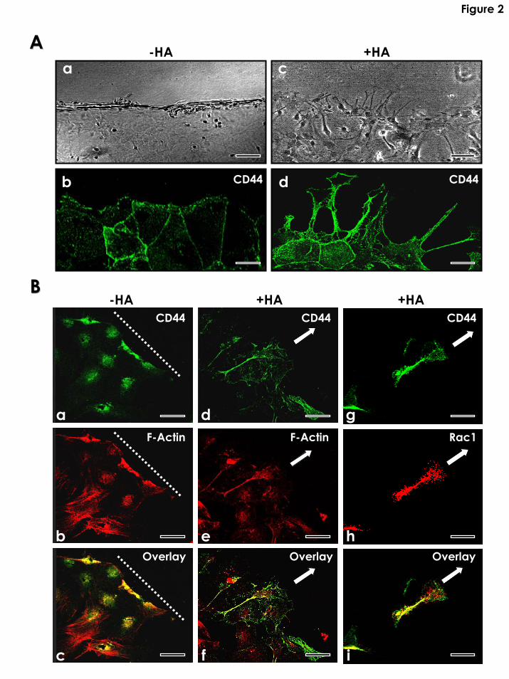
Figure 2
AA
BB-HA +HA +HA
a
b
c
d
e
f
a
b
c
dCD44 CD44
CD44 CD44 CD44
F-Actin F-Actin Rac1
Overlay Overlay Overlay
g
h
i
-HA +HA

Figure 3
A
-HA +HA Anti-CD44 + HA
0h
24h
B
100
0
200
300
Wou
nd C
losu
re(%
of c
ontro
l)
**
**
a b c-HA
(control)+HA Anti-CD44
+HA
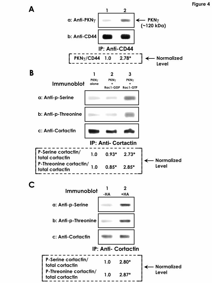
Figure 4AA 1 2
a: Anti-PKNγ PKNγ(~120 kDa)
b: Anti-CD44
IP: Anti-CD44
BB
PKNγ/CD44 1.0 2.78* NormalizedLevel
1 2 3PKNγ
+Rac1-GDP R
PKNγ+
ac1-GTP
PKNγaloneImmunoblot
a: Anti-p-Serine
b: Anti-p-Threonine
c: Anti-Cortactin
IP: AntiIP: Anti-- CortactinCortactinP-Serine cortactin/total cortactinP-Threonine cortactin/total cortactin
1.0
1.0
0.93* 2.73*
0.85* 2.85*NormalizedLevel
CC 1 2Immunoblot -HA +HA
a: Anti-p-Serine
b: Anti-p-Threonine
c: Anti-Cortactin
IP: AntiIP: Anti-- CortactinCortactin
P-Serine cortactin/total cortactinP-Threonine cortactin/total cortactin
1.0 2.80*
1.0 2.87*NormalizedLevel
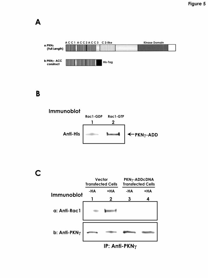
Figure 5
AA
a:PKNγ(Full Length)
a:PKNγ(Full Length)
b:PKNγ- ACCconstruct
b:PKNγ- ACCconstruct
His-Tag
A C C 1 A C C 2 A C C 3 C 2-like Kinase Domain
BB
Immunoblot
1 2
PKNγ-ADD
Rac1-GDP Rac1-GTP
Anti-His
CCVector
Transfected Cells
1 2 3 4Immunoblot
a: Anti-Rac1
b: Anti-PKNγ
IP: Anti-PKNγ
-HA
PKNγ-ADDcDNA Transfected Cells
+HA -HA +HA

Figure 6
AA
0
20
40
60
80
100
120
(% o
f con
trol)
a b c
Cor
tact
in-F
-Act
in B
indi
ng
0
50
300
100
150
200
250
(% o
f con
trol)
a b c
Cor
tact
in P
hosp
hory
latio
n
BB
**
**
**
**
**
d
**
d
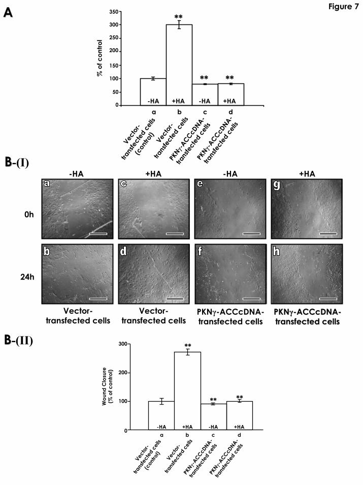
Figure 7AA
0
50
100
150
200
250
300
350
% o
f con
trol
-HA -HA +HAaa bb cc dd
+HA
**
** **
Vecto
r-
trans
fect
ed c
ells
(con
trol)
Vecto
r-
trans
fect
ed c
ells
PKNγ
-ACCcD
NA-
trans
fect
ed c
ells
PKNγ
-ACCcD
NA-
trans
fect
ed c
ells
-HA +HA -HABB-(I)
+HA
BB-(II)
Vector-transfected cells
Vector-transfected cells
PKNγ-ACCcDNA-transfected cells
PKNPKNγγ--ACCcDNAACCcDNA--transfected transfected cellscell
0h
24h
s
**
** **
a b c d
Vecto
r-
trans
fect
ed c
ells
(con
trol)
Vecto
r-
trans
fect
ed c
ells
PKNγ
-ACCcD
NA-
trans
fect
ed c
ells
PKNγ
-ACCcD
NA-
trans
fect
ed c
ells
-HA +HA -HA +HA0
300
200
100Wou
nd C
losu
re(%
of c
ontro
l)
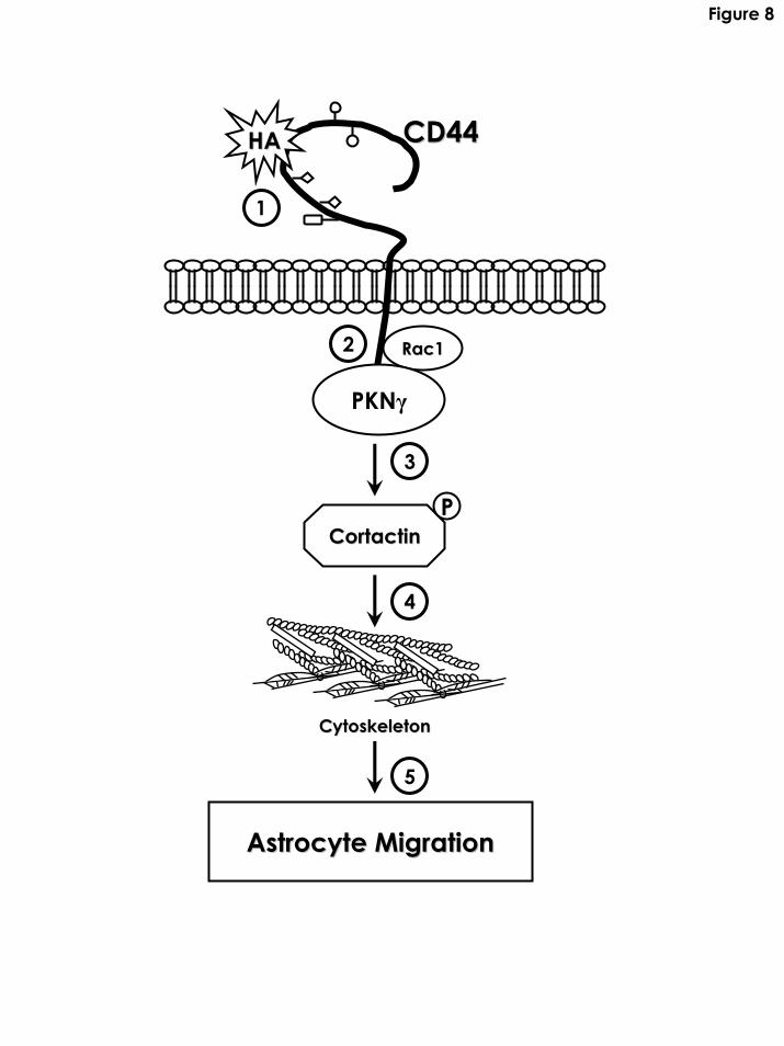
Figure 8
CD44CD44
2
HAHA
Rac1Rac1
1
PKNγ
3
CortactinCortactinP
4
CytoskeletonCytoskeleton
5
Astrocyte MigrationAstrocyte Migration