ubiquitin ligases: cell-cycle control and cancer - us oncotherapy
TRANSCRIPT

*Department of Molecular and Cellular Biology, Medical Institute of Bioregulation, Kyushu University, Fukuoka, Fukuoka 812-8582, Japan.‡Department of Developmental Biology, Center for Translational and Advanced Animal Research on Human Disease, Graduate School of Medicine, Tohoku University, 2-1 Seiryo, Aoba-ku, Sendai 980-8575, Japan.Correspondence to K.I.N. e-mail: [email protected]:10.1038/nrc1881
Cyclin-dependent kinaseA protein kinase that controls cell-cycle progression in all eukaryotes and requires physical association with cyclins to achieve full enzymatic activity.
CyclinThe positive regulatory subunits of cyclin-dependent kinases, showing oscillating expression in the cell cycle.
Ubiquitin ligases: cell-cycle control and cancerKeiichi I. Nakayama* and Keiko Nakayama‡
Abstract | A driving force of the cell cycle is the activation of cyclin-dependent kinases (CDKs), the activities of which are controlled by the ubiquitin-mediated proteolysis of key regulators such as cyclins and CDK inhibitors. Two ubiquitin ligases, the SKP1–CUL1–F-box-protein (SCF) complex and the anaphase-promoting complex/cyclosome (APC/C), are responsible for the specific ubiquitylation of many of these regulators. Deregulation of the proteolytic system might result in uncontrolled proliferation, genomic instability and cancer. Cumulative clinical evidence shows alterations in the ubiquitylation of cell-cycle regulators in the aetiology of many human malignancies. A better understanding of the ubiquitylation machinery will provide new insights into the regulatory biology of cell-cycle transitions and the development of anti-cancer drugs.
Deregulated cell-cycle control is a fundamental aspect of cancer. Normal cells only proliferate in response to developmental or other mitogenic signals that indicate a requirement for tissue growth, whereas the prolifera-tion of cancer cells proceeds essentially unchecked. This indicates that cancer cells proliferate because of defects in internal and external proliferation-inhibitory signals. However, there must also be defects in the negative-feedback systems that should prevent such unregulated proliferation. We now have a detailed molecular picture of normal cell-cycle control, in which a series of kinases promote progression through each phase of the cell cycle. The phosphorylation of a variety of proteins by members of the cyclin-dependent kinase (CDK) family is crucial to this progression. Each CDK is dependent on a particular cyclin; therefore, the activity of each CDK can be controlled by the availability of its cyclin partner, and also the expression of a specific CDK inhibitor (CKI).
Levels of cyclin expression during cell division are periodic1. This is the result of a constant synthetic rate coupled with a defined window in the cycle of specific proteolysis, which is executed by the ubiquitin-proteasome system (UPS) (REF. 2). CKIs, negative-regula-tors of cyclin–CDK kinase complexes, are also targeted for degradation by the UPS. Therefore, the cell cycle is predominantly regulated by two types of post-transla-tional protein modification — phosphorylation and ubiq-uitylation (BOX 1). The UPS comprises two discrete steps: the covalent attachment of multiple ubiquitin molecules to the protein substrate and degradation of the polyubiq-uitylated protein by the 26S proteasome complex3 (FIG. 1).
The first step is mediated by at least three enzymes: a ubiquitin-activating enzyme (E1), a ubiquitin-conjugating enzyme (E2) and a ubiquitin ligase (E3). An involvement of E1 in cancer has not been described, and only a few reports have linked E2 to cancer development4. By con-trast, a large amount of evidence indicates that deregula-tion of E3 ligase, which is involved in many biological systems, often results in cancer development.
A direct molecular link between the deregulation of cell-cycle control and cancer was first discov-ered when it was shown that the genes that encode retinoblastoma protein (RB) and p53, both of which restrain cell-cycle progression, are frequently mutated or deleted in human cancers. Decreased expression of p27 CKI is also frequently found in various human cancers, and is a strong indicator of poor prognosis5. In contrast to RB and p53, mutations or deletions in p27 were found to be rare in cancer patients. Instead, a decrease in the protein stability of p27 contributes to its reduced levels in cancer cells6,7. These clinical findings were later explained by the overexpression of S-phase kinase-associated protein 2 (SKP2), a component of E3 ligase that targets p27 for degrada-tion8. In contrast to the characterization of SKP2 as an oncogene, another component of E3 ligase, F-box and WD-40 domain protein 7 (FBW7), was biochemically and genetically shown to be a tumour suppressor8. Experimental and clinical data on SKP2 and FBW7, and also on other E3 ligases, indicates that the deregu-lation of the UPS in cell-cycle control is tightly linked to cancer development.
R E V I E W S
NATURE REVIEWS | CANCER VOLUME 6 | MAY 2006 | 369
© 2006 Nature Publishing Group

Ubiquitylating enzymes
Regulation by ubiquitylation(quantitative control)
Regulation by phosphorylation(qualitative control)
AcceleratorCDKs(protein kinases)
BrakeCDKIs
Cell cycle (DNA synthesis, chromosome separation, etc)
a
b
c
UbiquitinA 76-amino-acid polypeptide that is conjugated through an isopeptide linkage to other proteins. Such conjugates are most commonly targeted for degradation by the proteasome.
ProteasomeA large ~2.5-MDa multisubunit protein complex that binds to and subsequently degrades polyubiquitylated proteins in an ATP-dependent manner.
Ubiquitin-conjugating enzyme (E2)An enzyme that accepts ubiquitin from a ubiquitin-activating enzyme (E1) and, together with a ubiquitin ligase (E3), transfers it to a substrate protein.
Ubiquitin ligase (E3)A protein or protein complex that facilitates the transfer of ubiquitin from a ubiquitin-conjugating enzyme (E2) to a substrate. E3 enzymes provide platforms for binding E2 enzymes and specific substrates, thereby coordinating the ubiquitylation of the selected substrate.
CullinsA family of proteins that are characterized by the presence of a distinct globular C-terminal domain (cullin homology domain) and a series of N-terminal repeats of a five helix bundle (cullin repeats).
F-box proteinA variable component of SCF E3 ligase that binds to SKP1 through the F-box domain. FBPs recognize specific substrates and, with the help of other subunits of the E3 ubiquitin ligase, deliver them to the E2 ubiquitin-conjugating enzyme.
RING-finger proteinsA family of proteins structurally defined by a particular folded protein domain that binds Zn2+ through a four-point arrangement of cysteine and histidine amino acids. In most cases, a RING-finger protein interacts with an E2 and serves as an E3.
The recent approval of a proteasomal inhibi-tor, Bortezomib, by the US Food and Drugs Administration (FDA) for the treatment of relapsed and refractory multiple myeloma should further stim-ulate the search for specific E3 inhibitors. Targeting a specific E3 would selectively stabilize its target(s) and have an advantage over proteasomal inhibition by
avoiding unwanted effects on other cellular proteins. Therefore, E3 ubiquitin ligases are expected to draw much attention as a promising class of new drug tar-gets with a high level of specificity and less associated toxicity.
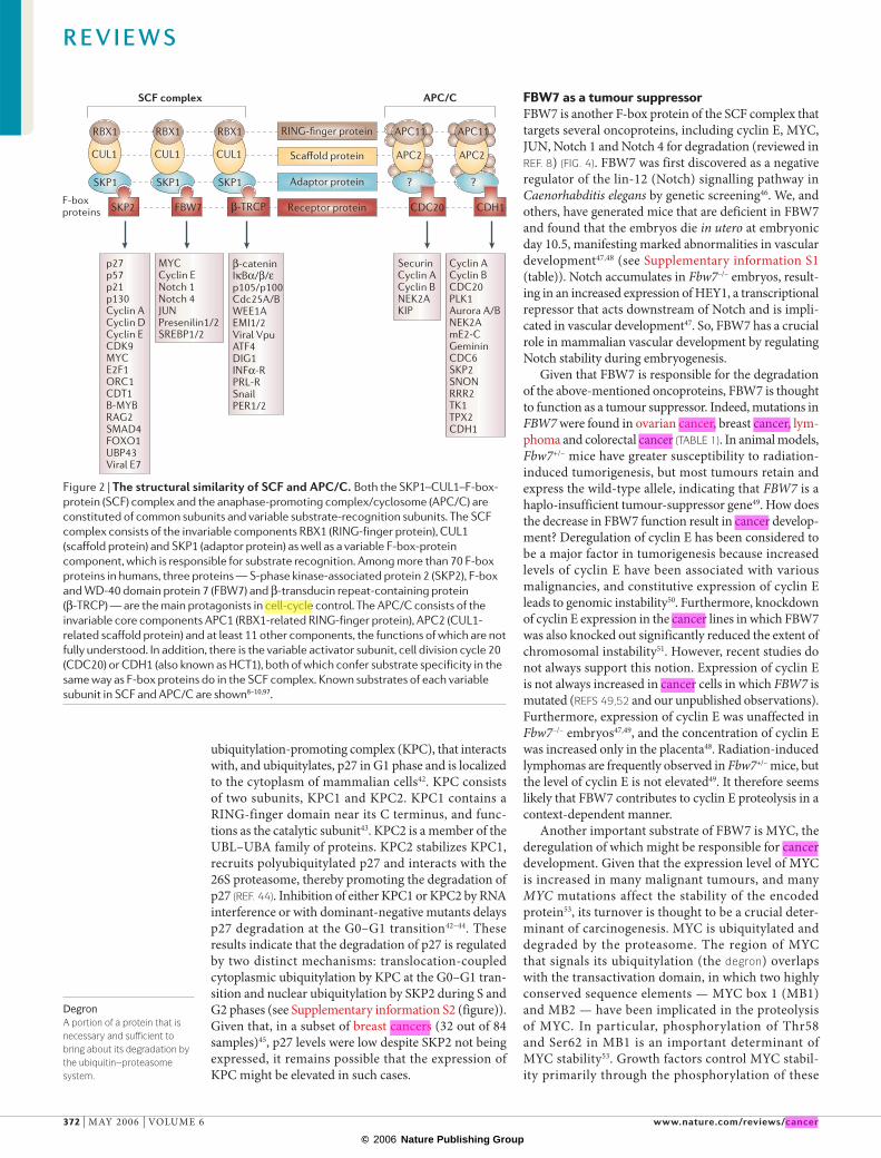
SCF and APC/C: similar but distinctE3 components in the UPS are thought to be pri-marily responsible for the specific recognition of a large number of target proteins3. This requires both specificity and versatility, which are provided by the existence of 500–1,000 different E3 ligases. These are now categorized into four major classes on the basis of their specific structural motif: HECT-type, RING-finger-type, U-box-type or PHD-finger-type (FIG. 1). RING-finger-type E3s are thought to be the largest family and are further divided into subfamilies; one of these, the cullin-based E3 subfamily, is one of the larg-est single classes of E3. There are seven cullin-based E3s, including the SKP1–CUL1–F-box-protein (SCF) complex8 and the anaphase-promoting complex/cyclosome (APC/C)9,10, both of which are involved in the proteolysis of core components of the cell-cycle machinery. The SCF complex consists of three invari-able components — RBX1 (RING-finger protein), CUL1 (scaffold protein), and SKP1 (adaptor protein) — as well as one variable component, known as an F-box protein, that binds through its F-box motif to SKP1 and is responsible for substrate recognition (FIG. 2). Approximately 70 F-box proteins have been identi-fied in humans, and they fall into three categories: those with WD40 repeats (FBXW), leucine-rich repeats (FBXL) or other domains (FBXO)11. Three F-box proteins — SKP2 (FBXL1), FBW7 (FBXW7) and β-transducin repeat-containing protein (β-TRCP) (FBXW1/11) — are thought to be involved in cell-cycle control (the functions of most other F-box proteins remain unknown).
At a glance
• Two major classes of ubiquitin ligases, the SKP1–CUL1–F-box-protein (SCF) complex and the anaphase-promoting complex/cyclosome (APC/C), have a central role in cell-cycle regulation.
• The SCF complex and APC/C are structurally similar. Each is constituted of common subunits and a variable substrate-recognition subunit (F-box proteins for the SCF complex and activators for the APC/C). Three F-box proteins in the SCF complex — S-phase kinase-associated protein 2 (SKP2), F-box and WD-40 domain protein 7 (FBW7) and β-transducin repeat-containing protein (β-TRCP) — and two activators in the APC/C — cell division cycle 20 (CDC20) and CDH1 (also known as HCT1) — are the most important in cell-cycle regulation.
• SKP2 targets negative regulators of the cell cycle such as p27, p21 and p57 for degradation, and thereby promotes cell-cycle progression during S and G2 phases. SKP2 is upregulated in many human cancers.
• FBW7 induces the degradation of positive regulators of the cell cycle, such as MYC, JUN, cyclin E and Notch. FBW7 is often mutated in a subset of human cancers.
• β-TRCP is a versatile F-box protein that recognizes several cell-cycle regulators — EMI1/2, WEE1A and CDC25A/B — in addition to its classical substrates, β-catenin and IκB. In some cancers, β-TRCP mutation or overexpression is found.
• CDC20 targets securin and mitotic cyclins for destruction, and thereby promotes sister-chromatid separation. CDC20 is the crucial mediator of the spindle checkpoint, which prevents aneuploidy and genomic instability. CDC20 is overexpressed in some cancers, although in others the CDC20 gene is mutated or deleted.
• CDH1 facilitates exit from M phase and maintains G1 phase by mediating the degradation of mitotic cyclins, non-CDK (cyclin-dependent kinase) mitotic kinases and some regulators of the formation of pre-replicative complexes. Deregulated expression or mutation of CDH1 as well as of most CDH1 targets have been described in human cancers.
Box 1 | Three-layer regulation of the cell cycle
Cell-cycle control can be described as a 3-layer process. The immediate phenomena of the cell cycle, including DNA synthesis and chromosome separation (layer a), are qualitatively controlled by phosphorylation. Movement through the cycle (layer b) depends on the activity of cyclin-dependent kinases (CDKs), which are promoted by accelerators — cyclins — and antagonized by brakes — CDK inhibitors (CKIs). The protein levels of cyclins, CKIs and many other cell-cycle-related regulators are quantitatively controlled by ubiquitylating enzymes (layer c). Accumulating clinical evidence shows various alterations in the ubiquitylation of cell-cycle regulators in the aetiology of many human malignancies.
R E V I E W S
370 | MAY 2006 | VOLUME 6 www.nature.com/reviews/cancer
© 2006 Nature Publishing Group

UbUb
E2
E1
E3
sco =
Ub
UbUb
UbUb
UbUb
UbUb
Ub
sco =
ATP AMP
Target protein
HECT-typeRING-finger typeU-box typePHD-finger type
SCF (Cul1-based E3)ECV (Cul2-based E3)CUL3-based E3CUL4-based E3ECS (Cul5-based E3)CUL7-based E3APC/COthers
Recycling
ADP
ATP
26S proteasome
Degradedproduct
WD40 repeatA protein-interaction domain consisting of 40 amino-acid repeats that form a propeller-like structure, in which each repeat contributes a blade.
Leucine-rich repeatA protein-sequence motif that contains regular occurrences of the amino acid leucine, which are present as tandem arrays in certain proteins. The back-to-back set of motifs was found to correspond to a small sub-domain structure in the protein that stacks next to adjacent repeats to form a parallel, β-sheet, arc-like structure.
The APC/C is structurally similar to the SCF complex, and consists of invariable core components — APC11 (RBX1-related RING-finger protein), APC2 (CUL1-related scaffold protein) and at least 11 other components without a defined role — as well as a vari-able component known as an activator. There are two such variable components in mitotically cycling cells — cell division cycle 20 (CDC20) and CDH1 (also known as HCT1) — and they confer substrate specificity in the same way that F-box proteins do in the SCF complex9,10. Additional APC/C activators function during meiosis and in non-dividing cells (reviewed in REF. 12).
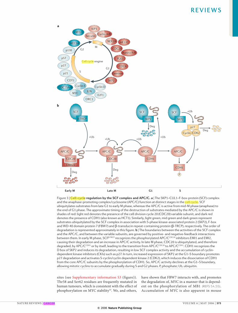
Despite the structural and biochemical similarities between the SCF complex and the APC/C, their cellular functions are different. This is highlighted by the timing of action of each E3 complex (FIG. 3a): the APC/C is active from mid-mitosis (anaphase) to the end of G1 phase, whereas the SCF complex, although originally thought to function mainly at the G1–S transition, is active from late G1 to early M phase. How are their roles clearly delineated? The most persuasive explanation is that they constitute a regulatory loop. Although the entire picture is not clear, several lines of evidence show that there is a tight interplay between the SCF complex and the APC/C13–16 (FIG. 3b). The SCF complex and the APC/C also differ in their frequency of genetic alterations in cancer: far more alterations connected with cancer have been found for the SCF complex than for the APC/C.
SKP2 is oncogenicSKP2 was originally discovered as a protein that associ-ates with cyclin A–CDK2 in transformed cells17, and is
now known to be an F-box protein of the SCF complex. In conjunction with SKP2, the SCF complex targets p27 (REFS 18–20), p21 (REFS 21,22) and p57 (REF. 23) CKIs for degradation (FIG. 4). To date, it has also been reported that SKP2 targets p130, cyclin A, cyclin D1, free cyclin E, E2F1, ORC1, CDT1, CDK9, MYC, B-MYB, SMAD4, RAG2, UBP43, FOXO1 and papillomavirus E7 (reviewed in REF. 8). However, it is p27 that seems to be the primary target of SKP2, given that Skp2–/– mice show a marked accumulation of p27 (REF. 24), and that prominent cellular phenotypes apparent in Skp2–/– mice (see Supplementary information S1 (table)) — includ-ing nuclear enlargement, polyploidy and an increased number of centrosomes that are probably caused by overreplication of chromosomes and centrosomes — disappear in Skp2–/– p27–/– double-mutant mice25,26.
It has been widely accepted that p27 is a tumour suppressor, not only because of its activity as a CKI, but also because of evidence from mouse models27–29 and the marked correlation between reduced p27 levels and poor prognosis found in clinical studies of patients with cancer5. Indeed, a reduction in the con-centration of p27 is common in many types of human malignancies. However, in contrast with other tumour suppressors such as p53 or RB, mutation or deletion of the p27 gene is an uncommon event in the develop-ment of human cancers, indicating that deregulation of p27 expression in human tumours is often due to post-transcriptional mechanisms. It has become evi-dent that SKP2 expression is inversely correlated with levels of p27 in many cancers, and also with the grade of malignancy in certain human tumours (TABLE 1). In addition, frequent amplification and overexpression of the SKP2 gene has been observed in lung cancers30,31 and in cell lines expressing high-risk human papil-loma virus32. Other components of the ubiquitylation machinery for p27, primarily CKS1, have oncogenic potential in patients with colorectal carcinoma33, and overexpression of SKP2 or CKS1 is strongly and inde-pendently associated with a loss of tumour differen-tiation and poor survival34. The oncogenic potential of SKP2 has also been shown in transgenic mouse models35,36. Furthermore, Ras signalling induces SKP2 expression through the binding of GA-binding pro-tein, an Ets-family transcription factor, to the SKP2 promoter37. Such evidence supports the notion that SKP2 is a growth promoter and an oncoprotein.
However, in the time since it became widely accepted that SKP2 mediates p27 degradation in G1 phase, sev-eral discrepancies have emerged. First, SKP2 is not expressed until early S phase, unequivocally later than the degradation of p27 apparent at G1 (REFS 17,38,39). Second, p27 is exported from the nucleus to the cyto-plasm at G1 (REFS 39,40), whereas SKP2 is restricted to the nucleus41. Third, the downregulation of p27 at the G0–G1 transition occurs normally in SKP2–/– cells and is sensitive to proteasome inhibitors38. These temporal, spatial and genetic discrepancies indicate that p27 is degraded at G1 in the cytoplasm by a proteasome-dependent, but SKP2-independent, mechanism. We have recently purified an E3 enzyme, designated KIP1
Figure 1 | Overview of the ubiquitin-proteasome pathway. Ubiquitin (Ub) is a small 8-kDa protein, which is first transferred to the ubiquitin-activating enzyme, E1, in an ATP-dependent manner. This activated ubiquitin is then transferred to the ubiquitin-conjugating enzyme, E2. Finally, the ubiquitin is covalently attached to the target protein by E3 ubiquitin ligase, leading to the formation of a polyubiquitin chain. The polyubiquitylated protein is recognized by the 26S proteasome, and is destroyed in an ATP-dependent manner. There are many E3 ubiquitin ligases, which are categorized into four major classes: HECT-type, RING-finger-type, U-box-type and PHD-finger-type. RING-finger-type E3s are further divided into subfamilies, including cullin-based E3s, which constitute one of the largest classes of E3s. There are seven cullin-based E3s including the SKP1–CUL1–F-box-protein (SCF) complex and the anaphase-promoting complex/cyclosome (APC/C).
R E V I E W S
NATURE REVIEWS | CANCER VOLUME 6 | MAY 2006 | 371
© 2006 Nature Publishing Group
Skp2
SKP1
SKP2
SKP1
FBW7 Skp2
?Skp1SKP1
β-TRCP
RING-finger protein
Scaffold protein
Adaptor protein
Receptor protein
RBX1
CUL1
RBX1
CUL1
APC11
APC2
APC11
APC2
RBX1
CUL1
CDC20 CDH1
p27p57p21p130Cyclin ACyclin DCyclin ECDK9MYCE2F1ORC1CDT1B-MYBRAG2SMAD4FOXO1UBP43Viral E7
MYCCyclin ENotch 1Notch 4JUNPresenilin1/2SREBP1/2
β-cateninIκBα/β/εp105/p100Cdc25A/BWEE1AEMI1/2Viral VpuATF4DIG1INFα-RPRL-RSnailPER1/2
SecurinCyclin ACyclin BNEK2AKIP
Cyclin ACyclin BCDC20PLK1Aurora A/BNEK2AmE2-CGemininCDC6SKP2SNONRRR2TK1TPX2CDH1
F-boxproteins
APC/CSCF complex
?
DegronA portion of a protein that is necessary and sufficient to bring about its degradation by the ubiquitin–proteasome system.
ubiquitylation-promoting complex (KPC), that interacts with, and ubiquitylates, p27 in G1 phase and is localized to the cytoplasm of mammalian cells42. KPC consists of two subunits, KPC1 and KPC2. KPC1 contains a RING-finger domain near its C terminus, and func-tions as the catalytic subunit43. KPC2 is a member of the UBL–UBA family of proteins. KPC2 stabilizes KPC1, recruits polyubiquitylated p27 and interacts with the 26S proteasome, thereby promoting the degradation of p27 (REF. 44). Inhibition of either KPC1 or KPC2 by RNA interference or with dominant-negative mutants delays p27 degradation at the G0–G1 transition42–44. These results indicate that the degradation of p27 is regulated by two distinct mechanisms: translocation-coupled cytoplasmic ubiquitylation by KPC at the G0–G1 tran-sition and nuclear ubiquitylation by SKP2 during S and G2 phases (see Supplementary information S2 (figure)). Given that, in a subset of breast cancers (32 out of 84 samples)45, p27 levels were low despite SKP2 not being expressed, it remains possible that the expression of KPC might be elevated in such cases.
FBW7 as a tumour suppressorFBW7 is another F-box protein of the SCF complex that targets several oncoproteins, including cyclin E, MYC, JUN, Notch 1 and Notch 4 for degradation (reviewed in REF. 8) (FIG. 4). FBW7 was first discovered as a negative regulator of the lin-12 (Notch) signalling pathway in Caenorhabditis elegans by genetic screening46. We, and others, have generated mice that are deficient in FBW7 and found that the embryos die in utero at embryonic day 10.5, manifesting marked abnormalities in vascular development47,48 (see Supplementary information S1 (table)). Notch accumulates in Fbw7–/– embryos, result-ing in an increased expression of HEY1, a transcriptional repressor that acts downstream of Notch and is impli-cated in vascular development47. So, FBW7 has a crucial role in mammalian vascular development by regulating Notch stability during embryogenesis.
Given that FBW7 is responsible for the degradation of the above-mentioned oncoproteins, FBW7 is thought to function as a tumour suppressor. Indeed, mutations in FBW7 were found in ovarian cancer, breast cancer, lym-phoma and colorectal cancer (TABLE 1). In animal models, Fbw7+/– mice have greater susceptibility to radiation-induced tumorigenesis, but most tumours retain and express the wild-type allele, indicating that FBW7 is a haplo-insufficient tumour-suppressor gene49. How does the decrease in FBW7 function result in cancer develop-ment? Deregulation of cyclin E has been considered to be a major factor in tumorigenesis because increased levels of cyclin E have been associated with various malignancies, and constitutive expression of cyclin E leads to genomic instability50. Furthermore, knockdown of cyclin E expression in the cancer lines in which FBW7 was also knocked out significantly reduced the extent of chromosomal instability51. However, recent studies do not always support this notion. Expression of cyclin E is not always increased in cancer cells in which FBW7 is mutated (REFS 49,52 and our unpublished observations). Furthermore, expression of cyclin E was unaffected in Fbw7–/– embryos47,49, and the concentration of cyclin E was increased only in the placenta48. Radiation-induced lymphomas are frequently observed in Fbw7+/– mice, but the level of cyclin E is not elevated49. It therefore seems likely that FBW7 contributes to cyclin E proteolysis in a context-dependent manner.
Another important substrate of FBW7 is MYC, the deregulation of which might be responsible for cancer development. Given that the expression level of MYC is increased in many malignant tumours, and many MYC mutations affect the stability of the encoded protein53, its turnover is thought to be a crucial deter-minant of carcinogenesis. MYC is ubiquitylated and degraded by the proteasome. The region of MYC that signals its ubiquitylation (the degron) overlaps with the transactivation domain, in which two highly conserved sequence elements — MYC box 1 (MB1) and MB2 — have been implicated in the proteolysis of MYC. In particular, phosphorylation of Thr58 and Ser62 in MB1 is an important determinant of MYC stability53. Growth factors control MYC stabil-ity primarily through the phosphorylation of these
Figure 2 | The structural similarity of SCF and APC/C. Both the SKP1–CUL1–F-box-protein (SCF) complex and the anaphase-promoting complex/cyclosome (APC/C) are constituted of common subunits and variable substrate-recognition subunits. The SCF complex consists of the invariable components RBX1 (RING-finger protein), CUL1 (scaffold protein) and SKP1 (adaptor protein) as well as a variable F-box-protein component, which is responsible for substrate recognition. Among more than 70 F-box proteins in humans, three proteins — S-phase kinase-associated protein 2 (SKP2), F-box and WD-40 domain protein 7 (FBW7) and β-transducin repeat-containing protein (β-TRCP) — are the main protagonists in cell-cycle control. The APC/C consists of the invariable core components APC1 (RBX1-related RING-finger protein), APC2 (CUL1-related scaffold protein) and at least 11 other components, the functions of which are not fully understood. In addition, there is the variable activator subunit, cell division cycle 20 (CDC20) or CDH1 (also known as HCT1), both of which confer substrate specificity in the same way as F-box proteins do in the SCF complex. Known substrates of each variable subunit in SCF and APC/C are shown8–10,97.
R E V I E W S
372 | MAY 2006 | VOLUME 6 www.nature.com/reviews/cancer
© 2006 Nature Publishing Group
APC/C APC/C
EMI1/2
WEE1A
p130
p57
p27
p21
CDT1
Cyclin ECDC25A/B
MYCJUN
ORC1
Cyclin D
E2F1
Securin
KIP1
Cyclin A/B
NEK2A mE2-C
CDC20Aurora A/B
PLK1
CDC6
Geminin
SKP2
Cell cycle engine
G2M
G1S
SCF
β-TRCP
SCF
SKP2
CDC20
EMI1/2
Deg
CDC20
APC/C
CDH1
Deg
Deg p27
SCF
Deg
APC/C
CDH1
+Ub+Ub
+Ub+Ub
+Ub
+P
Cyclin CDK2
P
a
b
Early M Late M G1 S
SKP2
sites (see Supplementary information S3 (figure)). Thr58 and Ser62 residues are frequently mutated in human tumours, which is consistent with the effect of phosphorylation on MYC stability53. We, and others,
have shown that FBW7 interacts with, and promotes the degradation of, MYC in a manner that is depend-ent on the phosphorylation of MB1 (REFS 54,55). Accumulation of MYC is also apparent in mouse
Figure 3 | Cell-cycle regulation by the SCF complex and APC/C. a | The SKP1–CUL1–F-box-protein (SCF) complex and the anaphase-promoting complex/cyclosome (APC/C) function at distinct stages in the cell cycle. SCF ubiquitylates substrates from late G1 to early M phase, whereas the APC/C is active from mid-M phase (anaphase) to the end of G1 phase. The approximate timing of the destruction of substrates mediated by the APC/C is shown in shades of red: light red denotes the presence of the cell division cycle 20 (CDC20) variable subunit, and dark red denotes the presence of CDH1 (also known as HCT1). Similarly, light green, mid green and dark green represent substrates ubiquitylated by the SCF complex in association with S-phase kinase-associated protein 2 (SKP2), F-box and WD-40 domain protein 7 (FBW7) and β-transducin repeat-containing protein (β-TRCP), respectively. The order of degradation is represented approximately in this figure. b | The boundaries between the activities of the SCF complex and the APC/C, and between the variable subunits, are governed by positive- and negative-feedback interactions between them. In early M phase, SCFβ-TRCP recognizes the phosphorylated APC/CCDC20 inhibitors EMI1 and EMI2, causing their degradation and an increase in APC/C activity. In late M phase, CDC20 is ubiquitylated, and therefore degraded, by APC/CCDH1 or by itself, leading to the transition from APC/CCDC20 to APC/CCDH1. CDH1 recognizes the D box of SKP2 and induces its degradation, resulting in low SCF complex activity and the accumulation of cyclin-dependent kinase inhibitors (CKIs) such as p27. In turn, increased expression of SKP2 at the G1–S boundary promotes p27 degradation and activates S-cyclin/cyclin dependent kinase 2 (CDK2), which induces the dissociation of CDH1 from the core APC/C subunits by the phosphorylation of CDH1. So, APC/C activity declines at the G1–S boundary, allowing mitotic cyclins to accumulate gradually during S and G2 phases. P, phosphate; Ub, ubiquitin.
R E V I E W S
NATURE REVIEWS | CANCER VOLUME 6 | MAY 2006 | 373
© 2006 Nature Publishing Group

KPC1
KPC2
p27 p21 p57 Cyclin E
Cell-cycle arrest Cell-cycle promotion
MYC Notch CDC25 WEE1 EMI1
RBX1
CUL1
SKP1
RBX1
CUL1
SKP1
RBX1
CUL1
SKP1SKP2
(oncoprotein)
F LRRs
FBW7(tumour suppressor)
F WDs
β-TRCP(both)
F WDs
+Ub +Ub +Ub +Ub
Fbw7–/– cells54, as well as in lymphomas from Fbw7+/– mice49. Surprisingly, SKP2 also binds to MYC through its MB2 and helix-loop-helix–leucine-zipper (HLH-Zip) domains, and thereby mediates its ubiquitylation and degradation56,57. However, SKP2 unexpectedly increases the transactivation activity of MYC, indicat-ing that SKP2 is a transcriptional cofactor. Consistent with this notion, MYC accumulates, but its transcrip-tional activity is reduced, in mouse Skp2–/– cells57. These observations indicate that two F-box proteins, FBW7 and SKP2, differentially regulate MYC stability by targeting MB1 and MB2 domains, respectively.
Increased expression of not only cyclin E and MYC but also other substrates of FBW7 might contribute to cancer development. Deregulation of wild-type Notch, Notch ligands or downstream targets has been detected in many human malignancies58. Truncated Notch proteins exhibit transforming activity both in vitro and in animal models59–62. Furthermore, Notch 1 was recently shown to be a downstream effector of oncogenic Ras, CDC20 and CDH1, and depletion of Notch 1 in Ras-transformed human cells was sufficient to abolish key elements of the neoplastic phenotype in vitro and in vivo63. Notch4 was originally identified as Int3, a proto-oncogene that is a frequent target for the integration of mouse-mammary-tumour virus in mammary carcinomas64. The JUN oncoprotein is a major component of the transcription factor AP1, the constitutive activation of which is apparent in various types of human tumour cells, indicating that AP1 has an important role in human oncogenesis65. Mutation of the human FBW7 gene might therefore result in the impaired degradation of these substrates and their subsequent accumulation, which might then contribute to carcinogenesis.
β-TRCP regulates many pathwaysβ-TRCP is a versatile F-box protein of the SCF com-plex that targets various substrates for degradation, including crucial cell-cycle regulators such as EMI1/2 (REFS 13,14,66,67), WEE1A (REF. 68) and CDC25A/B (REFS 69–71) (FIG. 4). The β-TRCP family is highly con-served and includes Drosophila melanogaster Slimb72 and Xenopus laevis β-TrCP73, as well as mammalian β-TRCP1 (also termed FBW1a or FWD1) and β-TRCP2 (also known as FBW1b or HOS). β-TRCPs also target β-catenin and IκB for ubiquitylation and degradation8, and β-TrCP1–/– mice have a partial defect in their ability to eliminate β-catenin and IκBα in certain situations74 (see Supplementary information S1 (table)). Other sub-strates that are degraded as a result of β-TRCP-depend-ent ubiquitylation include IκBβ and IκBε, the p105 and p100 subunits of nuclear factor κB (NFκB), ATF4 (CREB2), VPU protein of human immunodeficiency virus type 1, interferon receptor, prolactin receptor, mammalian circadian regulatory proteins PER1 and PER2, Snail and the discs large (hDLG) tumour sup-pressor (reviewed in REF. 8). β-TRCP recognizes the DSG(X)2+nS destruction motif, the serines of which are phosphorylated by specific kinases75.
There are many studies that show alterations of the genes that encode β-catenin and NFκB in various human cancers. Given that β-TRCP targets β-catenin and IκB for degradation, overexpression of β-TRCP is expected to have complex consequences: it inactivates the Wnt pathway, which stimulates cellular prolif-eration, but also activates the NFκB pathway, which antagonizes pro-apoptotic signals. Genetic alteration of β-TRCP genes in human cancers have been shown in several studies (TABLE 1). A nucleotide substitution in β-TRCP2 was identified in a gastric cancer cell line,
Figure 4 | Functions of the SKP1–CUL1–F-box-protein (SCF) complex. The variable components (F-box proteins) of the SCF complex — S-phase kinase-associated protein 2 (SKP2), F-box and WD-40 domain protein 7 (FBW7) and β-transducin repeat-containing protein (β-TRCP) — and their major targets are shown: red and green boxes indicate cell-cycle promoters and inhibitors, respectively. SKP2 promotes the cell cycle through the ubiquitylation of CDK inhibitors (CKIs), including p27, and therefore functions as an oncoprotein. The KIP1 ubiquitylation-promoting complex (KPC) also mediates p27 ubiquitylation. SKP2 is overexpressed in many cancers. By contrast, FBW7 targets cell-cycle promoters to inhibit the cell cycle, and serves as a tumour suppressor. Mutations or deletions in FBW7 are found in a subset of human cancers. β-TRCP is both a cell-cycle promoter and inhibitor. Both mutation and overexpression of β-TRCP are observed in human cancers. F, F-box domain; LRRs, leucine-rich repeats; Ub, ubiquitin; WDs, WD40 repeats.
R E V I E W S
374 | MAY 2006 | VOLUME 6 www.nature.com/reviews/cancer
© 2006 Nature Publishing Group

which led to an amino-acid substitution in the seventh WD40-repeat domain; this domain is conserved among β-TRCPs derived from human, mouse, X. laevis, and D. melanogaster76. In human prostate cancers, alterations
in β-TRCP1 were identified in 2 out of 22 samples77. Alterations in adenomatosis polyposis coli (APC), β-TRCP1 and the β-catenin regulatory domain were not found together, consistent with their equivalent effects
Table 1 | Alterations in the variable components of SCF and APC/C in human malignancies
Cancer type Observed alterations References
SKP2 (SCF)
Oro-pharyngo-laryngeal cancer
Correlation with poor prognosis; inverse correlation with p27 expression 127–131
Oesophageal cancer Correlation with poor prognosis; inverse correlation with p27 expression 132
Gastric cancer Correlation with poor prognosis; inverse correlation with p27 expression 133,134
Colon cancer Correlation with poor prognosis; inverse correlation with p27 expression 34,135,136
Biliary tract cancer Correlation with poor prognosis; no correlation with p27 expression 137
Lung cancer Correlation with poor prognosis; inverse correlation with p27 expression; gene amplification; no correlation with p27 expression
30,31,138–141
Melanoma Correlation with poor prognosis; inverse correlation with p27 expression 142,143
Glioma/Glioblastoma Correlation with poor prognosis; inverse correlation with p27 expression; gene amplification 144,145
Breast cancer Correlation with poor prognosis; inverse correlation with p27 expression 45
Renal cell cancer Correlation with poor prognosis 146
Prostate cancer Correlation with prognosis; inverse correlation with p27 expression 147–149
Transitional cell cancer Correlation with poor prognosis 150
Cervix cancer Correlation with poor prognosis; no correlation with p27 expression 151
Endometrial cancer Correlation with poor prognosis 152
Ovarian cancer Correlation with poor prognosis 153
Kaposi sarcoma Correlation with poor prognosis; no correlation with p27 expression 154
Soft Tissue sarcoma Correlation with poor prognosis; no correlation with p27 expression 155
Lymphoma and leukaemia Correlation with poor prognosis; inverse expression of p27; no correlation with p27 expression 35,156–159
FBW7(SCF)
Ovarian cancer Mutation (only ~2%) 160
Breast cancer Mutation and elevated level of cyclin E 161
Loss of periodic expression of cyclin E 52
Endometrial cancer Point mutation or deletion (>16%); inverse correlation with cyclin E expression; correlation with poor prognosis
162
LOH (30%); no correlation with cyclin E expression 163
Colon cancer Mutation 51
β-TRCP (SCF)
Gastric cancer Mutation of β-TRCP2 in cell line 76
Prostate cancer Mutation (~10%) 77
Pancreatic cancer Overexpression in cell line; correlation with NFκB activity and chemoresistanse 81
Colon cancer Elevation of mRNA & protein (56%); increase in β-catenin and NFκB; correlation with poor prognosis 79
Hepatoblastoma Elevation of mRNA; activation of Wnt signal 78
CDC20 (APC/C)
Pancreatic cancer Overexpression 164
Lung cancer Upregulation in cDNA array 165
Gastric cancer Upregulation in cDNA array 166
CDH1 (APC/C)
Lymphoma (mouse) Reduced expression 105APC/C, anaphase-promoting complex/cyclosome; β-TRCP, β-transducin repeat-containing protein; CDC20, cell division cycle 20; FBW7, F-box and WD40-domain protein 7; LOH, loss of heterozygosity; NFκB, nuclear factor κB; SCF, SKP1–CUL1–F-box-protein complex; SKP2, S-phase kinase-associated protein 2.
R E V I E W S
NATURE REVIEWS | CANCER VOLUME 6 | MAY 2006 | 375
© 2006 Nature Publishing Group

SpindleA highly dynamic, bipolar array of microtubules that forms during mitosis or meiosis and serves to move the duplicated chromosomes apart.
KinetochoreThe complicated protein assembly that links the specialized areas of condensed chromosomes, known as centromeres, to the microtubule-based mitotic spindle.
AneuploidyThe ploidy of a cell refers to the number of sets of chromosomes that it contains. Aneuploid karyotypes are those of which chromosome complements are not a simple multiple of the haploid set.
on β-catenin stability. These data indicate that loss-of-function of β-TRCP results in the activation of the Wnt-signalling pathway and cancer development.
Paradoxically, β-TRCP is highly expressed in some tumours. In hepatoblastoma (23 out of 23 cases), increased expression of β-TRCP1 was observed inde-pendent of the β-catenin mutational status78. β-TRCP1 overexpression was associated with the accumulation of intra-cytoplasmic and nuclear β-TRCP1 protein. Furthermore, β-TRCP1 mRNA and protein levels are increased compared with normal tissue in more than half of colorectal cancers (25 out of 45 cases)79. Increased β-TRCP1 levels were significantly associated with β-catenin activation and decreased apoptosis. Given that Wnt signalling increases the expression of β-TRCP mRNA and protein, probably by the sta-bilization of β-TRCP mRNA as a negative-feedback mechanism80, the overexpression of β-TRCP mRNA might not be the cause but the result of Wnt activation. Instead, activation of the NFκB pathway because of the overexpression of β-TRCP might be more important for cancer development. Chemoresistant pancreatic cancer cell lines exhibiting constitutive NFκB activ-ity express significantly increased levels of β-TRCP1 (REF. 81). Suppression of β-TRCP1 by RNA interfer-ence reduces NFκB activation and chemoresistance. The effect of β-TRCP deregulation seems complex and needs to be analysed in more samples of human cancers.
Mice transgenic for β-TRCP1 in mammary-gland epi-thelium show increased ductal blanching and increased proliferation of the epithelial cells, correlating with increased NFκB activity, and finally develop tumours82. These findings indicate that aberrantly increased expression of β-TRCP induces cancer development. By contrast, no increased rate of cancer development was observed in β-TrCP1–/– mice13,74, although the possibil-ity that β-TRCP2 compensates for the lack of functional β-TRCP1 still remains. A recent study of transgenic mice that specifically express either wild-type β-TrCP or a dominant-negative mutant of β-TrCP (∆F β-TrCP) showed that tumours develop in both types of trans-genic mice83, indicating that deregulation of β-TrCP, either upregulation or downregulation, might result in cancer development.
β-TRCP seems to participate in the regulation of cell division. D. Melanogaster Slimb mutants exhibit additional centrosomes and mitotic defects84. Defects in male fertility accompanied by testicular accumula-tion of spermatocytes in metaphase I were observed in β-TrCP1–/– mice13. Furthermore, β-TrCP1–/– fibrob-lasts manifest polyploidy, centrosome overduplication, impaired progression through mitosis and a reduced growth rate13,74. These abnormalities are attributable to the stabilization of EMI1, an inhibitor of the APC/C. EMI1 inhibits the APC/C in S and G2 phases of the cell cycle to ensure that the cyclin B–CDC20 complex does not prematurely activate the APC/C in early mitosis13,14. EMI2 (also called ERP1), an EMI1-related APC/C inhibitor, functions as a central mechanism of cytostatic factor in meiosis arrest (CSF, see below)66,67.
Other crucial substrates of β-TRCP in cell-cycle regulation are WEE1A and CDC25A, a kinase and a phosphatase that negatively and positively regulate CDK activity, respectively. WEE1A expression is decreased by ubiquitin-dependent degradation associated with its phosphorylation at the onset of M phase, and its deg-radation is required for the rapid activation of CDK1 (REF. 68). These findings suggest the existence of a feed-back loop between CDK1 and WEE1A that ensures the rapid activation of CDK1 when cells are ready to divide. CDC25A is phosphorylated by the checkpoint kinases CHK1 and CHK2 in response to DNA damage or stalled DNA replication. Phosphorylation of CDC25A on Ser82 allows the protein to be recognized by β-TRCP and ubiquitylated69,70. These data indicate that β-TRCP has a prominent role in controlling the timing of entry into M phase, and in mediating the response to DNA dam-age. However, the effect of the deregulation of EMI1/2, WEE1A and CDC25A on cancer development has not been as extensively studied as Wnt or NFκB.
CDC20: target of the spindle checkpoint and CSFCDC20 and CDH1 are believed to be primarily responsi-ble for substrate recognition by the APC/C85. CDC20 and CDH1 recognize proteins that have a destruction box (D box) with the consensus sequence RxxLxxxxN (REF. 2) or a KEN box with the consensus sequence KENxxxN (REFS 86,87). The windows of activity of APC/CCDC20 and APC/CCDH1 are clearly different (FIG. 5): the former is active from mid-mitosis (anaphase) to late mitosis, whereas the latter is activated at late mitosis, remains active through G1 phase and is extinguished at the G1–S boundary9,10. APC/CCDC20 contributes to the proteolysis of securin, and thereby triggers chromosomal separation at anaphase. After replication, sister chromatids are accompanied until anaphase by the multiprotein complex cohesin. Separase cleaves cohesin, but its activity is suppressed by securin. The degradation of securin by APC/CCDC20 acti-vates separase, resulting in the cleavage of cohesin and separation of sister chromatids88–90. Although CDC20 is expressed in G2 phase before the entry to M phase, the activity of APC/CCDC20 is suppressed until spindle attachment at kinetochores is completed in mitosis. This surveillance system, known as the spindle checkpoint, precludes precocious segregation of chromosomes, which would result in abnormal chromosome number (aneuploidy), a prevalent form of genetic instability in human cancers91. The main mediators of this system are mitotic arrest deficient (Mad) and budding uninhibited by benzimidazole (Bub) proteins. Among them, MAD2, BUBR1 and BUB3 form the mitotic checkpoint complex (MCC), which binds to, and inhibits, the function of CDC20 (REF. 91). Once all kinetochores are attached to spindles at the metaphase–anaphase transition the MCC is dissociated from CDC20, resulting in the activation of APC/CCDC20 and chromosome segregation9,10.
Given that the major targets of CDC20 are securin and mitotic cyclins, it is expected that deregulation of CDC20-dependent proteolysis might result in aneu-ploidy and, finally, cause cancer. Securin is frequently overexpressed in human neoplasms92. In addition,
R E V I E W S
376 | MAY 2006 | VOLUME 6 www.nature.com/reviews/cancer
© 2006 Nature Publishing Group

Securin Separase Mitoticcyclin CDK1 CDK1SeparaseEffectors
Chromosomesegregation Mitotic exit
RegulatorsBUBR1 BUB3
MAD2EMI1/2
CDC14
BUB2
CDC14
NET1/CFI1
APC/C
CDC20
APC/C
CDC20
APC/C
CDH1
CDH1
DegradationDegradation
P
Nucleolus
Release
Spindlecheckpoint
Cytostaticfactor
FEAR
MEN
Dephosphorylation
blocked proteolysis of securin by APC/CCDC20 leads to genomic instability in cultured cells93. On the other hand, cells that lack securin lose chromosomes at a high frequency because of abnormal anaphases, during which cells make repeated unsuccessful attempts to segregate their chromosomes94. This apparent discrepancy might be explained by the fact that securin is required to fully activate separase94; therefore, both overexpression and loss of securin lead to inactive separase, causing problems in sister chromatid separation and the loss of chromosomes. The deregulation of CDC20-dependent proteolysis is important and is likely to be involved in tumorigenesis. CDC20 is overexpressed in some human cancers, although there are also some reports that CDC20 is mutated or deleted (TABLE 1). However, our knowledge of the mechanisms that regulate APC/C is developing rapidly, and more is likely to emerge about the role of these mechanisms in the development of cancer.
CDC20 is a target of APC/C inhibition by the spin-dle checkpoint not only in mitosis but also in meiosis. Vertebrate eggs awaiting fertilization are arrested at the metaphase stage of meiosis II by cytostatic factor (CSF). This inhibits APC/CCDC20, triggering anaphase onset and mitotic/meiotic exit by targeting securin for destruction. On fertilization, a transient rise in free intracellular calcium releases the egg from CSF arrest and activates APC/C. Recent studies95,96 have uncovered the mecha-nism of release from CSF arrest: phosphorylation of
EMI2, an inhibitor of CDC20, by calcium-activated CaMKII leads to the recruitment of polo-like kinase 1 (PLK1). The recruitment of PLK1 then triggers the destruction of EMI2 by phosphorylating a site that is known to serve as a phosphorylation-dependent degra-dation signal (DSGX3S) for SCFβ–TRCP (FIG. 3b). Mad/Bub and EMI1/2 inhibitors have a crucial role in preventing the premature activation of APC/CCDC20 in response to spindle checkpoint and developmental signals.
CDH1 maintains G1 phaseWhereas CDC20 targets securin and mitotic cyclins during anaphase, CDH1 recognizes various proteins in late M and G1 phases, such as mitotic cyclins, CDC20, CDH1, Aurora A, Aurora B, PLK1, NEK2A, geminin, CDC6, mE2-C, SNON, ribonucleotide reductase R2, thymidine kinase 1, TPX2 and SKP2 (reviewed in REFS
10,97). Unlike CDC20, CDH1 levels are relatively con-stant throughout the cell cycle98–100. CDH1 activity is regulated by cell-cycle-dependent phosphorylation and dephosphorylation, being unphosphorylated in late M and G1 phases, and then phosphorylated during S, G2 and early M phases99,100. The phosphorylation of CDH1 by cyclin–CDK inhibits APC/C activation by prevent-ing CDH1 from binding to the core APC/C subunits, whereas dephosphorylation of CDH1 by CDC14 phos-phatase induces APC/C activation by allowing CDH1 to access the core APC/C subunits100,101. CDC14 activity is
Figure 5 | Regulation and function of the anaphase-promoting complex/cyclosome (APC/C). Until all spindles have attached to the kinetochores at mitosis metaphase, cell division cycle 20 (CDC20) is inhibited by binding to MAD2, BUBR1 and BUB3; this system is called spindle checkpoint. In meiosis, eggs are arrested at metaphase by cytostatic factor (CSF), in which EMI2 is essential for the inhibition of CDC20-dependent proteolysis. Release from spindle checkpoint or CSF-arrest activates APC/CCDC20 and promotes the proteolysis of securin to allow separase to cleave cohesin, resulting in chromosome separation. APC/CCDC20 also targets mitotic cyclins for degradation. APC/C switches its activator from CDC20 to CDH1 (also known as HCT1) at late M phase. CDH1 associates with the core APC/C subunits when dephosphorylated by CDC14 phosphatase. CDC14 is sequestered in the nucleolus during most of the cell cycle by its inhibitor NET1/CFI1. The release of CDC14 from its inhibitor is mediated by the mitotic exit network (MEN) and the CDC fourteen early anaphase release (FEAR) network.
R E V I E W S
NATURE REVIEWS | CANCER VOLUME 6 | MAY 2006 | 377
© 2006 Nature Publishing Group

tightly regulated by its localization (FIG. 5). It is bound to an inhibitor, NET1/CFI1, which keeps it inactive in the nucleolus for most of the cell cycle. However, from early anaphase until telophase, the interaction between the two proteins is lost and CDC14 becomes active, resulting in the dephosphorylation of CDH1 and, in turn, the forma-tion of active APC/CCDH1. The release of CDC14 from its inhibitor is mediated by two pathways: the mitotic exit network (MEN), a group of proteins forming a Ras-like signalling cascade102–104, and the CDC fourteen early anaphase release (FEAR) network104,105.
The deregulation of APC/C activity might result in the uncontrolled expression of its substrates that are involved in cell proliferation and genomic stability. Reduced expression of CDH1 has been described dur-ing the malignant progression of a B-lymphoma cell line106, and recently it has been shown that two essential subunits of the APC/C, APC6 (also known as CDC16) and APC8 (also known as CDC23), often have inactivat-ing mutations in human colon cancer cells107. Moreover, overexpression of a mutated APC8 gene in a colon cancer cell line leads to the deregulation of cyclin B and cell-cycle progression107. Indeed, overexpression of mitotic cyclins results in genomic instability and is found in various cancers107,108.
Precise control of DNA replication and prevention of re-replication by licensing origin firing are essential for the maintenance of genomic integrity. The formation of pre-replicative complexes (pre-RCs) at origins of DNA replication is antagonized by CDK-dependent phospho-rylation of the components of pre-RCs. G1 is the only cell-cycle phase with low CDK activity, which is essential for the loading of pre-RCs at origins of DNA replication. One of the major roles of APC/CCDH1 seems to be to ensure this low CDK activity. First, APC/CCDH1 mediates constitutive proteolysis of mitotic cyclins throughout the G1 phase99,109. Second, APC/CCDH1 recognizes SKP2 and mediates its proteolysis15,16, resulting in the accu-mulation of CKIs p27, p21 and p57, suppressing CDK activity8. Third, APC/CCDH1 promotes the degradation of geminin110, an inhibitor of the DNA replication factor CDT1, which is involved in the formation of pre-RCs. The downregulation of geminin by APC/CCDH1 allows CDT1 to access the origins. Therefore, APC/CCDH1 pro-motes the formation of pre-RCs at origins in G1 phase. Deregulated expression of CDT1 and its inhibitor geminin have been described in many types of human cancers111, indicating that deregulated control of pre-RC loading contributes to the development of genetically unstable cancer cells.
Not only cyclin-CDK, but also mitotic non-CDK kinases such as Aurora A, PLK1 and NEK2A are down-regulated by APC/CCDH1 during G1 phase9,10. Aurora A accumulates in G2 and M phase, contributing to centrosome duplication and separation, and to spin-dle assembly112,113. Increased levels of Aurora A have been described in many human cancers114–119, and lead to cells passing through mitosis without cytokinesis, producing tetraploid progeny, which can give rise to aneuploid cells in subsequent cell division, particularly in the absence of the tumour suppressor p53 (REF. 120).
Plks are key enzymes that control the mitotic entry of proliferating cells and regulate many aspects of mitosis necessary for successful cytokinesis121. PLK1 is the best characterized of the four Plks and is overexpressed in many tumour types, with overexpression frequently an indicator of poor prognosis. NEK2A is a member of the NIMA-related serine/threonine kinase family, the activity of which is cell-cycle-regulated, peaking in S and G2 phase122. NEK2 is a core component of the human centrosome throughout the cell cycle, and might trigger centrosome separation at the onset of mitosis through the phosphorylation of multiple linker components. Data are emerging that NEK2 is abnor-mally expressed in various human malignancies such as breast, cervical and prostate carcinomas, Ewing sar-coma and lymphomas123. Therefore, the dysfunction of APC/CCDH1 might result in the abnormal accumulation of both mitotic CDK and non-CDK kinases, leading to cancer development.
Conclusions and perspectivesTogether, biochemistry, cell biology, animal genetics and clinical science have produced a large amount of evidence that SCF and APC/C E3 ligases play an integral part in the highly ordered progression of the cell cycle, and that their deregulation contributes to tumorigenesis. The main goal of studying E3-ligase control of the cell cycle is to develop a drug that specifically targets these enzymes for cancer therapy. However, attaining this goal is expected to be difficult, although not impossible, given that they do not con-tain a canonical active site and their mode of action involves protein–protein interactions. For example, a small compound named Nutlins was shown to inhibit MDM2 (the ligase for p53) by binding to its p53-binding pocket, thereby blocking access and resulting in p53 accumulation124. This indicates that specific inhibitors that interfere with the interaction between the SCF complex or the APC/C and their substrates could be developed.
On the other hand, attempts to develop proteasome inhibitors for cancer therapy have been questioned because suppression of the proteasome is likely to be highly toxic to normal cells. However, cancer cells with uncontrolled proteolysis unexpectedly turned out to be more sensitive to such drugs than normal cells, making lower doses more effective and therefore limiting the side effects of these inhibitors125. The recent success in developing the proteasome inhibitor Bortezomib for the treatment of relapsed multiple myeloma is encouraging, even though the molecular mechanism of action has not been fully uncovered. Treatment with Bortezomib effectively blocks NFκB activation, which is a prominent instigator of chemoresistance to anti-apoptotic molecules125. However, several lines of evidence indicate that, while important, the downregu-lation of NFκB activity is not solely responsible for the activity of proteasome inhibitors. Proteasome inhibi-tors also induce upregulation of p27 and MYC, present-ing the cell with contradictory signals, and this conflict leads to the initiation of apoptotic pathways126.
R E V I E W S
378 | MAY 2006 | VOLUME 6 www.nature.com/reviews/cancer
© 2006 Nature Publishing Group

Two opposing strategies, the development of broad or specific inhibitors of the UPS, are being adopted in parallel in the hope of producing anti-tumour drugs. The successful development of the proteasome inhibitor Bortezomib for cancer therapy allows us to reconsider whether developing drugs that are highly specific is indeed required and worth the huge effort and cost. Broad inhibitors might attack cancer cells through multiple pathways such as cell cycle, apop-tosis and signal transduction, and might be more
effective than specific inhibitors. However, the broad nature of the drugs inevitably involves the risk of side effects because it also damages normal cells, and the administered dose needs to be carefully monitored. Specific inhibitors might overcome this problem. It is impossible to predict at present which strategy will be more successful, but for either strategy to succeed it is important to discover the molecular mechanisms that are responsible for cell-cycle regulation by ubiquitin ligases.
1. Evans, T., Rosenthal, E. T., Youngblom, J., Distel, D. & Hunt, T. Cyclin: a protein specified by maternal mRNA in sea urchin eggs that is destroyed at each cleavage division. Cell 33, 389–396 (1983).
2. Glotzer, M., Murray, A. W. & Kirschner, M. W. Cyclin is degraded by the ubiquitin pathway. Nature 349, 132–138 (1991).Shows that cyclins are degraded by the ubiquitin-proteasome pathway and defines the D-box degron.
3. Hershko, A. Ubiquitin: roles in protein modification and breakdown. Cell 34, 11–12 (1983).
4. Okamoto, Y. et al. UbcH10 is the cancer-related E2 ubiquitin-conjugating enzyme. Cancer Res. 63, 4167–4173 (2003).
5. Bloom, J. & Pagano, M. Deregulated degradation of the cdk inhibitor p27 and malignant transformation. Semin. Cancer Biol. 13, 41–47 (2003).
6. Pagano, M. et al. Role of the ubiquitin-proteasome pathway in regulating abundance of the cyclin-dependent kinase inhibitor p27. Science 269, 682–685 (1995).The first paper describing ubiquitylation of p27.
7. Shirane, M. et al. Down-regulation of p27Kip1 by two mechanisms, ubiquitin-mediated degradation and proteolytic processing. J. Biol. Chem. 274, 13886–13893 (1999).
8. Nakayama, K. I. & Nakayama, K. Regulation of the cell cycle by SCF-type ubiquitin ligases. Semin. Cell Dev. Biol. 16, 323–333 (2005).
9. Harper, J. W., Burton, J. L. & Solomon, M. J. The anaphase-promoting complex: it’s not just for mitosis any more. Genes Dev. 16, 2179–2206 (2002).
10. Castro, A., Bernis, C., Vigneron, S., Labbe, J. C. & Lorca, T. The anaphase-promoting complex: a key factor in the regulation of cell cycle. Oncogene 24, 314–325 (2005).
11. Jin, J. et al. Systematic analysis and nomenclature of mammalian F-box proteins. Genes Dev. 18, 2573–2580 (2004).
12. Aulia, S. & Tang, B. L. Cdh1-APC/C, cyclin B-Cdc2, and Alzheimer’s disease pathology. Biochem. Biophys. Res. Commun. 339, 1–6 (2006).
13. Guardavaccaro, D. et al. Control of meiotic and mitotic progression by the F box protein β-Trcp1 in vivo. Dev. Cell 4, 799–812 (2003).
14. Margottin-Goguet, F. et al. Prophase destruction of Emi1 by the SCFβTrCP/Slimb ubiquitin ligase activates the anaphase promoting complex to allow progression beyond prometaphase. Dev. Cell 4, 813–826 (2003).References 13 and 14 show the interplay between the SCF complex and the APC/C through the degradation of EMI1, an inhibitor of APC/C, by SCFβ–TRCP.
15. Bashir, T., Dorrello, N. V., Amador, V., Guardavaccaro, D. & Pagano, M. Control of the SCFSkp2–Cks1 ubiquitin ligase by the APC/CCdh1 ubiquitin ligase. Nature 428, 190–193 (2004).
16. Wei, W. et al. Degradation of the SCF component Skp2 in cell-cycle phase G1 by the anaphase-promoting complex. Nature 428, 194–198 (2004).References 15 and 16 show the interplay between the SCF complex and APC/C through the degradation of SKP2 by APC/CCDH1.
17. Zhang, H., Kobayashi, R., Galaktionov, K. & Beach, D. p19Skp1 and p45Skp2 are essential elements of the cyclin A-CDK2 S phase kinase. Cell 82, 915–925. (1995).
18. Carrano, A. C., Eytan, E., Hershko, A. & Pagano, M. SKP2 is required for ubiquitin-mediated degradation
of the CDK inhibitor p27. Nature Cell Biol. 1, 193–199 (1999).
19. Sutterluty, H. et al. p45SKP2 promotes p27Kip1 degradation and induces S phase in quiescent cells. Nature Cell Biol. 1, 207–214 (1999).
20. Tsvetkov, L. M., Yeh, K. H., Lee, S. J., Sun, H. & Zhang, H. p27Kip1 ubiquitination and degradation is regulated by the SCFSkp2 complex through phosphorylated Thr187 in p27. Curr. Biol. 9, 661–664 (1999).References 18–20 show that SKP2 targets p27 for degradation only when it is phosphorylated on Thr187.
21. Yu, Z. K., Gervais, J. L. & Zhang, H. Human CUL-1 associates with the SKP1/SKP2 complex and regulates p21CIP1/WAF1 and cyclin D proteins. Proc. Natl Acad. Sci. USA 95, 11324–11329 (1998).
22. Bornstein, G. et al. Role of the SCFSkp2 ubiquitin ligase in the degradation of p21Cip1 in S phase. J. Biol. Chem. 278, 25752–25757 (2003).
23. Kamura, T. et al. Degradation of p57Kip2 mediated by SCFSkp2-dependent ubiquitylation. Proc. Natl Acad. Sci. USA 100, 10231–10236 (2003).
24. Nakayama, K. et al. Targeted disruption of Skp2 results in accumulation of cyclin E and p27Kip1, polyploidy and centrosome overduplication. EMBO J. 19, 2069–2081 (2000).Describes SKP2-deficient mice, which show over-replication problems with a marked accumulation of p27.
25. Nakayama, K. et al. Skp2-mediated degradation of p27 regulates progression into mitosis. Dev. Cell 6, 661–672 (2004).Shows that p27 is one of the main substrates of SCFSKP2, as the SKP2–/– phenotype is rescued by p27 deficiency. It also shows that p27 inhibits CDK1 as well as CDK2.
26. Kossatz, U. et al. Skp2-dependent degradation of p27kip1 is essential for cell cycle progression. Genes Dev. 18, 2602–2607 (2004).
27. Fero, M. L. et al. A syndrome of multiorgan hyperplasia with features of gigantism, tumorigenesis, and female sterility in p27Kip1-deficient mice. Cell 85, 733–744 (1996).
28. Kiyokawa, H. et al. Enhanced growth of mice lacking the cyclin-dependent kinase inhibitor function of p27Kip1. Cell 85, 721–732 (1996).
29. Nakayama, K. et al. Mice lacking p27Kip1 display increased body size, multiple organ hyperplasia, retinal dysplasia, and pituitary tumors. Cell 85, 707–720 (1996).
30. Yokoi, S. et al. A novel target gene, SKP2, within the 5p13 amplicon that is frequently detected in small cell lung cancers. Am. J. Pathol. 161, 207–216 (2002).
31. Yokoi, S. et al. Amplification and overexpression of SKP2 are associated with metastasis of non-small-cell lung cancers to lymph nodes. Am. J. Pathol. 165, 175–180 (2004).
32. Dowen, S. E. et al. Amplification of chromosome 5p correlates with increased expression of Skp2 in HPV-immortalized keratinocytes. Oncogene 22, 2531–2540 (2003).
33. Shapira, M. et al. Alterations in the expression of the cell cycle regulatory protein cyclin kinase subunit 1 in colorectal carcinoma. Cancer 100, 1615–1621 (2004).
34. Shapira, M. et al. The prognostic impact of the ubiquitin ligase subunits Skp2 and Cks1 in colorectal carcinoma. Cancer 103, 1336–1346 (2005).
35. Latres, E. et al. Role of the F-box protein Skp2 in lymphomagenesis. Proc. Natl Acad. Sci. USA 98, 2515–2520 (2001).
36. Shim, E. H. et al. Expression of the F-box protein SKP2 induces hyperplasia, dysplasia, and low-grade carcinoma in the mouse prostate. Cancer Res. 63, 1583–1588 (2003).
37. Imaki, H. et al. Cell cycle-dependent regulation of the Skp2 promoter by GA-binding protein. Cancer Res. 63, 4607–4613 (2003).
38. Hara, T. et al. Degradation of p27Kip1 at the G0-G1 transition mediated by a Skp2-independent ubiquitination pathway. J. Biol. Chem. 276, 48937–48943 (2001).
39. Ishida, N. et al. Phosphorylation of p27Kip1 on serine 10 is required for its binding to CRM1 and nuclear export. J. Biol. Chem. 277, 14355–14358 (2002).
40. Rodier, G. et al. p27 cytoplasmic localization is regulated by phosphorylation on Ser10 and is not a prerequisite for its proteolysis. EMBO J. 20, 6672–6682 (2001).
41. Miura, M., Hatakeyama, S., Hattori, K. & Nakayama, K. I. Structure and expression of the gene encoding mouse F-box protein, Fwd2. Genomics 62, 50–58 (1999).
42. Kamura, T. et al. Cytoplasmic ubiquitin ligase KPC regulates proteolysis of p27Kip1 at G1 phase. Nature Cell Biol. 6, 1229–1235 (2004).Identifies the second E3 ligase for p27 that functions during G1 phase in the cytoplasm.
43. Kotoshiba, S., Kamura, T., Hara, T., Ishida, N. & Nakayama, K. I. Molecular dissection of the interaction between p27 and Kip1 ubiquitylation-promoting complex, the ubiquitin ligase that regulates proteolysis of p27 in G1 phase. J. Biol. Chem. 280, 17694–17700 (2005).
44. Hara, T. et al. Role of the UBL-UBA protein KPC2 in degradation of p27 at G1 phase of the cell cycle. Mol. Cell. Biol. 25, 9292–9303 (2005).
45. Signoretti, S. et al. Oncogenic role of the ubiquitin ligase subunit Skp2 in human breast cancer. J. Clin. Invest. 110, 633–641 (2002).
46. Hubbard, E. J., Wu, G., Kitajewski, J. & Greenwald, I. sel-10, a negative regulator of lin-12 activity in Caenorhabditis elegans, encodes a member of the CDC4 family of proteins. Genes Dev. 11, 3182–3193 (1997).
47. Tsunematsu, R. et al. Mouse Fbw7/Sel-10/Cdc4 is required for notch degradation during vascular development. J. Biol. Chem. 279, 9417–9423 (2004).
48. Tetzlaff, M. T. et al. Defective cardiovascular development and elevated cyclin E and Notch proteins in mice lacking the Fbw7 F-box protein. Proc. Natl Acad. Sci. USA 101, 3338–3345 (2004).
49. Mao, J. H. et al. Fbxw7/Cdc4 is a p53-dependent, haploinsufficient tumour suppressor gene. Nature 432, 775–779 (2004).
50. Spruck, C. H., Won, K. A. & Reed, S. I. Deregulated cyclin E induces chromosome instability. Nature 401, 297–300 (1999).
51. Rajagopalan, H. et al. Inactivation of hCDC4 can cause chromosomal instability. Nature 428, 77–81 (2004).
52. Ekholm-Reed, S. et al. Mutation of hCDC4 leads to cell cycle deregulation of cyclin E in cancer. Cancer Res. 64, 795–800 (2004).
53. Adhikary, S. & Eilers, M. Transcriptional regulation and transformation by Myc proteins. Nature Rev. Mol. Cell Biol. 6, 635–645 (2005).
54. Yada, M. et al. Phosphorylation-dependent degradation of c-Myc is mediated by the F-box protein Fbw7. EMBO J. 23, 2116–2125 (2004).
55. Welcker, M. et al. The Fbw7 tumor suppressor regulates glycogen synthase kinase 3 phosphorylation-dependent c-Myc protein degradation. Proc. Natl Acad. Sci. USA 101, 9085–9090 (2004).
R E V I E W S
NATURE REVIEWS | CANCER VOLUME 6 | MAY 2006 | 379
© 2006 Nature Publishing Group
56. Kim, S. Y., Herbst, A., Tworkowski, K. A., Salghetti, S. E. & Tansey, W. P. Skp2 regulates Myc protein stability and activity. Mol. Cell 11, 1177–1188 (2003).
57. von der Lehr, N. et al. The F-box protein Skp2 participates in c-Myc proteosomal degradation and acts as a cofactor for c-Myc-regulated transcription. Mol. Cell 11, 1189–1200 (2003).References 54 and 55 show that FBW7 targets MYC for degradation only when MB1 is phosphorylated, while references 56 and 57 show that SKP2 binds to, and activates, MYC.
58. Weng, A. P. & Aster, J. C. Multiple niches for Notch in cancer: context is everything. Curr. Opin. Genet. Dev. 14, 48–54 (2004).
59. Capobianco, A. J., Zagouras, P., Blaumueller, C. M., Artavanis-Tsakonas, S. & Bishop, J. M. Neoplastic transformation by truncated alleles of human NOTCH1/TAN1 and NOTCH2. Mol. Cell. Biol. 17, 6265–6273 (1997).
60. Pear, W. S. et al. Exclusive development of T cell neoplasms in mice transplanted with bone marrow expressing activated Notch alleles. J. Exp. Med. 183, 2283–2291 (1996).
61. Callahan, R. & Raafat, A. Notch signaling in mammary gland tumorigenesis. J. Mammary Gland Biol. Neoplasia 6, 23–36 (2001).
62. Hoemann, C. D., Beaulieu, N., Girard, L., Rebai, N. & Jolicoeur, P. Two distinct Notch1 mutant alleles are involved in the induction of T-cell leukemia in c-myc transgenic mice. Mol. Cell. Biol. 20, 3831–3842 (2000).
63. Weijzen, S. et al. Activation of Notch-1 signaling maintains the neoplastic phenotype in human Ras-transformed cells. Nature Med. 8, 979–986 (2002).
64. Gallahan, D. & Callahan, R. The mouse mammary tumor associated gene INT3 is a unique member of the NOTCH gene family (NOTCH4). Oncogene 14, 1883–1890 (1997).
65. Hartl, M., Bader, A. G. & Bister, K. Molecular targets of the oncogenic transcription factor jun. Curr. Cancer Drug Targets 3, 41–55 (2003).
66. Tung, J. J. et al. A role for the anaphase-promoting complex inhibitor Emi2/XErp1, a homolog of early mitotic inhibitor 1, in cytostatic factor arrest of Xenopus eggs. Proc. Natl Acad. Sci. USA 102, 4318–4323 (2005).
67. Schmidt, A. et al. Xenopus polo-like kinase Plx1 regulates XErp1, a novel inhibitor of APC/C activity. Genes Dev. 19, 502–513 (2005).References 66 and 67 show that EMI2, an inhibitor of the APC/C, is essential for the CSF-arrest of eggs.
68. Watanabe, N. et al. M-phase kinases induce phospho-dependent ubiquitination of somatic Wee1 by SCFβ–TrCP. Proc. Natl Acad. Sci. USA 101, 4419–4424 (2004).
69. Busino, L. et al. Degradation of Cdc25A by β-TrCP during S phase and in response to DNA damage. Nature 426, 87–91 (2003).
70. Jin, J. et al. SCFβ–TRCP links Chk1 signaling to degradation of the Cdc25A protein phosphatase. Genes Dev. 17, 3062–3074 (2003).
71. Kanemori, Y., Uto, K. & Sagata, N. β-TrCP recognizes a previously undescribed nonphosphorylated destruction motif in Cdc25A and Cdc25B phosphatases. Proc. Natl Acad. Sci. USA 102, 6279–6284 (2005).
72. Jiang, J. & Struhl, G. Regulation of the Hedgehog and Wingless signalling pathways by the F-box/WD40-repeat protein Slimb. Nature 391, 493–496 (1998).Shows that a mutation in a D. melanogaster homologue of β-TrCP (Slimb) induces accumulation of a D. melanogaster homologue of β-catenin (Armadillo).
73. Spevak, W., Keiper, B. D., Stratowa, C. & Castanon, M. J. Saccharomyces cerevisiae cdc15 mutants arrested at a late stage in anaphase are rescued by Xenopus cDNAs encoding N-ras or a protein with β-transducin repeats. Mol. Cell. Biol. 13, 4953–4966 (1993).
74. Nakayama, K. et al. Impaired degradation of inhibitory subunit of NF-κB (IκB) and β-catenin as a result of targeted disruption of the β-TrCP1 gene. Proc. Natl Acad. Sci. USA 100, 8752–8757 (2003).
75. Hattori, K., Hatakeyama, S., Shirane, M., Matsumoto, M. & Nakayama, K. I. Molecular dissection of the interactions among IκBα, FWD1, and Skp1 required for ubiquitin-mediated proteolysis of IκBα. J. Biol. Chem. 274, 29641–29647 (1999).
76. Saitoh, T. & Katoh, M. Expression profiles of βTRCP1 and βTRCP2, and mutation analysis of βTRCP2 in gastric cancer. Int. J. Oncol. 18, 959–964 (2001).
77. Gerstein, A. V. et al. APC/CTNNB1 (β-catenin) pathway alterations in human prostate cancers. Genes Chromosomes Cancer 34, 9–16 (2002).
78. Koch, A. et al. Elevated expression of Wnt antagonists is a common event in hepatoblastomas. Clin. Cancer Res. 11, 4295–4304 (2005).
79. Ougolkov, A. et al. Associations among β-TrCP, an E3 ubiquitin ligase receptor, β-catenin, and NF-κB in colorectal cancer. J. Natl Cancer Inst. 96, 1161–1170 (2004).
80. Spiegelman, V. S. et al. Wnt/β-catenin signaling induces the expression and activity of βTrCP ubiquitin ligase receptor. Mol. Cell 5, 877–882 (2000).
81. Muerkoster, S. et al. Increased expression of the E3-ubiquitin ligase receptor subunit βTRCP1 relates to constitutive nuclear factor-κB activation and chemoresistance in pancreatic carcinoma cells. Cancer Res. 65, 1316–1324 (2005).
82. Kudo, Y. et al. Role of F-box protein βTrcp1 in mammary gland development and tumorigenesis. Mol. Cell. Biol. 24, 8184–8194 (2004).
83. Belaidouni, N. et al. Overexpression of human βTrCP1 deleted of its F box induces tumorigenesis in transgenic mice. Oncogene 24, 2271–2276 (2005).
84. Wojcik, E. J., Glover, D. M. & Hays, T. S. The SCF ubiquitin ligase protein slimb regulates centrosome duplication in Drosophila. Curr. Biol. 10, 1131–1134 (2000).Compares the two APC/C degron motifs, the D-box and the KEN-box.
85. Pfleger, C. M., Lee, E. & Kirschner, M. W. Substrate recognition by the Cdc20 and Cdh1 components of the anaphase-promoting complex. Genes Dev. 15, 2396–2407 (2001).
86. Pfleger, C. M. & Kirschner, M. W. The KEN box: an APC recognition signal distinct from the D box targeted by Cdh1. Genes Dev. 14, 655–665 (2000).
87. Burton JL, Solomon MJ. D box and KEN box motifs in budding yeast Hsl1p are required for APC-mediated degradation and direct binding to Cdc20p and Cdh1p. Genes Dev. 15, 2381–2395 (2001)
88. Uhlmann, F., Lottspeich, F. & Nasmyth, K. Sister-chromatid separation at anaphase onset is promoted by cleavage of the cohesin subunit Scc1. Nature 400, 37–42 (1999).
89. Uhlmann, F., Wernic, D., Poupart, M. A., Koonin, E. V. & Nasmyth, K. Cleavage of cohesin by the CD clan protease separin triggers anaphase in yeast. Cell 103, 375–386 (2000).
90. Yanagida, M. Cell cycle mechanisms of sister chromatid separation; roles of Cut1/separin and Cut2/securin. Genes Cells 5, 1–8 (2000).
91. Bharadwaj, R. & Yu, H. The spindle checkpoint, aneuploidy, and cancer. Oncogene 23, 2016–2027 (2004).
92. Dominguez, A. et al. hpttg, a human homologue of rat pttg, is overexpressed in hematopoietic neoplasms. Evidence for a transcriptional activation function of hPTTG. Oncogene 17, 2187–2193 (1998).
93. Hagting, A. et al. Human securin proteolysis is controlled by the spindle checkpoint and reveals when the APC/C switches from activation by Cdc20 to Cdh1. J. Cell Biol. 157, 1125–1137 (2002).
94. Jallepalli, P. V. et al. Securin is required for chromosomal stability in human cells. Cell 105, 445–457 (2001).Shows that human cells without a securin gene lose chromosomes at a high frequency.
95. Rauh, N. R., Schmidt, A., Bormann, J., Nigg, E. A. & Mayer, T. U. Calcium triggers exit from meiosis II by targeting the APC/C inhibitor XErp1 for degradation. Nature 437, 1048–1052 (2005).
96. Liu, J. & Maller, J. L. Calcium elevation at fertilization coordinates phosphorylation of XErp1/Emi2 by Plx1 and CaMK II to release metaphase arrest by cytostatic factor. Curr. Biol. 15, 1458–1468 (2005).
97. Wasch, R. & Engelbert, D. Anaphase-promoting complex-dependent proteolysis of cell cycle regulators and genomic instability of cancer cells. Oncogene 24, 1–10 (2005).
98. Prinz, S., Hwang, E. S., Visintin, R. & Amon, A. The regulation of Cdc20 proteolysis reveals a role for APC components Cdc23 and Cdc27 during S phase and early mitosis. Curr. Biol. 8, 750–760 (1998).
99. Zachariae, W., Schwab, M., Nasmyth, K. & Seufert, W. Control of cyclin ubiquitination by CDK-regulated binding of Hct1 to the anaphase promoting complex. Science 282, 1721–1724 (1998).
100. Jaspersen, S. L., Charles, J. F. & Morgan, D. O. Inhibitory phosphorylation of the APC regulator Hct1 is controlled by the kinase Cdc28 and the phosphatase Cdc14. Curr. Biol. 9, 227–236 (1999).
101. Visintin, R. et al. The phosphatase Cdc14 triggers mitotic exit by reversal of Cdk-dependent phosphorylation. Mol. Cell 2, 709–718 (1998).
102. Bardin, A. J. & Amon, A. Men and sin: what’s the difference? Nature Rev. Mol. Cell Biol. 2, 815–826 (2001).
103. McCollum, D. & Gould, K. L. Timing is everything: regulation of mitotic exit and cytokinesis by the MEN and SIN. Trends Cell Biol. 11, 89–95 (2001).
104. Simanis, V. The mitotic exit and septation initiation networks. J. Cell Sci. 116, 4261–4262 (2003).
105. D’Amours, D. & Amon, A. At the interface between signaling and executing anaphase — Cdc14 and the FEAR network. Genes Dev. 18, 2581–2595 (2004).
106. Wang, C. X., Fisk, B. C., Wadehra, M., Su, H. & Braun, J. Overexpression of murine fizzy-related (fzr) increases natural killer cell-mediated cell death and suppresses tumor growth. Blood 96, 259–263 (2000).The only published study that analyses mutations in subunits of APC/C in human cancers.
107. Wang, Q. et al. Alterations of anaphase-promoting complex genes in human colon cancer cells. Oncogene 22, 1486–1490 (2003).
108. Sarafan-Vasseur, N. et al. Overexpression of B-type cyclins alters chromosomal segregation. Oncogene 21, 2051–2057 (2002).
109. Visintin, R., Prinz, S. & Amon, A. CDC20 and CDH1: a family of substrate-specific activators of APC-dependent proteolysis. Science 278, 460–463 (1997).
110. McGarry, T. J. & Kirschner, M. W. Geminin, an inhibitor of DNA replication, is degraded during mitosis. Cell 93, 1043–1053 (1998).
111. Melixetian, M. & Helin, K. Geminin: a major DNA replication safeguard in higher eukaryotes. Cell Cycle 3, 1002–1004 (2004).
112. Carmena, M. & Earnshaw, W. C. The cellular geography of aurora kinases. Nature Rev. Mol. Cell. Biol. 4, 842–854 (2003).
113. Giet, R., Petretti, C. & Prigent, C. Aurora kinases, aneuploidy and cancer, a coincidence or a real link? Trends Cell Biol. 15, 241–250 (2005).
114. Zhou, H. et al. Tumour amplified kinase STK15/BTAK induces centrosome amplification, aneuploidy and transformation. Nature Genet. 20, 189–193 (1998).
115. Sen, S. et al. Amplification/overexpression of a mitotic kinase gene in human bladder cancer. J. Natl Cancer Inst. 94, 1320–1329 (2002).
116. Li, D. et al. Overexpression of oncogenic STK15/BTAK/Aurora A kinase in human pancreatic cancer. Clin. Cancer Res. 9, 991–997 (2003).
117. Ewart-Toland, A. et al. Identification of Stk6/STK15 as a candidate low-penetrance tumor-susceptibility gene in mouse and human. Nature Genet. 34, 403–412 (2003).
118. Gritsko, T. M. et al. Activation and overexpression of centrosome kinase BTAK/Aurora-A in human ovarian cancer. Clin. Cancer Res. 9, 1420–1426 (2003).
119. Hamada, M. et al. Aurora2/BTAK/STK15 is involved in cell cycle checkpoint and cell survival of aggressive non-Hodgkin’s lymphoma. Br. J. Haematol. 121, 439–447 (2003).
120. Meraldi, P., Honda, R. & Nigg, E. A. Aurora-A overexpression reveals tetraploidization as a major route to centrosome amplification in p53–/– cells. EMBO J. 21, 483–492 (2002).
121. Takai, N., Hamanaka, R., Yoshimatsu, J. & Miyakawa, I. Polo-like kinases (Plks) and cancer. Oncogene 24, 287–291 (2005).
122. Fry, A. M. The Nek2 protein kinase: a novel regulator of centrosome structure. Oncogene 21, 6184–6194 (2002).
123. Hayward, D. G. & Fry, A. M. Nek2 kinase in chromosome instability and cancer. Cancer Lett. 2 Aug 2005 [epub ahead of print].
124. Vassilev, L. T. et al. In vivo activation of the p53 pathway by small-molecule antagonists of MDM2. Science 303, 844–848 (2004).
125. Ludwig, H., Khayat, D., Giaccone, G. & Facon, T. Proteasome inhibition and its clinical prospects in the treatment of hematologic and solid malignancies. Cancer 104, 1794–1807 (2005).
126. Cuasck, J. C. Rationale for the treatment of solid tumors with the proteasome inhibitor bortezomib. Cancer Treat. Rev. 29, (Suppl. 1) 21–31 (2003).
127. Kudo, Y. et al. High expression of S-phase kinase-interacting protein 2, human F-box protein, correlates with poor prognosis in oral squamous cell carcinomas. Cancer Res. 61, 7044–7047 (2001).
128. Shintani, S. et al. Skp2 and Jab1 expression are associated with inverse expression of p27KIP1 and poor
R E V I E W S
380 | MAY 2006 | VOLUME 6 www.nature.com/reviews/cancer
© 2006 Nature Publishing Group

prognosis in oral squamous cell carcinomas. Oncology 65, 355–362 (2003).
129. Ben-Izhak, O., Kablan, F., Laster, Z. & Nagler, R. M. Oropharyngeal cancer pathogenesis: ubiquitin proteolytic, apoptotic and epidermal growth factor related pathways act in concert — first report. Oral Oncol. 41, 851–860 (2005).
130. Dong, Y., Sui, L., Watanabe, Y., Sugimoto, K. & Tokuda, M. S-phase kinase-associated protein 2 expression in laryngeal squamous cell carcinomas and its prognostic implications. Oncol. Rep. 10, 321–325 (2003).
131. Gstaiger, M. et al. Skp2 is oncogenic and overexpressed in human cancers. Proc. Natl Acad. Sci. USA 98, 5043–5048 (2001).
132. Fukuchi, M. et al. Inverse correlation between expression levels of p27 and the ubiquitin ligase subunit Skp2 in early esophageal squamous cell carcinoma. Anticancer Res. 24, 777–783 (2004).
133. Masuda, T. A. et al. Clinical and biological significance of S-phase kinase-associated protein 2 (Skp2) gene expression in gastric carcinoma: modulation of malignant phenotype by Skp2 overexpression, possibly via p27 proteolysis. Cancer Res. 62, 3819–3825 (2002).
134. Honjo, S. et al. COX-2 correlates with F-box protein, Skp2 expression and prognosis in human gastric carcinoma. Int. J. Oncol. 26, 353–360 (2005).
135. Hershko, D. et al. Inverse relation between levels of p27Kip1 and of its ubiquitin ligase subunit Skp2 in colorectal carcinomas. Cancer 91, 1745–1751 (2001).
136. Li, J. Q. et al. Correlation of Skp2 with carcinogenesis, invasion, metastasis, and prognosis in colorectal tumors. Int. J. Oncol. 25, 87–95 (2004).
137. Sanada, T. et al. Skp2 overexpression is a p27Kip1-independent predictor of poor prognosis in patients with biliary tract cancers. Cancer Sci. 95, 969–976 (2004).
138. Zhu, C. Q. et al. Skp2 gene copy number aberrations are common in non-small cell lung carcinoma, and its overexpression in tumors with ras mutation is a poor prognostic marker. Clin. Cancer Res. 10, 1984–1991 (2004).
139. Goto, A. et al. Immunohistochemical study of Skp2 and Jab1, two key molecules in the degradation of P27, in lung adenocarcinoma. Pathol. Int. 54, 675–681 (2004).
140. Osoegawa, A. et al. Regulation of p27 by S-phase kinase-associated protein 2 is associated with aggressiveness in non-small-cell lung cancer. J. Clin. Oncol. 22, 4165–4173 (2004).
141. Takanami, I. The prognostic value of overexpression of Skp2 mRNA in non-small cell lung cancer. Oncol. Rep. 13, 727–731 (2005).
142. Li, Q., Murphy, M., Ross, J., Sheehan, C. & Carlson, J. A. Skp2 and p27kip1 expression in melanocytic nevi and melanoma: an inverse relationship. J. Cutan. Pathol. 31, 633–642 (2004).
143. Woenckhaus, C. et al. Expression of Skp2 and p27KIP1 in naevi and malignant melanoma of the skin and its
relation to clinical outcome. Histol. Histopathol. 20, 501–508 (2005).
144. Schiffer, D., Cavalla, P., Fiano, V., Ghimenti, C. & Piva, R. Inverse relationship between p27/Kip.1 and the F-box protein Skp2 in human astrocytic gliomas by immunohistochemistry and Western blot. Neurosci. Lett. 328, 125–128 (2002).
145. Saigusa, K. et al. Overexpressed Skp2 within 5p amplification detected by array-based comparative genomic hybridization is associated with poor prognosis of glioblastomas. Cancer Sci. 96, 676–683 (2005).
146. Langner, C., von Wasielewski, R., Ratschek, M., Rehak, P. & Zigeuner, R. Biological significance of p27 and Skp2 expression in renal cell carcinoma. A systematic analysis of primary and metastatic tumour tissues using a tissue microarray technique. Virchows Arch. 445, 631–636 (2004).
147. Yang, G. et al. Elevated Skp2 protein expression in human prostate cancer: association with loss of the cyclin-dependent kinase inhibitor p27 and PTEN and with reduced recurrence-free survival. Clin. Cancer Res. 8, 3419–3426 (2002).
148. Ben-Izhak, O. et al. Inverse relationship between Skp2 ubiquitin ligase and the cyclin dependent kinase inhibitor p27Kip1 in prostate cancer. J. Urol. 170, 241–245 (2003).
149. Drobnjak, M. et al. Altered expression of p27 and Skp2 proteins in prostate cancer of African-American patients. Clin. Cancer Res. 9, 2613–2619 (2003).
150. Langner, C., von Wasielewski, R., Ratschek, M., Rehak, P. & Zigeuner, R. Expression of p27 and its ubiquitin ligase subunit Skp2 in upper urinary tract transitional cell carcinoma. Urology 64, 611–616 (2004).
151. Dowen, S. E., Scott, A., Mukherjee, G. & Stanley, M. A. Overexpression of Skp2 in carcinoma of the cervix does not correlate inversely with p27 expression. Int. J. Cancer 105, 326–330 (2003).
152. Lahav-Baratz, S. et al. Decreased level of the cell cycle regulator p27 and increased level of its ubiquitin ligase Skp2 in endometrial carcinoma but not in normal secretory or in hyperstimulated endometrium. Mol. Hum. Reprod. 10, 567–572 (2004).
153. Shigemasa, K., Gu, L., O’Brien, T. J. & Ohama, K. Skp2 overexpression is a prognostic factor in patients with ovarian adenocarcinoma. Clin. Cancer Res. 9, 1756–1763 (2003).
154. Penin, R. M. et al. Over-expression of p45(SKP2) in Kaposi’s sarcoma correlates with higher tumor stage and extracutaneous involvement but is not directly related to p27KIP1 down-regulation. Mod. Pathol. 15, 1227–1235 (2002).
155. Oliveira, A. M., Okuno, S. H., Nascimento, A. G. & Lloyd, R. V. Skp2 protein expression in soft tissue sarcomas. J. Clin. Oncol. 21, 722–727 (2003).
156. Chiarle, R. et al. S-phase kinase-associated protein 2 expression in non-Hodgkin’s lymphoma inversely correlates with p27 expression and defines cells in S phase. Am. J. Pathol. 160, 1457–1466 (2002).
157. Seki, R. et al. Prognostic significance of the F-box protein Skp2 expression in diffuse large B-cell lymphoma. Am. J. Hematol. 73, 230–235 (2003).
158. Min, Y. H. et al. Elevated S-phase kinase-associated protein 2 protein expression in acute myelogenous leukemia: its association with constitutive phosphorylation of phosphatase and tensin homologue protein and poor prognosis. Clin. Cancer Res. 10, 5123–5130 (2004).
159. Lim, M. S. et al. Expression of Skp2, a p27Kip1 ubiquitin ligase, in malignant lymphoma: correlation with p27Kip1 and proliferation index. Blood 100, 2950–2956 (2002).
160. Kwak, E. L. et al. Infrequent mutations of Archipelago (hAGO, hCDC4, Fbw7) in primary ovarian cancer. Gynecol. Oncol. 98, 124–128 (2005).
161. Strohmaier, H. et al. Human F-box protein hCdc4 targets cyclin E for proteolysis and is mutated in a breast cancer cell line. Nature 413, 316–322 (2001).
162. Spruck, C. H. et al. hCDC4 gene mutations in endometrial cancer. Cancer Res. 62, 4535–4539 (2002).
163. Cassia, R. et al. Cyclin E gene (CCNE) amplification and hCDC4 mutations in endometrial carcinoma. J. Pathol. 201, 589–595 (2003).
164. Li, D. et al. Overexpression of oncogenic STK15/BTAK/Aurora A kinase in human pancreatic cancer. Clin. Cancer Res. 9, 991–997 (2003).
165. Singhal, S. et al. Alterations in cell cycle genes in early stage lung adenocarcinoma identified by expression profiling. Cancer Biol. Ther. 2, 291–298 (2003).
166. Kim, J. M. et al. Identification of gastric cancer-related genes using a cDNA microarray containing novel expressed sequence tags expressed in gastric cancer cells. Clin. Cancer Res. 11, 473–482 (2005).
AcknowledgementsThe authors wish to thank all members of our laboratories for their comments. We are also thankful to A. Ohta and M. Kimura for help in preparing this manuscript. This research was partly supported by a grant from CREST, Japan Science and Technology Agency.
Competing InterestsThe authors declare no competing financial interests.
DATABASESThe following terms in this article are linked online to:Entrez Gene: http://www.ncbi.nlm.nih.gov/entrez/query.fcgi?db=geneFBW7 | SKP2 | β-TRCPNational Cancer Institute: http://www.cancer.govbreast cancer | colorectal carcinoma | gastric cancer| lung cancer | lymphoma | myeloma | ovarian cancer | prostate cancer
FURTHER INFORMATIONKeiichi I. Nakayama’s homepage: www.bioreg.kyushu-u.ac.jp/saibou/index-e.html
SUPPLEMENTARY INFORMATIONSee online article: S1 (table) | S2 (figure) | S3 (figure)Access to this links box is available online.
R E V I E W S
NATURE REVIEWS | CANCER VOLUME 6 | MAY 2006 | 381
© 2006 Nature Publishing Group