u3 region in the hiv 1 genome adopts a g quadruplex …gsharma/papers/dorota_bi4016692.pdf · u3...
TRANSCRIPT

U3 Region in the HIV‑1 Genome Adopts a G‑Quadruplex Structure inIts RNA and DNA SequenceDorota Piekna-Przybylska,† Mark A. Sullivan,† Gaurav Sharma,‡ and Robert A. Bambara*,†
†Department of Microbiology and Immunology, School of Medicine and Dentistry, University of Rochester, Rochester, New York14642, United States‡Department of Electrical and Computer Engineering, University of Rochester, Rochester, New York 14627, United States
*S Supporting Information
ABSTRACT: Genomic regions rich in G residues are proneto adopt G-quadruplex structure. Multiple Sp1-binding motifsarranged in tandem have been suggested to form this structurein promoters of cancer-related genes. Here, we demonstratethat the G-rich proviral DNA sequence of the HIV-1 U3region, which serves as a promoter of viral transcription,adopts a G-quadruplex structure. The sequence contains threebinding elements for transcription factor Sp1, which isinvolved in the regulation of HIV-1 latency, reactivation, andhigh-level virus expression. We show that the three Sp1binding motifs can adopt different forms of G-quadruplexstructure and that the Sp1 protein can recognize and bind toits site folded into a G-quadruplex. In addition, a c-kit2 specificantibody, designated hf2, binds to two different G-quadruplexes formed in Sp1 sites. Since U3 is encoded at both viral genomicends, the G-rich sequence is also present in the RNA genome. We demonstrate that the RNA sequence of U3 forms dimers withcharacteristics known for intermolecular G-quadruplexes. Together with previous reports showing G-quadruplex dimers in thegag and cPPT regions, these results suggest that integrity of the two viral genomes is maintained through numerousintermolecular G-quadruplexes formed in different RNA genome locations. Reconstituted reverse transcription shows that thepotassium-dependent structure formed in U3 RNA facilitates RT template switching, suggesting that the G-quadruplexcontributes to recombination in U3.
Recent cellular research revealed that G-quadruplexesformed in promoter regions of cancer-related genes
regulate their expression.1−11 Formation of this structure isoften linked to inhibition of transcription, but stimulation ofpromoter activity was also demonstrated.1,12−19 A G-quad-ruplex is assembled from two or more G-quartets with compactsquare structure, in which four guanines from differentpositions in a G-rich strand are held together by Hoogsteenhydrogen bonding (Figure 1). The G-quadruplexes differ byfolding pattern, number of tetrads, size of nontetrad loops, andorientation of the strands in the quadruplex. In addition,whereas most reports show the structure core formed byguanines from G runs (two or more consecutive Gs),unprecedented and bulged G-quadruplexes were also reportedwith an isolated guanine involved in G-tetrad core for-mation.20,21 The DNA sequence can adopt this non-Bconfiguration when complementary strands are separated inthe DNA duplex during transcription and replication. Thegenomic regions prone to adopt this structure are rich in Gresidues, and include telomeres and gene promoters. In the caseof promoters, multiple Sp1-binding motifs arranged in tandemare often indicated by computational analyses to form G-
quadruplexes, and promoters of cancer-related genes wereshown to form this structure in Sp1 binding regions.1,12−17
For RNA sequences, G-quadruplexes were detected incoding and noncoding regions of mRNA, and found to regulateprotein synthesis.22−25 Formation of this structure in intronswas suggested to influence alternative splicing.26−29 In HIV-1,sequences prone to adopt G-quadruplex structure are in aregion of gag near DIS and in the central part of the genomenear the cPPT.30−34 In both locations, G-quadruplex formationwas associated with dimerization of the homologous templatesand increased rate of primer-strand transfers during reversetranscription, suggesting that in vivo the structure contributes todimerization of the viral genomes that promotes recombina-tion.The ability of the proviral DNA U3 region to adopt G-
quadruplexes was recently reported by Perrone and co-workers,who described two parallel-like intramolecular G-quadruplexes,and showed that G-rich sequences of an NF-κB site togetherwith G runs of Sp1 sites are involved in the quadruplex
Received: December 16, 2013Revised: March 31, 2014Published: April 7, 2014
Article
pubs.acs.org/biochemistry
© 2014 American Chemical Society 2581 dx.doi.org/10.1021/bi4016692 | Biochemistry 2014, 53, 2581−2593

structure.35 Going beyond this work, we explored therelationship between the formation of G-quadruplex structurein the U3 DNA Sp1 transcription factor binding site region andbinding of Sp1. Our native gel analyses, c-kit2 antibody bindinganalyses, and CD spectra show that the region fully transformsinto different forms of intramolecular G-quadruplexes, whichlikely include a mixture of parallel/antiparallel and/or hybridconfigurations that constitute the Sp1 binding site. Additionally,we investigated whether G-quadruplex formation in U3 RNApromotes viral recombination and the implications for a generalviral recombination mechanism mediated by periodicallyspaced genome linkages including those that occur in U3together with previously reported linkages in the gag and cPPTregions.30−32,34
■ EXPERIMENTAL PROCEDURESMaterials. DNA oligonucleotides and the HPLC purified
RNA strand used for CD spectra analyses were purchased fromIntegrated DNA Technologies, Inc. (Coralville, IA). HIV-1 NC(55 amino acids) was generously provided by Dr. Robert J.Gorelick (NCI, Frederick, MD). HIV-1 reverse transcriptase(p66/p51 heterodimer) (RT) was purified as describedpreviously.36 The [γ-32P]ATP was purchased from PerkinElmerLife Sciences. Recombinant Sp1 protein was purchased fromActive Motif (Carlsbad, CA). Sp1 polyclonal antibodies andantirabbit IgG, HRP-linked antibodies were purchased fromCell Signaling Technology, Inc. (Danvers, MA). The promotersequences of viruses are from the HIV database (www.hiv.lanl.gov).Preparation of RNA Templates. RNA molecules were
transcribed in vitro (Ambion T7-MEGAshortscript kit; AppliedBiosystems) from DNA templates amplified by PCR using VentDNA polymerase (New England BioLabs, Inc.) and twooverlapping oligomers with the sequence of the desired region.The following RNA strands were used in our studies: (a) Forthe reverse transcription assay, the RNA template with theregion of three Sp1 binding sites (8960−9051 in the RNAgenome) of NL4−3 HIV-1 was made from a DNA templatesynthesized with the oligomer pair 1/2. (b) For affinityselection analysis, the nontagged RNAs and poly(A) taggedRNAs were made from DNA generated using oligomers 1/3and 1/4 representing the U3 region (8960−9037) of NL4−3
HIV-1, 5/6 and 5/7 representing the cPPT region (4309−4396) of NL4−3 HIV-1, 8/9 and 8/10 representing the gagregion (290−403) of NL4−3 HIV-1, and 11/12 and 11/13representing the gag region (303−415) of MAL HIV-1. (c) Forthe analysis of dimerization in a native gel, the RNA template ofthe U3 region (8960−9037) in NL4−3 HIV-1 was made fromDNA generated using oligomers 1/3. (d) For strand transferassays, the donor and acceptor templates representing the U3region (8960−9051 and 8961−9033) of NL4−3 HIV-1 weremade from DNA generated using oligomers 1/2 and 14/15,respectively. After transcription in vitro, the RNA templateswere purified by polyacrylamide/urea gel electrophoresis andresuspended in water. RNAs were quantitated by UVabsorption using a GeneQuant II from Amersham Biosciences.
DNA and RNA Oligonucleotides. Sequences of oligonu-cleotides are in Table S1 (Supporting Information).
Preparation of the 5′-Radiolabeled RNA Templateand DNA Primer. DNA oligomers (16, a−e, c-kit, TBA, andTEL) were labeled at the 5′ end using T4 polynucleotide kinase(New England BioLab) and [γ-32P]ATP (6000 Ci/mmol).Preparation of the 5′-radiolabeled RNA template wasperformed as follows: The gel-cleaned RNA template wastreated with shrimp alkaline phosphatase (SAP, Fermentas) at37 °C for 60 min and then incubated at 65 °C for 25 min toinactivate the enzyme. Following cooling on ice, the reactionmixture was treated with [γ-32P]ATP (6000 Ci/mmol), 10×PNK buffer, and T4 polynucleotide kinase (New EnglandBioLab). After incubation for 1 h at 37 °C, the radiolabeledRNA and DNA primers were separated from unincorporatedradionucleotides using a Micro Bio-Spin column (Bio-Rad).
Reverse Transcriptase (RT) Progression Assay. Theassay was performed in two steps (1) folding/annealing, and(2) primer extension. For folding/annealing, the RNA or DNAtemplates (2 pmol) were mixed with 5′ end labeled primerDNA (2 pmol) in the presence of 0.5 M salt (KCl or LiCl) and50 mM of Tris HCl (pH 8.0) in a volume of 20 μL. Themixtures were heated to 95 °C for 5 min and cooled slowly toroom temperature. The RNA template with three Sp1 bindingsites was synthesized from the PCR product primed witholigomers 1 and 2 (see above). As a DNA template with threeSp1 binding sites, we used oligomer 15. After the annealing/folding step, 2 μL of the mixture was taken for the RT-catalyzedprimer extension reaction carried out in 25 μL at a finalconcentration of 50 mM Tris-HCl (pH 8.0), 50 mM KCl orLiCl, 1 mM DTT, 1 mM EDTA, 32 nM HIV-1 RT, 6 mMMgCl2, and 50 μM dNTPs. After 30 min of incubation at 37°C, reactions were stopped with 1 volume of termination buffer(10 mM EDTA, pH 8.0, 90% formamide (v/v), and 0.1% eachxylene cyanole and bromophenol blue). Extension productswere resolved on a 6% polyacrylamide−8 M urea gel andanalyzed using a PhosphorImager (GE Healthcare). Sizes ofDNA products were estimated by using a 5′-radiolabeled 10 bpDNA ladder (Invitrogen).
Native Gel Analysis of Monomer G-Quadruplexes. Amixture of the 5′ end 32P-labeled (about 300,000 cpm) andunlabeled DNA oligonucleotides (a−e, c-kit, TBA, TEL) at aconcentration of 4 μM and in a final volume of 25 μL washeated to 95 °C for 3 min, chilled, and incubated for 20 h atroom temperature in a buffer containing 10 mM Tris-HCl, pH7.5, 1 mM EDTA, and 100 mM KCl. Samples weresubsequently mixed with one volume of loading buffer (30%glycerol in Tris-EDTA buffer) and applied onto a 15%nondenaturing gel with 0.5× TBE, 10 mM KCl, and 1.5%
Figure 1. Guanine-rich sequence of the HIV-1 U3 region of theprovirus and in the RNA genome might fold into a G-quadruplex.According to QGRS Mapper, runs of G residues (shaded) in three Sp1binding sites (in bold) in the virus promoter are capable of forming aG-quadruplex. Four guanines are connected through hydrogenbonding to form a single G-quartet stabilized by a monovalent cation(left). Layering of two or more G-quartets forms a G-quadruplex(right).
Biochemistry Article
dx.doi.org/10.1021/bi4016692 | Biochemistry 2014, 53, 2581−25932582

glycerol, and run at 4 °C at 6.5 V/cm for 18 h. The gel wasdried onto Whatman 3MM Ch paper and analyzed using aPhosphorImager (GE Healthcare).Circular Dichroism. CD spectra were obtained at 25 °C
over a wavelength range of 210−340 nm using an AVIVCircular Dichroism Spectrometer, Model 202. The RNA(oligomers 17 and 23) and DNA (oligomers a−e) sampleswere at a concentration of 4 or 20 μM, in 10 mM Tris HCl, pH7.5, 0.3 mM EDTA, and 100 mM KCl. Before analysis, thesamples were heated to 90 °C for 10 min and gently cooled at arate of 1 °C/5 min, and incubated at 4 °C overnight. Spectrawere recorded using a quartz cell of 1 mm optical path length,with data collected every nanometer at a bandwidth of 1 nm.Each spectrum was recorded three times and baseline-correctedfor signal contributions from the buffer. The data wereprocessed with AVIV Biomedical Inc. software and reportedas ellipticity (mdeg) versus wavelength (nm).Production of Phage Displaying hf2 scFv. The plasmid
pIT2 with hf2 sequence was generously provided by Dr.Shankar Balasubramanian.37 The hf2 phagemid was used toproduce phages displaying the scFv after infection as previouslydescribed38 except that the VCS M13 helper phage (Agilent)was used instead of the KM13 helper. The phages were isolatedfrom the culture supernatants by PEG precipitation andresuspended in 50 mM K2HPO4 at pH 7.4 and 100 mM KClcontaining 3% BSA.Phage ELISA. The 10 μM stock solutions of biotinylated
oligos b−e (20, 24, 25, and 26), single stranded DNA control(27), and c-kit2 were prepared in 10 mM Tris-HCl at pH 7.4and 100 mM KCl. The samples were heated to 95 °C for 10min and annealed over 14 h (o/n) at a rate of 0.1 °C/mindown to room temperature in a buffer of 10 mM Tris-HCl atpH 7.4 and 100 mM KCl. In order to form a double strandedDNA, oligos 28 and 29 were mixed in a buffer of 10 mM Tris-HCl at pH 7.4 and 100 mM KCl, then heated at 95 °C for 10min and cooled to room temperature at a rate of 1 °C/min. Forphage ELISA, samples were diluted to 50 nM in 10 mM Tris-HCl at pH 7.4 and 100 mM KCl. The standard ELISA protocolwas followed, but 50 mM K2HPO4 at pH 7.4 and 100 mM KCl(ELISA buffer) instead of PBS was used to maintain the G-quadruplex conformation. Pierce Streptavidin coated highbinding capacity (HBC) strips were coated with biotinylatedoligonucleotides for 1 h, then washed three times with ELISAbuffer. Wells were blocked (3% BSA in ELISA buffer) for 1 hand then incubated with 50 μL of 2-fold serial dilutions ofphages for 1 h. The transductional titer (ampicillin resistance)of the phages was about 7 × 10e11 transducing units/mL. After6 washes (ELISA buffer), wells were incubated with a 1:3,000dilution of anti-M13-HRP antibody (GE Healthcare) in ELISAbuffer + 3% BSA for 1 h. After three washes (ELISA buffer), theELISA was developed with the substrate TMB. Colordevelopment was terminated after 5−10 min with acid, andthe absorbance at 450 nm was measured with a plate reader(Tecan).Pull-Down of Sp1 and Western Blotting. The Sp1 was
selected with biotinylated oligonucleotides and streptavidin-coated magnetic beads (Promega). The beads were washedthree times with 0.5 mL of 0.5 × SSC buffer and three timeswith buffer A (25 mM HEPES at pH 7.5, 12.5 mM MgCl2, 20%v/v glycerol, 0.1% v/v Nonidet P-40, 1 mM dithiothreitol, and100 mM KCl) containing 3% of BSA. Before the binding ofbiotinylated DNA to the beads, oligomers c (20) and c-kit (18)were incubated overnight in buffer A to form the G-quadruplex
structure. In order to form dsDNA for protein selection, thepairs of oligomers 18/19 and 20/21 were incubated in buffer Ain a ratio of 1:3. Biotinylated DNA (200 μL) samples (1 μM)were incubated with the beads for 30 min at room temperature.Beads were then washed three times with 500 μL of buffer Acontaining 3% of BSA and blocked with the same buffer for 30min. All subsequent procedures were performed at 4 °C. Sp1protein (80 ng from Active Motif) was added to 500 μL ofbuffer A with 3% of BSA. The mixture was added to the beadsand incubated for 20 min, followed by washing six times with200 μL of buffer A. The beads were resuspended in 20 μL ofLaemmli buffer and boiled for 2 min. After removing the beads,the samples were separated on a 4−12% gradient Tris−Glycinepolyacrylamide gel (BioRad) and transferred to a PVDFmembrane. SP1 was identified by immunoblotting using arabbit polyclonal antibody diluted 1:5000. A goat antirabbitsecondary antibody linked with HRP was used for chem-iluminescent detection. For competition binding with Sp1,oligomer c and a nonspecific sequence (oligomer 22) were firstincubated under G-quadruplex forming conditions (see above)and mixed with biotinylated dsDNA samples before incubationwith buffers containing Sp1.
Affinity Selection with Oligo d(T)25 Magnetic Beads.About 40 pmol of poly(A)-tagged RNA and nontagged RNAtemplates were mixed (ratio 1:1) in the presence of 50 mMTris-HCl (pH 8.0), 200 mM KCl, and 1 mM EDTA in a finalvolume of 20 μL. The mixtures were heated to 95 °C for 3 min,then chilled on ice and incubated at room temperature for 2 h.Before using oligo d(T)25 magnetic beads (New EnglandBioLabs), the suspension of 50 μL was washed once withbinding buffer (20 mM Tris-HCl, pH 7.5, 500 mM KCl, and 1mM EDTA), resuspended in 180 μL of binding buffer, andadded to the RNA. The mixture was agitated at roomtemperature for 10 min and then placed in a magnetic rackto separate the magnetic beads from solution. The beads werewashed once with binding buffer and three times with washbuffer (20 mM Tris-HCl, pH 7.5, 200 mM KCl, and 1 mMEDTA), each time for 1 min with gentle agitation. In order toelute the RNA, the beads were resuspended in 15 μL of elutionbuffer (20 mM Tris-HCl, pH 7.5, and 1 mM EDTA), incubatedfor 3 min in 95 °C, and placed in a magnetic rack to separatethe magnetic beads from solution. One volume of loadingbuffer (10 mM EDTA, pH 8.0, 90% formamide (v/v), and 0.1%each of xylene cyanole and bromophenol blue) was added tothe eluted solution, and the products were resolved in 6%polyacrylamide−8 M urea gels. The gels were stained withethidium bromide.
Cation-Dependent Dimerization and Thermal Disso-ciation Analysis. Dimerization and melting experiments withRNA dimers of the U3 region were conducted in parallel foreach reaction setting. The samples contained a mixture of 5′end 32P-labeled (about 500,000 cpm) and unlabeled RNA at aconcentration of 4 μM and a final volume of 6 μL. To form adimer, the RNA was heated to 95 °C for 3 min, chilled, andincubated for 60 min at room temperature in a buffercontaining 10 mM Tris-HCl, pH 7.5, 1 mM EDTA, and oneof three different salts (KCl, NaCl, or LiCl), each at 1 M. Afterincubation, the mixtures were placed on ice, and one volume of2× Tris-EDTA buffer was added. Aliquots of 15 μL weretransferred to new tubes and incubated for 8 min at a specifictemperature between 30 and 90 °C, then returned to the ice.Samples were mixed with one volume of loading dye (30%glycerol in 1× Tris-EDTA buffer and relevant dyes), and loaded
Biochemistry Article
dx.doi.org/10.1021/bi4016692 | Biochemistry 2014, 53, 2581−25932583

onto 6% nondenaturing gels run at 4 °C at 7 V/cm for 3−4 h.All gels were dried onto Whatman 3MM Chr paper andanalyzed using a PhosphorImager (GE Healthcare).Strand Transfer Assay. The 5′ end labeled DNA primer
16 was heat-annealed to donor RNA by incubation at 95 °C for5 min and slow cooling to 37 °C. The acceptor template wasalso present in the mixture. NC at 200% polymer substrate-coating level (100% NC is 7 nt of the polymer substrate perNC molecule) was added and incubated for 3 min. Next, theRT was added to the mixture and incubated for another 4 minto prebind the RT with the substrates, before reactions wereinitiated with MgCl2 and dNTPs. Primer, donor, and acceptorstrands were mixed at a ratio of 2:1:1. The final reactioncontained 50 mM Tris-HCl (pH 8.0), 50 mM KCl, 1 mMDTT, 1 mM EDTA, 32 nM HIV-1 RT, 6 mM MgCl2, 50 μMdNTPs, 16 nM donor RNA, 16 nM acceptor RNA, and 32 nMprimer. For reactions in the presence of lithium ions, KCl wasreplaced with 50 mM of LiCl. Reactions were incubated at 37°C, and terminated after 1, 5, 15, and 30 min with 1 volume oftermination dye (10 mM EDTA, pH 8.0, 90% formamide (v/v), and 0.1% each xylene cyanole and bromophenol blue).Products were then resolved by 6% polyacrylamide−8 M ureagels and analyzed using a PhosphorImager (GE Healthcare)and ImageQuant software (version 2.1). Sizes of DNA productswere estimated by using a 5′-radiolabeled 10 bp DNA ladder(Invitrogen).
■ RESULTS
G-Rich Sequences Capable of Forming G-QuadruplexStructure Are Present in the U3 Regions of Various HIVSpecies. Intensive research on G-quadruplexes showed thatgenomic regions prone to adopt this structure correlateparticularly with gene promoters and telomeres, both rich inG residues. More importantly, cellular research revealed that G-quadruplexes in promoter regions of cancer-related genesregulate their expression.1−11 Since sequences capable offorming G-quadruplexes were previously found in the HIV-1genome, we wanted to determine whether this structure mightalso be formed in the viral promoter.30−32,34 Our computationalanalyses with a software program designed to predict theformation of G-quadruplexes (QGRS Mapper39) revealed thatthe promoter sequence in HIV-1 NL4−3 U3 between −80 and−48 (+1 refers to transcription start site at the beginning of R),
has a high probability of adopting different forms of G-quadruplex structure (Figure 1). The same region was alsopredicted to form G-quadruplexes in the genomes of variousisolates of HIV-1 (A, B, C, D, F, G, H, U, N, and O), HIV-2,and several SIV species closely related to HIV-1 (Table 1). Thisregion shows a higher variation in primary sequence amongdifferent viruses than the protein coding regions. Yet, the regionfrom all of these viruses retains the ability to form G-quadruplex structure as indicated by QGRS Mapper. Thissuggests that the G-rich part of the promoter is prone to adoptG-quadruplex structure and that the structure regulates viralexpression. The G-rich promoter sequence in HIV-1 containsfrom five to seven G runs and contains three Sp1 binding sites.Some of these G runs are composed of only two guanines,suggesting that the putative G-quadruplex is composed of onlytwo G-quartets. However, single G residues near G runs mightalso be included in a structure, which is called the bulged G-quadruplex. An extra G tetrad in a core formed with an isolatedG residue (not a part of G run) will produce a bulge with non-G residue between tetrads.21 Since in retroviruses the U3 ispresent in both the proviral LTRs, the Sp1 binding sites shouldbe also capable of forming a G-quadruplex at the 3′ end of theRNA genome (8996−9028 in RNA HIV-1 NL4−3).
G-Rich Sequences in U3 of HIV-1 RNA and SingleStranded DNA Form G-Quadruplex Structure. Thedistinctive signature of cation-dependent pauses during RTprogression serves as a simple test to verify whether the G-richregion in a template sequence can form G-quadruplexstructure.33,34,40 In the presence of K+ or Na+, the G-quadruplexstructure is formed in the template, and it pauses RT duringcDNA synthesis. In the presence of lithium ions, the structure isnot formed, so G-quadruplex pausing does not occur. Withlithium, higher salt and template concentration can also induceG-quadruplex formation. The RT progression assay isperformed in a low salt (50 mM) and template concentration(16 nM) so that ion type-dependent pausing is clearly evident.Pausing caused by hairpin structures occurs with either ion,allowing it to be readily distinguished.In order to assess G-quadruplex formation in HIV-1 U3
RNA, a 92-nt long U3 sequence (8960−9051 in the RNAgenome) was synthesized by transcription in vitro. For analysisof G-quadruplex formation, in the corresponding singlestranded DNA we made an oligonucleotide with the sequence
Table 1. G-Rich Sequences in the Sp1 Binding Region in Different Immunodeficiency Virusesa
aNumbers indicate locations with reference to the transcription start site. G runs are shown in gray boxes.
Biochemistry Article
dx.doi.org/10.1021/bi4016692 | Biochemistry 2014, 53, 2581−25932584
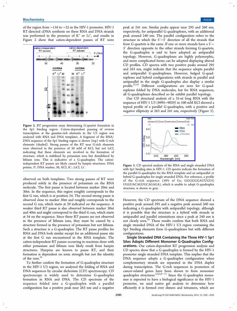
of the region from −116 to −25 in the HIV-1 promoter. HIV-1RT-directed cDNA synthesis on these RNA and DNA strandswas performed in the presence of K+ or Li+, and results inFigure 2 show that cation-dependent pauses of RT were
observed on both templates. Two strong pauses of RT wereproduced solely in the presence of potassium on the RNAmolecule. The first pause is located between marker 20nt and30nt. In the sequence, this region roughly corresponds to thefirst G run, which is at position 24. The second strong pause isobserved close to marker 30nt and roughly corresponds to thesecond G run, which starts at 29 indicated on the sequence. Aweaker third RT pause is also observed between marker 30ntand 40nt and might correspond to the third G run, which startsat 34 on the sequence. Since these RT pauses are not observedin the presence of lithium ions, they must be caused by astructure formed in the presence of potassium but not lithium.Such a structure is a G-quadruplex. The RT pause profiles forRNA and DNA look similar except for an additional pause siteat the first G run encountered in the RNA template. Thecation-independent RT pauses occurring in reactions done witheither potassium and lithium ions likely result from hairpinstructures. Hairpins are known to pause RT, and theirformation is dependent on ionic strength but not the identityof the ions.33
To further confirm the formation of G-quadruplex structurein the HIV-1 U3 region, we analyzed the folding of RNA andDNA sequences by circular dichroism (CD) spectroscopy. CDspectroscopy is widely used to determine G-quadruplexformation in RNA and DNA. The CD spectrum of thesequence folded into a G-quadruplex with a parallelconfiguration has a positive peak near 263 nm and a negative
peak at 241 nm. Similar peaks appear near 295 and 260 nm,respectively, for antiparallel G-quadruplexes, with an additionalpeak around 240 nm. The parallel configuration refers to thestructure in which the 5′−3′ direction of all the strands thatform G-quartets is the same. If one or more strands have a 5′−3′ direction opposite to the other strands forming G-quartets,the G-quadruplex is said to have adopted an antiparalleltopology. However, G-quadruplexes are highly polymorphic,and more complicated forms can be adopted displaying alteredCD profiles. CD spectra with two positive peaks around 295and 263 nm, might indicate that the sequence adopts paralleland antiparallel G-quadruplexes. However, bulged G-quad-ruplexes and hybrid configurations with strands in parallel andantiparallel in the single G-quadruplex also display a similarprofile.21,41 Different configurations are seen for G-quad-ruplexes folded by DNA molecules, but for RNA sequences,all G-quadruplexes described so far exhibit parallel topology.Our CD structural analysis of a 35-nt long RNA with the
sequence of HIV-1 U3 (8995−9029) in 100 mM KCl showed atypical profile of a parallel G-quadruplex, with a positive andnegative ellipticity at 263 and 241 nm, respectively (Figure 3).
However, the CD spectrum of the DNA sequence showed apositive peak around 295 and a negative peak around 260 nmindicating a G-quadruplex with antiparallel topology, althoughit is possible that the structure is a hybrid with strands inantiparallel and parallel orientations since a peak at 240 nm isnot clearly seen.41 These results confirm that both RNA andsingle stranded DNA of the HIV-1 U3 region containing theSp1 binding elements form G-quadruplexes but with differentconfigurations.
Single Stranded DNA Containing the Three HIV-1 Sp1Sites Adopts Different Monomer G-Quadruplex Config-urations. Our cation-dependent RT progression analysis andCD spectra show that a G-quadruplex is formed by the HIV-1promoter single stranded DNA template. This implies that theDNA sequence adopts a G-quadruplex configuration whencomplementary strands are separated in the DNA duplexduring transcription. The G-rich sequences in promoters ofcancer-related genes have been shown to form monomerquadruplex structures.2,4,5,8−11 Since the G-quadruplex mono-mer is expected to have a biological significance in the HIV-1promoter, we used native gel analysis to determine howefficiently it is formed over dimers and tetramers, which are
Figure 2. RT progression assay determining G-quartet formation inthe Sp1 binding region. Cation-dependent pausing of reversetranscription at the guanine-rich elements in the U3 region wasanalyzed with RNA and DNA templates. A fragment of the RNA/DNA sequence of the Sp1 binding region is shown (top) with G-richelements (shaded). Strong pauses of the RT near G-rich elementswere observed in the presence of 50 mM of KCl, but not LiCl,indicating that these elements are involved in the formation ofstructure, which is stabilized by potassium ions but destabilized bylithium ions. This is indicative of a G-quadruplex. The cation-independent RT pauses are likely caused by hairpin structures. DNAprimer, P; DNA marker, M; KCl, K+; LiCl, Li+.
Figure 3. CD spectral analysis of the RNA and single stranded DNAwith Sp1 binding sites in HIV-1. CD spectra indicate the formation ofthe parallel G-quadruplex for the RNA template and an antiparallel orhybrid G-quadruplex for single stranded DNA. For reference, a profileof the G-rich sequence (50% of Gs; GGGGGGAUUGUGUGGUACAGUGCAGAGA), which is unable to adopt G-quadruplexstructure, is shown in gray.
Biochemistry Article
dx.doi.org/10.1021/bi4016692 | Biochemistry 2014, 53, 2581−25932585
intermolecular forms of G-quadruplex that do not requiremultiple G runs. In this approach, the 32P-labeled strand is firstincubated under G-quadruplex folding conditions and thenloaded into a native gel to separate monomers, dimers, andtetramers from nonfolded molecules. The gel contains KCl at aconcentration of at least 10 mM to maintain the integrity offolded structures. The monomer G-quadruplex appears in thegel as the fastest migrating band, then the nonfolded sequence,whereas dimers and tetramers are slower migrating forms.Because the Sp1 binding elements of the HIV-1 promoter
have seven G runs, to gain information on their individual rolesin a G-quadruplex formation, we analyzed several DNA
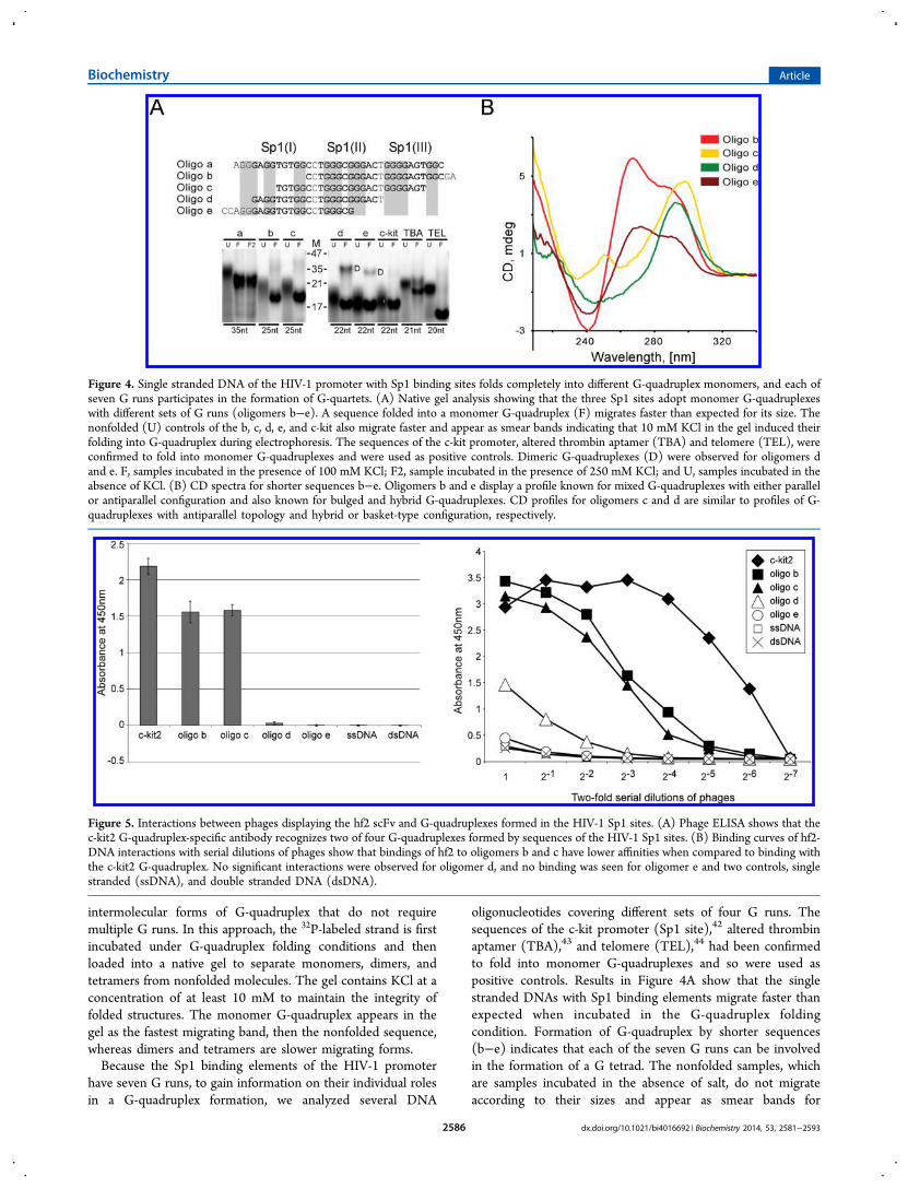
oligonucleotides covering different sets of four G runs. Thesequences of the c-kit promoter (Sp1 site),42 altered thrombinaptamer (TBA),43 and telomere (TEL),44 had been confirmedto fold into monomer G-quadruplexes and so were used aspositive controls. Results in Figure 4A show that the singlestranded DNAs with Sp1 binding elements migrate faster thanexpected when incubated in the G-quadruplex foldingcondition. Formation of G-quadruplex by shorter sequences(b−e) indicates that each of the seven G runs can be involvedin the formation of a G tetrad. The nonfolded samples, whichare samples incubated in the absence of salt, do not migrateaccording to their sizes and appear as smear bands for
Figure 4. Single stranded DNA of the HIV-1 promoter with Sp1 binding sites folds completely into different G-quadruplex monomers, and each ofseven G runs participates in the formation of G-quartets. (A) Native gel analysis showing that the three Sp1 sites adopt monomer G-quadruplexeswith different sets of G runs (oligomers b−e). A sequence folded into a monomer G-quadruplex (F) migrates faster than expected for its size. Thenonfolded (U) controls of the b, c, d, e, and c-kit also migrate faster and appear as smear bands indicating that 10 mM KCl in the gel induced theirfolding into G-quadruplex during electrophoresis. The sequences of the c-kit promoter, altered thrombin aptamer (TBA) and telomere (TEL), wereconfirmed to fold into monomer G-quadruplexes and were used as positive controls. Dimeric G-quadruplexes (D) were observed for oligomers dand e. F, samples incubated in the presence of 100 mM KCl; F2, sample incubated in the presence of 250 mM KCl; and U, samples incubated in theabsence of KCl. (B) CD spectra for shorter sequences b−e. Oligomers b and e display a profile known for mixed G-quadruplexes with either parallelor antiparallel configuration and also known for bulged and hybrid G-quadruplexes. CD profiles for oligomers c and d are similar to profiles of G-quadruplexes with antiparallel topology and hybrid or basket-type configuration, respectively.
Figure 5. Interactions between phages displaying the hf2 scFv and G-quadruplexes formed in the HIV-1 Sp1 sites. (A) Phage ELISA shows that thec-kit2 G-quadruplex-specific antibody recognizes two of four G-quadruplexes formed by sequences of the HIV-1 Sp1 sites. (B) Binding curves of hf2-DNA interactions with serial dilutions of phages show that bindings of hf2 to oligomers b and c have lower affinities when compared to binding withthe c-kit2 G-quadruplex. No significant interactions were observed for oligomer d, and no binding was seen for oligomer e and two controls, singlestranded (ssDNA), and double stranded DNA (dsDNA).
Biochemistry Article
dx.doi.org/10.1021/bi4016692 | Biochemistry 2014, 53, 2581−25932586

oligomers d, e, and c-kit indicating that 10 mM KCl in a gel issufficient to induce their folding into G-quadruplex duringelectrophoresis. The 10 mM KCl is sufficient to induce G-quadruplex folding in RNA and single stranded DNA.4,34,45 Inaddition to monomers, oligomers d and e can also form dimers.The CD spectral analysis confirms the formation of G-
quadruplexes for sequences b, c, and e (Figure 4B); however,the profiles differ for each sequence indicating that they exhibitdifferent configurations. The CD spectra for oligomers b and ehave two peaks around 295 and 260 nm, which might representa mixture of parallel and antiparallel G-quadruplexes.46
However, such profiles are also described for hybrid (3 + 1)and bulged G-quadruplexes.21,41 The profile for oligomer d hasa strong maximum around 295 nm and a minimum around 250nm, and such a profile is generally not observed for G-quadruplex structures, although few reports attributed thesespectral characteristics to hybrid conformations containing amixture of both parallel and antiparallel strand orientations andbasket-type G-quadruplexes.47,48 Other techniques, like NMRspectroscopy would need to be used to determine whatstructure is formed by oligomer d. The CD spectrum foroligomer c is similar to the profile describing G-quadruplexeswith antiparallel configuration. These results indicate that theSp1 binding region can easily and fully adopt differentmonomer G-quadruplexes. The ability of the sequence toadopt different G-quadruplex configurations was previouslyreported and might reflect that G-quadruplexes with differenttopology could have different functions.49
hf2 Antibody Recognizes Two G-QuadruplexesFormed in the Sp1 Binding Region of the HIV-1Promoter. Engineered proteins, such as single-chain antibod-ies and the Gq1 zinc finger protein, have been previouslygenerated as molecular probes to study G-quadruplexstructures.37,50 Using phage display technology, Fernando andco-workers generated an hf2 antibody with high binding affinityto the G-quadruplex formed by c-kit2 found in the promoter ofthe c-kit proto-oncogene.37 Recent studies determined that thisantibody also has binding affinity to G-quadruplexes formed inother genomic regions.46 In order to determine whether hf2would bind to structures formed in the Sp1 binding sites ofHIV-1, we produced phages displaying hf2 antibodies on theirsurface and performed a phage ELISA against structures formedby oligomers b−e. Results showed that the hf2 displaying phagebinds to G-quadruplexes formed by two HIV-1 sequences, theoligomers b and c (Figure 5A). Serial dilutions of phagesshowed that the highest affinity of hf2 antibody was seen for theG-quadruplex formed by the c-kit2 sequence, as expected(Figure 5B). A lower affinity was observed for structuresformed by sequences of oligomers b and c, indicating that theseG-quadruplexes must contain some structural features similar tothose of the G-quadruplex formed by the c-kit2 sequence. Avery weak binding affinity was detected for the structure formedby oligomer d, whereas no binding was detected for oligomer eand two controls, single stranded and double stranded DNA. Asan additional control, we performed a phage ELISA for phagesdisplaying unrelated antibodies. No binding was observed forall analyzed sequences (data not shown). In summary, ourresults show that the hf2 antibody is able to recognize and bindto two G-quadruplex structures of the Sp1 binding region inHIV-1 promoter. This directly proves that oligomers b and cadopt G-quadruplex configurations. The lack of interactionsbetween antibody hf2 and oligomers d and e suggest that bothsequences do not form G-quadruplexes. However, although the
hf2 antibody displayed ability to recognize different G-quadruplexes, likely they do not recognize all configurations.Thus, other methods would have to be used to provide directevidence that oligomers d and e adopt this structure.
Sp1 Protein Binds to a G-Quadruplex Formed in theHIV-1 Promoter. Recent studies showed that the Sp1 bindingsites of the c-kit promoter fold into a G-quadruplex that isrecognized and bound by Sp1 protein.42 The G-rich sequenceof the c-kit Sp1 site and distribution of G runs are differentfrom Sp1 sites in HIV-1. In order to determine whether Sp1can also bind to an HIV-1 sequence folded into a G-quadruplex,we used a previously described affinity selection approach.42 Inthis method, the pull-down of protein is performed with the 3′-biotinylated oligonucleotide with the sequence folded into G-quadruplex and immobilized to streptavidin-coated magneticbeads. The Sp1 protein used for affinity selection is at aconcentration of 1.98 nM.As indicated above, the three Sp1 sites have the ability to
adopt different G-quadruplex configurations, in which differentsets of G runs participate in the formation of a G-quartet.However, for some configurations, the folding might involve Gruns of two Sp1 binding sites leaving one site unfolded. Toensure that only one Sp1 site is available for protein binding ina pull-down of Sp1 and that it is folded, we used as bait a 21-ntsequence of oligomer c with the Sp1 site II surrounded by twoG runs from Sp1 sites I and III (Figure 4). The native gelanalysis shows that this sequence transforms completely into amonomer G-quadruplex (Figure 4A), and CD spectra indicatethat the G-quadruplex is likely in an antiparallel configuration(Figure 4B).In assessing Sp1 binding to a G-quadruplex in the HIV-1
promoter, we used, as a positive control, an oligonucleotidewith the sequence of the c-kit promoter for which the bindingof Sp1 to its site folded as a G-quadruplex had beenconfirmed.42 As a negative control, selection of Sp1 proteinwas done with beads not coupled to any DNA. As seen in theWestern blots in Figure 6A, the Sp1 protein was pulled downwith the HIV-1 Sp1 binding site II folded into a G-quadruplexwith the same efficiency as with a G-quadruplex of the c-kitpromoter. When the dsDNA of this sequence was used for Sp1selection, the interaction of protein with the DNA wasdisrupted by the presence of the 3 and 6 molar excess of
Figure 6. Sp1 binds to a G-quadruplex in the HIV-1 promoter. (A)Sp1 is selected by a G-quadruplex (ss-GQ) with the sequence of theSp1 site II of the HIV-1 promoter (top) with the same efficiency asthat selected by a G-quadruplex with the sequence of the c-kitpromoter. Sp1 is also selected with the same efficiency by thesesequences in dsDNA form. (B) Three and six molar excess of oligomerc with Sp1 site II (top) folded into a G-quadruplex competesefficiently with dsDNA sequence for binding with Sp1. Sp1 binding tothe dsDNA is not affected in the presence of 6 molar excess of anonspecific 18-nt sequence (T)15CTA.
Biochemistry Article
dx.doi.org/10.1021/bi4016692 | Biochemistry 2014, 53, 2581−25932587
oligomer c with the Sp1 site II folded into a G-quadruplex(Figure 6B). This indicates that the Sp1 site in a G-quadruplexform competes efficiently with dsDNA for binding with Sp1.Importantly, the interaction of Sp1 with dsDNA was notaffected by the presence of a 6 molar excess of the nonspecificsequence (T15CTA; ds/ss line in Figure 6B). In summary, theseresults confirm that Sp1 protein can recognize and bind to itsbinding element in the HIV-1 promoter folded into a G-quadruplex configuration.Dimerization of RNA Strands in the HIV-1 U3 Region.
Since the two copies of the HIV-1 RNA genomes are heldtogether at DIS, the ability of the RNA strands to fold into a G-quadruplex configuration raises the possibility that multiple G-quadruplex structures formed between the two viral RNAgenomes support additional dimer contacts during reversetranscription. In order to determine whether an intermolecularG-quadruplex in the U3 region can be formed between twoRNA strands, we first used our previously developed affinityselection approach to test if two homologous sequences of U3can interact.34 In this method, the RNA strands tagged withpoly(A) tail are incubated with nontagged RNA molecules, andthen the mixture is subjected to affinity selection with magneticbeads conjugated with oligo-d(T)25. The interaction betweennucleic acids is measured by analyzing selected RNA molecules.The interacting partners are distinguished by their size in adenaturing gel stained with ethidium bromide. The quantity ofselected nontagged template is expected to be lower sinceinteractions will also occur between two tagged RNAs and twonontagged RNAs. In addition, the formation of an intermo-lecular G-quadruplex between two templates likely competeswith the formation of intramolecular G-quadruplexes in bothtemplates.The RNA strands corresponding to positions 8960−9037 of
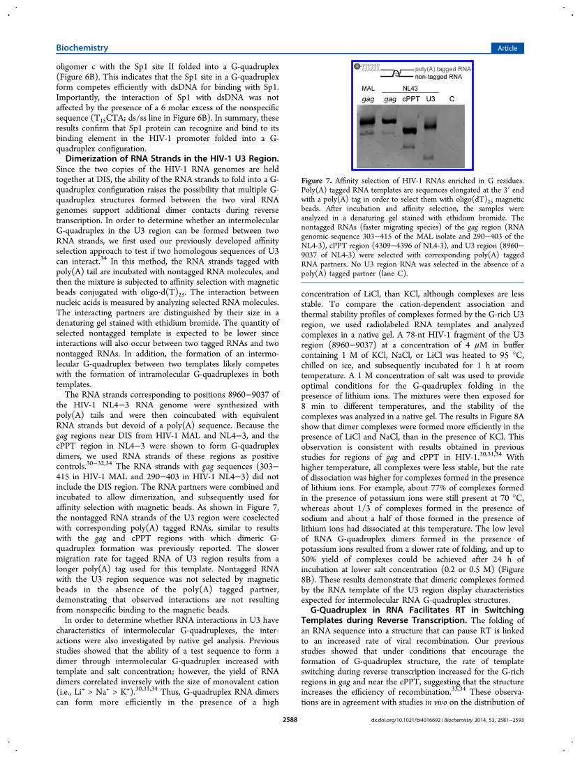
the HIV-1 NL4−3 RNA genome were synthesized withpoly(A) tails and were then coincubated with equivalentRNA strands but devoid of a poly(A) sequence. Because thegag regions near DIS from HIV-1 MAL and NL4−3, and thecPPT region in NL4−3 were shown to form G-quadruplexdimers, we used RNA strands of these regions as positivecontrols.30−32,34 The RNA strands with gag sequences (303−415 in HIV-1 MAL and 290−403 in HIV-1 NL4−3) did notinclude the DIS region. The RNA partners were combined andincubated to allow dimerization, and subsequently used foraffinity selection with magnetic beads. As shown in Figure 7,the nontagged RNA strands of the U3 region were coselectedwith corresponding poly(A) tagged RNAs, similar to resultswith the gag and cPPT regions with which dimeric G-quadruplex formation was previously reported. The slowermigration rate for tagged RNA of U3 region results from alonger poly(A) tag used for this template. Nontagged RNAwith the U3 region sequence was not selected by magneticbeads in the absence of the poly(A) tagged partner,demonstrating that observed interactions are not resultingfrom nonspecific binding to the magnetic beads.In order to determine whether RNA interactions in U3 have
characteristics of intermolecular G-quadruplexes, the inter-actions were also investigated by native gel analysis. Previousstudies showed that the ability of a test sequence to form adimer through intermolecular G-quadruplex increased withtemplate and salt concentration; however, the yield of RNAdimers correlated inversely with the size of monovalent cation(i.e., Li+ > Na+ > K+).30,31,34 Thus, G-quadruplex RNA dimerscan form more efficiently in the presence of a high
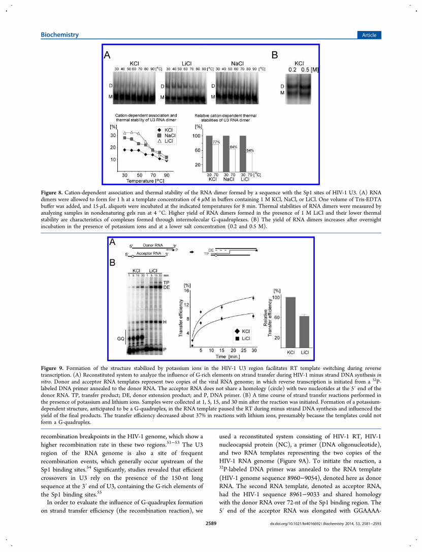
concentration of LiCl, than KCl, although complexes are lessstable. To compare the cation-dependent association andthermal stability profiles of complexes formed by the G-rich U3region, we used radiolabeled RNA templates and analyzedcomplexes in a native gel. A 78-nt HIV-1 fragment of the U3region (8960−9037) at a concentration of 4 μM in buffercontaining 1 M of KCl, NaCl, or LiCl was heated to 95 °C,chilled on ice, and subsequently incubated for 1 h at roomtemperature. A 1 M concentration of salt was used to provideoptimal conditions for the G-quadruplex folding in thepresence of lithium ions. The mixtures were then exposed for8 min to different temperatures, and the stability of thecomplexes was analyzed in a native gel. The results in Figure 8Ashow that dimer complexes were formed more efficiently in thepresence of LiCl and NaCl, than in the presence of KCl. Thisobservation is consistent with results obtained in previousstudies for regions of gag and cPPT in HIV-1.30,31,34 Withhigher temperature, all complexes were less stable, but the rateof dissociation was higher for complexes formed in the presenceof lithium ions. For example, about 77% of complexes formedin the presence of potassium ions were still present at 70 °C,whereas about 1/3 of complexes formed in the presence ofsodium and about a half of those formed in the presence oflithium ions had dissociated at this temperature. The low levelof RNA G-quadruplex dimers formed in the presence ofpotassium ions resulted from a slower rate of folding, and up to50% yield of complexes could be achieved after 24 h ofincubation at lower salt concentration (0.2 or 0.5 M) (Figure8B). These results demonstrate that dimeric complexes formedby the RNA template of the U3 region display characteristicsexpected for intermolecular RNA G-quadruplex structures.
G-Quadruplex in RNA Facilitates RT in SwitchingTemplates during Reverse Transcription. The folding ofan RNA sequence into a structure that can pause RT is linkedto an increased rate of viral recombination. Our previousstudies showed that under conditions that encourage theformation of G-quadruplex structure, the rate of templateswitching during reverse transcription increased for the G-richregions in gag and near the cPPT, suggesting that the structureincreases the efficiency of recombination.33,34 These observa-tions are in agreement with studies in vivo on the distribution of
Figure 7. Affinity selection of HIV-1 RNAs enriched in G residues.Poly(A) tagged RNA templates are sequences elongated at the 3′ endwith a poly(A) tag in order to select them with oligo(dT)25 magneticbeads. After incubation and affinity selection, the samples wereanalyzed in a denaturing gel stained with ethidium bromide. Thenontagged RNAs (faster migrating species) of the gag region (RNAgenomic sequence 303−415 of the MAL isolate and 290−403 of theNL4-3), cPPT region (4309−4396 of NL4-3), and U3 region (8960−9037 of NL4-3) were selected with corresponding poly(A) taggedRNA partners. No U3 region RNA was selected in the absence of apoly(A) tagged partner (lane C).
Biochemistry Article
dx.doi.org/10.1021/bi4016692 | Biochemistry 2014, 53, 2581−25932588
recombination breakpoints in the HIV-1 genome, which show ahigher recombination rate in these two regions.51−53 The U3region of the RNA genome is also a site of frequentrecombination events, which generally occur upstream of theSp1 binding sites.54 Significantly, studies revealed that efficientcrossovers in U3 rely on the presence of the 150-nt longsequence at the 3′ end of U3, containing the G-rich elements ofthe Sp1 binding sites.55
In order to evaluate the influence of G-quadruplex formationon strand transfer efficiency (the recombination reaction), we
used a reconstituted system consisting of HIV-1 RT, HIV-1nucleocapsid protein (NC), a primer (DNA oligonucleotide),and two RNA templates representing the two copies of theHIV-1 RNA genome (Figure 9A). To initiate the reaction, a32P-labeled DNA primer was annealed to the RNA template(HIV-1 genome sequence 8960−9054), denoted here as donorRNA. The second RNA template, denoted as acceptor RNA,had the HIV-1 sequence 8961−9033 and shared homologywith the donor RNA over 72-nt of the Sp1 binding region. The5′ end of the acceptor RNA was elongated with GGAAAA-
Figure 8. Cation-dependent association and thermal stability of the RNA dimer formed by a sequence with the Sp1 sites of HIV-1 U3. (A) RNAdimers were allowed to form for 1 h at a template concentration of 4 μM in buffers containing 1 M KCl, NaCl, or LiCl. One volume of Tris-EDTAbuffer was added, and 15-μL aliquots were incubated at the indicated temperatures for 8 min. Thermal stabilities of RNA dimers were measured byanalyzing samples in nondenaturing gels run at 4 °C. Higher yield of RNA dimers formed in the presence of 1 M LiCl and their lower thermalstability are characteristics of complexes formed through intermolecular G-quadruplexes. (B) The yield of RNA dimers increases after overnightincubation in the presence of potassium ions and at a lower salt concentration (0.2 and 0.5 M).
Figure 9. Formation of the structure stabilized by potassium ions in the HIV-1 U3 region facilitates RT template switching during reversetranscription. (A) Reconstituted system to analyze the influence of G-rich elements on strand transfer during HIV-1 minus strand DNA synthesis invitro. Donor and acceptor RNA templates represent two copies of the viral RNA genome; in which reverse transcription is initiated from a 32P-labeled DNA primer annealed to the donor RNA. The acceptor RNA does not share a homology (circle) with two nucleotides at the 5′ end of thedonor RNA. TP, transfer product; DE, donor extension product; and P, DNA primer. (B) A time course of strand transfer reactions performed inthe presence of potassium and lithium ions. Samples were collected at 1, 5, 15, and 30 min after the reaction was initiated. Formation of a potassium-dependent structure, anticipated to be a G-quadruplex, in the RNA template paused the RT during minus strand DNA synthesis and influenced theyield of the final products. The transfer efficiency decreased about 37% in reactions with lithium ions, presumably because the templates could notform a G-quadruplex.
Biochemistry Article
dx.doi.org/10.1021/bi4016692 | Biochemistry 2014, 53, 2581−25932589

AAAAAA so that transfer products (TP) could be separatedand distinguished on a denaturing gel from DNA fully extendedon the donor RNA (DE). End transfer of the DE was preventedbecause our acceptor RNA did not share homology with twonucleotides at the 5′ end of the donor RNA (a circle in Figure9A). Thus, all transfers to the acceptor RNA originated onlyfrom internal regions of the donor template, as they do in vivoduring reverse transcription over the U3 region.The strand transfer assays were performed in the presence of
a low concentration of either K+ or Li+. The monovalentcations added to the reaction do not significantly affect theenzymatic activity of RT.56,57 The major cation-dependentpause sites of RT synthesis were clearly visible within the G-rich region in the presence of K+ but not Li+ (Figure 9B). Anonion-dependent RT pause, likely resulting from a hairpinstructure, was visible. The strong pauses in potassium,presumed to result from G-quadruplex formation, evidentlycaused dissociation of the RT since fewer final products weremade when compared with that in reactions in lithium, whereno RT pauses were seen in the G-rich region. The transferefficiency of reactions was calculated by comparison of values ofdonor extension products and transfer products using thefollowing formula: transfer efficiency = 100 × TP/(TP + DE).As expected, the elimination of ion-dependent RT pauses bylithium caused a drop in transfer efficiency of about 37%. Sinceion-dependent RT pauses are associated with G-quadruplexformation, the effect of increased template switching likelyresulted from RT encountering G-quadruplex. This isconsistent with a general observation that a structure capableof pausing the RT also facilitates transfer reactions; however,the effect observed here is less striking than for two other G-quadruplex forming sequences in a region of gag and cPPT.33,34
In summary, these results show that G-quadruplex(es) formedat the 3′ end of the viral RNA genome are likely contributors tothe increased recombination rate in the U3 region.
■ DISCUSSIONMany promoters have multiple runs of G-residues, includingthose with several G-rich Sp-1 transcription factor bindingmotifs in tandem. Using genome-wide computational analysis,it has been suggested that such tandem Sp1 binding sites formG-quadruplex structures.58,59 Direct evidence that Sp1 bindingelements form G-quadruplexes that regulate gene expressionhas already be derived from studies of the c-mycpromoter.1,12,15−17 The HIV-1 promoter has three Sp1 bindingelements, and our analysis of the region in U3 revealed that thesequence readily adopts different G-quadruplex configurationswithin both its RNA and single stranded DNA forms. Theseresults predict that during viral infection the G-quadruplexstructure forms in both the RNA genome and in the promoterof the HIV-1 provirus, and presumably has important biologicalfunctions in both environments. In fact, recently Perrone andco-workers published results showing that HIV-1 promoteractivity is impaired in the presence of a G-quadruplex bindingligand, indicating that indeed the structures have a role inregulating virus expression.35 The group also demonstrated thatdifferent G-quadruplexes are formed in the U3 region of DNAand that some of them involve G runs of Sp1 sites togetherwith a G run in the NF-κB site, which was not a subject of ourstudies. However, these and our results together show that theentire G-rich transcription factor binding region in the HIV-1proximal promoter adopts different forms of G-quadruplexstructure.
The variation of genomic sequence among various viralspecies is higher for the U3 region having Sp1 binding sitesthan for the protein coding regions. However, according to ourcomputational analysis, the sequence variants all retain theability of the Sp1 binding sites to form G-quadruplex structure.This observation additionally supports the conclusion that G-quadruplex structure is an important element of the HIV-1promoter region that is maintained by a strong evolutionarypressure.Because the G-quadruplex structure forms in the Sp1
transcription factor binding region, it is likely that it participatesin the regulation of promoter function. Recent studies of the c-kit promoter showed that Sp1 protein can bind to its sequencefolded into a G-quadruplex.42 The sequence and G rundistribution in HIV-1 Sp1 sites differ from those of the Sp1binding region in the c-kit promoter. However, our results alsoshow Sp1-G-quadruplex interactions, indicating that Sp1 couldregulate HIV-1 promoter activity through binding dsDNA orG-quadruplex structure and that these binding characteristicsmight be linked with different functions of Sp1. Moreover, wefound that the G-rich HIV-1 sequence with three Sp1 bindingsites can adopt alternative configuration G-quadruplexes withdifferent interacting sets of G runs and that antibodies specificto the c-kit2 G-quadruplex recognize two of these forms. Suchdiversity suggests that the G-rich Sp1 binding region uses itsability to adopt several non-B DNA configurations as a complexswitching mechanism involving subtle Sp1 interaction differ-ences to fine-tune transcriptional output.Why might such complex alternative protein−DNA inter-
actions be necessary? Sp1 is a strong activator of HIV-1expression, but Sp1 activity is also associated with maintainingvirus latency.60,61 Importantly, the Sp1 binding elements arewithin a region of the HIV-1 promoter that remainsnucleosome-free despite hypermethylation of two CpG islandsthat flank the HIV-1 transcription start site.62,63 As a result, theregion remains accessible to transcription regulators, any ofwhich might act through G-quadruplex structure. G-quadruplexbinding agents and proteins were shown to suppress cellularpromoter activity by stabilizing a G-quadruplex formed in theSp1 binding region, but unwinding of the structure reactivatedgene expression.1,12−17 Interestingly, stimulation of cellularpromoter activity through G-quadruplex structure was recentlyalso demonstrated.18,19 However, recently published results byPerrone and co-workers indicate that G-quadruplexes formed inHIV-1 promoter have a rather inhibitory effect on viraltranscription.35
Formation of G-quadruplexes in the HIV-1 RNA genome haspreviously been demonstrated in gag near the RNA 5′ end andrecently in the central region of the genome near thecPPT.30−32,34 Significantly, both sequences can form dimercomplexes through intermolecular G-quadruplex structure,indicating that G-quartets might be formed that link the twoRNA genomes. Our results show that short RNA moleculeswith HIV-1 sequences containing the Sp1 binding sites can alsodimerize and that their interactions have characteristics of theintermolecular G-quadruplex. This suggests that the U3 regionis an additional point of contact in a multiply linked genomedimer and together with G-rich sequences in gag and cPPThelps to maintain interaction of the HIV-1 genomes along theirwhole sequence. Apart from dimerization through the DISregion, additional interactions through intermolecular G-quadruplexes would explain the ability of viral RNA genomesto maintain a dimeric configuration when binding-disruptive
Biochemistry Article
dx.doi.org/10.1021/bi4016692 | Biochemistry 2014, 53, 2581−25932590
mutations are introduced in DIS.64,65 Moreover, additionalcontacts would also be necessary to keep the two 9kb-longRNA sequences in proximity for efficient reverse transcriptionand the observed widely distributed hot spots for recombina-tion. The ability of the G-rich sequence in U3 to form a dimeralso means that a contact point at the 3′ end provides equalaccessibility of either of the 3′ ends of the copackaged RNAgenomes for minus strong stop DNA transfer. Indeed, studiesshowed that transfers of the minus strand DNA primer occurwith the same frequency to each 3′ genomic end.54
Consistent with these expectations, our results demonstratethat the G-quadruplex formed by the Sp1 binding sites is likelya structural element contributing to the increased recombina-tion rate in U3. Detailed measurements of strand transfers atthe ends of HIV-1 showed that 11 of 86 analyzed clones (12%)underwent homologous recombination in the U3 region, andall crossovers occurred upstream of the G-rich region of theSp1 binding sites.54 Other results showed that recombination inU3 dropped significantly in tests conducted with a templateconstruct missing a 150-nt long sequence containing theputative dimerization site at the 3′ end, suggesting the existenceof sequence elements in this region that are crucial for efficienthomologous recombination in this region.55
Our analysis of minus strand transfer using a reconstitutedsystem shows that the transfer is cation-dependent and thattransfer efficiency decreases in the presence of lithium ions,which destabilize G-quadruplex structure, although the effect isless dramatic than observed for G-rich regions of gag and cPPT.This suggests that G-quadruplex(es) formed in the region ofSp1 binding sites are one of the factors causing increasedrecombination in U3. The correlation between potential G-quadruplex formation and hot spots for recombination was alsofound in the gag and cPPT regions and is in an agreement withprevious analyses in vitro showing that G-quadruplexes facilitateRT template switching during reverse transcription.33,34,66
In summary, our current studies, combined with previouswork, show that G-rich sequences in the HIV-1 genome arecapable of forming G-quadruplexes in both RNA and DNAforms of the genome. The distribution of recombination hotspots correlates with the sites where G-quadruplexes areformed, and reconstituted systems confirm that these structuresfacilitate strand transfers. The concept of G-quadruplexesregulating the activity of the HIV-1 promoter is new, anddetermining how and when G-quadruplexes regulate HIV-1transcription would enhance our understanding of HIV-1latency and reactivation, which might help to identify a newmolecular target for therapeutic reactivation of virus replication.
■ ASSOCIATED CONTENT
*S Supporting InformationDNA and RNA oligonucleotides. This material is available freeof charge via the Internet at http://pubs.acs.org.
■ AUTHOR INFORMATION
Corresponding Author*Department of Microbiology and Immunology, School ofMedicine and Dentistry, University of Rochester, 601 ElmwoodAve., Rochester, NY 14642, USA. Phone: +1 585 275-2764.Fax: +1 585 275-6007. E-mail: [email protected].
FundingThis work was supported by the National Institutes of Health(NIH) [GM049573 to R.A.B. and GM097334 to G.S.]. Wereceived additional support from the University of RochesterDevelopmental Center for AIDS Research grant P30 AI078498(National Institutes of Health/National Institute of Allergy andInfectious Diseases) and the University of Rochester School ofMedicine and Dentistry. Funding for open access charge:National Institutes of Health.NotesThe authors declare no competing financial interest.
■ ACKNOWLEDGMENTSWe are grateful to Dr. Robert J. Gorelick, of the NationalCancer Institute, for the NC used in these studies. We thankDr. Shankar Balasubramanian for sharing with us the pIT2plasmid construct with hf2 sequence.
■ ABBREVIATIONSDIS, dimer initiation site; cPPT, central polypurine tract; NC,nucleocapsid protein; TP, transfer product; DE, donorextension product; nt, nucleotide(s)
■ REFERENCES(1) Siddiqui-Jain, A., Grand, C. L., Bearss, D. J., and Hurley, L. H.(2002) Direct evidence for a G-quadruplex in a promoter region andits targeting with a small molecule to repress c-MYC transcription.Proc. Natl. Acad. Sci. U.S.A. 99, 11593−11598.(2) Phan, A. T., Modi, Y. S., and Patel, D. J. (2004) Propeller-typeparallel-stranded G-quadruplexes in the human c-myc promoter. J. Am.Chem. Soc. 126, 8710−8716.(3) Rankin, S., Reszka, A. P., Huppert, J., Zloh, M., Parkinson, G. N.,Todd, A. K., Ladame, S., Balasubramanian, S., and Neidle, S. (2005)Putative DNA quadruplex formation within the human c-kit oncogene.J. Am. Chem. Soc. 127, 10584−10589.(4) Sun, D., Guo, K., Rusche, J. J., and Hurley, L. H. (2005)Facilitation of a structural transition in the polypurine/polypyrimidinetract within the proximal promoter region of the human VEGF geneby the presence of potassium and G-quadruplex-interactive agents.Nucleic Acids Res. 33, 6070−6080.(5) De Armond, R., Wood, S., Sun, D., Hurley, L. H., andEbbinghaus, S. W. (2005) Evidence for the presence of a guaninequadruplex forming region within a polypurine tract of the hypoxiainducible factor 1alpha promoter. Biochemistry 44, 16341−16350.(6) Ambrus, A., Chen, D., Dai, J., Jones, R. A., and Yang, D. (2005)Solution structure of the biologically relevant G-quadruplex element inthe human c-MYC promoter. Implications for G-quadruplexstabilization. Biochemistry 44, 2048−2058.(7) Phan, A. T., Kuryavyi, V., Gaw, H. Y., and Patel, D. J. (2005)Small-molecule interaction with a five-guanine-tract G-quadruplexstructure from the human MYC promoter. Nat. Chem. Biol. 1, 167−173.(8) Dai, J., Dexheimer, T. S., Chen, D., Carver, M., Ambrus, A., Jones,R. A., and Yang, D. (2006) An intramolecular G-quadruplex structurewith mixed parallel/antiparallel G-strands formed in the human BCL-2promoter region in solution. J. Am. Chem. Soc. 128, 1096−1098.(9) Cogoi, S., and Xodo, L. E. (2006) G-quadruplex formation withinthe promoter of the KRAS proto-oncogene and its effect ontranscription. Nucleic Acids Res. 34, 2536−2549.(10) Fernando, H., Reszka, A. P., Huppert, J., Ladame, S., Rankin, S.,Venkitaraman, A. R., Neidle, S., and Balasubramanian, S. (2006) Aconserved quadruplex motif located in a transcription activation site ofthe human c-kit oncogene. Biochemistry 45, 7854−7860.(11) Xu, Y., and Sugiyama, H. (2006) Formation of the G-quadruplexand i-motif structures in retinoblastoma susceptibility genes (Rb).Nucleic Acids Res. 34, 949−954.
Biochemistry Article
dx.doi.org/10.1021/bi4016692 | Biochemistry 2014, 53, 2581−25932591

(12) Seenisamy, J., Rezler, E. M., Powell, T. J., Tye, D., Gokhale, V.,Joshi, C. S., Siddiqui-Jain, A., and Hurley, L. H. (2004) The dynamiccharacter of the G-quadruplex element in the c-MYC promoter andmodification by TMPyP4. J. Am. Chem. Soc. 126, 8702−8709.(13) Sun, D., Liu, W. J., Guo, K., Rusche, J. J., Ebbinghaus, S.,Gokhale, V., and Hurley, L. H. (2008) The proximal promoter regionof the human vascular endothelial growth factor gene has a G-quadruplex structure that can be targeted by G-quadruplex-interactiveagents. Mol. Cancer Ther. 7, 880−889.(14) Gonzalez, V., Guo, K., Hurley, L., and Sun, D. (2009)Identification and characterization of nucleolin as a c-myc G-quadruplex-binding protein. J. Biol. Chem. 284, 23622−23635.(15) Thakur, R. K., Kumar, P., Halder, K., Verma, A., Kar, A., Parent,J. L., Basundra, R., Kumar, A., and Chowdhury, S. (2009) Metastasessuppressor NM23-H2 interaction with G-quadruplex DNA within c-MYC promoter nuclease hypersensitive element induces c-MYCexpression. Nucleic Acids Res. 37, 172−183.(16) Gonzalez, V., and Hurley, L. H. (2010) The C-terminus ofnucleolin promotes the formation of the c-MYC G-quadruplex andinhibits c-MYC promoter activity. Biochemistry 49, 9706−9714.(17) Brown, R. V., Danford, F. L., Gokhale, V., Hurley, L. H., andBrooks, T. A. (2011) Demonstration that drug-targeted down-regulation of MYC in non-Hodgkins lymphoma is directly mediatedthrough the promoter G-quadruplex. J. Biol. Chem. 286, 41018−41027.(18) Baral, A., Kumar, P., Halder, R., Mani, P., Yadav, V. K., Singh, A.,Das, S. K., and Chowdhury, S. (2012) Quadruplex-single nucleotidepolymorphisms (Quad-SNP) influence gene expression differenceamong individuals. Nucleic Acids Res. 40, 3800−3811.(19) Wei, P. C., Wang, Z. F., Lo, W. T., Su, M. I., Shew, J. Y., Chang,T. C., and Lee, W. H. (2013) A cis-element with mixed G-quadruplexstructure of NPGPx promoter is essential for nucleolin-mediatedtransactivation on non-targeting siRNA stress. Nucleic Acids Res. 41,1533−1543.(20) Phan, A. T., Kuryavyi, V., Burge, S., Neidle, S., and Patel, D. J.(2007) Structure of an unprecedented G-quadruplex scaffold in thehuman c-kit promoter. J. Am. Chem. Soc. 129, 4386−4392.(21) Mukundan, V. T., and Phan, A. T. (2013) Bulges in g-quadruplexes: broadening the definition of g-quadruplex-formingsequences. J. Am. Chem. Soc. 135, 5017−5028.(22) Kumari, S., Bugaut, A., Huppert, J. L., and Balasubramanian, S.(2007) An RNA G-quadruplex in the 5′ UTR of the NRAS proto-oncogene modulates translation. Nat. Chem. Biol. 3, 218−221.(23) Brown, V., Jin, P., Ceman, S., Darnell, J. C., O’Donnell, W. T.,Tenenbaum, S. A., Jin, X., Feng, Y., Wilkinson, K. D., Keene, J. D.,Darnell, R. B., and Warren, S. T. (2001) Microarray identification ofFMRP-associated brain mRNAs and altered mRNA translationalprofiles in fragile X syndrome. Cell 107, 477−487.(24) Beaudoin, J. D., and Perreault, J. P. (2010) 5′-UTR G-quadruplex structures acting as translational repressors. Nucleic AcidsRes. 38, 7022−7036.(25) Beaudoin, J. D., and Perreault, J. P. (2013) Exploring mRNA 3′-UTR G-quadruplexes: evidence of roles in both alternativepolyadenylation and mRNA shortening. Nucleic Acids Res. 41, 5898−5911.(26) Marcel, V., Tran, P. L., Sagne, C., Martel-Planche, G., Vaslin, L.,Teulade-Fichou, M. P., Hall, J., Mergny, J. L., Hainaut, P., and VanDyck, E. (2011) G-quadruplex structures in TP53 intron 3: role inalternative splicing and in production of p53 mRNA isoforms.Carcinogenesis 32, 271−278.(27) Gomez, D., Lemarteleur, T., Lacroix, L., Mailliet, P., Mergny, J.L., and Riou, J. F. (2004) Telomerase downregulation induced by theG-quadruplex ligand 12459 in A549 cells is mediated by hTERT RNAalternative splicing. Nucleic Acids Res. 32, 371−379.(28) Didiot, M. C., Tian, Z., Schaeffer, C., Subramanian, M., Mandel,J. L., and Moine, H. (2008) The G-quartet containing FMRP bindingsite in FMR1 mRNA is a potent exonic splicing enhancer. Nucleic AcidsRes. 36, 4902−4912.
(29) Fisette, J. F., Montagna, D. R., Mihailescu, M. R., and Wolfe, M.S. (2012) A G-rich element forms a G-quadruplex and regulatesBACE1 mRNA alternative splicing. J. Neurochem. 121, 763−773.(30) Marquet, R., Baudin, F., Gabus, C., Darlix, J. L., Mougel, M.,Ehresmann, C., and Ehresmann, B. (1991) Dimerization of humanimmunodeficiency virus (type 1) RNA: stimulation by cations andpossible mechanism. Nucleic Acids Res. 19, 2349−2357.(31) Sundquist, W. I., and Heaphy, S. (1993) Evidence forinterstrand quadruplex formation in the dimerization of humanimmunodeficiency virus 1 genomic RNA. Proc. Natl. Acad. Sci. U.S.A.90, 3393−3397.(32) Marquet, R., Paillart, J. C., Skripkin, E., Ehresmann, C., andEhresmann, B. (1994) Dimerization of human immunodeficiency virustype 1 RNA involves sequences located upstream of the splice donorsite. Nucleic Acids Res. 22, 145−151.(33) Shen, W., Gao, L., Balakrishnan, M., and Bambara, R. A. (2009)A recombination hot spot in HIV-1 contains guanosine runs that canform a G-quartet structure and promote strand transfer in vitro. J. Biol.Chem. 284, 33883−33893.(34) Piekna-Przybylska, D., Sharma, G., and Bambara, R. A. (2013)Mechanism of HIV-1 RNA dimerization in the central region of thegenome, and significance for viral evolution. J. Biol. Chem. 288,24140−24150.(35) Perrone, R., Nadai, M., Frasson, I., Poe, J. A., Butovskaya, E.,Smithgall, T. E., Palumbo, M., Palu, G., and Richter, S. N. (2013) Adynamic G-quadruplex region regulates the HIV-1 long terminalrepeat promoter. J. Med. Chem. 56, 6521−6530.(36) Pandey, V. N., Kaushik, N., Rege, N., Sarafianos, S. G., Yadav, P.N., and Modak, M. J. (1996) Role of methionine 184 of humanimmunodeficiency virus type-1 reverse transcriptase in the polymerasefunction and fidelity of DNA synthesis. Biochemistry 35, 2168−2179.(37) Fernando, H., Rodriguez, R., and Balasubramanian, S. (2008)Selective recognition of a DNA G-quadruplex by an engineeredantibody. Biochemistry 47, 9365−9371.(38) de Wildt, R. M., Mundy, C. R., Gorick, B. D., and Tomlinson, I.M. (2000) Antibody arrays for high-throughput screening of antibody-antigen interactions. Nat. Biotechnol. 18, 989−994.(39) Kikin, O., D’Antonio, L., and Bagga, P. S. (2006) QGRSMapper: a web-based server for predicting G-quadruplexes innucleotide sequences. Nucleic Acids Res. 34, W676−682.(40) Schaeffer, C., Bardoni, B., Mandel, J. L., Ehresmann, B.,Ehresmann, C., and Moine, H. (2001) The fragile X mentalretardation protein binds specifically to its mRNA via a purine quartetmotif. EMBO J. 20, 4803−4813.(41) Vorlickova, M., Kejnovska, I., Sagi, J., Renciuk, D., Bednarova,K., Motlova, J., and Kypr, J. (2012) Circular dichroism and guaninequadruplexes. Methods 57, 64−75.(42) Raiber, E. A., Kranaster, R., Lam, E., Nikan, M., andBalasubramanian, S. (2012) A non-canonical DNA structure is abinding motif for the transcription factor SP1 in vitro. Nucleic AcidsRes. 40, 1499−1508.(43) Fialova, M., Kypr, J., and Vorlickova, M. (2006) The thrombinbinding aptamer GGTTGGTGTGGTTGG forms a bimolecularguanine tetraplex. Biochem. Biophys. Res. Commun. 344, 50−54.(44) Zaug, A. J., Podell, E. R., and Cech, T. R. (2005) Human POT1disrupts telomeric G-quadruplexes allowing telomerase extension invitro. Proc. Natl. Acad. Sci. U.S.A. 102, 10864−10869.(45) Arora, A., and Suess, B. (2011) An RNA G-quadruplex in the 3′UTR of the proto-oncogene PIM1 represses translation. RNA Biol. 8,802−805.(46) Lam, E. Y., Beraldi, D., Tannahill, D., and Balasubramanian, S.(2013) G-quadruplex structures are stable and detectable in humangenomic DNA. Nat. Commun. 4, 1796.(47) Smargiasso, N., Gabelica, V., Damblon, C., Rosu, F., De Pauw,E., Teulade-Fichou, M. P., Rowe, J. A., and Claessens, A. (2009)Putative DNA G-quadruplex formation within the promoters ofPlasmodium falciparum var genes. BMC Genomics 10, 362.(48) Ambrus, A., Chen, D., Dai, J., Bialis, T., Jones, R. A., and Yang,D. (2006) Human telomeric sequence forms a hybrid-type intra-
Biochemistry Article
dx.doi.org/10.1021/bi4016692 | Biochemistry 2014, 53, 2581−25932592

molecular G-quadruplex structure with mixed parallel/antiparallelstrands in potassium solution. Nucleic Acids Res. 34, 2723−2735.(49) Qin, Y., Rezler, E. M., Gokhale, V., Sun, D., and Hurley, L. H.(2007) Characterization of the G-quadruplexes in the duplex nucleasehypersensitive element of the PDGF-A promoter and modulation ofPDGF-A promoter activity by TMPyP4. Nucleic Acids Res. 35, 7698−7713.(50) Isalan, M., Patel, S. D., Balasubramanian, S., and Choo, Y.(2001) Selection of zinc fingers that bind single-stranded telomericDNA in the G-quadruplex conformation. Biochemistry 40, 830−836.(51) Zhuang, J., Jetzt, A. E., Sun, G., Yu, H., Klarmann, G., Ron, Y.,Preston, B. D., and Dougherty, J. P. (2002) Human immunodeficiencyvirus type 1 recombination: rate, fidelity, and putative hot spots. J.Virol. 76, 11273−11282.(52) Dykes, C., Balakrishnan, M., Planelles, V., Zhu, Y., Bambara, R.A., and Demeter, L. M. (2004) Identification of a preferred region forrecombination and mutation in HIV-1 gag. Virology 326, 262−279.(53) Fan, J., Negroni, M., and Robertson, D. L. (2007) Thedistribution of HIV-1 recombination breakpoints. Infect. Genet. Evol. 7,717−723.(54) Yu, H., Jetzt, A. E., Ron, Y., Preston, B. D., and Dougherty, J. P.(1998) The nature of human immunodeficiency virus type 1 strandtransfers. J. Biol. Chem. 273, 28384−28391.(55) Moumen, A., Polomack, L., Roques, B., Buc, H., and Negroni,M. (2001) The HIV-1 repeated sequence R as a robust hot-spot forcopy-choice recombination. Nucleic Acids Res. 29, 3814−3821.(56) Filler, A. G., and Lever, A. M. (1997) Effects of cationsubstitutions on reverse transcriptase and on human immunodefi-ciency virus production. AIDS Res. Hum. Retroviruses 13, 291−299.(57) Yong, W. H., Wyman, S., and Levy, J. A. (1990) Optimalconditions for synthesizing complementary DNA in the HIV-1endogenous reverse transcriptase reaction. AIDS 4, 199−206.(58) Eddy, J., and Maizels, N. (2008) Conserved elements withpotential to form polymorphic G-quadruplex structures in the firstintron of human genes. Nucleic Acids Res. 36, 1321−1333.(59) Todd, A. K., and Neidle, S. (2008) The relationship of potentialG-quadruplex sequences in cis-upstream regions of the human genometo SP1-binding elements. Nucleic Acids Res. 36, 2700−2704.(60) Jiang, G., Espeseth, A., Hazuda, D. J., and Margolis, D. M.(2007) c-Myc and Sp1 contribute to proviral latency by recruitinghistone deacetylase 1 to the human immunodeficiency virus type 1promoter. J. Virol. 81, 10914−10923.(61) Marban, C., Suzanne, S., Dequiedt, F., de Walque, S., Redel, L.,Van Lint, C., Aunis, D., and Rohr, O. (2007) Recruitment ofchromatin-modifying enzymes by CTIP2 promotes HIV-1 transcrip-tional silencing. EMBO J. 26, 412−423.(62) Verdin, E., Paras, P., Jr., and Van Lint, C. (1993) Chromatindisruption in the promoter of human immunodeficiency virus type 1during transcriptional activation. EMBO J. 12, 3249−3259.(63) Kauder, S. E., Bosque, A., Lindqvist, A., Planelles, V., andVerdin, E. (2009) Epigenetic regulation of HIV-1 latency by cytosinemethylation. PLoS Pathog. 5, e1000495.(64) Song, R., Kafaie, J., Yang, L., and Laughrea, M. (2007) HIV-1viral RNA is selected in the form of monomers that dimerize in athree-step protease-dependent process; the DIS of stem-loop 1initiates viral RNA dimerization. J. Mol. Biol. 371, 1084−1098.(65) Shen, N., Jette, L., Liang, C., Wainberg, M. A., and Laughrea, M.(2000) Impact of human immunodeficiency virus type 1 RNAdimerization on viral infectivity and of stem-loop B on RNAdimerization and reverse transcription and dissociation of dimerizationfrom packaging. J. Virol. 74, 5729−5735.(66) Shen, W., Gorelick, R. J., and Bambara, R. A. (2011) HIV-1nucleocapsid protein increases strand transfer recombination bypromoting dimeric G-quartet formation. J. Biol. Chem. 286, 29838−29847.
Biochemistry Article
dx.doi.org/10.1021/bi4016692 | Biochemistry 2014, 53, 2581−25932593