u2 snrna-protein contacts in puriï¬ed human 17s u2 snrnps and in
TRANSCRIPT

MOLECULAR AND CELLULAR BIOLOGY, Apr. 2006, p. 2803–2816 Vol. 26, No. 70270-7306/06/$08.00�0 doi:10.1128/MCB.26.7.2803–2816.2006Copyright © 2006, American Society for Microbiology. All Rights Reserved.
U2 snRNA-Protein Contacts in Purified Human 17S U2 snRNPsand in Spliceosomal A and B Complexes
Olexandr Dybkov, Cindy L. Will, Jochen Deckert, Nastaran Behzadnia, Klaus Hartmuth,and Reinhard Luhrmann*
Department of Cellular Biochemistry, Max Planck Institute for Biophysical Chemistry, D-37077 Gottingen, Germany
Received 28 November 2005/Returned for modification 23 December 2005/Accepted 13 January 2006
The 17S U2 snRNP plays an essential role in branch point selection and catalysis during pre-mRNA splicing.Much remains to be learned about the molecular architecture of the U2 snRNP, including which proteinscontact the functionally important 5� end of the U2 snRNA. Here, RNA-protein interactions within immuno-affinity-purified human 17S U2 snRNPs were analyzed by lead(II)-induced RNA cleavage and UV cross-linking.Contacts between the U2 snRNA and SF3a60, SF3b49, SF3b14a/p14 and SmG and SmB were detected. SF3b49appears to make multiple contacts, interacting with the 5� end of U2 and nucleotides in loops I and IIb. SF3a60also contacted different regions of the U2 snRNA, including the base of stem-loop I and a bulge in stem-loopIII. Consistent with it contacting the pre-mRNA branch point adenosine, SF3b14a/p14 interacted with the U2snRNA near the region that base pairs with the branch point sequence. A comparison of U2 cross-linkingpatterns obtained with 17S U2 snRNP versus purified spliceosomal A and B complexes revealed that RNA-protein interactions with stem-loop I and the branch site-interacting region of U2 are dynamic. These studiesprovide important insights into the molecular architecture of 17S U2 snRNPs and reveal U2 snRNP remod-eling events during spliceosome assembly.
Pre-mRNA splicing is catalyzed by the spliceosome, a largedynamic macromolecular machinery, which assembles by thehighly coordinated, sequential association of four small nuclearRNPs (snRNPs) and a large number of non-snRNP proteinsplicing factors with conserved sequences of the pre-mRNA(reviewed in reference 51). A number of spliceosome assemblyintermediates can be detected in vitro by biochemical means.First, U1 snRNP interacts with the 5� splice site to form thespliceosomal E (early) complex. The U2 snRNP is weaklyassociated with this complex (9), and subsequently it stablyinteracts with the branch point sequence (BPS) of the pre-mRNA in an ATP-dependent fashion, forming complex A. Inthe next step the U4/U6.U5 tri-snRNP is incorporated into thespliceosome, generating complex B. Significant RNA and pro-tein rearrangements, which lead to destabilization (or re-lease) of the U1 and U4 snRNPs, generate the catalyticallyactivated spliceosome (B*) (reviewed in reference 41). Thesubsequent catalytic steps of splicing involve two sequentialtransesterification reactions (reviewed in reference 51). Inthe first step, the branch point adenosine carries out a nu-cleophilic attack at the 5� splice site, which generates thecleaved 5� exon and intron 3� exon. In the second, the 3�splice site is cleaved, resulting in the excision of the intronand ligation of the 5� and 3� exons. The spliced-out intron issubsequently degraded, and the snRNPs are released toparticipate in new rounds of splicing.
The U2 snRNP plays a pivotal role in splicing. It participatesin proper selection and subsequent positioning of the branchpoint adenosine (the nucleophile for the first catalytic step)
within the catalytic core of the spliceosome. The U2 snRNAfirst establishes a short duplex with the BPS of the intron, inwhich the branch adenosine is bulged out (reference 35 andreferences therein). It then forms several short duplexes withthe U6 snRNA (e.g., helices I, II, and III) (19, 27, 42). To-gether with U6, which also base pairs with the 5� splice site, U2forms part of the RNA network that brings into close proximitythe reactive sites of the pre-mRNA and is thus at the catalyticcore of the spliceosome (reviewed in reference 31).
The human U2 snRNP, which consists of the U2 snRNA andseveral proteins, has a modular structure. It was first describedas a 12S particle that consists of the seven Sm proteins, com-mon to all but the U6 and U6atac spliceosomal snRNPs, andtwo U2-specific proteins A� and B� (reviewed in reference 50).The splicing-active 17S U2 snRNP additionally contains theheteromeric splicing factors SF3a and SF3b (2); they consist ofthree proteins (SF3a120, SF3a66, and SF3a60) and seven pro-teins (SF3b155, SF3b145, SF3b130, SF3b49, SF3b14a/p14,SF3b14b, and SF3b10), respectively (8, 15, 25, 53, 55). SF3aand SF3b are required for A complex formation (3) and con-tribute to the selection of the BPS by stabilizing the U2snRNP-BPS interaction. Components of SF3a and SF3b inter-act either directly with the branch adenosine (i.e., SF3b14a/p14) or with a region �25 nucleotides (nt) upstream or 5 ntdownstream of the BPS (15, 16, 53). Due to their close prox-imity to the branch adenosine, several SF3b proteins are likelyto be core components of the spliceosome. Orthologues ofnearly all SF3a and SF3b components have also been identifiedin the yeast Saccharomyces cerevisiae (5, 12, 22, 47). Morerecently, a number of other proteins were found to associatewith the human 17S U2 snRNP, the majority of which arepresent in substoichiometric amounts (55).
Although a great deal has been learned about the organiza-tion of components of the U2 snRNP, a complete picture of its
* Corresponding author. Mailing address: Department of CellularBiochemistry, Max Planck Institute for Biophysical Chemistry, AmFassberg 11, 37077 Gottingen, Germany. Phone: 49-551-2011407. Fax:49-551-2011197. E-mail: [email protected].
2803
Dow
nloa
ded
from
http
s://j
ourn
als.
asm
.org
/jour
nal/m
cb o
n 31
Oct
ober
202
1 by
114
.69.
93.4
8.
molecular architecture is currently missing. The core RNPstructure of the U2 snRNP consists of the seven Sm proteinsG, F, E, D1, D2, D3, and B/B� that interact with the conservedSm site of the U2 snRNA to form a seven-membered ring, theinner surface of which contacts the Sm site (23, 45). The ma-jority of U2-specific proteins, including SF3a, SF3b, and U2-B�/U2-A�, form stable heteromeric complexes in the absenceof the U2 snRNA. Currently only the U2-B�/U2-A� het-erodimer has been shown to directly bind the U2 snRNA,specifically recognizing stem-loop IV (SLIV) (reviewed in ref-erence 46). The atomic structure of the human U2-B�/U2-A�heterodimer complexed with an RNA hairpin comprising U2SLIV was determined previously (33).
Recently the three-dimensional structure of purified humanSF3b, the largest U2 subunit, was obtained by single-particleelectron microscopy (13). The relative orientations of severalSF3b proteins and their domains could be determined. In par-ticular, the SF3b155 C-terminal HEAT repeats are located onthe outer shell of SF3b and curve around the entire complex,whereas the two RRMs of SF3b49 are found on the peripheryand the SF3b14a/p14 RRM is localized in the central cavity. AsSF3b14a/p14 was shown to directly interact with the branchpoint adenosine of pre-mRNA upon stable incorporation ofU2 into the spliceosome (36, 53), SF3b likely undergoes adramatic conformational change either upon integration intothe U2 snRNP or during U2 association with the pre-mRNA.The former is supported by a recent electron cryomicroscopystudy of the U11/U12 di-snRNP. U11 and U12 are the func-tional counterparts of U1 and U2, respectively, in the minorU12-type spliceosome that is responsible for splicing a rareclass of pre-mRNA introns (reviewed in reference 32). SF3balso associates with the U11/U12 di-snRNP (54), and it isfound in a more open conformation in this particle comparedto isolated SF3b (14).
Several protein-protein interactions within the SF3a andSF3b complexes are well documented, including highly stableinteractions between SF3b155 and SF3b14a/p14 (53), SF3b145and SF3b49 (6), SF3a120 and SF3a60 (7), and SF3a120 andSF3a66 (24). However, relatively little is known about howthese functionally important complexes are tethered to the U2snRNP. Electron microscopy studies suggest that the 17S U2snRNP consists of two 10- to 12-nm globular domains, of whichone contains SF3b and a 5� portion of the U2 snRNA whereasthe other contains the Sm core, U2-B�/U2-A�, SF3a, and the 3�part of the U2 snRNA (2, 25).
Here, we have investigated RNA-protein interactions withinthe human 17S U2 snRNP. Native human 17S U2 snRNPs,immunoaffinity purified from HeLa nuclear extract, were ana-lyzed by RNA cleavage with lead(II) and UV-induced cross-linking followed by immunoprecipitation and primer extensionanalyses. Data from these studies revealed contacts betweenthe U2 snRNA and SF3a60, SF3b49, and SF3b14a/p14. UVcross-linking studies with in vitro-reconstituted U2 snRNPsconfirmed an interaction between SF3b49 and the extreme5� end of the U2 snRNA. Subsequent UV cross-linking withaffinity-purified spliceosomal A and B complexes revealed re-modeling events involving RNA-protein contacts at the 5� endof the U2 snRNA upon stable incorporation of the U2 snRNPinto the A complex. Our data thus provide important insightinto the molecular architecture of the 17S U2 snRNP and
reveal that the U2 snRNP is partially remodeled during spli-ceosome assembly.
MATERIALS AND METHODS
Antibodies. The following rabbit polyclonal antibodies were used in this study:anti-SF3a120 (directed against a peptide comprising amino acids [aa] 437 to 451)(53), anti-SF3b155 (aa 99 to 113) (53), anti-SF3b14a/p14 (aa 111 to 125) (53),anti-SF3a66 (aa 444 to 458) (55) and anti-G (raised against a glutathioneS-transferase–SmG fusion protein) (21). Rabbit anti-SF3a60, anti-SF3b145, andanti-SF3b49 antibodies and mouse anti-SF3a120 monoclonal antibodies weregenerated against peptides DYTDRVKPLQDQNEL, AMTQKYEEHVREQQA,MAAGPISERNQDATV, and RWLEQRDRSIREKQS (encompassing aa 195to 209, 816 to 830, 1 to 15, and 437 to 451, respectively). The mouse monoclonalantibody Ana125 was used to precipitate SmB/B� (34). Rabbit antibodies wereaffinity purified as described previously (55) and reacted specifically with thecorresponding protein on immunoblots containing HeLa nuclear extract proteins(data not shown).
Purification of the human 17S U2 snRNP. HeLa cell nuclear extract wasprepared according to the method described in reference 10 and dialyzed againstG150 buffer (20 mM HEPES-KOH, 150 mM KCl, 1.5 mM MgCl2, 5% glycerol,0.5 mM dithioerythritol, 0.5 mM phenylmethylsulfonyl fluoride, pH 7.9). It wasthen diluted with an equal volume of the same buffer and centrifuged at 15,000� g for 10 min to remove precipitates. The supernatant was passed over animmunoaffinity chromatography column containing monoclonal mouse antipep-tide antibodies directed against the SF3a120 protein or polyclonal rabbit anti-peptide antibodies against the SF3a66 protein (55), covalently coupled to proteinG- or A-Sepharose with dimethylpimelimidate, respectively (52). After extensivewashes, 17S U2 snRNPs were eluted from the column with 3 column volumes ofG150 buffer containing 0.4 mg/ml of the cognate peptide. Purified U2 snRNPswere fractionated on a 10% to 30% glycerol gradient as described previously(55). The RNA and protein components of the U2 snRNP were analyzed bydenaturing gel electrophoresis and sodium dodecyl sulfate-polyacrylamide gelelectrophoresis (SDS-PAGE), respectively.
In vitro reconstitution of 17S U2 snRNPs. Reconstitution of 17S U2 snRNPswas performed as described previously (11, 38). Briefly, chimeric U2 snRNA wasprepared by ligation of a synthetic oligoribonucleotide comprising U2 nt 1 to 24to in vitro-transcribed U2 snRNA encompassing nt 25 to 187 (29). RNAs werethen incubated with native, purified total snRNP proteins in vitro and subse-quently with HeLa nuclear extract depleted of endogenous 12S U2 snRNPs.
Purification of human spliceosomal A and B complexes. Spliceosomal A andB complexes formed on MINX pre-mRNA were purified from splicing reactionsby using the previously described tobramycin affinity selection method (18).Briefly, MINX pre-mRNA containing the J6f1 tobramycin binding RNAaptamer was first bound to tobramycin covalently coupled to Sepharose and thenincubated with HeLa nuclear extract under splicing conditions for 45 min. Fol-lowing several wash steps, complexes were eluted from the matrix by incubationwith 5 mM tobramycin. A and B complexes were separated by centrifugation at374,000 � g for 107 min in a TH660 rotor (Sorvall), using a linear 10% to 30%(vol/vol) glycerol gradient, which was fractionated manually from the top. Frac-tions 11 to 12 and 15 to 16 were enriched in complexes A and B, respectively.Additionally, complex B was assembled on a pre-mRNA tagged with threeMS2-binding sites and purified via the MS2-MBP affinity purification method(9). Briefly, MINX pre-mRNA was prebound with the MS2-MBP fusion proteinand spliceosomes were allowed to form by performing splicing in HeLa nuclearextract for 8 min. Spliceosomal complexes were fractionated by gradient centrif-ugation on 10%-to-30% (vol/vol) glycerol gradients (in 20 mM HEPES-KOH[pH 7.9], 1.5 mM MgCl2, 150 mM NaCl) in a Centrikon TST 41.14 rotor for 16 hat 80,000 � g, and gradient fractions were harvested manually from the top. Peakgradient fractions containing spliceosomal B complex were pooled and applied toamylose beads (NEB) preequilibrated with 20 mM HEPES-KOH (pH 7.9), 150mM NaCl, and 1.5 mM MgCl2. Amylose beads were then washed with 15 columnvolumes of preequilibration buffer, and bound spliceosomal B complexes wereeluted with preequilibration buffer containing 12 mM maltose. RNA was recov-ered from the purified complexes by extraction with phenol:chloroform:isoamy-lalcohol (PCI), analyzed on a 7 M urea–10% polyacrylamide gel, and visualizedby staining with silver.
Lead(II)-induced U2 snRNA cleavage. 17S U2 snRNPs, and U2 snRNA ex-tracted from 17S U2 particles with PCI at 4°C, were dialyzed against buffer N1(20 mM HEPES-NaOH, 5 mM magnesium acetate, 100 mM potassium acetate,pH 7.5). RNA cleavage was induced by addition of 5 to 45 mM lead(II) acetateand subsequent incubation for 5 min at 20°C (4). Reactions were terminated byaddition of 50 mM EDTA. Samples were precipitated with ethanol, dissolved in
2804 DYBKOV ET AL. MOL. CELL. BIOL.
Dow
nloa
ded
from
http
s://j
ourn
als.
asm
.org
/jour
nal/m
cb o
n 31
Oct
ober
202
1 by
114
.69.
93.4
8.
PK buffer (100 mM Tris-HCl, 150 mM NaCl, 12.5 mM EDTA, 1% SDS, pH 7.5),and incubated with 1 mg/ml proteinase K for 1 h at 42°C. Following phenol-chloroform extraction, RNAs were precipitated twice with ethanol. The RNAwas dissolved in CE buffer (20 mM cacodylic acid-KOH, 0.2 mM EDTA, pH 7.0),and RNA cleavage sites were analyzed by primer extension.
UV cross-linking of 17S U2 snRNPs, spliceosomal A and B complexes, andnaked U2 snRNA. Approximately 14 pmol of immunoaffinity-purified 17S U2snRNPs or purified spliceosomal A and B complexes was pipetted in a thin layerinto a 24-well microtiter plate and then subjected to 254 nm UV irradiation for30 to 120 s on ice essentially as described previously (44). In parallel, U2 snRNAthat had been extracted with PCI from approximately 2 pmol of 17S U2 snRNPsor spliceosomal complexes at 4°C was cross-linked under the same conditions.Irradiated and nonirradiated control samples of U2 snRNP and U2 RNA wereprecipitated with 3 volumes of ethanol in the presence of 0.3 M sodium acetate,pH 5.2. The pellets were dissolved in 25 �l of 20 mM sodium phosphate buffercontaining 130 mM NaCl (PBS; pH 8.0) supplemented with 3% SDS, incubatedfor 2 min at 96°C followed by 10 min at 70°C, and allowed to cool to roomtemperature. Triton X-100 was added to a final concentration of 5%. The volumewas adjusted to 375 �l with PBS containing 0.5 mM dithioerythritol. Prior toprimer extension analysis, 12 pmol of the sample was subjected to immunopre-cipitation (see below) and 2 pmol was directly digested with proteinase K,extracted with PCI, precipitated twice with ethanol, and dissolved in CE buffer.
Immunoprecipitation of RNA-protein cross-links. Polyclonal rabbit antibodiesdirected against a given U2 snRNP-specific protein were bound to proteinA-Sepharose beads. After being washed with PBS, the antibody-bound beadswere incubated with PBS containing 50 �g/ml yeast tRNA, 0.5 mg/ml bovineserum albumin, and 0.01% NP-40 for 2 h. Beads were subsequently washed threetimes with PBS and incubated for 1 h at 4°C with UV cross-linked or non-cross-linked 17S U2 snRNPs that had been disrupted by heating at 96°C in thepresence of 3% SDS (see above). After being washed extensively with PBS, beadswere incubated with 2� PK buffer for 5 min at 95°C and then diluted with anequal volume of water. Proteins were digested on the beads with proteinase K(end concentration of 0.5 mg/ml) for 1 h at 42°C. The RNA and peptide-RNAcross-linked species were extracted with PCI, precipitated twice with ethanol,and dissolved in CE buffer.
Primer extension analysis. Oligodeoxynucleotide primers complementary topositions 77 to 97 (k91), 97 to 117 (J1), or 149 to 169 (k31) of the human U2snRNA were 32P labeled at their 5� end by use of T4 polynucleotide kinase, and�1 to 2 � 105 cpm of primer was added to U2 snRNA-containing samples. Thesamples were denatured by heating at 96°C for 1 min, allowed to anneal bycooling to room temperature, and reverse transcribed with 1.5 U of AMV reversetranscriptase (USB) per reaction for 50 min at 42.5°C. Sequencing ladders wereobtained from in vitro-transcribed U2 RNA under identical conditions exceptthat 0.5 mM dideoxynucleoside triphosphates were added. Reverse transcriptswere separated on an 8.3 M urea–9.6% polyacrylamide sequencing gel andvisualized by autoradiography.
RESULTS
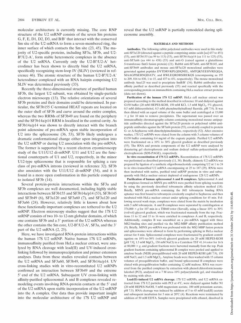
To learn more about the molecular architecture of the 17SU2 snRNP, we examined RNA-protein interactions within thisparticle by lead(II) cleavage footprinting and direct UV cross-linking. For this purpose, native human 17S U2 snRNPs wereimmunoaffinity-purified from HeLa nuclear extract using anti-SF3a120 monoclonal antibodies bound to protein G-Sepharose.Particles were eluted under native conditions with an excess ofcognate peptide, and their RNA and protein compositionswere analyzed. As shown in Fig. 1, highly pure U2 snRNPscould be isolated, as evidenced by the presence of almostexclusively U2 snRNA in the eluate (Fig. 1A), and they possessthe same protein composition—as evidenced by SDS-PAGE(Fig. 1B) and mass spectrometry (not shown)—as human 17SU2 snRNPs previously immunoaffinity purified with anti-SF3a66 antibodies (55). Importantly, immunoaffinity-purified17S U2 snRNPs exhibited a homogeneous sedimentation be-havior during glycerol gradient centrifugation, with nearly allparticles sedimenting in fractions 13 to 15 (Fig. 1C), whichcorrespond to the 17S region of the gradient.
Lead(II)-induced cleavage of U2 snRNA. Previous structureprobing of the human 17S U2 snRNP was limited to chemicalprobing with DMS and kethoxal (2). To obtain more informa-tion about the secondary structure of the U2 snRNA and thosenucleotides that might be involved in RNA-protein interac-tions within the 17S U2 snRNP, we compared the lead(II)-induced RNA cleavage pattern of naked U2 snRNA and U2snRNA within native 17S U2 particles. Lead(II) cations inducecleavages of RNA molecules in single-stranded and bulgedregions that are not spatially constrained or protected by pro-teins (4). The reactivity of the sugar-phosphate backbone ofnaked U2 snRNA towards lead(II), as determined by reverse
FIG. 1. Characterization of immunoaffinity-purified human 17S U2snRNPs. (A and B) The RNA (A) and protein (B) compositions of 17SU2 snRNPs immunoaffinity purified with antibodies against SF3a120were analyzed by denaturing and SDS-PAGE, respectively. RNA wasvisualized by silver staining and protein by staining with Coomassieblue. The identity of RNA or protein is shown on the right. NE,nuclear extract. (C) Purified 17S U2 snRNPs were fractionated on a10% to 30% glycerol gradient, and the RNA composition of odd-numbered and bottom gradient fractions (as indicated above) wasdetermined as described for panel A.
VOL. 26, 2006 RNA-PROTEIN CONTACTS IN THE HUMAN 17S U2 snRNP 2805
Dow
nloa
ded
from
http
s://j
ourn
als.
asm
.org
/jour
nal/m
cb o
n 31
Oct
ober
202
1 by
114
.69.
93.4
8.

FIG. 2. Lead(II)-induced cleavage of native human 17S U2 snRNPs. (A and B) Isolated U2 snRNA (RNA; lanes 1 to 4) or immunoaffinity-purified 17S U2 snRNP (RNP; lanes 5 to 8) was probed with increasing concentrations of lead(II) acetate (0, 5, 15, and 45 mM; lanes 1 to 4 and5 to 8). Cleavage sites were assayed by primer extension using oligonucleotides complementary to nt 97 to 117 (A) or 149 to 169 (B) and werevisualized by autoradiography. A sequencing ladder (on the left or right) was obtained by primer extension of in vitro-transcribed U2 RNA in thepresence of dideoxynucleotides. Those nucleotides that are strongly cleaved in the naked RNA are indicated by arrows to the right of lane 4.Regions of U2 snRNA with increased or strongly reduced reactivity towards lead(II) when probed in the 17S U2 snRNP are marked by an openor closed bar, respectively. Positions of the nucleotides are shown on the right of each panel. (C) Schematic representation of the comparativereactivity of U2 snRNA in the purified 17S U2 snRNP versus isolated U2 snRNA. Increased reactivity towards lead(II) of U2 in 17S U2 snRNPscompared to the results seen with isolated U2 snRNA is indicated by open arrowheads and weakly and strongly decreased reactivity by small andlarge black boxes, respectively. The degree of reactivity was determined by eye by comparing the relative intensity of the bands in the RNA versusRNP lanes, also taking into account the intensity of background in the absence of lead(II). The branch site-interacting region (BSiR) is highlightedby a bar above the corresponding nucleotides, and the Sm site is underlined.
2806 DYBKOV ET AL. MOL. CELL. BIOL.
Dow
nloa
ded
from
http
s://j
ourn
als.
asm
.org
/jour
nal/m
cb o
n 31
Oct
ober
202
1 by
114
.69.
93.4
8.

transcription with primers complementary to positions 97 to117 and 149 to 169 of U2 snRNA, is shown in Fig. 2A and B.Regions efficiently cleaved in the presence of lead(II) (indi-cated by closed arrows to the right of lane 4 in Fig. 2A and B),as evidenced by greatly enhanced reverse transcription stops(lanes 2 to 4) relative to the control reaction without lead(II)(lane 1), included nucleotides located in (i) the loop of SLI(U17-Gm19) or just upstream of SLIIa (C45-U46), (ii) stemIIa (U49-C50), (iii) the loop of SLIIa (A56), (iv) the regionbetween SLIIa and SLIIb (G63-G68), (v) the loop of SLIIb(A75-C78), (vi) the Sm site (U100-U104), (vii) the loop orbulge of SLIII (U129-G131 and C135-G136), and (viii) a trinu-cleotide downstream of it (A145-G147). Weakly accessible po-sitions included nucleotides at the 5� end of U2, comprising thestem of SLI, at or near the U2 branch site-interacting region,in loop IIa, at the base of SLIIb and directly downstream of it,and several nucleotides just upstream or within SLIII. Theoverall cleavage pattern is in good agreement with the previ-ously proposed secondary structure of U2 snRNA (1) (Fig.2C), as the most reactive sites (with the exception of severalnucleotides in the stems of SLIIa and SLIIb) are located insingle-stranded regions.
When 17S U2 snRNPs were subjected to lead(II) cleavage,U2 snRNA showed an altered reactivity compared to nakedU2 snRNA (Fig. 2A and B, lanes 5 to 8). The comparativeanalysis of lead(II)-induced cleavage of naked U2 snRNA ver-sus U2 in 17S U2 RNPs is schematically summarized in Fig.2C. Cleavage of nucleotides in the branch site-interacting re-gion, SLIIa, as well as in loops IIb and III was greatly reducedor even abolished in the case of the Sm site (Fig. 2B; indicatedby solid bars to the right of lane 8). Other regions of U2snRNA weakly reactive in the naked RNA were also less effi-ciently cleaved in the 17S U2 snRNP, including the 5� end ofU2 and SLI. These results suggest either that RNA was directlyprotected by the presence of proteins in these regions or thatits structure was altered (e.g., the formation of base pairs wasstabilized) via the interaction with proteins. Interestingly, se-quences adjacent to the Sm site (nucleotides 94 to 98 and 110to 112) were more susceptible to lead(II)-induced cleavage(marked by open bars in Fig. 2B). Taken together, these resultssuggest that multiple regions of the U2 snRNA are likelycontacted by protein in the 17S U2 snRNP.
Mapping of RNA-protein interaction sites via UV cross-linking. To more precisely map the RNA-protein interactionsites in native human 17S U2 snRNPs, we performed directUV cross-linking, followed by immunoprecipitation and primerextension analysis as described previously (44). Exposure to254-nm UV light is known to induce direct (zero-length) cross-links between nitrogenous bases of nucleic acids and aminoacid side chains when they are in a favorable configuration.First we analyzed UV cross-links in native 17S U2 snRNPspurified from HeLa cell nuclear extract (“RNP” lanes), as wellas in the naked U2 snRNA (“RNA” lanes) extracted from 17SU2 particles under mild conditions, which should preserve thenative secondary structure of the RNA. Putative RNA-proteincontact sites were determined by comparing the patterns ofreverse transcription (Fig. 3 and 4, lanes 1 to 4). Cross-linkednucleotides interfere with primer extension. Therefore, strongreverse transcriptase stops induced upon UV irradiation thatare present in the “RNP” lanes but absent or less intense in
“RNA” lanes should indicate the position of RNA-proteincross-links, with an actual interaction site situated 1 nucleotideupstream from the stop site. In contrast, stops that appearirrespectively of UV irradiation are naturally occurring reversetranscriptase stops and do not result from protein cross-links.For example, major naturally occurring stops were consistentlyobserved at nucleotides 30 to 31, 48, 62, 66, 136, 140, and 145(Fig. 3 and 4).
A number of putative RNA-protein cross-links could bediscerned. Enhanced reverse transcriptase stops were observedat nucleotides C3 and �7, in SLI (i.e., at nucleotides U9-Gm11, U16-Gm19, and A23), in the branch point-interactingregion (A35 and 38) and just downstream of it (�41 andUm47), in SLIIa (U55, A56, and U62), in loop IIb (A75, U76,and C78), upstream of and at the Sm site (A88, A90, U92, A93,U101, and U103), and in SLIII (A123, C128, U130, G131, andC135) (Fig. 3A and B and 4A and B [cf. lanes 1 and 3]; pleasenote that the order of the RNA and RNP lanes is reversed inFig. 3B). Thus, proteins are potentially cross-linked one nucle-otide upstream of these sites in the 17S U2 snRNP (see Fig. 5for summary). Some of these stops were enhanced in the UV-irradiated versus nonirradiated, naked U2 snRNA (e.g., U17or A56) (Fig. 3A and 4A [cf. lanes 3 and 4]), and thus theycould alternatively represent RNA-RNA cross-links, due toeither long-range internal RNA interactions or the formationof pyrimidine dimers. As the most downstream primer that weused annealed to positions 149 to 169, no information aboutprotein-RNA contact sites downstream of C149 was obtained.Taken together, these data indicate that the U2 snRNA iscontacted at multiple positions apparently over its entirelength by proteins.
Identification of the protein-RNA cross-links within the U2snRNA Sm site. To identify proteins contacting the U2snRNA, cross-linked RNA-protein species were immunopre-cipitated with antibodies against a subset of 17S U2 snRNPproteins, including SmG and SmB, all SF3a subunits (SF3a60,SF3a66, and SF3a120), and several SF3b proteins (i.e., SF3b14a/p14, SF3b49, SF3b145, and SF3b155). These antibodies re-acted specifically with their cognate proteins on immunoblotscontaining HeLa nuclear extract proteins (data not shown).Precipitated cross-links were subsequently identified by primerextension analysis. Prior to immunoprecipitation, 17S U2 snRNPswere disrupted by incubating at 96°C in the presence of 3%SDS (for details, see Materials and Methods). Under theseconditions, only SF3b49, SF3b14a/p14, or SF3a60, and noother U2 proteins (as evidenced by silver staining after SDS-PAGE), were precipitated with anti-SF3b49, anti-SF3b14a/p14,or anti-SF3a60 antibodies, respectively, that were covalently cou-pled to protein A-Sepharose, indicating that the vast majority ofU2 snRNPs and their subcomplexes had been dissociated (datanot shown). Furthermore, U2 RNA from the nonirradiated U2snRNP samples was not precipitated by any of the antibodiesthat were tested (Fig. 3 and 4 and data not shown), furtherindicating that all non-cross-linked, protein-RNA interactionshad been disrupted and also did not reform during immuno-precipitation.
UV irradiation generated at least two RNA-protein cross-links within the Sm site at positions U100 and U102 (Fig. 3Band 4B [cf. lanes 1 and 3]). Primer extension analysis of cross-links immunoprecipitated with anti-SmG antibodies revealed
VOL. 26, 2006 RNA-PROTEIN CONTACTS IN THE HUMAN 17S U2 snRNP 2807
Dow
nloa
ded
from
http
s://j
ourn
als.
asm
.org
/jour
nal/m
cb o
n 31
Oct
ober
202
1 by
114
.69.
93.4
8.

that they precipitated nearly exclusively the cross-link at U100(i.e., a major reverse transcriptase stop at U101 was observed)(Fig. 3B, lane 7), confirming that SmG contacts this site. Sim-ilarly, the cross-link at U102 was highly enriched in the anti-SmB/B� immunoprecipitate, indicating that it is contacted by
SmB/B� (Fig. 3B, lane 9). These data are in accordance withprevious UV cross-linking studies performed with a short oli-gonucleotide derived from the Sm site of the U4 snRNA,which indicated that the first and third residues of the Sm siteare contacted by SmG and SmB/B�, respectively (45).
FIG. 3. Identification of UV-induced U2 snRNA-protein cross-linking sites within native human 17S U2 snRNPs. (A) Primer extensionanalyses of U2 snRNA extracted from UV-irradiated (lane 1) or nonirradiated (lane 2) immunoaffinity-purified 17S U2 snRNPs (RNP) andUV-irradiated (lane 3) or nonirradiated (lane 4) naked U2 snRNA (RNA). (B) Primer extension analyses of UV-irradiated (lane 1) ornonirradiated (lane 2) naked U2 snRNA or U2 snRNA extracted from UV-irradiated (lane 3) or nonirradiated (lane 4) immunoaffinity-purified17S U2 snRNPs (RNP). Immunoprecipitation (IP) with anti-SF3b49, anti-SmG, anti-SmB/B�, or anti-SF3a66 antibodies (as indicated above eachlane), followed by primer extension analyses, was performed after denaturation of UV-irradiated (lanes 5, 7, and 9) or nonirradiated (lanes 6, 8,and 10) 17S U2 snRNPs. Primer extension was performed with oligonucleotides complementary to nt 77 to 97 (A) and 149 to 169 (B). Dideoxysequencing markers were generated as described for Fig. 2. Nucleotide positions are shown on the left. Reverse transcriptase stops due to putativeRNA-protein cross-links (closed circles), specific protein-RNA cross-links (open diamonds), potential RNA-RNA cross-links (asterisks), andmajor background stops (squares) are indicated.
2808 DYBKOV ET AL. MOL. CELL. BIOL.
Dow
nloa
ded
from
http
s://j
ourn
als.
asm
.org
/jour
nal/m
cb o
n 31
Oct
ober
202
1 by
114
.69.
93.4
8.

SF3a60 contacts SLI and SLIII of the U2 snRNA. Anti-SF3a60 antibodies precipitated multiple U2 cross-link species,including some not readily apparent in the input irradiatedRNP lane (Fig. 4A and B [cf. lanes 1 and 7]). Several of these,including U16-U18, �34, �37, U55, Cm61, U92, and U100,were precipitated equally well by other antibodies and werenot clearly enriched in the anti-SF3a60 immunoprecipitate(Fig. 3 and 4). Thus, they could not unequivocally be desig-
nated as SF3a60 cross-links. The precipitation of multiplecross-links by several antibodies might be due to cross-linkingof two different proteins to the same RNA molecule or arisefrom incomplete disruption or reassociation of a small fractionof the U2 particles during immunoprecipitation. However, sixcross-links were either specifically precipitated by anti-SF3a60antibodies (i.e., C8-U9 and U22-A23 within SLI as well as C28downstream of it) or they were significantly enriched relative to
FIG. 4. Identification of protein-RNA contact sites within native human 17S U2 snRNPs. (A and B) Primer extension analyses of U2 snRNAextracted from UV-irradiated (lane 1) or nonirradiated (lane 2) immunoaffinity-purified 17S U2 snRNPs (RNP) and UV-irradiated (lane 3) ornonirradiated (lane 4) naked U2 snRNA (RNA). Immunoprecipitation (IP) with anti-SF3b14a/p14 (lanes 5 to 6) or anti-SF3a60 (lanes 7 to 8)antibodies, followed by primer extension analyses, was performed after denaturation of UV-irradiated (lanes 5 and 7) or nonirradiated (lanes 6and 8) 17S U2 snRNPs. Primer extension was performed with oligonucleotides complementary to nt 77 to 97 (A) and 149 to 169 (B) and visualizedby autoradiography. Dideoxy sequencing markers were generated as described for Fig. 2. Nucleotide positions are shown on the left. Reversetranscriptase stops due to putative RNA-protein cross-links (closed circles), specific protein-RNA cross-links (open diamonds), potential RNA-RNA cross-links (asterisks), and major background stops (squares) are indicated.
VOL. 26, 2006 RNA-PROTEIN CONTACTS IN THE HUMAN 17S U2 snRNP 2809
Dow
nloa
ded
from
http
s://j
ourn
als.
asm
.org
/jour
nal/m
cb o
n 31
Oct
ober
202
1 by
114
.69.
93.4
8.
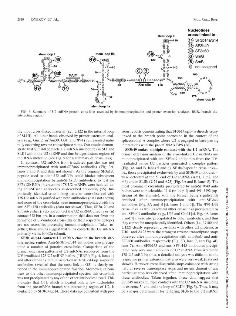
the input cross-linked material (i.e., U122 in the internal loopof SLIII). All other bands observed by primer extension anal-ysis (e.g., Gm12, m6Am30, G31, and �41) represented natu-rally occurring reverse transcriptase stops. Our results demon-strate that SF3a60 contacts U2 snRNA nucleotides in SLI andSLIII within the U2 snRNP and thus bridges distant regions ofthe RNA molecule (see Fig. 5 for a summary of cross-links).
In contrast, U2 snRNA from irradiated particles was notimmunoprecipitated with anti-SF3a66 antibodies (Fig. 3A,lanes 7 and 8, and data not shown). As the cognate SF3a120peptide used to elute U2 snRNPs could hinder subsequentimmunoprecipitation by anti-SF3a120 antibodies, to test forSF3a120-RNA interactions 17S U2 snRNPs were isolated us-ing anti-SF3a66 antibodies as described previously (55). Im-portantly, identical cross-linking patterns were observed with17S U2 snRNPs purified with both antibodies (data not shown)and none of the cross-links were immunoprecipitated with theanti-SF3a120 antibodies (data not shown). Thus, SF3a120 andSF3a66 either (i) do not contact the U2 snRNA directly or (ii)contact U2 but are in a conformation that does not favor theformation of UV-induced cross-links or their respective epitopesare not accessible, preventing immunoprecipitation. Taken to-gether, these results suggest that SF3a contacts the U2 snRNAprimarily via its 60-kDa subunit.
SF3b14a/p14 contacts U2 snRNA close to the branch site-interacting region. Anti-SF3b14a/p14 antibodies also precipi-tated a number of putative cross-links. Comparison of theprimer extension patterns of U2 snRNAs recovered from theUV-irradiated 17S U2 snRNP before (“RNP”; Fig. 4, lanes 1)and after (lanes 5) immunoselection with SF3b14a/p14-specificantibodies revealed that the cross-link at G31 is clearly en-riched in the immunoprecipitated fraction. Moreover, in con-trast to the other immunoprecipitated species, this cross-linkwas not precipitated by any of the other antibodies tested. Thisindicates that G31, which is located only a few nucleotidesfrom the pre-mRNA branch site-interacting region of U2, iscontacted by SF3b14a/p14. These data are consistent with pre-
vious reports demonstrating that SF3b14a/p14 is directly cross-linked to the branch point adenosine in the context of thespliceosomal A complex where U2 is engaged in base-pairinginteractions with the pre-mRNA’s BPS (36).
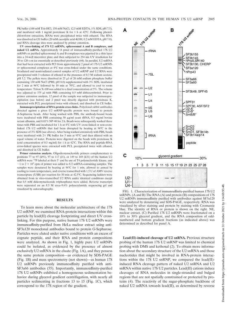
SF3b49 makes multiple contacts with the U2 snRNA. Theprimer extension analysis of the cross-linked U2 snRNAs im-munoprecipitated with anti-SF3b49 antibodies from the UV-irradiated native U2 particles generated a complex pattern(Fig. 3A and B, lanes 5 and 6). SF3b49-specific cross-links—i.e., those precipitated exclusively by anti-SF3b49 antibodies—were detected at the 5� end of U2 snRNA (Am1, Um2, and�6) and in SLIIb (U74 and A75) (Fig. 3A and B, lanes 5). Themost prominent cross-links precipitated by anti-SF3b49 anti-bodies were to nucleotides U18 (in loop I) and �91-U92 (up-stream of the Sm site), with the former being significantlyenriched after immunoprecipitation with anti-SF3b49antibodies (Fig. 3A and B [cf. lanes 1 and 5]). The �91-U92cross-links, as well as several other cross-links precipitated byanti-SF3b49 antibodies (e.g., U55 and Cm61 [cf. Fig. 4A, lanes5 and 7]), were also precipitated by other antibodies, and thusthey cannot be unequivocally identified. Others (i.e., U100 andU122) clearly represent cross-links with other U2 proteins, asU101 and A123 were the strongest reverse transcriptase stopsobserved after immunoprecipitation with anti-SmG and anti-SF3a60 antibodies, respectively (Fig. 3B, lane 7, and Fig. 4B,lane 7). Anti-SF3b155 and anti-SF3b145 antibodies precipi-tated only very small amounts of U2 snRNA from irradiated17S U2 snRNPs; thus, a detailed analysis was difficult, as therespective primer extension patterns were very weak (data notshown). However, most discernible stops coincided with strongnatural reverse transcriptase stops and no enrichment of anyparticular stop was observed after immunoprecipitation withthese antibodies. Taken together, these data suggest thatSF3b49 makes multiple contacts with the U2 snRNA, includingits extreme 5� end and the loop of SLIIb (Fig. 5). Thus, it maybe a major determinant for tethering SF3b to the U2 snRNP.
FIG. 5. Summary of U2 snRNA-protein cross-links revealed by immunoprecipitation and primer extension analyses. BSiR, branch site-interacting region.
2810 DYBKOV ET AL. MOL. CELL. BIOL.
Dow
nloa
ded
from
http
s://j
ourn
als.
asm
.org
/jour
nal/m
cb o
n 31
Oct
ober
202
1 by
114
.69.
93.4
8.

SF3b49 interacts with the 5� end of U2 snRNA in reconsti-tuted 17S U2 snRNPs. To obtain independent evidence thatSF3b49 contacts nucleotides at the very 5� end of the U2snRNA, we performed UV cross-linking with in vitro-recon-stituted U2 snRNPs. Previous reports indicated that modifica-tions within the first 24 nucleotides of the human U2 snRNAare essential and sufficient for its activity in splicing (11). Wethus prepared a chimeric U2 snRNA by ligating together asynthetic oligonucleotide, 32P labeled at its 5� end, that encom-passes the 5� terminal 24 nucleotides of the U2 snRNA andcontains all modifications (Fig. 6A), with an unmodified in vitro-transcribed RNA comprising the remaining part of U2. Thischimeric U2 RNA was first incubated with purified totalsnRNP proteins to form 12S U2 snRNPs and later with HeLanuclear extract specifically depleted of 12S U2 snRNPs togenerate 17S U2 snRNPs (38). U2 snRNPs reconstituted inthis way support pre-mRNA splicing in U2-depleted HeLanuclear extract and thus are functionally active (11).
Reconstituted U2 particles were UV irradiated and treatedwith RNase T1 to generate a radiolabeled fragment comprisingsolely the first four nucleotides of U2, thereby allowing us toidentify proteins cross-linked to nucleotides 1 to 4 of U2snRNA. Gel analysis of the digested RNA revealed that morethan 95% of the U2 RNA had been digested to completion(data not shown), with only a small amount of a partiallydigested fragment observed (Fig. 6B). A double band migrat-ing at �50 kDa on SDS-polyacrylamide gels was observedupon UV irradiation of 17S U2 snRNPs but not in the absenceof UV (Fig. 6B [cf. lanes 1 and 2]). This cross-link was sensitiveto proteinase K and was not observed with in vitro-reconsti-tuted 12S U2 snRNPs or naked U2 snRNA (data not shown),suggesting that it was a cross-link between the SF3b49 proteinand U2 snRNA. To determine the identity of the cross-linkedprotein, immunoprecipitations were performed with anti-SF3b49and anti-SF3a60 antibodies after disruption of RNP complexesas described above. This cross-linked species was precipitatedsolely by anti-SF3b49 antibodies (Fig. 6B, lanes 3 to 6), con-firming that it contains SF3b49. Note that the cross-link mi-grated faster and appeared as a single band after immunopre-cipitation, most likely due to the presence of large amounts ofheavy chain immunoglobulin G in the lanes. These resultscorroborate our cross-linking data obtained with native 17S U2snRNPs, according to which SF3b49 could be cross-linked to ntAm1 and Um2 and thus provide independent evidence that theSF3b49 protein contacts the 5� end of the U2 snRNA.
RNA-protein cross-linking in spliceosomal A and B com-plexes. Next we addressed whether rearrangements occur inprotein-RNA interactions within the U2 snRNP upon its inte-gration into the spliceosomal A complex and/or upon the sub-sequent integration of the U4/U6.U5 tri-snRNP during B com-plex formation. For this purpose, spliceosomal complexes werepurified using aptamer-tagged, adenovirus-derived MINX pre-mRNA and a tobramycin affinity selection method (18) fol-lowed by glycerol gradient centrifugation. Gradient fractions11 to 12 were enriched in complex A (as evidenced by thenearly equimolar amounts of pre-mRNA and U1 and U2 snRNA),and fractions 15 to 16 contained predominantly complex B (asevidenced by the nearly equimolar amounts of pre-mRNA andU1, U2, U4, U5, and U6 snRNA) (Fig. 7A). B complexes werealso isolated using an MS2 affinity purification method and a
MINX pre-mRNA containing MS2 binding sites (9). Afterincubation with HeLa nuclear extract under splicing condi-tions, spliceosomal complexes were separated by glycerol gra-dient centrifugation, and the B complex peak was further pu-
FIG. 6. Identification of proteins cross-linked to the 5� end of U2snRNA in 17S U2 snRNPs reconstituted in vitro. (A) Schematic rep-resentation of the chimeric U2 snRNA prepared by ligation of a32P-5�-end-labeled synthetic oligoribonucleotide, corresponding to thefirst 24 nucleotides of human U2 snRNA (shown in black), to invitro-transcribed U2 encompassing nucleotides 25 to 187 (shown ingray). The radioactive label (asterisk) and the 5�-most RNase T1 cleav-age site (arrowhead) are indicated. The 4-nt-long radioactive fragmentof U2 generated by complete RNase T1 digestion of U2 snRNA isunderlined. (B) 17S U2 snRNP particles were in vitro reconstitutedwith the chimeric U2 snRNA and subjected to UV irradiation (lanes 1,3, and 5). U2 snRNPs in the even-numbered lanes and in lane 7 werenot irradiated. All samples (except the lane 7 sample) were digested byRNase T1 and either directly analyzed by SDS-PAGE (“input”; lanes1 and 2) or after immunoprecipitation with anti-SF3b49 (lanes 3 and 4)or anti-SF3a60 (lanes 5 and 6) antibodies. Protein-RNA cross-linkedspecies were visualized by autoradiography. The positions of full-length U2, the SF3b49-U2 snRNA cross-link, and partially digested U2snRNA are indicated by closed arrowheads on the right; note that the32P-labeled 4-nt fragment has run out of the gel. The positions ofmolecular mass markers are shown on the left.
VOL. 26, 2006 RNA-PROTEIN CONTACTS IN THE HUMAN 17S U2 snRNP 2811
Dow
nloa
ded
from
http
s://j
ourn
als.
asm
.org
/jour
nal/m
cb o
n 31
Oct
ober
202
1 by
114
.69.
93.4
8.
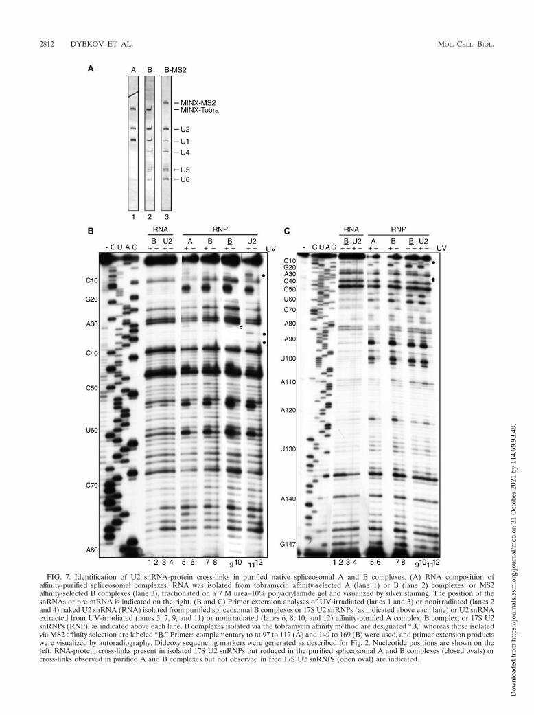
FIG. 7. Identification of U2 snRNA-protein cross-links in purified native spliceosomal A and B complexes. (A) RNA composition ofaffinity-purified spliceosomal complexes. RNA was isolated from tobramycin affinity-selected A (lane 1) or B (lane 2) complexes, or MS2affinity-selected B complexes (lane 3), fractionated on a 7 M urea–10% polyacrylamide gel and visualized by silver staining. The position of thesnRNAs or pre-mRNA is indicated on the right. (B and C) Primer extension analyses of UV-irradiated (lanes 1 and 3) or nonirradiated (lanes 2and 4) naked U2 snRNA (RNA) isolated from purified spliceosomal B complexes or 17S U2 snRNPs (as indicated above each lane) or U2 snRNAextracted from UV-irradiated (lanes 5, 7, 9, and 11) or nonirradiated (lanes 6, 8, 10, and 12) affinity-purified A complex, B complex, or 17S U2snRNPs (RNP), as indicated above each lane. B complexes isolated via the tobramycin affinity method are designated “B,” whereas those isolatedvia MS2 affinity selection are labeled “B.” Primers complementary to nt 97 to 117 (A) and 149 to 169 (B) were used, and primer extension productswere visualized by autoradiography. Dideoxy sequencing markers were generated as described for Fig. 2. Nucleotide positions are shown on theleft. RNA-protein cross-links present in isolated 17S U2 snRNPs but reduced in the purified spliceosomal A and B complexes (closed ovals) orcross-links observed in purified A and B complexes but not observed in free 17S U2 snRNPs (open oval) are indicated.
2812 DYBKOV ET AL. MOL. CELL. BIOL.
Dow
nloa
ded
from
http
s://j
ourn
als.
asm
.org
/jour
nal/m
cb o
n 31
Oct
ober
202
1 by
114
.69.
93.4
8.
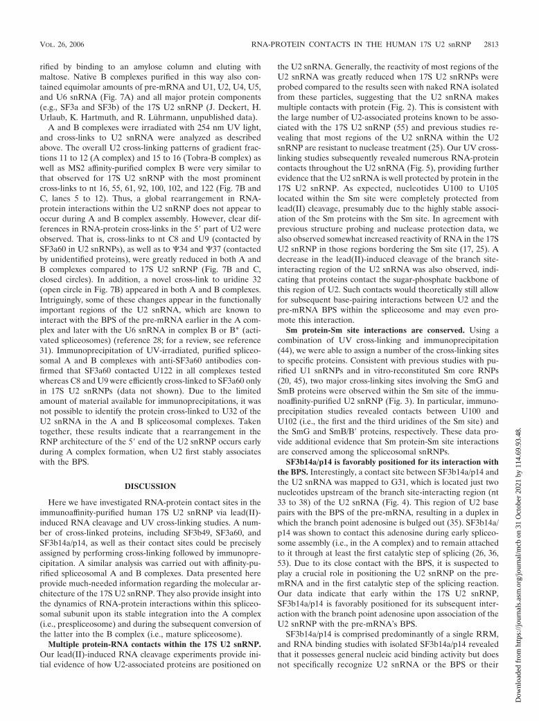
rified by binding to an amylose column and eluting withmaltose. Native B complexes purified in this way also con-tained equimolar amounts of pre-mRNA and U1, U2, U4, U5,and U6 snRNA (Fig. 7A) and all major protein components(e.g., SF3a and SF3b) of the 17S U2 snRNP (J. Deckert, H.Urlaub, K. Hartmuth, and R. Luhrmann, unpublished data).
A and B complexes were irradiated with 254 nm UV light,and cross-links to U2 snRNA were analyzed as describedabove. The overall U2 cross-linking patterns of gradient frac-tions 11 to 12 (A complex) and 15 to 16 (Tobra-B complex) aswell as MS2 affinity-purified complex B were very similar tothat observed for 17S U2 snRNP with the most prominentcross-links to nt 16, 55, 61, 92, 100, 102, and 122 (Fig. 7B andC, lanes 5 to 12). Thus, a global rearrangement in RNA-protein interactions within the U2 snRNP does not appear tooccur during A and B complex assembly. However, clear dif-ferences in RNA-protein cross-links in the 5� part of U2 wereobserved. That is, cross-links to nt C8 and U9 (contacted bySF3a60 in U2 snRNPs), as well as to �34 and �37 (contactedby unidentified proteins), were greatly reduced in both A andB complexes compared to 17S U2 snRNP (Fig. 7B and C,closed circles). In addition, a novel cross-link to uridine 32(open circle in Fig. 7B) appeared in both A and B complexes.Intriguingly, some of these changes appear in the functionallyimportant regions of the U2 snRNA, which are known tointeract with the BPS of the pre-mRNA earlier in the A com-plex and later with the U6 snRNA in complex B or B* (acti-vated spliceosomes) (reference 28; for a review, see reference31). Immunoprecipitation of UV-irradiated, purified spliceo-somal A and B complexes with anti-SF3a60 antibodies con-firmed that SF3a60 contacted U122 in all complexes testedwhereas C8 and U9 were efficiently cross-linked to SF3a60 onlyin 17S U2 snRNPs (data not shown). Due to the limitedamount of material available for immunoprecipitations, it wasnot possible to identify the protein cross-linked to U32 of theU2 snRNA in the A and B spliceosomal complexes. Takentogether, these results indicate that a rearrangement in theRNP architecture of the 5� end of the U2 snRNP occurs earlyduring A complex formation, when U2 first stably associateswith the BPS.
DISCUSSION
Here we have investigated RNA-protein contact sites in theimmunoaffinity-purified human 17S U2 snRNP via lead(II)-induced RNA cleavage and UV cross-linking studies. A num-ber of cross-linked proteins, including SF3b49, SF3a60, andSF3b14a/p14, as well as their contact sites could be preciselyassigned by performing cross-linking followed by immunopre-cipitation. A similar analysis was carried out with affinity-pu-rified spliceosomal A and B complexes. Data presented hereprovide much-needed information regarding the molecular ar-chitecture of the 17S U2 snRNP. They also provide insight intothe dynamics of RNA-protein interactions within this spliceo-somal subunit upon its stable integration into the A complex(i.e., prespliceosome) and during the subsequent conversion ofthe latter into the B complex (i.e., mature spliceosome).
Multiple protein-RNA contacts within the 17S U2 snRNP.Our lead(II)-induced RNA cleavage experiments provide ini-tial evidence of how U2-associated proteins are positioned on
the U2 snRNA. Generally, the reactivity of most regions of theU2 snRNA was greatly reduced when 17S U2 snRNPs wereprobed compared to the results seen with naked RNA isolatedfrom these particles, suggesting that the U2 snRNA makesmultiple contacts with protein (Fig. 2). This is consistent withthe large number of U2-associated proteins known to be asso-ciated with the 17S U2 snRNP (55) and previous studies re-vealing that most regions of the U2 snRNA within the U2snRNP are resistant to nuclease treatment (25). Our UV cross-linking studies subsequently revealed numerous RNA-proteincontacts throughout the U2 snRNA (Fig. 5), providing furtherevidence that the U2 snRNA is well protected by protein in the17S U2 snRNP. As expected, nucleotides U100 to U105located within the Sm site were completely protected fromlead(II) cleavage, presumably due to the highly stable associ-ation of the Sm proteins with the Sm site. In agreement withprevious structure probing and nuclease protection data, wealso observed somewhat increased reactivity of RNA in the 17SU2 snRNP in those regions bordering the Sm site (17, 25). Adecrease in the lead(II)-induced cleavage of the branch site-interacting region of the U2 snRNA was also observed, indi-cating that proteins contact the sugar-phosphate backbone ofthis region of U2. Such contacts would theoretically still allowfor subsequent base-pairing interactions between U2 and thepre-mRNA BPS within the spliceosome and may even pro-mote this interaction.
Sm protein-Sm site interactions are conserved. Using acombination of UV cross-linking and immunoprecipitation(44), we were able to assign a number of the cross-linking sitesto specific proteins. Consistent with previous studies with pu-rified U1 snRNPs and in vitro-reconstituted Sm core RNPs(20, 45), two major cross-linking sites involving the SmG andSmB proteins were observed within the Sm site of the immu-noaffinity-purified U2 snRNP (Fig. 3). In particular, immuno-precipitation studies revealed contacts between U100 andU102 (i.e., the first and the third uridines of the Sm site) andthe SmG and SmB/B� proteins, respectively. These data pro-vide additional evidence that Sm protein-Sm site interactionsare conserved among the spliceosomal snRNPs.
SF3b14a/p14 is favorably positioned for its interaction withthe BPS. Interestingly, a contact site between SF3b14a/p14 andthe U2 snRNA was mapped to G31, which is located just twonucleotides upstream of the branch site-interacting region (nt33 to 38) of the U2 snRNA (Fig. 4). This region of U2 basepairs with the BPS of the pre-mRNA, resulting in a duplex inwhich the branch point adenosine is bulged out (35). SF3b14a/p14 was shown to contact this adenosine during early spliceo-some assembly (i.e., in the A complex) and to remain attachedto it through at least the first catalytic step of splicing (26, 36,53). Due to its close contact with the BPS, it is suspected toplay a crucial role in positioning the U2 snRNP on the pre-mRNA and in the first catalytic step of the splicing reaction.Our data indicate that early within the 17S U2 snRNP,SF3b14a/p14 is favorably positioned for its subsequent inter-action with the branch point adenosine upon association of theU2 snRNP with the pre-mRNA’s BPS.
SF3b14a/p14 is comprised predominantly of a single RRM,and RNA binding studies with isolated SF3b14a/p14 revealedthat it possesses general nucleic acid binding activity but doesnot specifically recognize U2 snRNA or the BPS or their
VOL. 26, 2006 RNA-PROTEIN CONTACTS IN THE HUMAN 17S U2 snRNP 2813
Dow
nloa
ded
from
http
s://j
ourn
als.
asm
.org
/jour
nal/m
cb o
n 31
Oct
ober
202
1 by
114
.69.
93.4
8.

duplex on its own (39). As high-affinity binding of SF3b14a/p14to a U2 oligonucleotide comprising the branch site-interactingregion was not observed in the aforementioned studies, itwould appear that SF3b14a/p14 is not the main determinantfor tethering other SF3b proteins to the U2 snRNP. Our cross-linking studies with purified A and B complexes did not pro-vide clear information as to whether or not SF3b14a/p14 re-mains in contact with G31 of the U2 snRNA in spliceosomalcomplexes where it has been shown to contact the branch pointadenosine. Thus, it is presently not clear whether SF3b14a/p14contacts simultaneously both the pre-mRNA and the U2snRNA within the spliceosome. Interestingly, a novel uniden-tified cross-link at U32 of U2 was observed in these complexesand could thus potentially be SF3b14a/p14. However, addi-tional studies are required to clarify this point.
SF3a60 contacts SLI and SLIII of the U2 snRNA. Of thethree proteins comprising SF3a, we identified cross-links be-tween U2 snRNA and SF3a60 only, suggesting that this proteinplays a key role in tethering SF3a to the U2 snRNP. In vitrostudies indicate that SF3a incorporation into the U2 snRNPrequires the prior assembly of a 15S U2 snRNP that containsSF3b and components of the 12S U2 snRNP (i.e., the Smproteins and A�/B�) (25). In vitro reconstitution studies withpurified, individual SF3a subunits indicate that SF3a60 (as wellas the other SF3a proteins) can bind the 15S U2 snRNP ontheir own (30). Deletion analyses revealed that association ofSF3a60 requires an intact zinc finger domain at its C terminus(30), which is the only conserved motif found in SF3a60. Zincfinger domains are known to bind nucleic acids; thus, it istempting to speculate that this region of SF3a60 contacts theU2 snRNA. Taken together, our data indicate that the inter-action of SF3a to form the 17S U2 snRNP is stabilized, at leastin part, by direct U2 snRNA-SF3a60 contacts.
In addition to contacting the bulge in SLIII, SF3a60 mademultiple contacts with the lower stem of SLI (Fig. 4). AsUV-induced RNA-protein cross-links are generally not formedwhen nucleotides are engaged in base-pairing interactions, ourresults suggest that within the U2 snRNP, the lower part of SLIdoes not form. Likewise, previous structure-probing studiessuggested that the base of stem I is unpaired in 17S U2 snRNPs(2). Formation of stem I is not essential, whereas its hyper-stabilization has deleterious effects on splicing (56). Thus,SF3a60, together with other U2 proteins such as SF3b49, whichwas also cross-linked nearby, may help to maintain it in anunpaired conformation. This in turn likely facilitates the base-pairing interaction between this region of U2 and the U6snRNA to form U2/U6 helix I and II, a crucial RNA structureat the catalytic core of the spliceosome (see below for detaileddiscussion).
SF3b49 makes multiple contacts with the U2 snRNA. Inter-estingly, SF3b49 was found to contact U2 snRNA at multiplesites, including its extreme 5� end and loops I and IIb. Itcontains two N-terminal RRMs; thus, its domain structuresuggests that it should possess RNA binding activity. SF3b49was shown to play a role in tethering the U2 snRNP to the BPSduring spliceosomal A complex assembly, contacting the pre-mRNA just upstream of the BPS (15) and interacting withanother component of the U2 snRNP, the SF3b145 protein(6). The Caenorhabditis elegans homologue of SF3b49 wasshown to bind RNA in vitro primarily via its RRM2 (43). In
yeast, the protein-protein interaction site of HSH49 and Cus1p,the yeast homologues of the human SF3b49 and SF3b145 pro-teins, respectively (49), was mapped to the first RRM of HSH49.Further mutational analyses indicated that the RNA and pro-tein binding surfaces of the RRM1 of HSH49 are most likelydistinct and that both RRMs are required for viability in yeast(22). Based on the detection of multiple cross-links involvingSF3b49, it is likely that the observed cross-linking pattern (in-volving different regions of the U2 snRNA) would require thecontribution of both RRMs. However, as cross-linking impliesclose contact and not necessarily binding per se, regions otherthan SF3b49’s RRMs could also be cross-linked.
The interactions of SF3b49 and SF3a60 with the 5� end ofthe U2 snRNA (including SLI) described here might be ofprimary importance for the integrity of 17S U2 snRNPs. Thatis, it was observed previously that removal of 10 to 15 nucleo-tides from the 5� end of U2 snRNA results in a loss of themajority of proteins, changing the sedimentation value of theparticle to 10S to 12S (2). Thus, SF3b49 and SF3a60 couldpotentially play a crucial role in tethering the SF3a/b com-plexes to U2 snRNP. Within the spliceosome, SF3b49, as wellas SF3a60, contacts a relatively short region (about 25 nt) ofthe pre-mRNA upstream of the BPS (6, 15, 40). As withSF3b14a/p14, it is presently not clear whether these proteinscontact simultaneously both the pre-mRNA and the U2 snRNAin spliceosomal complexes. However, as the U2 snRNA cross-linking pattern in purified A and B complexes was nearly identicalto that in purified 17S U2 snRNPs (with notable exceptions at the5� end of U2; see below), several SF3b49 and SF3a60 contactswith the U2 snRNA (e.g., at nucleotides 74, 75, and 122) appearto remain unchanged. Thus, it is likely that these proteins simul-taneously contact the U2 snRNA and the pre-mRNA in thevicinity of the BPS.
As SF3b is also a component of the U11/U12 snRNP, ourdata may provide first insights into how SF3b is incorporatedinto this snRNP particle. That is, it is likely that SF3b49 alsocontacts the U12 snRNA within the U11/U12 snRNP. As thesequences of U12 and U2 are essentially unrelated but thegeneral structures of their 5� halves are similar, SF3b49 couldbe envisioned to bind in a sequence-independent manner toanalogous regions of the U12 snRNA. However, additionalstudies (i.e., UV cross-linking of purified U11/U12 snRNPs)are clearly required to answer this question.
Additional proteins likely contact the U2 snRNA. The pro-tein moiety of a number of U2 snRNA-protein cross-linkscould not be identified, due to lack of antibodies or the inac-cessibility of the epitope recognized by the antibodies at hand(Fig. 5). Thus, additional U2-associated proteins appear tocontact the U2 snRNA. Furthermore, some RNA-protein in-teractions may have escaped detection, as only those in aconformation favorable for UV cross-linking would be ob-served. Indeed, previous studies suggested that SF3b145 con-tacts the U2 snRNA. The cross-links to nucleotides Cm40 andU46, as well as those to SLIIa, might be due to interactionswith SF3b145, as several genetic studies performed with yeastlinked this protein to these regions of the U2 snRNA (49, 57).Likewise, Prp9-Prp11-Prp21 (the yeast homologues of SF3a60,SF3a66, and SF3a120, respectively) and Prp5 were found togenetically interact with nucleotides just upstream of SLIIa, aswell as with SLIIa and SLIIb, suggesting that these factors
2814 DYBKOV ET AL. MOL. CELL. BIOL.
Dow
nloa
ded
from
http
s://j
ourn
als.
asm
.org
/jour
nal/m
cb o
n 31
Oct
ober
202
1 by
114
.69.
93.4
8.

might bind to these regions of U2 (37, 48, 57). Finally, on thebasis of the U2 model presented by Kramer et al. (25) (seebelow), the cross-links in the loop of SLIII might involveSF3a120 and/or SF3a66.
A more compact RNP structure for the 17 U2 snRNP? Pre-vious biochemical and electron microscopy studies (2, 25) sug-gested a model of 17S U2 snRNP organization in which SF3bproteins interact with the 5� half of the U2 snRNA whereas theSm proteins, SF3a and U2-A�/U2-B�, bind to its 3� half andform a second domain of the U2 snRNP. Consistent with thismodel, two SF3b subunits (SF3b49 and SF3b14a/p14) werecross-linked to the 5� part of the U2 snRNA (i.e., within nt 1 to31) whereas one of the contact sites of SF3a60 was mapped tothe bulge in SLIII (Fig. 3 and 4). Intriguingly, a second site ofinteraction with SF3a60 was found at the base of SLI, indicat-ing that SF3a60 spans the 5� and 3� halves of the U2 snRNA.Thus, the 17S U2 snRNP might have a more compact structurethan previously thought (2, 25). Indeed, electron microscopyimaging of the 17S U2 particles used in this study revealed thatthey predominantly have a globular shape (B. Sander, M. M.Golas, C. L. Will, B. Kastner, H. Stark, and R. Luhrmann,unpublished data) rather than two smaller domains connectedwith a thin filament (i.e., a dumbbell shape) as previouslyreported (2, 25). This difference in appearance most likelyarises from the different purification conditions used in eachstudy and suggests that some of the interactions within the 17SU2 snRNP are not particularly stable.
Evidence for U2 snRNP remodeling upon its stable integra-tion into the spliceosome. Examination of U2 snRNA-proteininteractions in purified spliceosomal A and B complexes re-vealed that the RNP structure involving the 5� end of the U2snRNA undergoes a conformational change during spliceo-some assembly. That is, cross-links to nucleotides 8 and 9 (SLI)and to nucleotides 34 and 37 (branch site-interacting region)were significantly reduced in both spliceosomal complexescompared to the results seen with the free 17S U2 snRNP (Fig.7). In contrast, enhanced cross-link formation was observed atnucleotide 32 in spliceosomal complexes. The observed changeswere quantitative in nature (e.g., residual cross-linking to nt 8, 9,34, and 37 was consistently observed), suggesting that the com-plexes analyzed may be structurally heterogeneous or containsmall amounts of contaminating 17S U2 snRNPs. Nonetheless,our data indicate that the U2 snRNP has already been remodeledat the time of its stable interaction with the BPS of the pre-mRNA.
Interestingly, the 5� end of the U2 snRNA is involved in anextensive RNA network, formed by U2, U6, and the pre-mRNA, that is essential for splicing (reviewed in reference 31).While the U2 snRNA-BPS base-pairing interaction is known tobe first established during A complex formation (i.e., in theprespliceosome), U2-U6 interactions occur after tri-snRNPaddition (after B complex formation). However, it is presentlynot clear whether these interactions have already been estab-lished in the B complex or occur only at the time of spliceo-some activation (for a recent discussion, see reference 28). Atpresent, three different U2/U6 base-pairing interactions havebeen reported: helix I, consisting of helix Ia and Ib separatedby two unpaired adenosines of U2 (27), helix II (19), and helixIII (42).
Intriguingly, the observed changes in the U2 snRNA cross-linking pattern in spliceosomal complexes involve protein con-tacts with nucleotides of U2 forming U2/U6 helix II (C8 andU9) and nucleotides upstream (U32) or within the branchpoint helix (�34 and �37). Our finding that SF3a60 is boundto nucleotides 8 and 9 of the U2 snRNA and that these con-tacts are significantly reduced upon association of U2 with theBPS in the A complex indicates that the RNP rearrangementsinvolve SF3a60. Immunoprecipitations with anti-SF3a60 anti-bodies of the A and B complexes after UV irradiation showedthat this protein does not completely leave the U2 snRNP(data not shown); thus, it likely remains attached to the bulgein SLIII in spliceosomal complexes. As UV-induced RNA-protein cross-links are typically not formed when nucleotidesare engaged in base-pairing interactions, it is currently notclear whether the unknown protein(s) contacting �34 and �37(which are base paired already in the A complex) are displacedfrom the U2 snRNA or whether the relative protein-RNAorientation was changed due to base-pairing interactions suchthat cross-linking is prevented. Likewise, the loss of the C8 andU9 cross-links in the purified B complexes could be due to theformation of the U2/U6 helix II, which involves these nucleo-tides. However, as the cross-links to C8-U9 disappear and thecross-link to U32 has already appeared in the A complex, whenno U6 is present (18), these RNP rearrangements occur earlyon and are not a consequence of U2/U6 helix formation. In-deed, based on their location, one or more of these rearrange-ments may facilitate or be a prerequisite for subsequently es-tablishing U2/U6 base-pairing interactions. Even though theidentity of the protein(s) cross-linked to U32, �34, and �37has not been determined, it is tempting to speculate that it isSF3b14a/p14, which might “move” towards the branch pointadenosine, switching from G31 in the isolated 17S U2 snRNPto U32 in the spliceosome.
ACKNOWLEDGMENTS
We are grateful to Gabi Heyne, Thomas Conrad, Hossein Kohansal,and Irene Ochsner for excellent technical assistance. We thank Hen-ning Urlaub for helpful discussions and Gizem Donmez for kindlyproviding native snRNP proteins and synthetic RNA oligonucleotidesfor U2 snRNP reconstitutions.
This work was supported by grants from the DFG Forschergruppe,Fonds der Chemischen Industrie, and the Ernst-Jung Stiftung to R.L.
REFERENCES
1. Ares, M., Jr., and A. H. Igel. 1990. Lethal and temperature-sensitive muta-tions and their suppressors identify an essential structural element in U2small nuclear RNA. Genes Dev. 4:2132–2145.
2. Behrens, S. E., K. Tyc, B. Kastner, J. Reichelt, and R. Luhrmann. 1993.Small nuclear ribonucleoprotein (RNP) U2 contains numerous additionalproteins and has a bipartite RNP structure under splicing conditions. Mol.Cell. Biol. 13:307–319.
3. Brosi, R., K. Groning, S. E. Behrens, R. Luhrmann, and A. Kramer. 1993.Interaction of mammalian splicing factor SF3a with U2 snRNP and relationof its 60-kD subunit to yeast PRP9. Science 262:102–105.
4. Brunel, C., and P. Romby. 2000. Probing RNA structure and RNA-ligandcomplexes with chemical probes. Methods Enzymol. 318:3–21.
5. Caspary, F., A. Shevchenko, M. Wilm, and B. Seraphin. 1999. Partial puri-fication of the yeast U2 snRNP reveals a novel yeast pre-mRNA splicingfactor required for pre-spliceosome assembly. EMBO J. 18:3463–3474.
6. Champion-Arnaud, P., and R. Reed. 1994. The prespliceosome componentsSAP 49 and SAP 145 interact in a complex implicated in tethering U2 snRNPto the branch site. Genes Dev. 8:1974–1983.
7. Chiara, M. D., P. Champion-Arnaud, M. Buvoli, B. Nadal-Ginard, and R.Reed. 1994. Specific protein-protein interactions between the essential mam-malian spliceosome-associated proteins SAP 61 and SAP 114. Proc. Natl.Acad. Sci. USA 91:6403–6407.
VOL. 26, 2006 RNA-PROTEIN CONTACTS IN THE HUMAN 17S U2 snRNP 2815
Dow
nloa
ded
from
http
s://j
ourn
als.
asm
.org
/jour
nal/m
cb o
n 31
Oct
ober
202
1 by
114
.69.
93.4
8.

8. Das, B. K., L. Xia, L. Palandjian, O. Gozani, Y. Chyung, and R. Reed. 1999.Characterization of a protein complex containing spliceosomal proteinsSAPs 49, 130, 145, and 155. Mol. Cell. Biol. 19:6796–6802.
9. Das, R., Z. Zhou, and R. Reed. 2000. Functional association of U2 snRNPwith the ATP-independent spliceosomal complex E. Mol. Cell 5:779–787.
10. Dignam, J. D., R. M. Lebovitz, and R. G. Roeder. 1983. Accurate transcrip-tion initiation by RNA polymerase II in a soluble extract from isolatedmammalian nuclei. Nucleic Acids Res. 11:1475–1489.
11. Donmez, G., K. Hartmuth, and R. Luhrmann. 2004. Modified nucleotides atthe 5� end of human U2 snRNA are required for spliceosomal E-complexformation. RNA 10:1925–1933.
12. Dziembowski, A., A. P. Ventura, B. Rutz, F. Caspary, C. Faux, F. Halgand,O. Laprevote, and B. Seraphin. 2004. Proteomic analysis identifies a newcomplex required for nuclear pre-mRNA retention and splicing. EMBO J.23:4847–4856.
13. Golas, M. M., B. Sander, C. L. Will, R. Luhrmann, and H. Stark. 2003.Molecular architecture of the multiprotein splicing factor SF3b. Science300:980–984.
14. Golas, M. M., B. Sander, C. L. Will, R. Luhrmann, and H. Stark. 2005.Major conformational change in the complex SF3b upon integration into thespliceosomal U11/U12 di-snRNP as revealed by electron cryomicroscopy.Mol. Cell 17:869–883.
15. Gozani, O., R. Feld, and R. Reed. 1996. Evidence that sequence-independentbinding of highly conserved U2 snRNP proteins upstream of the branch siteis required for assembly of spliceosomal complex A. Genes Dev. 10:233–243.
16. Gozani, O., J. Potashkin, and R. Reed. 1998. A potential role for U2AF-SAP155 interactions in recruiting U2 snRNP to the branch site. Mol. Cell. Biol.18:4752–4760.
17. Hartmuth, K., V. A. Raker, J. Huber, C. Branlant, and R. Luhrmann. 1999.An unusual chemical reactivity of Sm site adenosines strongly correlates withproper assembly of core U snRNP particles. J. Mol. Biol. 285:133–147.
18. Hartmuth, K., H. Urlaub, H.-P. Vornlocher, C. L. Will, M. Gentzel, M. Wilm,and R. Luhrmann. 2002. Protein composition of human prespliceosomesisolated by a tobramycin affinity-selection method. Proc. Natl. Acad. Sci.USA 99:16719–16724.
19. Hausner, T. P., L. M. Giglio, and A. M. Weiner. 1990. Evidence for base-pairing between mammalian U2 and U6 small nuclear ribonucleoproteinparticles. Genes Dev. 4:2146–2156.
20. Heinrichs, V., W. Hackl, and R. Luhrmann. 1992. Direct binding of smallnuclear ribonucleoprotein G to the Sm site of small nuclear RNA. Ultra-violet light cross-linking of protein G to the AAU stretch within the Sm site(AAUUUGUGG) of U1 small nuclear ribonucleoprotein reconstituted invitro. J. Mol. Biol. 227:15–28.
21. Hermann, H., P. Fabrizio, V. A. Raker, K. Foulaki, H. Hornig, H. Brahms,and R. Luhrmann. 1995. snRNP Sm proteins share two evolutionarily con-served sequence motifs which are involved in Sm protein-protein interac-tions. EMBO J. 14:2076–2088.
22. Igel, H., S. Wells, R. Perriman, and M. Ares, Jr. 1998. Conservation ofstructure and subunit interactions in yeast homologues of splicing factor 3b(SF3b) subunits. RNA 4:1–10.
23. Kambach, C., S. Walke, R. Young, J. M. Avis, E. de la Fortelle, V. A. Raker,R. Luhrmann, J. Li, and K. Nagai. 1999. Crystal structures of two Sm proteincomplexes and their implications for the assembly of the spliceosomalsnRNPs. Cell 96:375–387.
24. Kramer, A., F. Mulhauser, C. Wersig, K. Groning, and G. Bilbe. 1995.Mammalian splicing factor SF3a120 represents a new member of the SURPfamily of proteins and is homologous to the essential splicing factor PRP21pof Saccharomyces cerevisiae. RNA 1:260–272.
25. Kramer, A., P. Gruter, K. Groning, and B. Kastner. 1999. Combined bio-chemical and electron microscopic analyses reveal the architecture of themammalian U2 snRNP. J. Cell Biol. 145:1355–1368.
26. MacMillan, A. M., C. C. Query, C. R. Allerson, S. Chen, G. L. Verdine, andP. A. Sharp. 1994. Dynamic association of proteins with the pre-mRNAbranch region. Genes Dev. 8:3008–3020.
27. Madhani, H. D., and C. Guthrie. 1992. A novel base-pairing interactionbetween U2 and U6 snRNAs suggests a mechanism for the catalytic activa-tion of the spliceosome. Cell 71:803–817.
28. Makarova, O. V., E. M. Makarov, H. Urlaub, C. L. Will, M. Gentzel, M.Wilm, and R. Luhrmann. 2004. A subset of human 35S U5 proteins, includ-ing Prp19, function prior to catalytic step 1 of splicing. EMBO J. 23:2381–2391.
29. Moore, M. J., and P. A. Sharp. 1992. Site-specific modification of pre-mRNA: the 2�-hydroxyl groups at the splice sites. Science 256:992–997.
30. Nesic, D., and A. Kramer. 2001. Domains in human splicing factors SF3a60and SF3a66 required for binding to SF3a120, assembly of the 17S U2 snRNP,and prespliceosome formation. Mol. Cell. Biol. 21:6406–6417.
31. Nilsen, T. W. 1998. RNA-RNA interactions in nuclear pre-mRNA splicing,p. 279–307. In R. W. Simons and M. Grunberg-Manago (ed.), RNA structureand function. Cold Spring Harbor Laboratory Press, Cold Spring Harbor,N.Y.
32. Patel, A. A., and J. A. Steitz. 2003. Splicing double: insights from the secondspliceosome. Nat. Rev. Mol. Cell Biol. 4:960–970.
33. Price, S. R., P. R. Evans, and K. Nagai. 1998. Crystal structure of thespliceosomal U2B�-U2A� protein complex bound to a fragment of U2 smallnuclear RNA. Nature 394:645–650.
34. Pruijn, G. J. M., F. Schoute, J. P. H. Thijssen, R. J. T. Smeenk, and W. J. vanVenrooij. 1997. Mapping of SLE-specific Sm B cell epitopes using murinemonoclonal antibodies. J. Autoimmun. 10:127–136.
35. Query, C. C., M. J. Moore, and P. A. Sharp. 1994. Branch nucleophileselection in pre-mRNA splicing: evidence for the bulged duplex model.Genes Dev. 8:587–597.
36. Query, C. C., S. A. Strobel, and P. A. Sharp. 1996. Three recognition eventsat the branch-site adenine. EMBO J. 15:1392–1402.
37. Ruby, S. W., T. H. Chang, and J. Abelson. 1993. Four yeast spliceosomalproteins (PRP5, PRP9, PRP11, and PRP21) interact to promote U2 snRNPbinding to pre-mRNA. Genes Dev. 7:1909–1925.
38. Segault, V., C. L. Will, B. S. Sproat, and R. Luhrmann. 1995. In vitroreconstitution of mammalian U2 and U5 snRNPs active in splicing: Smproteins are functionally interchangeable and are essential for the formationof functional U2 and U5 snRNPs. EMBO J. 14:4010–4021.
39. Spadaccini, R., U. Reidt, O. Dybkov, C. L. Will, R. Frank, G. Stier, L.Corsini, M.C. Wahl, R. Luhrmann, and M. Sattler. Biochemical and NMRanalyses of an SF3b155-p14-U2AF-RNA interaction network involved inbranch point definition during pre-mRNA splicing. RNA, in press.
40. Staknis, D., and R. Reed. 1994. Direct interactions between pre-mRNA andsix U2 small nuclear ribonucleoproteins during spliceosome assembly. Mol.Cell. Biol. 14:2994–3005.
41. Staley, J. P., and C. Guthrie. 1998. Mechanical devices of the spliceosome:motors, clocks, springs, and things. Cell 92:315–326.
42. Sun, J. S., and J. L. Manley. 1995. A novel U2-U6 snRNA structure isnecessary for mammalian mRNA splicing. Genes Dev. 9:843–854.
43. Tanaka, Y., A. Ohta, K. Terashima, and H. Sakamoto. 1997. Polycistronicexpression and RNA-binding specificity of the C. elegans homologue of thespliceosome-associated protein SAP49. J. Biochem. (Tokyo) 121:739–745.
44. Urlaub, H., K. Hartmuth, S. Kostka, G. Grelle, and R. Luhrmann. 2000. Ageneral approach for identification of RNA-protein cross-linking sites withinnative human spliceosomal small nuclear ribonucleoproteins (snRNPs).Analysis of RNA-protein contacts in native U1 and U4/U6.U5 snRNPs.J. Biol. Chem. 275:41458–41468.
45. Urlaub, H., V. A. Raker, S. Kostka, and R. Luhrmann. 2001. Sm protein-Smsite RNA interactions within the inner ring of the spliceosomal snRNP corestructure. EMBO J. 20:187–196.
46. Varani, G., and K. Nagai. 1998. RNA recognition by RNP proteins duringRNA processing. Annu. Rev. Biophys. Biomol. Struct. 27:407–445.
47. Wang, Q., and B. C. Rymond. 2003. Rds3p is required for stable U2 snRNPrecruitment to the splicing apparatus. Mol. Cell. Biol. 23:7339–7349.
48. Wells, S. E., and M. Ares, Jr. 1994. Interactions between highly conservedU2 small nuclear RNA structures and Prp5p, Prp9p, Prp11p, and Prp21pproteins are required to ensure integrity of the U2 small nuclear ribonucleo-protein in Saccharomyces cerevisiae. Mol. Cell. Biol. 14:6337–6349.
49. Wells, S. E., M. Neville, M. Haynes, J. Wang, H. Igel, and M. Ares, Jr. 1996.CUS1, a suppressor of cold-sensitive U2 snRNA mutations, is a novel yeastsplicing factor homologous to human SAP 145. Genes Dev. 10:220–232.
50. Will, C. L., and R. Luhrmann. 2001. Spliceosomal UsnRNP biogenesis,structure and function. Curr. Opin. Cell Biol. 13:290–301.
51. Will, C. L., and R. Luhrmann. 2006. Spliceosome structure and function,p. 369–400. In R. F. Gesteland, T. R. Cech, and J. F. Atkins (ed.), The RNAworld, 3rd ed. Cold Spring Harbor Laboratory Press, Cold Spring Harbor,New York.
52. Will, C. L., B. Kastner, and R. Luhrmann. 1994. Analysis of ribonucleopro-tein interactions, p. 141–177. In S. J. Higgins and B. D. Hames (ed.), RNAprocessing: a practical approach, vol. 1. IRL Press, Oxford, United Kingdom.
53. Will, C. L., C. Schneider, A. M. MacMillan, N. F. Katopodis, G. Neubauer,M. Wilm, R. Luhrmann, and C. C. Query. 2001. A novel U2 and U11/U12snRNP protein that associates with the pre-mRNA branch site. EMBO J.20:4536–4546.
54. Will, C. L., C. Schneider, R. Reed, and R. Luhrmann. 1999. Identification ofboth shared and distinct proteins in the major and minor spliceosomes.Science 284:2003–2005.
55. Will, C. L., H. Urlaub, T. Achsel, M. Gentzel, M. Wilm, and R. Luhrmann.2002. Characterization of novel SF3b and 17S U2 snRNP proteins, includinga human Prp5p homologue and an SF3b DEAD-box protein. EMBO J.21:4978.
56. Wu, J., and J. L. Manley. 1992. Multiple functional domains of human U2small nuclear RNA: strengthening conserved stem I can block splicing. Mol.Cell. Biol. 12:5464–5473.
57. Yan, D. and M. Ares, Jr. 1996. Invariant U2 RNA sequences bordering thebranchpoint recognition region are essential for interaction with yeast SF3aand SF3b subunits. Mol. Cell. Biol. 16:818–828.
2816 DYBKOV ET AL. MOL. CELL. BIOL.
Dow
nloa
ded
from
http
s://j
ourn
als.
asm
.org
/jour
nal/m
cb o
n 31
Oct
ober
202
1 by
114
.69.
93.4
8.