tyrosinase inhibitors from natural and synthetic sources
TRANSCRIPT

REVIEWS OPENACCESS
Please cite this article as Masum et al., Reviews in Agricultural Science, 7:41-58, 2019 https://dx.doi.org/10.7831/ras.7.41
Received December 2, 2018, Revised February 8, 2019, Accepted February 8, 2019. 41 Published on line: April 19, 2019. Correspondence to T.M.: [email protected] ©2019 Reviews in Agricultural Science
Tyrosinase Inhibitors from Natural and Synthetic Sources as Skin-lightening Agents Mohammad N. Masum1, Kosei Yamauchi1 and Tohru Mitsunaga1*
1 The united graduate school of agricultural science, Gifu University, 1-1 Yanagido, 501-1193, Gifu, Japan.
ABSTRACT
Melanin, a major pigment in mammalian skin, is known to protect the skin against harmful effects of ultraviolet (UV) irradiation,
oxidative stress, and DNA damage. The accumulation or over production of melanin can cause esthetic problem as well as serious
diseases related to hyperpigmentation. Tyrosinase, is a copper-containing enzyme which catalyses two rate–limiting reactions in
melanogenesis: the hydroxylation of monophenols to o-diphenols, and the oxidation of o-diphenols to o-quinones. Therefore, inhibi-
tion of tyrosinase, is the prime target for researchers to regulate melanin production. Tyrosinase inhibitors with high efficacy and
less adverse side effects, have huge demand in cosmetic and medicinal industries due to their preventive effect on pigmentation dis-
orders as well as skin-whitening effect. In this review, we focus on the recent advances of tyrosinase inhibitors from all sources, in-
cluding synthesized compounds, natural products, virtual screening and structure-based molecular docking studies; by categorized
into two parts, mushroom and human tyrosinase inhibitors.
Keywords anti-tyrosinase; kojic acid; melanogenesis; skin-whitening
Introduction
Cosmetic skin concerns have a major impact on the quality of life of an individual (Salsberg et al., 2016). Skin lightening is de-
fined as the procedure of using natural or synthetic products to lighten the skin tone, or provide an even skin complexion by reducing
the melanin content in the skin and thus skin lightening products helps people to treat their skin problems such as acne scars, age
spots, freckles, or discoloration. Desire for flawless and radiant skin, the demand for skin lightening products is not only limited to
women but also a significant rise from men as well, particularly in countries of Asia-Pacific. Skin whitening products are largely
popular in Asian countries including China, India, Japan, Korea, and Indonesia. As projected by the latest research report published
by Fact.MR, the global market for skin lightening products are estimated to account for over US$ 23,000 million by 2022-end
(www.factmr.com).
Melanin, a major pigment in mammalian skin, is produced form melanocytes by the enzymatic oxidation of tyrosine. It is known
to protect the skin against harmful effects of ultraviolet (UV) irradiation, oxidative stress, and DNA damage. Moreover, it is also
thought to play an important role in the prevention of skin cancer by protecting cells from UV rays. However, it is believed that
melanin is also a reason for sunburns and mottle; therefore, compounds inhibiting melanin synthesis are expected to have cosmetic
applications as whitening agents (Yamauchi and Mitsunaga, 2016; Baek et al., 2015). Melanin is synthesized in melanosomes and
subsequently transferred to the surrounding epidermal keratinocytes (Ando et al., 2007). Melanogenesis is regulated by several mel-
anocyte-specific enzymes such as tyrosinase, tyrosinase-related protein 1 (TRP1), and tyrosinase-related protein 2 (TRP2) (Koba-
yashi et al., 1994; Yokoyama et al., 1994; Pawelek and Korner, 1982).
Tyrosinase, a binuclear copper enzyme, is a key regulatory enzyme of melanogenesis. Tyrosinase catalyzes the rate limiting reac-
tions of melanin synthesis: the hydroxylation of tyrosine and the oxidation of 3,4-dihydroxyphenylalanine (L-DOPA) to
o-dopaquinone. The color of skin is determined by the type and amount of melanin synthesized, and its distribution in the surround-
ing keratinocytes. Inhibition of tyrosinase with high efficacy and less adverse side effects, has been a long standing challenge in
dermatological and cosmetological sciences (Briganti et al., 2003).
Properties of tyrosinase and mechanism of action Tyrosinase (EC 1.14.18.1), also known as phenol oxidase, is a copper-containing enzyme which catalyses a similar phenol oxida-
tion reaction to peroxidase. This enzyme is widely distributed in nature. Tyrosinase catalyses two main reactions. The first reaction
is the hydroxylation of monophenols leading to o-diphenols, often known as monophenolase or cresolase. The second reaction is the
oxidation of o-diphenols to o-quinones, often referred to as o-diphenolase or catecholase. In the both of these oxidation reactions,

Please cite this article as
Masum et al., Reviews in Agricultural Science, 7:41-58, 2019 https://dx.doi.org/10.7831/ras.7.41
42 ©2019 Reviews in Agricultural Science
oxygen is used as an oxidant.
Bourquelot and Bertrand in 1895, first isolated tyrosinase from mushroom. After that, tyrosinase has been isolated and purified
from a number of bacteria, fungi, plant and animal sources (Table 1). The molecular weight of tyrosinase ranges from 13.4 kDa to
128 kDa depending on the sources (Duckworth and Coleman, 1970; Solomon et al., 1996). The structure of tyrosinase contains three
domains: the central domain, the N�terminal domain, and the C�terminal domain (Van Gelder et al., 1997). The central domain
composed of six conserved histidine residues, coordinating the two copper ions (CuA and CuB) (Figure 1). The spectroscopic prop-
erties of the copper ions are similar in all type�3 copper proteins suggesting that the differences in activity may be attributed to the
architecture and substrate accessibility of the different members (Decker et al., 2006). Matoba et al. determined the
three-dimensional structure of Streptomyces castaneoglobisporus tyrosinase in complex with caddie and suggested that two copper
ions are transported to the tyrosinase catalytic center with the assistance of caddie protein which act as a metallochaperone.
Table 1. Properties of various known tyrosinase (Tepper et al., 2011) Scientific name/Common name Number of Amino acids Molecular weight (kDa)
Homo sapiens (Human)
529 60.4
Mus musculus (Domestic mouse)
533 60.6
Rana nigromaculata (Black spotted frog)
60.6 60.1
Oryzias latipes (Rice fish)
540 61.3
Agaricus bisporus (Button mushroom)
556 64
Neurospora crassa (Filamentous fungus)
685 75.9
Lycopersicon esclentum (Tomato)
585–630 70.5
Streptomyces antibioticus (Bacteria Gram+)
272 30.5
Rhizobium melitot (Bacteria Gram-)
494 54
Ipomea batatas (Sweet potato)
496 39
Figure 1. Active site of Agaricus bisporus tyrosinase (modified from Ramsden et al., 2014). Cu, O and His are Cupper ion, Oxygen
and Histidine.
[From 3D structure, we have drawn 2D and ignored the binding of other amino acids and atoms except Cu, O and His]
Please cite this article as
Masum et al., Reviews in Agricultural Science, 7:41-58, 2019 https://dx.doi.org/10.7831/ras.7.41
©2019 Reviews in Agricultural Science 43
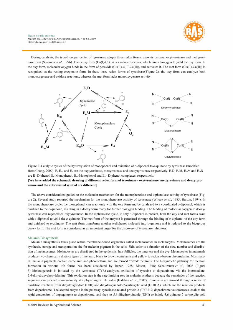
During catalysis, the type-3 copper center of tyrosinase adopts three redox forms: deoxytyrosinase, oxytyrosinase and mettyrosi-
nase form (Solomon et al., 1996). The deoxy form (Cu(I)-Cu(I)) is a reduced species, which binds dioxygen to yield the oxy form. In
the oxy form, molecular oxygen binds in the form of peroxide (Cu(II)-O22−-Cu(II)), and activates it. The met form (Cu(II)-Cu(II)) is
recognized as the resting enzymatic form. In these three redox forms of tyrosinase(Figure 2), the oxy form can catalyze both
monooxygenase and oxidase reactions, whereas the met form lacks monooxygenase activity.
The above considerations guided to the molecular mechanism for the monophenolase and diphenolase activity of tyrosinase (Fig-
ure 2). Several study reported the mechanism for the monophenolase activity of tyrosinase (Wilcox et al., 1985; Burton, 1994). In
the monophenolase cycle, the monophenol can react only with the oxy form and be catalyzed to a coordinated o-diphenol, which is
oxidized to the o-quinone, resulting in a deoxy form ready for further dioxygen binding. The binding of molecular oxygen to deoxy-
tyrosinase can regenerated oxytyrosinase. In the diphenolase cycle, if only o-diphenol is present, both the oxy and met forms react
with o-diphenol to yeild the o-quinone. The met form of the enzyme is generated through the binding of o-diphenol to the oxy form
and oxidized to o-quinone. The met form transforms another o-diphenol molecule into o-quinone and is reduced to the bicuprous
deoxy form. The met form is considered as an important target for the discovery of tyrosinase inhibitors.
Melanin Biosynthesis Melanin biosynthesis takes place within membrane-bound organelles called melanosomes in melanocytes. Melanosomes are the
synthesis, storage and transportation site for melanin pigment in the cells. Skin color is a function of the size, number and distribu-
tion of melanosomes. Melanocytes are distributed in the epidermis, hair follicles, the inner ear and the eye. Melanocytes in mammals
produce two chemically distinct types of melanin, black to brown eumelanin and yellow to reddish-brown pheomelanin. Most natu-
ral melanin pigments contain eumelanin and pheomelanin and are termed 'mixed' melanins. The biosynthetic pathway for melanin
formation in various life forms has been elucidated by Raper, 1928; Mason, 1948; Schallreuter et al., 2008 (Figure
3). Melanogenesis is initiated by the tyrosinase (TYR)-catalyzed oxidation of tyrosine to dopaquinone via the intermediate,
3,4-dihydroxyphenylalanine. This oxidation step is the rate-limiting step in melanin synthesis because the remainder of the reaction
sequence can proceed spontaneously at a physiological pH value (Halaban et al., 2002). Eumelanin are formed through a series of
oxidation reactions from dihydroxyindole (DHI) and dihydroxyindole-2-carboxylic acid (DHICA), which are the reaction products
from dopachrome. The second enzyme in the pathway, tyrosinase-related protein 2 (TYRP-2; dopachrome tautomerase), enables the
rapid conversion of dopaquinone to dopachrome, and then to 5,6-dihydroxyindole (DHI) or indole 5,6-quinone 2-carboxylic acid
Figure 2. Catalytic cycles of the hydroxylation of monophenol and oxidation of o-diphenol to o-quinone by tyrosinase (modified
from Chang, 2009). E, Em, and Ed are the oxytyrosinase, mettyrosinase and deoxytyrosinase respectively. EoD, EoM, EmM and EmD
are Eo-Diphenol, Eo-Monophenol, Em-Monophenol and Em- Diphenol complexes, respectively.
[We have added the schematic drawing of different redox form of tyrosinase - oxytyrosinase, mettyrosinase and deoxytyro-
sinase and the abbreviated symbol are different]

Please cite this article as
Masum et al., Reviews in Agricultural Science, 7:41-58, 2019 https://dx.doi.org/10.7831/ras.7.41
44 ©2019 Reviews in Agricultural Science
(DHICA). TYRP-1 (DHICA oxidase) then catalyzes the oxidation of DHICA to produce eumelanin. In the presence of cysteine or
glutathione, dopaquinone is converted to cysteinyldopa or glutathionyldopa and finally can produce pheomelanin. Although three
enzymes (TYR, TRP-1 and TRP-2) are involved in the melanogenesis pathway, only TYR is exclusively necessary as rate limiting
catalyst for melanogenesis.
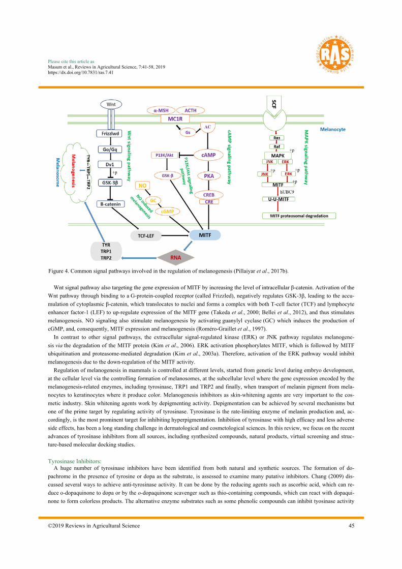
Signaling pathways to regulate melanogenesis Cutaneous pigmentation is under complex genetic control regulated by more than 150 alleles spread over 90 loci. Protein products
of these loci acting as enzymes, structural proteins, transcriptional regulators, transporters, receptors, and growth factors have a wide
array of functions and cellular targets (Silvers, 1979). UV exposure or other environmental stimulations, can triggered melanogene-
sis via different homones or cytokines. Figure 4 shows the most commonly known signal pathways involved in the regulation of
melanogenesis. All signaling pathways involve MITF, a master regulator of melanogenesis, which upregulates melanogenesis en-
zymes TYR, TRP-1 and TRP-2 via binding to the M-box motif in their promoter regions . The transcription factor MITF, which has
been reported to activate more than 25 genes in pigment cells, has emerged as an essential regulator not only for melanocyte devel-
opment, proliferation and survival, but also for the expression of enzymes and structural proteins ensuring the production of melanin
(Vachtenheim and Borovanský, 2010). The up-regulation or down-regulation of MITF activity activates or suppress the expression
of the melanogenesis-related enzymes, thus stimulating or inhibit melanogenesis.
Alpha melanocyte-stimulating hormone (α-MSH) regulates melanogenesis via cyclic adenosine monophosphate/protein kinase A
(cAMP/PKA) mediated pathway (Busca and Ballotti, 2000). When α-MSH binds to its receptor, melanocortin 1 receptor (MC1R),
on melanocyte membrane activates adenylate cyclase (AC) to produce cAMP, which activates protein kinase A (PKA)
by phosphorylation. Phosporylated PKA phosphorylates cAMP-response element binding (CREB) protein in the nucleus and in turn,
upregulates MITF (Gonzalez and Montminy, 1989). Ultimately, MITF efficiently activates the melanogenesis-related enzymes and
stimulates melanogenesis. β-MSH and adrenocorticotropic hormone (ACTH), also stimulate melanogenesis via the same pathway.
cAMP also affects the post-translational modification of MITF through inhibition of phosphatidylinositol 3-kinase (PI3K), which
results in reduction of Akt phosphorylation and thereby stimulating glycogen synthase kinase 3β (GSKβ) activity by reducing
p-GSKβ levels. The activated GSKβ, phosphorylates MITF and leading to stimulation of melanogenesis by upregution of
MITF (Saha et al., 2006).
Figure 3. Biosynthetic pathway of melanin (Yamauchi and Mitsunaga, 2016). Abbreviations:
TYR, tyrosinase; l-Dopa, l-3,4-dihydroxyphenylalanine; DHICA, 5,6-dihydroxyindole-2 carboxylic acid; DHI, 5,6-dihydroxyindole
Please cite this article as
Masum et al., Reviews in Agricultural Science, 7:41-58, 2019 https://dx.doi.org/10.7831/ras.7.41
©2019 Reviews in Agricultural Science 45
Wnt signal pathway also targeting the gene expression of MITF by increasing the level of intracellular β-catenin. Activation of the
Wnt pathway through binding to a G-protein-coupled receptor (called Frizzled), negatively regulates GSK-3β, leading to the accu-
mulation of cytoplasmic β-catenin, which translocates to nuclei and forms a complex with both T-cell factor (TCF) and lymphocyte
enhancer factor-1 (LEF) to up-regulate expression of the MITF gene (Takeda et al., 2000; Bellei et al., 2012), and thus stimulates
melanogenesis. NO signaling also stimulate melanogenesis by activating guanylyl cyclase (GC) which induces the production of
cGMP, and, consequently, MITF expression and melanogenesis (Roméro-Graillet et al., 1997).
In contrast to other signal pathways, the extracellular signal-regulated kinase (ERK) or JNK pathway regulates melanogene-
sis via the degradation of the MITF protein (Kim et al., 2006). ERK activation phosphorylates MITF, which is followed by MITF
ubiquitination and proteasome-mediated degradation (Kim et al., 2003a). Therefore, activation of the ERK pathway would inhibit
melanogenesis due to the down-regulation of the MITF activity.
Regulation of melanogenesis in mammals is controlled at different levels, started from genetic level during embryo development,
at the cellular level via the controlling formation of melanosomes, at the subcellular level where the gene expression encoded by the
melanogenesis-related enzymes, including tyrosinase, TRP1 and TRP2 and finally, when transport of melanin pigment from mela-
nocytes to keratinocytes where it produce color. Melanogenesis inhibitors as skin-whitening agents are very important to the cos-
metic industry. Skin whitening agents work by depigmenting activity. Depigmentation can be achieved by several mechanisms but
one of the prime target by regulating activity of tyrosinase. Tyrosinase is the rate-limiting enzyme of melanin production and, ac-
cordingly, is the most prominent target for inhibiting hyperpigmentation. Inhibition of tyrosinase with high efficacy and less adverse
side effects, has been a long standing challenge in dermatological and cosmetological sciences. In this review, we focus on the recent
advances of tyrosinase inhibitors from all sources, including synthesized compounds, natural products, virtual screening and struc-
ture-based molecular docking studies.
Tyrosinase Inhibitors: A huge number of tyrosinase inhibitors have been identified from both natural and synthetic sources. The formation of do-
pachrome in the presence of tyrosine or dopa as the substrate, is assessed to examine many putative inhibitors. Chang (2009) dis-
cussed several ways to achieve anti-tyrosinase activity. It can be done by the reducing agents such as ascorbic acid, which can re-
duce o-dopaquinone to dopa or by the o-dopaquinone scavenger such as thio-containing compounds, which can react with dopaqui-
none to form colorless products. The alternative enzyme substrates such as some phenolic compounds can inhibit tyosinase activity
Figure 4. Common signal pathways involved in the regulation of melanogenesis (Pillaiyar et al., 2017b).

Please cite this article as
Masum et al., Reviews in Agricultural Science, 7:41-58, 2019 https://dx.doi.org/10.7831/ras.7.41
46 ©2019 Reviews in Agricultural Science
through preventing the dopachrome formation. Some nonspecific enzyme inactivators such as acids or bases can also represent an-
ti-tyrosinase activity by denaturing the enzyme. The specific tyrosinase inactivators such as mechanism-based inhibitors, which can
inhibit tyrosinase activity by inducing the enzyme catalyzing ‘suicide reaction’. And finally the most significant, specific tyrosinase
inhibitors that reversibly bind to tyrosinase and reduce its catalytic capacity. Among them, only specific tyrosinase inactivators and
inhibitors are called as ‘true inhibitors’ because they actually bind to the enzyme and inhibit its activity. Based on inhibition mecha-
nism, true inhibitors are classified into four types: competitive, un-competitive, mixed type and non-competitive inhibitors.
Tyrosinase, the key regulatory enzyme of melanogenesis, is the prime target to inhibit melanin production. Commercially availa-
ble skin lighting agents are mostly tyrosinase inhibitors. Many tyrosinase inhibitors have been used as skin-whitening agents (Figure
5) but most of them contains specific drawbacks (Pillaiyar et al., 2017a). Hydroquinone, the most frequently prescribed ingredients
for skin-lighting agent have adverse effect such as skin irritation and burning, mutagenic to mammalian cells and cytotoxic to mela-
nocytes (Parvez et al., 2006; Curto et al., 1999). Kojic acid and arbutin also show poor efficacy in vivo, low formulation stability and
poor skin penetration (Hermanns et al., 2000). The use of kojic acid in skin treatment is limited due to its carcinogenicity (Fuyuno,
2004). Ascorbic acid is liable to degradation (Spínola et al., 2013) and the bioavailability of ellagic acid is poor (Arulmozhi et al.,
2013). Thus, it is in great need of developing new tyrosinase inhibitors from different sources with high efficacy and less adverse
side effect. Here, the tyrosinase inhibitors were mainly categorized into two parts, mushroom and human tyrosinase inhibitors and
their recent advances in both natural and synthetic sources are going to be discussed.
Mushroom tyrosinase inhibitors Kojic acid, a fungal metabolite used as skin-whitening agent is the most studied inhibitor of tyrosinase. Kojic acid can chelate
copper at the active site of the enzyme and show competitive inhibitory effect. The popular whitening agent kojic acid together with
arbutin, hydroquinone or tropolone used as a positive control to screen for finding inhibitors. In most of the study, the tyrosinase ac-
tivity is expressed as the half-maximal inhibitory concentration (IC50), which is the concentration of the samples producing 50% in-
hibition. B16F10 murine melanoma cells also frequently used for in vitro screening as they share most of the melanogenic mecha-
nisms of normal human melanocytes and they are relatively easy to culture in vitro. Recently, for in vitro screening, reconstituted
3-dimensional (3D) human skin equivalent model also used. Zebrafish used as an in vivo model to evaluate anti-pigmentary effect as
their genetic make-up and organ system are similar to human. Moreover, it’s easy to maintain and high efficacy in drug penetration
through skin.
There is an ongoing effort to search for tyrosinase inhibitors from natural sources particularly from plants as they are a rich source
of bioactive chemicals and are mostly free from harmful side effects. A number of research have been dedicated to identify tyrosi-
nase inhibitors from plants, fungal metabolites and marine algae. Polyphenols are widely distributed in nature and are the largest
groups in tyrosinase inhibitors. Best-studied polyphenols are flavonoids, that may be subdivided into seven major groups, including
Figure 5. Chemical structure of standard tyrosinase inhibitors as skin lightening agents

Please cite this article as
Masum et al., Reviews in Agricultural Science, 7:41-58, 2019 https://dx.doi.org/10.7831/ras.7.41
©2019 Reviews in Agricultural Science 47
flavones, flavonols, flavanones, flavanols, isoflavonoids, chalcones, and catechin. In addition to flavonoids, stilbenes and coumarin
derivatives; long-chain lipids and steroids; benzaldehyde and benzoate derivatives also identified as tyrosinase inhibitors. A large
number of compounds have been identified from the natural products and investigated for mushroom tyrosinase inhibitory activity;
these compounds differ from one another in the potency and type of inhibition imposed on the enzyme. Here listed some tyrosinase
inhibitors (Figure 6) identified from natural sources whose potency is equal to or better than the kojic acid, a well-known positive
control for tyosinase inhibitor.
The availability of efficient technology to rationally design efficient synthetic analogs, many synthetic tyrosinase inhibitors with
the novel structural moiety together with derivatives of natural compounds have been prepared. Lee et al. (2016) reviewed on natural,
semisynthetic and synthetic tyrosinase inhibitors, and mentioned that the development of many synthetic tyrosinase inhibitors (such
as N-hydroxy-N′-phenylthiourea, N-cyclopentyl-N-nitrosohydroxylamine, sildenafil methyl ether, 2,5-disubstituted-oxadiazoles, ox-
azolones, tetraketones, 4,4′-dihydroxybiphenyl, S-phenyl N-aryl S-alkylthiocarbamates, triazolothiadiazoles, benzaldehyde thio-
semicarbazones, 4-hydroxybenzaldehyde derivatives, bis-salicylaldehydes, phenylurenyl chalcones, polyphenolic curcumin deriva-
tives, bis-benzyl glycosides, rhodanine derivatives etc.) have been reported, and the properties of those synthetic inhibitors were
thoroughly reviewed by Loizzo et al.(2012) & Kim and Uyama (2005). Figure 7, showed some selective tyrosinase inhibitors from
synthetic sources whose potency is better than kojic acid in terms of IC50 value.
Carvacrol is a naturally occurring monoterpene phenol, having odoriferous and antimicrobial activities, used as meat preservatives
or flavoring agents in the food industry. Ashraf et al. (2017) synthesized carvacrol derivatives 1-6 (benzoic acid substituted)
and 7-10 (cinnamic acid substituted), with the aim to possess potent tyrosinase inhibitory activity (Figure 8). Among the deriva-
tivecompounds, 9(2-[2-methyl-5-(propan-2-yl)phenoxy]-2-oxoethyl(2E)-3-(2,4dihydroxyphenyl)prop-2-enoate) found to possess
Figure 6. Chemical structure of some potent tyrosinase inhibitors from natural sources

Please cite this article as
Masum et al., Reviews in Agricultural Science, 7:41-58, 2019 https://dx.doi.org/10.7831/ras.7.41
48 ©2019 Reviews in Agricultural Science
strongest anti-tyrosinase activity (IC50 0.0167μM) compare with standard kojic acid (IC50 16.69μM). The derivative com-
pounds 3 and 8 also showed good tyrosinase inhibitory activity with IC50 6.7 μM and 6.5 μM respectively. The kinetic analysis re-
vealed that compounds 3 and 8 showed mixed-type inhibition while 9 is a non-competitive inhibitor having Ki values 19μM, 10μM,
and 0.05μM respectively. Docking studies showed that compound 9 have maximum binding affinity against mushroom tyrosinase
with binding energy value (-7.90 kcal/mol) as compared to others. From structure–activity relationship, the hydroxy substituted de-
rivatives showed better tyrosinase inhibitory activity. The key factor of inhibitory activity is the substitution pattern of hydroxyl
groups at phenyl ring. The derivatives with hydroxy substituted cinnamic acid residue possess greater tyrosinase inhibitory potential
as compared to benzoic acids. The compound 9 exhibited excellent tyrosinase inhibitory activity (IC50 0.0167μM) bearing
2,4-dihydroxy substituted cinnamic acid residue. The study proposed that the hydroxy substitution pattern on phenyl ring in case of
compound 9 impedes the molecule to interact well with the active sites of enzyme.
Polyphenol compounds containing sulfur atom can interact with cupper ion in the active side of mushroom tyrosinase and inhibits
the activity. Chen et al. (2015) extracted bis(4-hydroxybenzyl)sulfide, from Gastrodia elata and found outstanding inhibitory po-
tency against mushroom tyrosinase (IC50 = 0.53 µM, Ki = 58 ± 6 nM), which is more effective than β-arbutin, kojic acid, short pep-
tides and other known natural compounds. Bis(4-hydroxybenzyl)sulfide (Figure 9A) may have function as a copper chelator to abol-
ish the tyrosinase activity and a strong competitive inhibitor of mushroom tyrosinase. In docking study, the sulfur atom of
bis(4-hydroxybenzyl)sulfide makes close contacts with the copper ions of tyrosinase. In addition, two hydrogen bonds were ob-
served between the hydroxyl groups of bis(4-hydroxybenzyl)sulfide and Asn260 and His224, respectively. The side chains of resi-
dues Glu256, Phe90, Val238, and Phe264 show tightly hydrophobic contacts and/or π-π interactions with the molecule.
Bis(4-hydroxybenzyl)sulfide adapts the same orientation as interacting with mushroom tyrosinase to interact with human tyrosinase
and more specific to mushroom instead of human tyrosinase. The in vitro and in vivo assay, reveals that this compound effectively
reduces melanogenesis without any adverse side effects and is free of discernable cytotoxicity in mice.
Tan et al. (2016), studied on tyrosinase inhibitory methanol extract of Campylotropis hirtella, and isolated fourteen phenolic
compounds, among which neorauflavane emerged as a lead structure for tyrosinase inhibition with IC50 in nanomolar concentration
(Figure 9C). Neorauflavane (16) inhibited monophenolase (IC50 = 30 nM), which was 400-fold more active than kojic acid
(IC50 = 13.2 μM). The geranylated isoflavanone (14) also inhibited monophenolase and diphenolase activity with IC50values 2.9 and
128.2 μM, respectively. Isoflavanone (17) bearing hydroxyl group on C-3 position inhibited tyrosinase efficiently with 18.4 μM of
Figure 7. Chemical structure of some potent synthetic tyrosinase inhibitors

Please cite this article as
Masum et al., Reviews in Agricultural Science, 7:41-58, 2019 https://dx.doi.org/10.7831/ras.7.41
©2019 Reviews in Agricultural Science 49
IC50. Compounds 14 and 16, with best candidate inhibitors among them, manifested competitive, simple reversible slow-binding in-
hibition against tyrosinase. Moreover, molecular docking analysis showesd that the interaction energies between the enzyme and two
inhibitors, compound 14 and 16, were −50.59 kcal/mol and −53.92 kcal/mol, respectively.
Figure 8. (A) Chemical structure of carvacrol derivatives 1-10
(B) Tyrosinase inhibitory activity of carvacrol derivatives 1-10
Figure 9. (A) Chemical structure of bis(4-hydroxybenzyl)sulfide isolated from Gastrodia elata
(B) Chemical structure of artocarpanone and artocaepin E isolated from Artocarpus heterophyllous
(C) Chemical structure and tyrosinase inhibitory activity of compounds 14-17 isolated from Campylotropis hirtella

Please cite this article as
Masum et al., Reviews in Agricultural Science, 7:41-58, 2019 https://dx.doi.org/10.7831/ras.7.41
50 ©2019 Reviews in Agricultural Science
Artocarpus heterophyllous (AH), popularly known as jackfruit; and is cultivated for its edible fruits, while the wood has been used
for its pharmacological values including anti-tyrosinase activities. Nguyen et al. (2016), reported that flavonoids isolated from
MeOH extract of the wood of AH showed potent tyrosinase inhibitory activity. They isolated seven compounds from AH which
were artocaepin E, artocaepin F, norartocarpetin, artocarpanone, liquiritigenin, steppogenin and dihydromorin with an IC50 of 6.7 ±
0.8, ˃50, ˃50, 2.0 ± 0.1, 22.0 ± 2.5, 7.5 ± 0.5 and ˃50 μM, respectively. Artocarpanone (Figure 9B) had the most potent tyrosinase
inhibitory effect, with an IC50 of 2.0 ± 0.1 μM, followed by artocaepin E, steppogenin and liquiritigenin, with IC50 values of
6.7 ± 0.8, 7.5 ± 0.5 and 22.0 ± 2.5 μM respectively, compare with kojic acid (IC50 44.6 ± 0.4 μM) as positive control. Struc-
ture–activity relationship showed that for artocaepin E , the presence of one hydroxyl group at C-2′ and a trans-p-coumaroyl unit
connected to the hydroxyprenyl through an ester linkage at C-6 of the flavone skeleton, led to significantly stronger inhibitory activ-
ity than that of norartocarpetin. So, the absence of the side-chain at C-6 of the B-ring leads to a significant loss of activity, and the
presence of a side-chain such as trans-p-coumaroyl connected to the hydroxyprenyl may positively influence the tyrosinase inhibi-
tory activity. For artocarpanone, which possesses a methoxyl group at C-7 of ring A, had the strongest inhibitory activity. Dihy-
dromorin which has four hydroxyl groups at C-2′, C-3, C-5, and C-7, had weak activity. The study suggested that the methoxyl and
hydroxyl groups in the main flavanone skeleton play an important role in tyrosinase inhibition.
Chaita et al.(2017), investigated 900 extracts from Greek plants for potential tyrosinase inhibition properties. Among the five
most potent extracts, the methanol extract of Morus alba wood (MAM) demonstrated a significant reduction in intracellular tyrosi-
nase and melanin content in B16F10 melanoma cells. Bioassay-guided isolation led to the acquisition of twelve compounds (Figure
10A). Among these, 2,4,3′-trihydroxydihydrostilbene(24) and dihydrooxyresveratrol(22) constitutes a potent tyrosinase inhibitor
with an IC50 0.8 ± 0.15 and IC50 0.3 ± 0.05 compare to kojic acid(IC50 16.1 ± 1.4). MAM extract and com-
pounds oxyresveratrol(18), trans-dihydromorin(23) and 2,4,3′-trihydroxydihydrostilbene(24) also significantly suppress in vivo
melanogenesis during zebrafish embryogenesis. Two dihydrostilbenes, dihydrooxyresveratrol(22) and
2,4,3′-trihydroxydihydrostilbene(24) showed the stronger inhibition than their stilbene analog, oxyresveratrol (IC50 1.7 μM). Study
suggested that the enhancement of the tyrosinase inhibitory properties may be responsible for the reduction of the double bond in the
stilbene structure, as the subsequent bibenzyl structure allows the molecule to approach and interact with the active center more ef-
fectively.
Condensed tannins are important class of plant-derived natural products and have potential to be leads for new drugs due to their
Figure 10. (A) Compounds isolated from Morus alba and their tyrosinase inhibitory activity
(B) Chemical structure of oxyresveratrol, dihydrooxyresveratrol, trans-dihydromorin and 2,4,3′-trihydroxydihydrostilbene isolated
from Morus alba
Please cite this article as
Masum et al., Reviews in Agricultural Science, 7:41-58, 2019 https://dx.doi.org/10.7831/ras.7.41
©2019 Reviews in Agricultural Science 51
biological activities such as antioxidant, anticancer, antimicrobial and anti-cardiovascular activities. Condensed tannins are com-
posed of flavan-3-ol sub-units linked mainly through C4→C8 (or C4→C6) bonds. Chen et al. (2014), isolated condensed tannins
from Ficus virens (F. virens) leaves, fruit, and stem bark. The extracts were prepared using acetone as a solvent. The condensed tan-
nins from different extracts of F. virens, were complex mixtures of homo- and heteropolymers of B-type procyanidins and prodel-
phinidins(11A) with degrees of polymerization up to hexamer, dodecamer, and pentadecamer, respectively. The condensed tannins
exhibited efficiency in inhibiting both mono- and diphenolase activities of the mushroom tyrosinase, and were found to be mixed
type inhibitors of the enzyme. The IC50 value for the leaves, fruit, and stem bark condensed tannins determined to 131.67, 99.89, and
106.22 μg/ml on monophenolase activity, and 128.42, 43.07, and 74.27 μg/ml on diphenolase activity (Figure 11B). Moreover, fluo-
rescence quenching, copper interacting, and molecular docking techniques were utilized to find out the molecular mechanisms of the
inhibition and the results showed that the inhibition was carried out mainly through the interaction of the hydroxyl groups in the ar-
omatic ring B of the condensed tannins with the active center of the enzyme by chelating dicopper ions. Additionally, the condensed
tannins could directly scavenge the o-quinones through oxidoreduction reaction. This study group suggested that, the condensed tan-
nins could be used to design and screen for potent novel tyrosinase inhibitors. Recently, another study (Chai et al., 2018) suggested
that condensed tannins from longan bark might be a good source of tyrosinase inhibitor and could be used as novel food preserva-
tives and medicines of skin diseases. Results showed good inhibitions on proliferation, intracellular enzyme activity and melano-
genesis of mouse melanoma cells.
Recently, Wang et al. (2018) studied on the inhibitory effect and mechanism of lignin on tyrosinase activity and founded lignin as
a novel tyrosinase inhibitor. In nature, lignin is the most abundant aromatic biopolymer. This study isolated six lignin samples by
alkali and ethanol organosolv processes from three typical lignocellulosic feedstocks. The ethanol organosolv lignins showed
stronger anti-tyrosinase activity than alkali lignins. For lignins from different sources, corn stalk lignin presented highest inhibitory
effect with an IC50 value of 0.276 mg/mL, compare to that of positive control p-hydroxy benzaldehyde (0.233 mg/mL). Moreover,
the kinetics study showed that the ethanol organosolv lignin from corn stalk was a reversible mixed-type inhibitor. This study group
suggested that lignin possesses antityrosinase activity and can be potentially used as an enzyme inhibitor in overtyrosinase activity
control fields.
Human tyrosinase inhibitors Tyrosinase inhibition is a well-established strategy for controlling melanin production in vivo and recently the development of
human tyrosinase inhibitors raises considerable interest in dermocosmetics. There is significant differences at cellular and structural
Figure 11. (A) Chemical structure of the condensed tannins (B) Inhibition constants of condensed tannins from the leaves, fruit, and
stem bark of F. virens. (Chen et al., 2017)

Please cite this article as
Masum et al., Reviews in Agricultural Science, 7:41-58, 2019 https://dx.doi.org/10.7831/ras.7.41
52 ©2019 Reviews in Agricultural Science
levels among the tyrosinases from distinct sources, especially plant, mushroom, bacterial, and human (Fogal et al., 2015). Mush-
room tyrosinase and bacterial tyrosinase are generally soluble oligomeric enzymes present in the cytosol, while human tyrosinase
exists as a highly glycosylated monomeric melanosomal transmembrane protein (van Gelder et al., 1997; Wang and Hebert, 2006).
There is a significant differences between mushroom and human tyrosinase enzyme in respect to the catalytic activities and substrate
specificities (Hearing et al., 1980). An abundant literature is dedicated to mushroom tyrosinase inhibitors for human applications but
unfortunately, currently used inhibitors lack the affinity and selectivity required for human tyrosinase targeting applications. Moreo-
ver, harmful toxicity has often been reported (Haudecoeur et al., 2016). Therefore, there is an urgent need for novel selective human
tyrosinase inhibitors that match efficacy and safety standards required for the development of products aimed to human use.
Recently, researchers are trying to identify tyrosinase inhibitors targeting human tyrosinase (hTYR). Pillaiyar et al.(2017b) re-
viewed on medicinal perspective of tyrosinase inhibitors, and discussed about the inhibitory potency and mechanisms of thujaplicins
(α, β and γ isomers), 4-butyl resorcinol, linderanolide B and subamolide A, as a human tyrosinase inhibitor. To find out novel human
tyrosinase inhibitor, Yoshimori et al., (2014) studied the inhibitory effects of three isomers of thujaplicin (α, β, and γ) on human ty-
rosinase and analyzed their binding modes using homology model and docking studies, and found that γ-thujaplicin
(IC50 = 1.15 μM) is the most potent inhibitor of human tyrosinase among thujaplicins compare to a well-known tyrosinase inhibitor
kojic acid (IC50 = 571.17 μM). Comparison of inhibitory activities of thujaplicins and kojic acid against hTYR and mTYR is illus-
trated in Figure 12A.
Wang et al.,(2011) isolated linderanolide B and subamolide A from the stems of Cinnamomum subavenium. Both of the com-
pounds exhibited mushroom tyrosinase inhibition and also non-cytotoxic to normal human skin cells and zebrafish sys-
tem. Linderanolide B and subamolide A (Figure 12B) reduced human tyrosinase activities (50%) at a dose of 1μm after 48h of
treatment and effectively inhibited the melanin production in HEMn�MP cells (40% reduction). Through molecular docking, in a
virtual model of human tyrosinase, it revealed that both of the compounds block copper ions within the tyrosinase active site. This
study suggested that linderanolide B and subamolide A are effective novel tyrosinase inhibitors to be considered as skin�lightening
agents.
In another study, An et al.(2010) compared the inhibitory effects of p�coumaric acid (p�CA), arbutin and kojic acid, on the cat-
alytic activities of mushroom, murine and human tyrosinases in vitro, using tyrosine and 3,4�dihydroxyphenylalanine as substrates.
The results showed that p�CA (Figure 12C) is a stronger inhibitor of human or murine than mushroom tyrosinase, in comparison
Figure 12. (A) Chemical structure and tyrosinase inhibitory activity of thujaplicin analagoues
(B) Chemical structure of Linderanolide B and Subamolide A
(C) Chemical structure of p�coumaric acid

Please cite this article as
Masum et al., Reviews in Agricultural Science, 7:41-58, 2019 https://dx.doi.org/10.7831/ras.7.41
©2019 Reviews in Agricultural Science 53
with kojic acid and arbutin as a positive control. Moreover, p�CA inhibited human TYR at much lower concentrations than those
required for the inhibition of murine or mushroom TYRs.
4-Butyl resorcinol, a resorcinol derivative that has an inhibitory effect on TYR and TRP-1 , was introduced in 1995 as a hy-
popigmenting agent (Okubo et al., 1995). Kolbe et al.(2013) examined the inhibitory effectsof4-butyl resorcinolonhTYRcompare
with kojic acid, hydroquinone and arbutin as positive control. 4�butylresorcinol with an IC50 of 21 μmol/L represented one of the
potent inhibitor of human tyrosinase. 4-Butyl resorcinol exhibited 20-times more-potent inhibitory activity than kojic acid, which
showed an IC50 of 500 μM. Arbutin and hydroquinone only weakly inhibit human tyrosinase with a IC50 in the millimolar range.
Aurones (2-benzylidenebenzofuran-3(2H)-ones), naturally occurring flavonoids act as inhibitors of melanin biosynthesis in human
melanocytes. It also act as effectors of mushroom tyrosinase and as molecular probes for the investigation of the binding-site struc-
tural homology between mushroom and bacterial tyrosinase (Haudecoeur et al., 2016). On the other hand, HOPNO, a cate-
chol-mimicking, nonoxidizable moiety, is a potent inhibitor of mushroom tyosinase (Ki = 1.8 μM). Haudecoeur et al.(2016) synthe-
sized aurone derivatives (36-38) whose B-ring replaced by a non-oxidizable 2-hydroxypyridine-N-oxide (HOPNO) moiety (Figure
13). The synthesized compounds tested for tyrosinase and melanogenesis inhibitory activity using purified human tyrosinase and
human melanoma MNT-1 cells, respectively, compare with kojic acid as positive control. The result showed that compounds 37 and
38 shared a similar inhibition potency (Ki = 1.02 and 1.2 μM, respectively), whereas analogue 36 (Ki = 0.35 μM) was found 3.5
times more active (Figure13). Compound 36 (IC50 = 16.6 μM) showed better efficiency in suppressing melanogenesis in MNT-1
cells compare to others. This study suggested that HOPNO-embedded 6-hydroxyaurone is one of the best effective inhibitor of iso-
lated human tyosinase.
Mann et al. (2018), used recombinant human tyrosinase to screen a library of 50,000 compounds and compared the active screen-
ing hits with well-known whitening ingredients. Among them, resorcinol derivatives thiamidol (isobutylamido thiazolyl resorcinol),
4-butylresorcinol, 4-hexylresorcinol, and 4-phenylethylresorcinol were the most promising inhibitors of the diphenolase (l-dopa ox-
idase) activity of human tyrosinase with IC50 values of 1.1 μmol/L, 21 μmol/L, 94 μmol/L, and 131 μmol/L, respectively (Figure 14).
Kojic acid, a well-known tyrosinase inhibotor was 500 times less potent than thiamidol, with an IC50 of about 500 μmol/L. But, thi-
amidol only weakly inhibited mushroom tyrosinase with an IC50 value of 108 μmol/L. kinetic analysis of the inhibition of human ty-
rosinase by thiamidol yielded a strictly competitive type of inhibition with an inhibitor constant (Ki) of 0.25 μmol/L, whereas the
Ki values for 4-butylresorcinol (9 μmol/L), 4-hexylresorcinol (39 μmol/L), and 4-phenylethylresorcinol (24 μmol/L) were markedly
higher than the Ki value of thiamidol. Thiamidol strongly and reversibly inhibited melanin production (IC50 = 0.9 μmol/L) in Mela-
noDerm skin models. In monolayer cultures, it visibly reduced melanin formation. 4-Butylresorcinol and hydroquinone inhibited
Figure 13. Chemical structure and tyrosinase inhibitory activity of aurones derivatives

Please cite this article as
Masum et al., Reviews in Agricultural Science, 7:41-58, 2019 https://dx.doi.org/10.7831/ras.7.41
54 ©2019 Reviews in Agricultural Science
melanin synthesis in MelanoDerm skin models with an IC50 of 13.5 and 15 μmol/L, respectively. In
long-term melanocyte monolayer cultures, thiamidol (1μmol/L) also reduced melanin production to less than 60% after 2 weeks.
Virtual docking studies revealed the possible binding modes of thiamidol to human tyrosinase, and found a strong hydrophobic sub-
pocket mainly by the side chains of I368, V377, and F347. The 1-hydroxy group of the aromatic ring of thiamidol makes extensive
contacts with the di-copper center of human tyrosinase, and the 3-hydroxy group is involved in hydrogen bonds with the side chain
of S380. To examine the in vivo efficacy of thiamidol, a clinical study was also carried out. The elderly subjects treated age spots
on their skin twice daily with a formula containing 0.2% thiamidol or with the vehicle only as a control. After 4 weeks of treatment,
the treated age spots were significantly lighter than the untreated control age spots and more improvement continued over the 12
weeks of treatment. A follow-up study showed that concentrations of thiamidol as low as 0.1% effectively reduced the visibility of
age spots. From this study it was found that thiamidol (isobutylamido thiazolyl resorcinol) was a potent inhibitor of human tyrosi-
nase with a remarkable efficacy in vitro and in vivo. But the efficacy is distinctively different in mushroom tyrosinase,
where 4-butylresorcinol, 4-hexylresorcinol, and 4-phenylethylresorcinol, and even kojic acid, are superior to thiamidol in inhibiting
the enzyme (Figure 14). The 4-substituted resorcinol motif has been known for an efficient chemical moiety that inhibits tyrosinase
activity. From structure-activity relationship, the thiazolylamide moiety of thiamidol conveys a much better inhibition of human ty-
rosinase than do the hydrocarbon side chains present in three other derivatives of resorcinol (4-butyl-, 4-hexyl-, and 4-phenyl ethyl-
resorcinol).
Conclusions The impact of cosmetic skin concerns on quality of life and the abnormal pigmentation causes serious esthetic problems as well as
life threating diseases. Hyperpigmentation is an important issue and the most prominent target for inhibiting hyperpigmentation is
tyrosinase, the rate-limiting enzyme in melanogenesis. Tyrosinase inhibitors have a huge demand in cosmetic and medicinal indus-
tries due to their preventive effect on pigmentation disorders as well as skin-whitening effect. A number of tyrosinase inhibitors have
been identified from both natural and synthetic sources, but only a few of them are used as skin-whitening agents, primarily due to
various safety concerns such as cytotoxicity, solubility, effective cutaneous absorption, etc. Many tyrosinase inhibitors presently
used as skin-whitening agents have found specific drawbacks including carcinogenicity. According to the WHO, skin lighteners are
found to cause cancers in human, as well as some manufacturers employ harmful toxins such as mercury in skin lightening products
to block melanin production (www.globenewswire.com). So, consumers are now inclining towards adoption of organic and natural
products. Moreover, there is a significant differences between mushroom and human tyrosinase enzyme in respect to the catalytic
activities and substrate specificities. Currently used inhibitors that was identified using mushroom tyrosinase, lack the affinity and
selectivity required for human tyrosinase. Therefore, there is an urgent need for novel selective human tyrosinase inhibitors that
Figure 14. Chemical structure and tyrosinase inhibitory activity of thiamidol and resorcinol derivatives

Please cite this article as
Masum et al., Reviews in Agricultural Science, 7:41-58, 2019 https://dx.doi.org/10.7831/ras.7.41
©2019 Reviews in Agricultural Science 55
match efficacy and safety standards required for the development of products aimed to human use including validation of
skin-whitening efficacy. So, more concrete studies with a human clinical point of view are required for the found inhibitors.
REFERENCES An SM, Koh JS and Boo YC (2010) p-coumaric acid not only inhibits human tyrosinase activity in vitro but also melanogenesis in
cells exposed to UVB. Phytherapy Research, 24:1175-1180
Ando H, Kondoh H, Ichihashi M and Hearing VJ (2007) Approaches to identify inhibitors of melanin biosynthesis via the quality
control of tyrosinase. J. Investig. Dermatol 127: 751–761
Arulmozhi V, Pandian K and Mirunalini S (2013) Ellagic acid encapsulated chitosan nanoparticles for drug delivery system in hu-
man oral cancer cell line (KB). Colloids Surf B Biointerfaces, 110:313–20
Ashraf Z, Rafiq M, Nadeem H, Hassan M, Afzal S, Waseem M, Afzal K and Latip J (2017) Carvacrol derivatives as mushroom ty-
rosinase inhibitors; synthesis, kinetics mechanism and molecular docking studies. PLoS One., 12(5):e0178069
Available from: https://www.factmr.com/report/309/skin-lightening-products-market
Aavailable from:
https://globenewswire.com/news-release/2018/07/10/1535161/0/en/Key-Insights-on-Skin-Lightening-Products-Market-through-2
022-by-Fact-MR.html
Baek SH, Nam IJ, Kwak HS, Kim KC and Lee SH (2015) Cellular Anti-Melanogenic Effects of a Euryale ferox Seed Extract Ethyl
Acetate Fraction via the Lysosomal Degradation Machinery. Int. J. Mol. Sci 16:9217-9235
Bellei B, Pitisci A, Izzo E and Picardo M (2012) Inhibition of melanogenesis by the pyridinyl imidazole class of compounds: Possi-
ble involvement of the Wnt/beta-catenin signaling pathway. PLoS One., 7:e33021
Bourquelot E and Bertrand A (1895) Le bleuissement et le noircissement des champignons. Comp Rend Soc Biol, 2:582–4
Briganti S, Camera E and Picardo M (2003) Chemical and instrumental approaches to Hyperpigmentation. Pigment. Cell Res.,
16:101-110
Burton SG (1994) Biocatalysis with polyphenol oxidase: a review. Catal. Today, 22: 459–487
Busca R and Ballotti R (2000) Cyclic AMP a key messenger in the regulation of skin pigmentation. Pigment Cell Res., 13:60–69
Chai WM, Huang Q, Lin MZ, Ou-Yang C, Huang WY, Wang YX, Xu KL and Feng HL (2018) Condensed Tannins from Longan
Bark as Inhibitor of Tyrosinase: Structure, Activity, and Mechanism. J. Agric. Food Chem., 66: 908–917
Chaita E, Lambrinidis G, Cheimonidi C, Agalou A, Beis D, Trougakos I, Mikros E, Skaltsounis AL and Aligiannis N (2017) An-
ti-Melanogenic Properties of Greek Plants. A Novel Depigmenting Agent from Morus alba Wood. Molecules, 22: 514
Chang TS, Ding HY, Tai SSK and Wu CY (2007) Mushroom tyrosinase inhibitory effects of isoflavones isolated from soygerm
koji fermented with Aspergillus oryzae BCRC 32288. Food Chem, 105:1430–1438
Chang TS (2009) An Updated Review of Tyrosinase Inhibitors. Int J Mol Sci., 10: 2440–2475
Chen WC, Tseng TS, Hsiao NW, Lin YL, Wen ZH, Tsai CC, Lee YC, Lin HH and Tsai KC(2015) Discovery of Highly Potent Ty-
rosinase Inhibitor, T1, with Significant Anti-Melanogenesis Ability by zebrafish in vivo Assay and Computational Molecular
Modeling. Sci Rep., 5:7995
Chen XX, Shi Y, Chai WM, Feng HL, Zhuang JX and Chen QX (2014) Condensed Tannins from Ficus virens as Tyrosinase Inhib-
itors: Structure, Inhibitory Activity and Molecular Mechanism. PLOS, 9: e91809. https://doi.org/10.1371/journal.pone.0091809
Choudhary MI, Sultan S, Khan MT and Rahman AU (2005) Microbial transformation of 17alpha-ethynyl- and
17alpha-ethylsteroids, and tyrosinase inhibitory activity of transformed products. Steroids, 70:798–802
Criton M and Le Mellay-Hamon V (2008) Analogues of N-hydroxy-N′-phenylthiourea and N-hydroxy-N′-phenylurea as inhibitors
of tyrosinase and melanin formation. Bioorg. Med. Chem. Lett., 18:3607–3610
Curto EV, Kwong C, Hermersdörfer H, Glatt H, Santis C, Virador V, Hearing VJ Jr and Dooley TP (1999) Inhibitors of mammalian
melanocyte tyrosinase: in vitro comparisons of alkyl esters of gentisic acid with other putative inhibitors. Biochem Pharmacol,
57: 663–672
Decker H, Schweikardt T and Tuczek F (2006) The first crystal structure of tyrosinase: all questions answered?. Angew Chem Int
Ed, 45:4546–4550
Duckworth H W and Coleman J E (1970) Physicochernical and Kinetic properties of mushroom tyrosinase. The Jour.of Bio. Che,
245: 1613-1625
Fogal S, Carotti M, Giaretta L, Lanciai F, Nogara L, Bubacco L and Bergantino E (2015) Human tyrosinase produced in insect
cells: a landmark for the screening of new drugs addressing its activity. Mol. Biotechnol., 57: 45−57
Please cite this article as
Masum et al., Reviews in Agricultural Science, 7:41-58, 2019 https://dx.doi.org/10.7831/ras.7.41
56 ©2019 Reviews in Agricultural Science
Fuyuno I (2004) Spotlight turns on cosmetics for Asian skin. Nature, 432: 938, 10.1038/432938a
Gonzalez G A and Montminy M R (1989) Cyclic AMP stimulates somatostatin gene transcription by phosphorylation of CREB at
serine 133. Cell, 59 :675-680
Halaban R, Patton RS, Cheng E, Svedine S, Trombetta ES, Wahl ML, Ariyan S and Hebert DN (2002) Abnormal acidification of
melanoma cells induces tyrosinase retention in the early secretory pathway. J. Biol. Chem., 277:14821–14828
Han YK, Park YJ, Ha YM, Park D, Lee JY, Lee N, Yoon JH, Moon HR and Chung HY (2012) Characterization of a novel tyrosi-
nase inhibitor, (2RS,4R)-2-(2,4-dihydroxylphenyl) thiazolidine-4-carboxylic acid (MHY384). Biochim Biophys Ac-
ta, 1820:542–549
Haudecoeur R, Carotti M, Gouron A, Maresca M, Buitrago E, Hardré R, Bergantino E, Jamet H, Belle C, Réglier M
(2016) 2-Hydroxypyridine-N-oxide-Embedded Aurones as Potent Human Tyrosinase Inhibitors. ACS Med. Chem. Lett., 8:
55–60
Hearing VJ Jr, Ekel TM, Montague PM and Nicholson JM (1980) Mammalian tyrosinase. Stoichiometry and measurement of reac-
tion products. Biochim Biophys Acta, 611: 251-268
Hermanns JF, Pierard-Franchimont C and Pierard GE (2000) Skin colour assessment in safety testing of cosmetics. An over-
view. Int J Cosmet Sci, 22: 67–71
Jeong SH, Ryu YB, Curtis-Long MJ, Ryu HW, Baek YS, Kang JE, Lee WS and Park KH (2009) Tyrosinase Inhibitory Polyphenols
from Roots of Morus lhou. J. Agric. Food Chem., 57:1195–1203
Jun N, Hong G and Jun K (2007) Synthesis and evalution of 2′,4′,6′-trihydroxychalcones as a new class of tyrosine inhibi-
tors. Bioorg. Med. Chem., 15:2396–2402
Kang HS, Choi JH, Cho WK, Park JC and Choi JS (2004) A sphingolipid and tyrosinase inhibitors from the fruiting body
of Phellinus linteus. Arch. Pharm. Res., 27:742–750
Khan SB, Azhar-Ul-Haq, Afza N, Malik A, Khan MT, Shah MR and Choudhary MI (2005) Tyrosinase-inhibitory long-chain esters
from Amberboa ramosa. Chem. Pharm. Bull., 53:86–89
Kim DS, Hwang ES, Lee JE, Kim SY, Kwon SB and Park KC (2003a) Sphingosine-1-phosphate decreases melanin synthe-
sis via sustained ERK activation and subsequent MITF degradation. J. Cell Sci., 116:1699–1706
Kim DS, Park SH, Kwon SB, Park ES, Huh CH, Youn SW and Park KC (2006) Sphingosylphosphorylcholine-induced ERK activa-
tion inhibits melanin synthesis in human melanocytes. Pigment Cell Res., 19:146–153
Kim JH, Baek SH, Kim DH, Choi TY, Yoon TJ, Hwang JS, Kim MR, Kwon HJ and Lee CH (2008) Downregulation of melanin
synthesis by haginin A and its application to in vivo lightening model. J. Invest. Dermatol., 128:1227–1235
Kim HJ, Seo SH, Lee BG, Lee YS (2005a) Identification of tyrosinase inhibitors from Glycyrrhiza uralensis. Planta Med.,
71:785–787
Kim SJ, Son KH, Chang HW, Kang SS and Kim HP (2003b) Tyrosinase inhibitory prenylated flavonoids from Sophora fla-
vescens. Biol. Pharm. Bull., 26:1348–1350
Kim YJ and Uyama H (2005) Tyrosinase inhibitors from natural and synthetic sources: structure, inhibition mechanism and per-
spective for the future. Cell Mol Life Sci., 62:1707-23
Kim YJ, No JK, Lee JH and Chung HY (2005b) 4,4′-Dihydroxybiphenyl as a new potent tyrosinase inhibitor. Biol Pharm
Bull, 28:323–7
Kim YM, Yun J, Lee CK, Lee H, Min KR and Kim Y (2002) Oxyresveratrol and hydroxystilbene compounds. Inhibitory effect on
tyrosinase and mechanism of action. J. Biol. Chem., 277:16340–16344
Kobayashi T, Urabe K, Winder A, Jimenez-Cervantes C, Imokawa G, Brewington T, Solano F, Garcia-Borron JC and Hearing VJ
(1994) Tyrosinase related protein 1(TRP1) functions as DHICA oxidase in melanin biosynthesis. EMBO J 13:5818–5825
Kolbe L, Mann T, Gerwat W, Batzer J, Ahlheit S, Scherner C, Wenck H and Stäb F (2013) 4-n-butylresorcinol, a highly effective
tyrosinase inhibitor for the topical treatment of hyperpigmentation. J. Eur. Acad. Dermatol. Venereol., 1:19-23
Lam KW, Syahida A, Ul-Haq Z, Abdul Rahman MB and Lajis NH (2010) Synthesis and biological activity of oxadiazole and tria-
zolothiadiazole derivatives as tyrosinase inhibitor. Bioorg Med Chem Lett, 20:3755–3759
Lee KH, Koketsu M, Choi SY, Lee KJ, Lee P, Ishihara H and Kim SY (2005) Potent inhibitory effects
of N-aryl S-alkylthiocarbamate derivatives on the dopa oxidase activity of mushroom tyrosinase. Chem Pharm Bull, 53:747–749
Lee SY, Baek N and Nam TG (2016) Natural, semisynthetic and synthetic tyrosinase inhibitors. J Enzyme Inhib Med Chem.,
31:1-13
Please cite this article as
Masum et al., Reviews in Agricultural Science, 7:41-58, 2019 https://dx.doi.org/10.7831/ras.7.41
©2019 Reviews in Agricultural Science 57
Likhitwitayawuid K, Sornsute A, Sritularak B and Ploypradith P (2006) Chemical transformations of oxyresveratrol
(trans-2,4,3′,5′-tetrahydroxystilbene) into a potent tyrosinase inhibitor and a strong cytotoxic agent. Bioorg. Med. Chem.
Lett., 16:5650–5653
Loizzo MR, Tundis R and Menichini F (2012) Natural and synthetic tyrosinase inhibitors as antibrowning agents: an update. Compr
Rev Food Sci Food Safety, 11:378–398
Mann T, Gerwat W, Batzer J, Eggers K, Scherner C, Wenck H, Stäb F, Hearing VJ, Röhm KH and Kolbe L (2018) Inhibition of
Human Tyrosinase Requires Molecular Motifs Distinctively Different from Mushroom Tyrosinase. J. Inv. Der., 138: 1601-1608
Mason HS (1948) The chemistry of melanin. III. Mechanism of the oxidation of trihydroxyphenylalanine by tyrosinase. J. Biol.
Chem., 172:83–99
Matoba Y, Bando N, Oda K, Noda M, Higashikawa F, Kumagai T and Sugiyama M (2011) A Molecular Mechanism for Copper
Transportation to Tyrosinase That Is Assisted by a Metallochaperone, Caddie Protein. J Biol Chem. 286:30219-31
Miyazawa M and Tamura N (2007) Inhibitory compound of tyrosinase activity from the sprout of Polygonum hydropiper L.
(Benitade). Biol. Pharm. Bull., 30:595–597
Nguyen HX, Nguyen NT, Nguyen MH, Le TH, Van Do TN, Hung TM and Nguyen MT (2016) Tyrosinase inhibitory activity of
flavonoids from Artocarpus heterophyllous. Che. Cen. J., 10:2
Ohguchi K, Tanaka T, Iliya I, Ito T, Iinuma M, Matsumoto K, Akao Y and Nozawa Y (2003) Gnetol as a potent tyrosinase inhibitor
from genus Gnetum. Biosci. Biotechnol. Biochem., 67:663–665
Okubo T, Oyohikawa M, Futaki K, Matsukami M and Fujii A (1995) The inhibitory effects of 4-N-butyl-resorcinol on melanogene-
sis. J. Dermatol. Sci., 10: 88
Park JW, Ha YM, Moon KM, Kim SR, Jeong HO, Park YJ, Lee HJ, Park JY, Song YM, Chun P (2013) De novo tyrosinase inhibi-
tor: 4-(6,7-dihydro-5H-indeno[5,6-d]thiazol-2-yl)benzene-1,3-diol(MHY1556). Bioorg Med Chem Lett, 23:4172–4176
Parvez S, Kang M, Chung HS, Cho C, Hong MC, Shin MK and Bae H (2006) Survey and mechanism of skin depigmenting and
lightening agents. Phytother Res, 20dy
Pawelek JM & Korner AM (1982) The biosynthesis of mammalian melanin. Am Sci 70:136–145
Piao XL, Baek SH, Park MK and Park JH (2004) Tyrosinase-inhibitory furanocoumarin from Angelica dahurica. Biol. Pharm.
Bull., 27:1144–1146
Pillaiyar T, Manickam M and Namasivayam V (2017a) Skin whitening agents: medicinal chemistry perspective of tyrosinase in-
hibitors. Jour. of Enz. Inhibition and Medicinal Che., 32: 403-425
Pillaiyar T, Manickam M and Jung SH (2017b) Downregulation of melanogenesis: drug discovery and therapeutic options. Drug
Discovery Today., 22: 282-298
Ramsden CA and Riley PA (2014) Tyrosinase: The four oxidation states of the active site and their relevance to enzymatic activa-
tion, oxidation and inactivation. Bioorganic & Medicinal Chemistry, 22:82388-2395
Raper HS (1928) The anaerobic oxidases. Physiol. Rev., 8:245–282
Roméro-Graillet C, Aberdam E, Clément M, Ortonne J P and Ballotti R (1997) Nitric oxide produced by ultraviolet-irradiated
keratinocytes stimulates melanogenesis. J. Clin. Invest., 99:635-642
Ryu YB, Ha TJ, Curtis-Long MJ, Ryu HW, Gal SW and Park KH (2008) Inhibitory effects on mushroom tyrosinase by flavones
from the stem barks of Morus lhou (S.) Koidz. J. Enzyme Inhib. Med. Chem., 23:922–930
Saha B, Singh SK, Sarkar C, Bera R, Ratha J, Tobin DJ and Bhadra R (2006) Activation of the Mitf promoter by lipid-stimulated
activation of p38-stress signaling to CREB. Pigment. Cell Res., 19: 595-605
Salsberg JM, Weinstein M, Shear N, Lee M and Pope E (2016) Impact of Cosmetic Camouflage on the Quality of Life of Children
with Skin Disease and Their Families. J Cutan Med Surg 20:211-215
Schallreuter KU, Kothari S, Chavan B and Spencer JD (2008) Regulation of melanogenesis-controversies and new concepts. Exp.
Dermatol., 17:395–404
Silvers WK (1979) The Coat Colors of Mice. A Model for Mammalian Gene Action and Interaction. New York: Springer Verlag.
Solomon EI, Sundaram UM and Machonkin TE (1996) Multicopper Oxidases and Oxygenases. Chem. Rev., 96:2563–2606
Spínola V, Mendes B, Câmara JS and Castilho PC (2013) Effect of time and temperature on vitamin C stability in horticultural ex-
tracts. UHPLC-PDA vs. iodometric titration as analytical methods. LWT-Food Sci Technol, 50:489–495
Takeda K, Yasumoto K, Takada R, Takada S, Watanabe K, Udono T, Saito H, Takahashi K and Shibahara S (2000) Induction of
melanocyte-specific microphthalmiaassociated transcription factor by Wnt-3a. J. Biol. Chem., 275:14013–14016
Please cite this article as
Masum et al., Reviews in Agricultural Science, 7:41-58, 2019 https://dx.doi.org/10.7831/ras.7.41
58 ©2019 Reviews in Agricultural Science
Tan X, Song YH, Park C, Lee KW, Kim JY, Kim DW, Kim KD, Lee KW, Curtis-Long MJ and Park KH (2016) Highly potent ty-
rosinase inhibitor, neorauflavane from Campylotropis hirtella and inhibitory mechanism with molecular docking. Bioorg Med
Chem., 24: 153-159
Tepper AWJW, Lonardi E, Bubacco L and Canters GW (2011) Structure, Spectroscopy, and Function of Tyrosinase; Comparison
with Hemocyanin and Catechol Oxidase. https://doi.org/10.1002/9781119951438.eibc0683
Ullah F, Hussain H, Hussain J, Bukhari IA, Khan MT, Choudhary MI, Gilani AH and Ahmad VU (2007) Tyrosinase inhibitory
pentacyclic triterpenes and analgesic and spasmolytic activities of methanol extracts of Rhododendron collettianum. Phytother.
Res., 21:1076–1081
Vachtenheim J and Borovanský J (2010) "Transcription physiology" of pigment formation in melanocytes: central role of MITF.
Exp Dermatol., 19:617-27
Van Gelder CW, Flurkey WH and Wichers HJ (1997) Sequence and structural features of plant and fungal tyrosi-
nases. Phytochemistry 45:1309–1323
Wang G, Xia Y, Sui W and Si C (2018) Lignin as a Novel Tyrosinase Inhibitor: Effects of Sources and Isolation Processes. ACS
Sustainable Chem. and Eng., 6: 9510–9518
Wang HM, Chen CY and Wen ZH (2011) Identifying melanogenesis inhibitors from cinnamomum subavenium with in vitro and in
vivo screening systems by targeting the human tyrosinase. Exp Dermatol, 20:242–8
Wang N and Hebert DN (2006) Tyrosinase maturation through the mammalian secretory pathway: bringing color to life. Pigm. Cell
Res., 19: 3−18
Wilcox DE, Porras AG, Hwang YT, Lerch K, Winkler ME and Solomon EI (1985) Substrate analogue binding to the coupled binu-
clear copper active site in tyrosinase. J. Am. Chem. Soc., 107: 4015–4027
Yamauchi K, Mitsunaga T and Batubara I (2011) Isolation, Identification and Tyrosinase Inhibitory Activities of the Extractives
from Allamanda cathartica. Natural Resources 2: 167-172
Yamauchi K and Mitsunaga T (2016) Melanogenesis and Melanosome Transportation Modulators from Medicinal Plants. Letters in
Drug Design & Discovery, 13:742-751
Yokoyama K, Suzuki H, Yasumoto K, Tomita Y and Shibahara S (1994) Molecular cloning and functional analysis of cDNA cod-
ing for human DOPAchrome tautomerase/tyrosinase-related protein-2. Biochim. Biophys. Acta., 1217:317–321
Yoshimori A, Oyama T, Takahashi S, Abe H, Kamiya T, Abe T and Tanuma S (2014) Structure–activity relationships of the thu-
japlicins for inhibition of human tyrosinase. Bioorg. Med. Chem., 22:6193-6200
Zhang X, Hu X, Hou A and Wang H (2009) Inhibitory effect of 2,4,2′,4′-tetrahydroxy-3-(3-methyl-2-butenyl)-chalcone on tyrosi-
nase activity and melanin biosynthesis. Biol. Pharm. Bull., 32:86–90