types and classification of fractures
TRANSCRIPT
Fracture Description & Classification
By Daaneyal J. Dilawar
Anatomic description of fractures
Type Comminution Location Displacement

Anatomic description - Type
Type is the overall fracture pattern
Examples are: – Simple
– Spiral
– segmental

Anatomic description - Communition
Comminution is the measure of the number of pieces of broken bone that there are.
Examples are: non-comminuted or mildly comminuted or severely comminuted
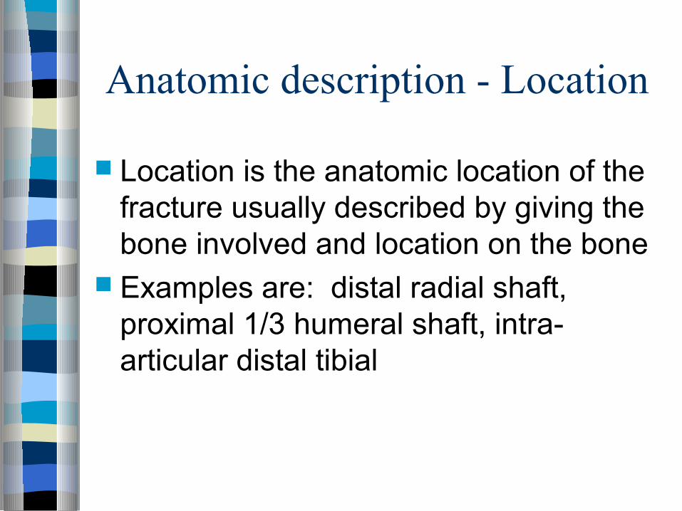
Anatomic description - Location
Location is the anatomic location of the fracture usually described by giving the bone involved and location on the bone
Examples are: distal radial shaft, proximal 1/3 humeral shaft, intra-articular distal tibial

Anatomic description - Displacement
Displacement is the amount the pieces of a fracture have moved from their normal location
Can be displaced or non-displaced Subdivided into 3 sub-categories:
translation, angulation, and shortening
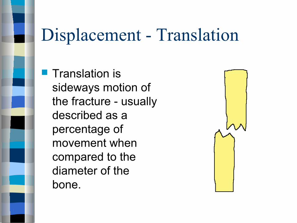
Displacement - Translation
Translation is sideways motion of the fracture - usually described as a percentage of movement when compared to the diameter of the bone.
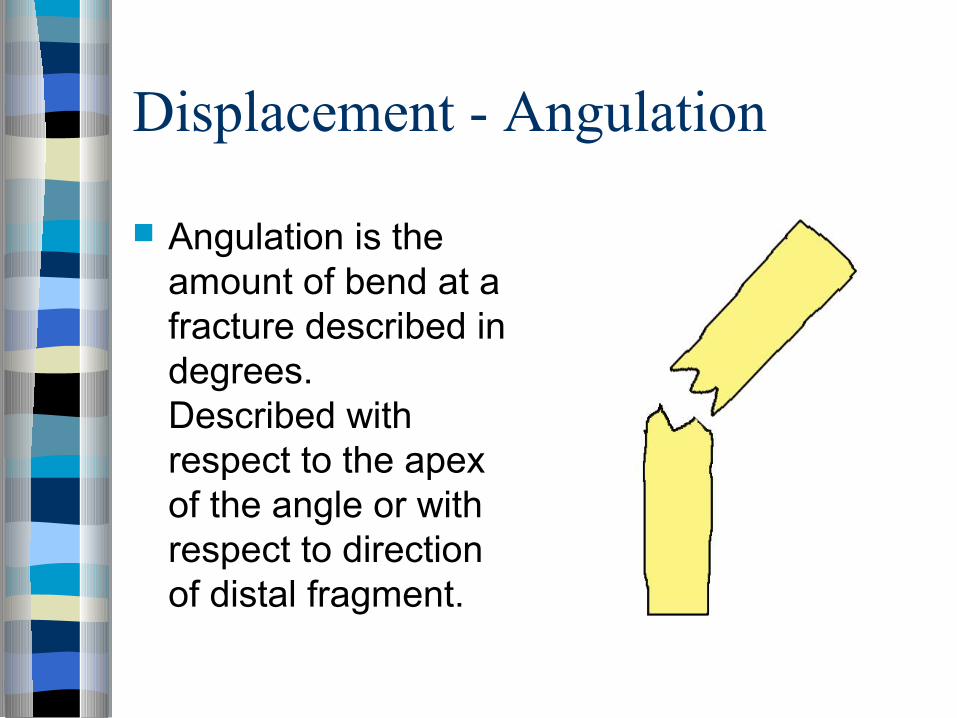
Displacement - Angulation
Angulation is the amount of bend at a fracture described in degrees. Described with respect to the apex of the angle or with respect to direction of distal fragment.

Displacement - Shortening
Shortening is the amount a fracture is collapsed expressed in centimeters. Sometimes called bayonette apposition.
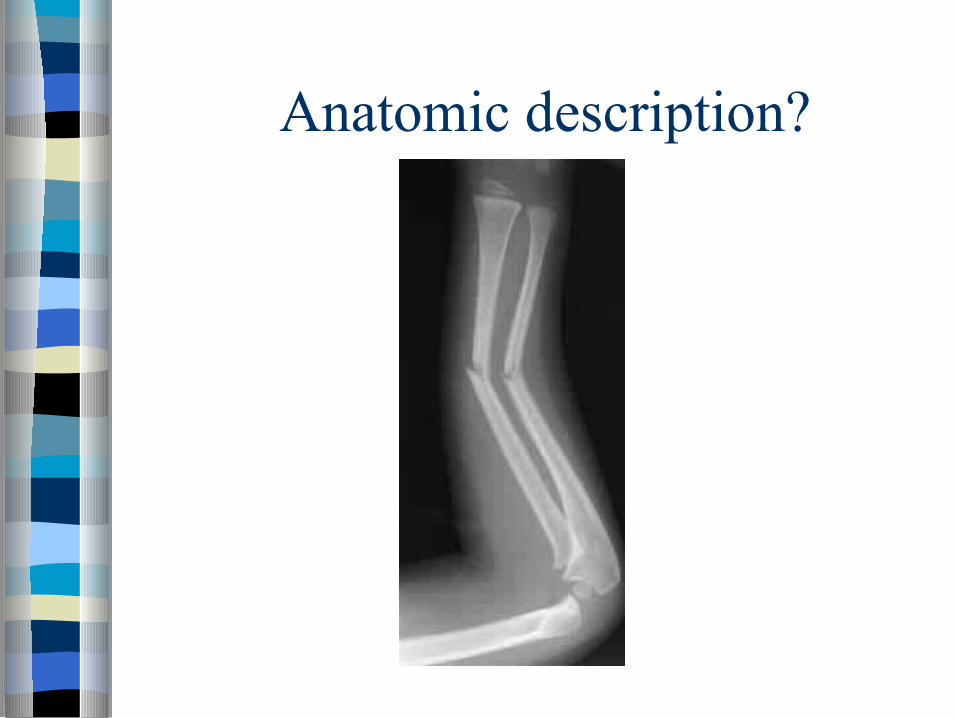
Anatomic description?

Anatomic description
Simple, transverse, non-communited midshaft radial and ulnar fracture with 30 degrees apex radial angulation.

Anatomic description??
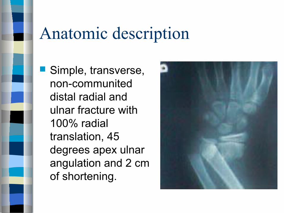
Anatomic description
Simple, transverse, non-communited distal radial and ulnar fracture with 100% radial translation, 45 degrees apex ulnar angulation and 2 cm of shortening.

Why classify fractures?
Classification or description of fractures is only used when the classification or description is useful in providing treatment or outcomes
Types of classifications
Anatomic description AO classification Salter-Harris classification Gustillo open fracture classification Fracture specific classifications
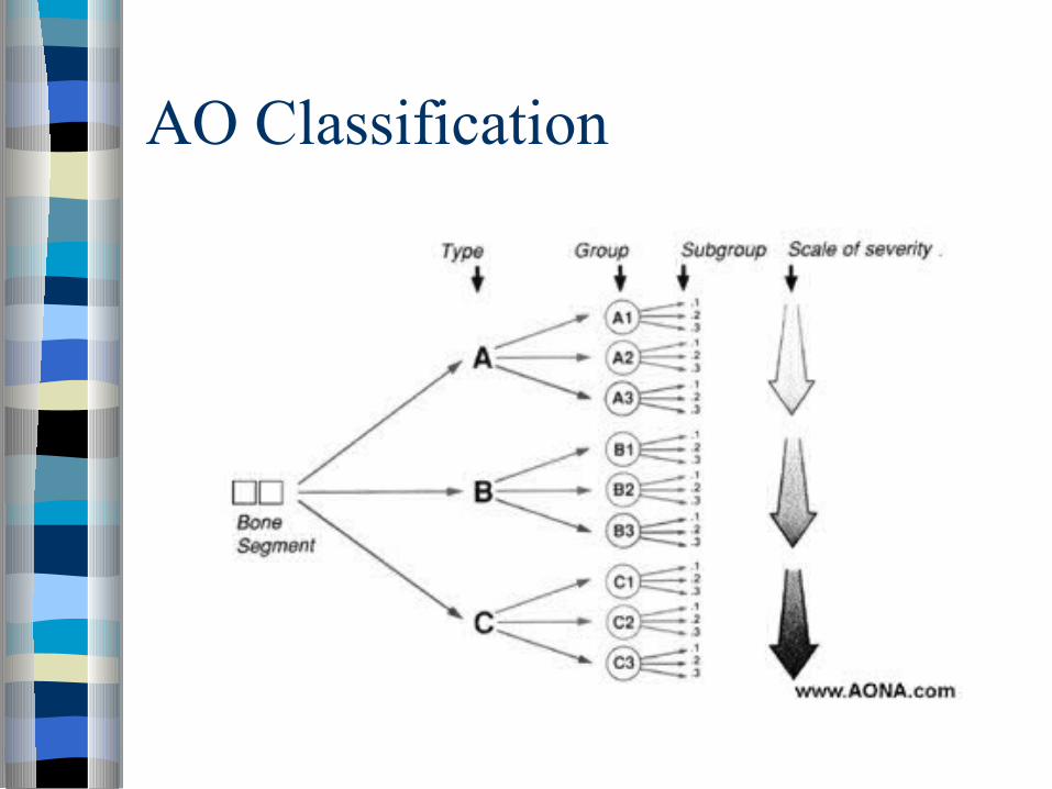
AO Classification

AO Classification
1st number = long bone 2nd number = bone segment Letter = fracture type (A,B,C) Then 3rd & 4th numbers classify fracture group
& subgroup

Salter-Harris Classification
Only used for pediatric fractures that involve the growth plate (physis)
Five types (I-V)

Salter-Harris type I fracture
Type I fracture is when there is a fracture across the physis with no metaphysial or epiphysial injury

Salter-Harris type II fracture
Type II fracture is when there is a fracture across the physis which extends into the metaphysis

Salter-Harris type III fracture
Type III fracture is when there is a fracture across the physis which extends into the epiphysis

Salter-Harris type IV fracture
Type IV fracture is when there is a fracture through metaphysis, physis, and epiphysis
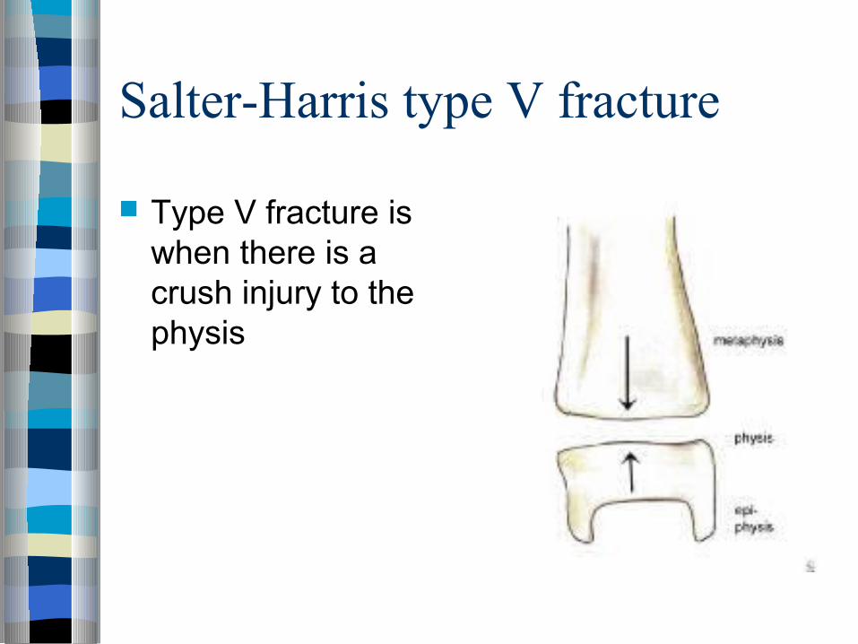
Salter-Harris type V fracture
Type V fracture is when there is a crush injury to the physis

Gustillo classification
The Gustillo classification is used to classify open fracture - ones in which the skin has been disrupted
Three grades that try to quantify the amount of soft tissue damage associated with the fracture

Open fractures - grade 1
wound less than 1 cm w/ minimal soft tissue injury
wound bed is clean bone injury is simple w/ minimal
comminution w/ IM nailing, average time to union is
21-28 weeks

Open fractures - grade 2
wound is greater than 1 cm w/ moderate soft tissue injury
wound bed is moderately contaminated fracture contains moderate
comminution w/ IM nailing, average time to union is
26-28 weeks

Open fractures - grade 3A
wound greater than 10 cm w/ crushed tissue and contamination
soft tissue coverage of bone is usually possible
w/ IM nailing, average time to union is 30-35 weeks

Open fractures - grade 3B
wound greater than 10 cm w/ crushed tissue and contamination
soft tissue is inadequate and requires regional or free flap
w/ IM nailing, average time to union is 30-35 weeks

Open fractures - grade 3C
is fracture in which there is a major vascular injury requiring repair for limb salvage
fractures can be classified using the MESS
in some cases it will be necessary to consider BKA following tibial fracture

Types Of Fractures Oblique: Break Occurs Diagonally Across the Bone Comminuted: Bone Is Broken, Splintered or Crushed
Into a Number of Pieces Spiral: The Break Travels Around the Bone. Compound: The Bone Sticks Through the Skin.

Types Of Fractures Greenstick
– Bone Cracks One Side Only– Not All the Way Through – Usually Only Seen in Children Due to Softness of Their Bones
Transverse: Complete Fracture in Which the Break Is Straight Across the Bone
Simple Or Closed– Partial Break on Bone– No Open Skin Wound
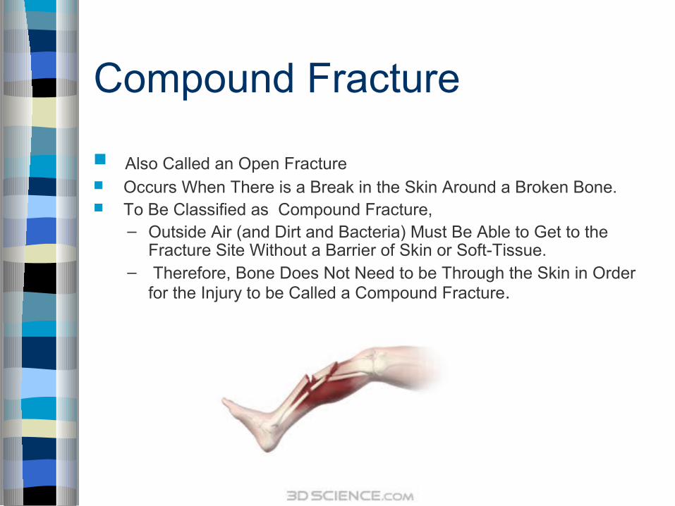
Compound Fracture
Also Called an Open Fracture Occurs When There is a Break in the Skin Around a Broken Bone. To Be Classified as Compound Fracture,
– Outside Air (and Dirt and Bacteria) Must Be Able to Get to the Fracture Site Without a Barrier of Skin or Soft-Tissue.
– Therefore, Bone Does Not Need to be Through the Skin in Order for the Injury to be Called a Compound Fracture.

The Fuss About Compound Fractures Injuries Are Open to the Outside World
– There Is a Very Significant Risk of Developing an Infection Around the Fracture.
– If Infection Develops, There Can Be Problems with Healing Process
Therefore, Compound Fractures Are Generally Treated with Surgery to Clean the Site of Injury and Stabilize the Fracture.

Compound Fracture X-Ray
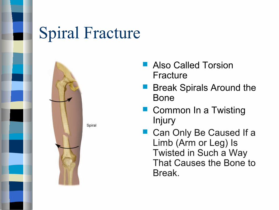
Spiral Fracture
Also Called Torsion Fracture
Break Spirals Around the Bone
Common In a Twisting Injury
Can Only Be Caused If a Limb (Arm or Leg) Is Twisted in Such a Way That Causes the Bone to Break.
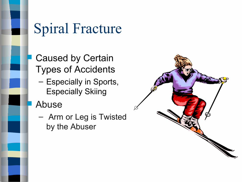
Spiral Fracture
Caused by Certain Types of Accidents– Especially in Sports,
Especially Skiing
Abuse– Arm or Leg is Twisted
by the Abuser

Causes Of Spiral Fracture
In Skiing - skiers lock their feet into the skis in sturdy ski boots, if a ski breaks or the skier loses control and the ski rotates, the leg may be violently twisted in one direction, creating a textbook spiral fracture.
Spiral Fracture has become famous as a warning sign of abuse, especially in children, because the twisting motion necessary could be caused by something such as a parent or guardian grabbing and twisting the arm or leg of a child. When doctors see spiral fractures in children, it may set off warning bells.

Sprial Fracture X-Ray
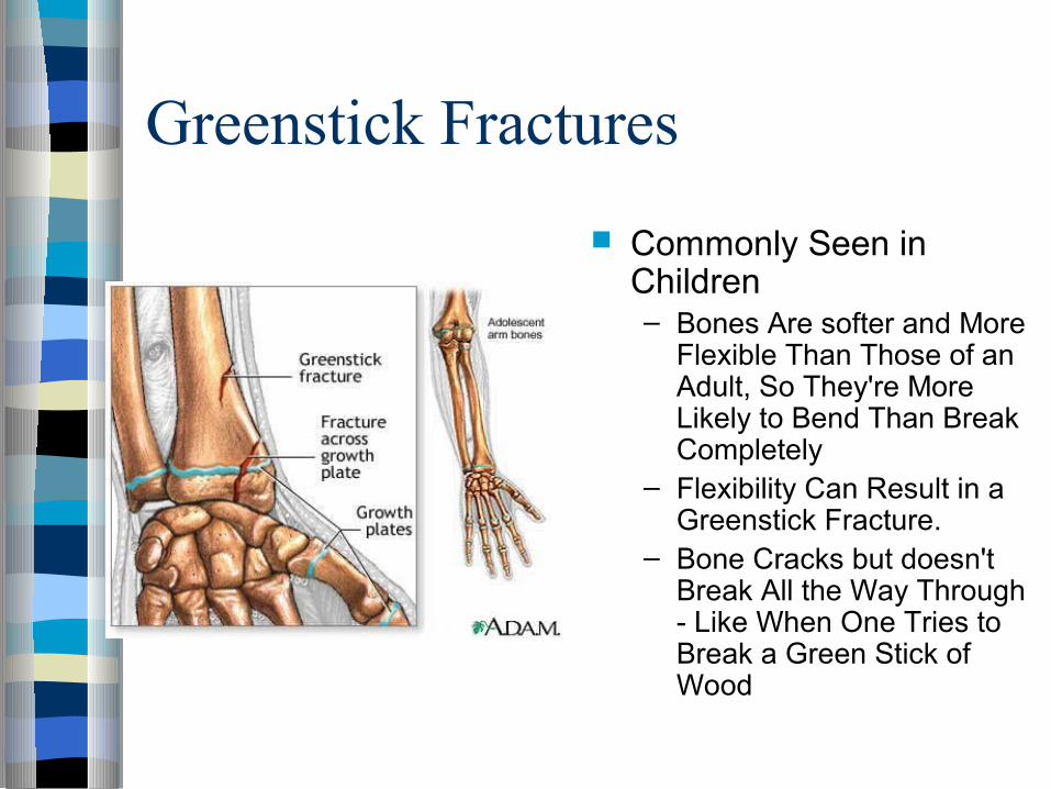
Greenstick Fractures
Commonly Seen in Children– Bones Are softer and More
Flexible Than Those of an Adult, So They're More Likely to Bend Than Break Completely
– Flexibility Can Result in a Greenstick Fracture.
– Bone Cracks but doesn't Break All the Way Through - Like When One Tries to Break a Green Stick of Wood

Greenstick Fractures
May Occur When a Child Falls While Playing or Participating in Sports.
Arm Bones Are the Most Likely to be Harmed Because of the Instinct to Throw out the Arms to Catch Fall.
Can Be Difficult to Diagnose, Because It May Not Cause All the Classic Signs and Symptoms of a Broken Bone

Greenstick Fracture X-Ray

Thank You!