two new species of arthrinium apiosporaceae xylariales...
TRANSCRIPT

Submitted 20 October 2016, Accepted 4 December 2016, Published online 28 December 2016 Corresponding Author: Dongqin Dai – e-mail – [email protected] 1332
Two new species of Arthrinium (Apiosporaceae, Xylariales) associated
with bamboo from Yunnan, China
Dai DQ1,*, Jiang HB1, Tang LZ1, Bhat DJ2,3
1 Center for Yunnan Plateau Biological Resources Protection and Utilization, College of Biological Resource and Food Engineering, Qujing Normal University, Qujing, Yunnan 655011, China 2 Formerly, Department of Botany, Goa University, Goa, India 3 No. 128/1-J, Azad Housing Society, Curca, Goa Velha-403108, India
Corresponding author: Dong-Qin Dai, email: [email protected]
Dai DQ, Jiang HB, Tang LZ, Bhat DJ 2016 – Two new species of Arthrinium (Apiosporaceae,
Xylariales) associated with bamboo from Yunnan, China. Mycosphere 7 (9), 1332–1345, Doi
10.5943/mycosphere/7/9/7
Abstract
Arthrinium, a globally distributed genus, is characterized by basauxic conidiogenesis in its
asexual morph, with globose to subglobose conidia, which are usually lenticular in side view and
obovioid and brown to dark brown. The sexual morph develops multi-locular perithecial stromata
with hyaline apiospores usually surrounded by a thick gelatinous sheath. Four Arthrinium species
collected in Kunming, China, are described and illustrated in this paper. Based on the morphology
and analyses of ITS sequence data, we introduce two new species of Arthrinium. Arthrinium
garethjonesii and A. neosubglobosa spp. nov. are provided with descriptions of sexual morphs and
compared with phylogenetically close taxa. Arthrinium hydei and A. hyphopodii are new records on
their sexual morphs.
Keywords – bambusicolous fungi – phylogeny – Sordariomycetes – taxonomy
Introduction
Arthrinium Kunze ex Fr. (sexual morph: Apiospora Sacc.) is the type genus of
Apiosporaceae and has a widespread distribution occurring in various habitats (Sharma et al. 2014).
Taxa are endophytes, pathogens or saprobes, usually isolated from soil debris, plants, lichens and
marine algae (Senanayake et al. 2015), or occasionally from erythematous nodules of human beings
(Sharma et al. 2014). Some Arthrinium species such as A. arundinis (Corda) Dyko & B. Sutton has
been utilized in the pharmaceutical industry as antifungal agents. Crous & Groenewald (2013)
synonymized Apiospora under Arthrinium. The genus Arthrinium currently contains 64 epithets
with 53 names under Apiospora in Index Fungorum (2016). However, most of existing taxa lack
sequence data and detailed morphological descriptions. Crous & Groenewald (2013) reviewed the
genus Arthrinium and included 16 species in the phylogenetic analysis. Sequence data from fresh
collections are however, needed to better resolve their natural classifications and establish species
numbers. We are studying bambusicolous fungi (Hyde et al. 2002, Dai et al. 2007) and collected
four new Arthrinium species from Yunnan Province. These are introduced in this paper with
molecular data.
Mycosphere 7 (9): 1332–1345 (2016) www.mycosphere.org ISSN 2077 7019
Article – special issue
Doi 10.5943/mycosphere/7/9/7
Copyright © Guizhou Academy of Agricultural Sciences

1333
Materials and methods
Collection, isolation and specimen examination
Dead bamboo culms were collected from Yunnan Province in China. The samples were
placed in plastic Zip lock bags and brought to the laboratory. The specimens were examined under
stereo- and compound microscopes. Single spore isolates were obtained following the method of
Chomnunti et al. (2014), and the specimens were incubated in a moist chamber for seven days at
room temperature, to induce sporulation. Microscopic observations were made from slides prepared
in distilled water. The micro-morphological characters were examined by differential interference
contrast (DIC) using a Nikon ECLIPSE 80i microscope with a 600D digital camera. Measurements
were made using the Tarosoft (R) Image Frame Work program. Type material is deposited at the
herbarium of Mae Fah Luang University, Chiang Rai, Thailand (MFLU) and Herbarium of
Kunming Institute of Botany, Chinese Academy of Sciences (KUN). The living cultures are
deposited at Mae Fah Luang University Culture Collection (MFLUCC), Kunming Culture
Collection (KUMCC). Facesoffungi (Jayasiri et al. 2015) and Index Fungorum (2016) numbers
were provided for newly described taxa.
DNA extraction, PCR amplification and sequencing
Pure cultures were grown on PDA for 30 days at 27˚C and genomic DNA was extracted
from fresh mycelia, following the specifications of the Biospin Fungus Genomic DNA Extraction
Kit (BioFlux®). ITS5 and ITS4, NS1 and NS4 (White et al. 1990) and LROR and LR5 (Vilgalys &
Hester 1990) primers were used for the amplification of internal transcribed spacers (ITS), small
subunit rDNA (SSU) and large subunit rDNA (LSU) respectively. Polymerase chain reaction (PCR)
amplification was carried out following Dai et al. (2017). Amplified PCR fragments were
sequenced at Shanghai Majorbio Bio-Pharm Technology Co., Ltd. and BGI Tech Solutions Co.,
Ltd. (BGI-Tech), P.R. China. The sequences generated from the ITS, LSU and SSU regions are
deposited in GenBank.
Phylogenetic analyses
A blast search in GenBank, using ITS gene was carried out to determine the closest
Arthrinium taxa. Additional sequences (Crous & Groenewald 2013, Dai et al. 2017) were
downloaded from GenBank. A single gene sequence alignment was generated with MAFFT v.
7.215 (Katoh & Standley, 2013, http://mafft.cbrc.jp/alignment/server/index.html) and improved
when necessary in BioEdit v. 7.0 (Hall 2004) by manual adjustment. In the analyses, gaps were
treated as missing data, and all characters were unordered and of equal weight (Dai et al. 2012).
Maximum-likelihood (ML) analysis including 1000 bootstrap replicates was performed in
raxmlGUI v.1.0. (Stamatakis 2006; Silvestro & Michalak 2011). Alignments in PHYLIP format
were exchanged and loaded (http://sing.ei.uvigo.es/ALTER/), and the online tool Findmodel
(http://www.hiv.lanl.gov/content/sequence/findmodel/findmodel.html) was used to determine the
best nucleotide substitution model for sequence data. GTR+G was chosen as the best model.
Maximum-parsimony (MP) analysis was run using PAUP v. 4.0b10 (Swofford 2002) with
1000 replications. Maxtrees were set to 1000, branches of zero length were collapsed, and all
multiple equally most parsimonious trees were saved. The robustness of the most parsimonious
trees was evaluated from 1 000 bootstrap replications (Phillips et al. 2013).
Bayesian analyses were performed using MrBayes v. 3.0b4 (Ronquist & Huelsenbeck 2003).
The model of evolution was performed by using MrModeltest v. 2.3 (Nylander 2004). Posterior
probabilities (PP) (Rannala & Yang 1996, Zhaxybayeva & Gogarten 2002) were determined by
Markov Chain Monte Carlo sampling (MCMC) in MrBayes v. 3.0b4. Six simultaneous Markov
chains were run for 5,000,000 generations and trees were sampled every 100th generation. The
burn-in was set to 0.25, and the run was automatically stopped when the average standard deviation
of split frequencies reached below 0.01 (Maharachchikumbura et al. 2015).

1334
Trees were visualized with TreeView (Page 1996), and additionally layouts were carried out
with Adobe Illustrator CS v. 5. Maximum-parsimony bootstrap values (MPBP) and Maximum-
likelihood bootstrap values (MLBP) equal or greater than 50 % are given. Bayesian posterior
probabilities (BYPP) greater than 0.80 are given. The alignment and trees were submitted in
TreeBASE (www.treebase.org, submission ID: 20359)
Table 1 List of strains and sequences used in the phylogenetic tree in this study.
Species name Strain substrate location collector ITS
Arthrinium arundinis CBS 106.12 / Germany: Bromberg E. Schaffnit KF144883
Arthrinium arundinis CBS 114316 Leaf of Hordeum vulgare
Iran: Shabestar B. Askari KF144884
Arthrinium arundinis CBS 124788 Living leaves of Fagus sylvatica
Switzerland: Basel M. Unterseher KF144885
Arthrinium arundinis CBS 133509 Aspergillus flavus sclerotium buried in sandy field
USA: Kilbourne / KF144886
Arthrinium arundinis CBS 449.92 Culm of cultivated Sasa Canada: Vancouver R.J. Bandoni KF144887
Arthrinium arundinis CBS 464.83 Dead culms of Phragmites australis
The Netherlands: Harderbos
W. Gams KF144888
Arthrinium arundinis CBS 732.71 Dung India B.C. Lodha KF144889 Arthrinium aureum CBS 244.83 Dead culms of
Phragmites australis Netherlands: Harderbos
W. Gams KF144888
Arthrinium garethjonesii KUMCC 16-0202
Dead culms of bamboo China: Kunming H.B. Jiang & D.Q. Dai
KY356086
Arthrinium hydei CBS 114990 Culms of Bambusa tuldoides
Hong Kong: Tai Po Kau
K.D. Hyde KF144890
Arthrinium hydei KUMCC 16-0204
Dead culms of bamboo China: Kunming H.B. Jiang & D.Q. Dai
KY356087
Arthrinium hyphopodii MFLUCC
15-0003
Culms of Bambusa tuldoides
China: Kunming D.Q. Dai & D.Q. Dai
KR069110
Arthrinium hyphopodii KUMCC 16-0201
Culms of Bamboo China: Kunming H.B. Jiang & D.Q. Dai
KY356088
Arthrinium kogelbergense CBS 113332 Dead culms of Cannomois virgata
South Africa S. Lee KF144891
Arthrinium
kogelbergense
CBS 113333 Dead culms of Restionaceae
South Africa S. Lee KF144892
Arthrinium kogelbergense CBS 113335 Dead culms of Restio quadratus
South Africa S. Lee KF144893
Arthrinium kogelbergense CBS 114734 Juncus gerardi Sweden: Börstil par. K. & L. Holm KF144894 Arthrinium kogelbergense CBS 117206 Unknown algae Croatia E. Eguereva KF144895
Arthrinium longistromum MFLUCC
11-0479
Dead culms of bamboo Thailand: Chiang Rai
D.Q. Dai KU940142
Arthrinium longistromum MFLUCC 11-0481
Dead culms of bamboo Thailand: Chiang Rai
D.Q. Dai KU940141
Arthrinium malaysianum CBS 102053 Macaranga hullettii stem colonised by ants
Malaysia: Gombak W. Federle KF144896
Arthrinium malaysianum CBS 251.29 Stembase of Cinnamomum camphora
/ / KF144897
Arthrinium marii CBS 113535 Oats Sweden C. Svenson KF144898 Arthrinium marii CBS 114803 Culm of Arundinaria
hindsi Hong Kong: Lung Fu Shan
K.D. Hyde KF144899
Arthrinium marii CBS 200.57 Leaf of Beta vulgaris Netherlands / KF144900 Arthrinium marii CBS 497.90 Beach sand Spain: Barcelona J.V. Larrondo &
A. Calvo AB220252
Arthrinium marii CPC 18902 Stems of Phragmites australis
Italy: Bomarzo W. Gams KF144901
Arthrinium marii CPC 18904 Stems of Phragmites
australis
Italy: Bomarzo W. Gams KF144902
Arthrinium montagnei ToD.7.1 Insect: Ips typographus Sweden / FJ824610 Arthrinium montagnei VL170 Stump of burned Pinus
mugo Lithuania V. Lygis JF440582
Arthrinium neosubglobosa JHB006 Dead culms of bamboo China: Kunming H.B. Jiang & D.Q. Dai
KY356089
Arthrinium KUMCC Dead culms of bamboo China: Kunming H.B. Jiang & D.Q. KY356090

1335
Species name Strain substrate location collector ITS
neosubglobosa 16-0203 Dai
Arthrinium ovatum CBS 115042 Arundinaria hindsii Hong Kong K.D. Hyde KF144903
Arthrinium
paraphaeospermum
MFLUCC
13-0644
Dead culms of bamboo Thailand: Chiangmai C. Senanayake KX822128
Arthrinium phaeospermum
CBS 114314 Leaf of Hordeum vulgare
Iran: Marand B. Askari KF144904
Arthrinium phaeospermum
CBS 114315 Leaf of Hordeum vulgare
Iran: Shabestar B. Askari KF144905
Arthrinium
phaeospermum
CBS 114317 Leaf of Hordeum
vulgare
Iran: Marand B. Askari KF144906
Arthrinium phaeospermum
CBS 114318 Leaf of Hordeum vulgare
Iran: Marand B. Askari KF144907
Arthrinium phragmites CPC 18900 Culms of Phragmites australis
Italy: Bomarzo W. Gams KF144909
Arthrinium
pseudosinense
CPC 21546 Leaf of bamboo Netherlands: Utrecht U. Damm KF144910
Arthrinium
pseudospegazzinii
CBS 102052 Macaranga hullettii stem
colonised by ants
Malaysia: Gombak W. Federle KF144911
Arthrinium pterospermum CBS 123185 Leaf lesion of Machaerina sinclairii
New Zealand: Auckland
C.F. Hill KF144912
Arthrinium pterospermum CPC 20193 Leaf of Lepidosperma gladiatum
Australia: Adelaide W. Quaedvlieg KF144913
Arthrinium rasikravindrae MFLUCC 11-0616
Dead culms of bamboo Thailand: Krabi K.D. Hyde KU940144
Arthrinium rasikravindrae MFLUCC
15-0203
Dead culms of bamboo Thailand: Krabi K.D. Hyde KU940143
Arthrinium rasikravindrae CBS 337.61 Cissus Netherlands H.A. van der Aa KF144914
Arthrinium
rasikravindrae
NFCCI 2144 Soil Norway: Ny-Alesund
S.M.Singh JF326454
Arthrinium sacchari CBS 212.30 Phragmites australis UK: Cambridge E.W. Mason KF144916 Arthrinium sacchari CBS 301.49 Bamboo Indonesia K.B. Boedijn & J.
Reitsma KF144917
Arthrinium sacchari CBS 372.67 Air / / KF144918
Arthrinium sacchari CBS 664.74 Soil under Calluna vulgaris
Netherlands H. Linder KF144919
Arthrinium saccharicola CBS 191.73 Air Netherlands H.A. van der Aa KF144920 Arthrinium saccharicola CBS 463.83 Dead culms of
Phragmites australis Netherlands: Harderbos
W. Gams KF144921
Arthrinium saccharicola CBS 831.71 / Netherlands M. van Schothorst KF144922 Arthrinium saccharicola CPC 18977 Phragmites australis Netherlands P.W. Crous KF144923 Arthrinium subglobosa MFLUCC
11-0397
Dead culms of bamboo Thailand: Chiang
Rai
D.Q. Dai KR069112
Arthrinium thailandicum MFLUCC 15-0199
Dead culms of bamboo Thailand: Krabi K.D. Hyde KU940146
Arthrinium thailandicum MFLUCC
15-0202
Dead culms of bamboo Thailand: Krabi K.D. Hyde KU940145
Arthrinium xenocordella CBS 478.86 Soil from roadway Zimbabwe: Matopos J.C. Krug KF144925 Arthrinium xenocordella CBS 595.66 Soil Austria:
Plaseckerjoch M.A.A. Schipper KF144926
Arthrinium yunnanum MFLU 15-
0002
Dead culms of Phyllostachys nigra
China: Kunming D.Q. Dai KU940147
Arthrinium yunnanum DDQ00281 Dead culms of Phyllostachys nigra
China: Kunming D.Q. Dai KU940148
Seiridium phylicae CPC 19965 Phylica arborea UK: Inaccessible Island
P. Ryan KC005787
Ex-type strains are in bold, new sequences are in red. Symbol “/” means the information is lacking. Abbreviations:
CBS: Centraalbureau voor Schimmelcultures, Utrecht, The Netherlands; CPC: Culture collection of Pedro Crous,
housed at CBS. MFLUCC: Mae Fah Luang University Culture Collection, Chiang Rai, Thailand; DDQ: D.Q. Dai; JHB:
H.B. Jiang; VL:V. Lygis.
Results
Phylogeny
An ITS dataset was used to determine the taxonomic placement of our strains. The dataset
comprised 63 strains including five newly sequenced strains plus one outgroup taxon (Table 1).

1336
Fig. 1 – One of the parsimonious trees resulting from 1000 bootstrap replicates obtained from the
ITS sequence data (CI 0.634, RI 0.876, RC 0.555), The tree is rooted to Seiridium phylicae (CPC
19965). Ex-type strains are in bold and newly generated sequences are highlighted with a blue
background.
The best tree is shown in Fig. 1. The phylogenetic trees generated by maximum-parsimony (MP),
maximum likelihood (ML) and Bayesian analyses using the ITS region demonstrate that new
strains clustered within the genus Arthrinium, Apiosporaceae, Xylariales. Arthrinium hydei
(JHB012) and A. hyphopodii (JHB004) grouped with the type strain CBS114990 and MFLUCC
15–0003 with high support (MPBS/MLBS/BYPP 81/90/0.90) and (MPBS/MLBS/BYPP
100/100/1.00) respectively. The new species, A. garethgonesii is closest to A. hyphopodii, while the
branch length can separate these two species. A second novel species A. neosuboglobosa groups

1337
with A. subglobosa in the same clade, however, they can be phylogenetically distinguished with
high bootstrap support (MPBS/MLBS/BYPP 100/100/1.00). Bootstrap support values of MP, ML
and the Bayesian posterior probabilities from MCMC analyses are shown in Fig 1. All the strains
used in this paper together with their GenBank associate numbers are listed in Table 1. The new
sequences of ITS, LSU and SSU regions are deposited in GenBank for further study (Table 2).
Table 2 List of strains sequenced in this study.
Species name Strain GenBank accession numbers
LSU SSU ITS
Arthrinium garethjonesii KUMCC 16-0202 KY356091 KY356096 KY356086
Arthrinium hydei KUMCC 16-0204 KY356092 KY356097 KY356087
Arthrinium hyphopodii KUMCC 16-0201 KY356093 KY356098 KY356088
Arthrinium neosubglobosa JHB006 KY356094 KY356099 KY356089
Arthrinium neosubglobosa KUMCC 16-0203 KY356095 KY356100 KY356090
Taxonomy
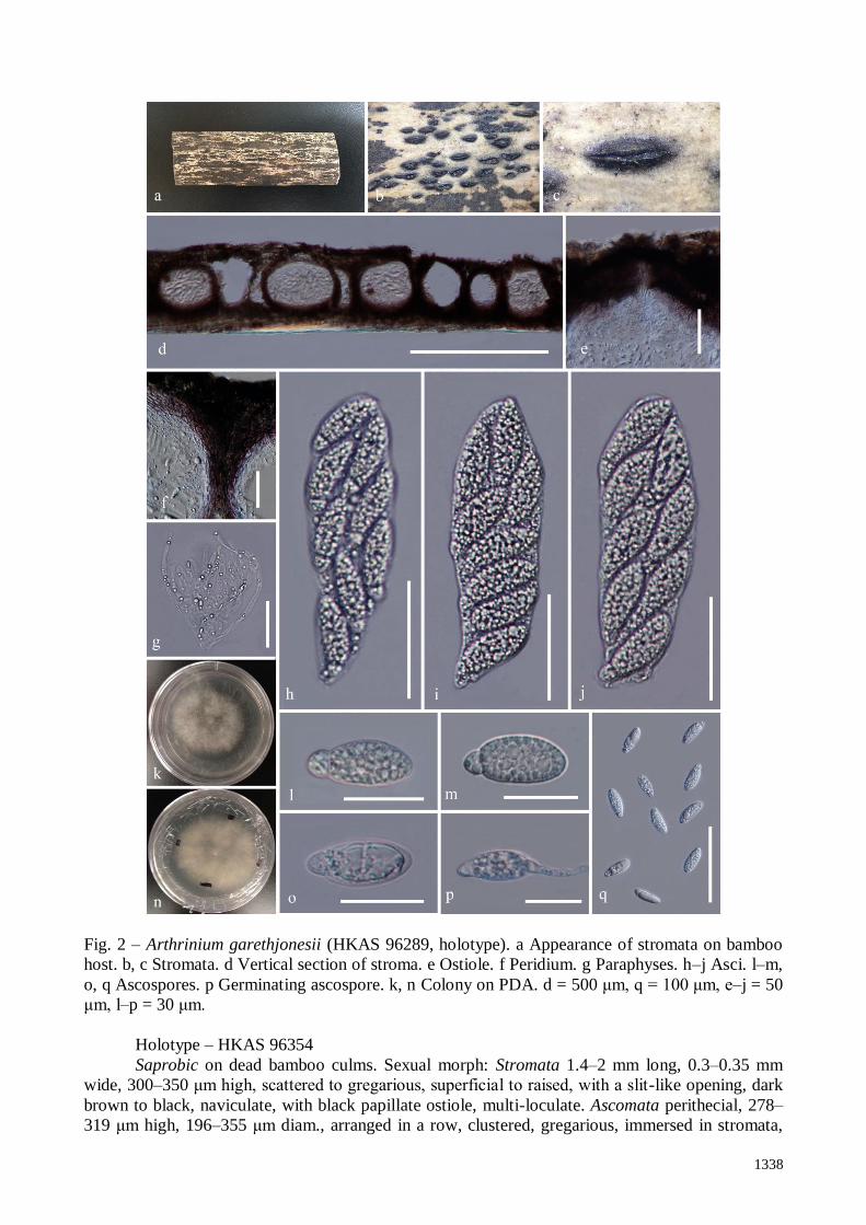
Arthrinium garethjonesii D.Q. Dai & H.B. Jiang, sp. nov.
Index Fungorum number: IF 552679; Facesoffungi number: FoF 02759, Fig. 2
Etymology – The specific epithet garethjonesii refers to the significant contribution of
Professor E.B. Gareth Jones made to mycology.
Holotype – HKAS 96289
Saprobic on dead bamboo culms. Sexual morph: Stromata 1.3–2 mm long, 0.3–0.5 mm
wide, 188–282 μm high, scattered to gregarious, partly immersed, becoming erumpent to
superficial, raised, dark brown, in linear rows, with a slit-like opening, multi-loculate. Ascomata
perithecial, 177–235 μm high, 141–232 μm diam., arranged in rows, clustered, gregarious, with 3–
10 perithecia forming groups immersed in stromata to erumpent through host surface, ellipsoidal to
subglobose, dark brown, membranous. Ostiole raised from centre of ascomata, internally lined with
periphyses. Peridium 22–35 μm wide, composed of dark brown to reddish brown to hyaline cells of
textura angularis. Hamathecium composed of dense paraphyses, 3.9–6.1 μm wide, filamentous,
unbranched and not anastomosed, indistinctly septate, constricted at the septum. Asci 125–154 ×
35–42 μm (x̅ = 139 × 38 μm, n = 20), 8-spored, unitunicate, clavate, apedicellate, apically rounded.
Ascospores 30–42 × 11–16 μm (x̅ = 39 × 13 μm, n = 20), 2-seriate, 1-septate, ellipsoidal, with a
small lower cell and a large upper cell, with many guttules, hyaline, smooth-walled, with a faint
gelatinous sheath. Asexual morph: Undetermined.
Culture characters – Ascospores germinating on WA within 24 h and germ tubes developing
from the upper cell. Colonies fast growing on PDA, reaching 40 mm in 1 week at 23 °C, under 12 h
light/12 h dark, cottony, circular, with irregular edge, white from above and below. Mycelium
superficial to immersed in media, with branched, septate, smooth hyphae.
Materials examined – CHINA, Yunnan Province, Kunming, Southwest Forest University
campus, on dead culms of bamboo, 20 July 2016, Hong-Bo Jiang & Dong-Qin Dai JHB004 (KUN,
HKAS 96289, holotype); Ibid. (MFLU, isotype), living cultures, MFLUCC, KUMCC 16-0202.
Notes – Arthrinium garethjonesii is phylogenetically close to A. hyphopodii based on
maximum-parsimony (MP) analysis (Fig. 1). Both of these species have dark brown ascostromata
in linear rows on bamboo, with a slit-like opening. However, A. garethjonesii has shorter stromata
(188–282 μm high vs. 369–488 μm high) and smaller 1-septate ascospores (30–42 × 11–16 μm vs.
61–80 × 14–18.5 μm), whereas A. hyphopodii has comparatively larger 1-septate ascospores.
Furthermore, they are phylogenetically separated by branch length (Fig. 1).
Arthrinium neosubglobosa D.Q. Dai & H.B. Jiang, sp. nov. Fig. 3
Index Fungorum number: IF552680; Faceoffungi number: FoF02760
Etymology – Refers to the morphological similarity with Arthrinium subglobosa.
1338
Fig. 2 – Arthrinium garethjonesii (HKAS 96289, holotype). a Appearance of stromata on bamboo
host. b, c Stromata. d Vertical section of stroma. e Ostiole. f Peridium. g Paraphyses. h–j Asci. l–m,
o, q Ascospores. p Germinating ascospore. k, n Colony on PDA. d = 500 μm, q = 100 μm, e–j = 50
μm, l–p = 30 μm.
Holotype – HKAS 96354
Saprobic on dead bamboo culms. Sexual morph: Stromata 1.4–2 mm long, 0.3–0.35 mm
wide, 300–350 μm high, scattered to gregarious, superficial to raised, with a slit-like opening, dark
brown to black, naviculate, with black papillate ostiole, multi-loculate. Ascomata perithecial, 278–
319 μm high, 196–355 μm diam., arranged in a row, clustered, gregarious, immersed in stromata,
1339
Fig. 3 – Arthrinium neosubglobosa (HKAS 96354, holotype). a Appearance of stromata on bamboo
host. b, c Stromata. d Vertical section of stroma. e Ostiole. f Peridium. g Paraphyses. i–k Asci. m–o
Ascospores surrounded by gelatinous sheath. p Germinating spore. h, l Colony on PDA. d = 250
μm, e–g, i–k = 50 μm, m–p = 30 μm.
later becoming erumpent through host surface to superficial, obpyriform to ampulliform, dark
brown, membranous. Ostiole raised from centre of ascomata, internally lined with periphyses.
Peridium 26–44 μm thick, with 2 layers; outer layer composed of dark brown, cells of textura
angularis; inner layer thin, with hyaline cells of textura angularis. Hamathecium comprising dense
paraphyses, 4.2–6.3 μm wide, indistinctly septate, unbranched, not anastomosing, filamentous-
clustered embedded in gelatinous matrix. Asci 130–170 × 30–43 μm (x̅ = 150 × 37 μm, n = 20), 8-
1340
spored, unitunicate, clavate, with a short pedicel, apically rounded. Ascospores 39–47 × 13–16 μm
(x̅ = 43 × 15 μm, n = 20), 2-seriate, elliptical, hyaline, 1-septate, constricted at the septum, mostly
curved at the lower cell, rarely straight, with a large upper cell and a small lower cell, smooth-
walled, guttulate, with a shallow 10 μm thick gelatinous sheath. Asexual morph: Undetermined.
Culture characters – Ascospores germinating on WA within 48 h and germ tubes developing
from the upper cell. Colonies slow growing on PDA, reaching 20 mm in 1 week at 23 °C, under 12
h light/12 h dark, velvety, circular, with regular edge, yellowish, sparse in the centre, white, dense
at the margin from above; yellowish brown from below. Mycelium superficial to immersed in
media, with branched, septate, smooth hyphae.
Materials examined – CHINA, Yunnan Province, Kunming, Southwest Forest University
campus, on dead culms of bamboo, 20 July 2016, Hong-Bo Jiang & Dong-Qin Dai JHB007 (KUN,
HKAS 96354, holotype); Ibid. (MFLU, isotype), living cultures, MFLUCC, KUMCC 16-0203;
JHB006.
Notes – Arthrinium neosubglobosa is similar to A. subglobosa in having ascospores with a
curved lower cell and yellowish brown culture on PDA (Senanayake et al. 2015). These two
species original isolated from bamboo, and form an individual clade in the phylogenetic analysis
with high bootstrap support (MPBS/MLBS/BYPP 100/100/1.00) (Fig. 1). However, A.
neosubglobosa differs by having higher stromata (300–350 μm high vs. 150–300 μm high) and
longer asci (130–170 μm long vs. 75–150 μm long) than A. subglobosa. In addition, A.
neosubglobosa has obpyriform to ampulliform ascomata, whereas A. neosubglobosa occurs has
subglobose ascomata with a flattened base.
Arthrinium hydei Crous, IMA Fungus 4(1): 142 (2013)
Facesoffungi number: FoF02761, Fig 4
Saprobic on rotting bamboo culms, forming black, filiform areas, arranged in a compact
way, with stromata breaking through raised cracks at the black centre. Sexual morph: Stromata 10–
20 mm long, 3.5–4 mm wide, 215–265 μm high, gregarious, initially immersed and raised, later
becoming erumpent through host tissue to superficial, shaping a slit-like axis at the top, filiform,
dark brown, multi-loculate. Ascomata perithecial, 135–230 μm high, 180–215 μm diam., gregarious,
immersed in stromata, reddish brown, ampulliform to subglobose usually with flattened base,
membranous, with a central, periphysate ostiole. Peridium 19–30 μm thick, composed of brown to
reddish brown to hyaline cells of textura angularis. Hamathecium comprising septate, unbranched
paraphyses. Asci 110–130 × 17–24 μm (x̅ = 118 × 21 μm, n = 20), 8-spored, unitunicate, cylindrical
to ellipsoidal, no pedicel, apically truncate. Ascospores 35–45 × 8.5–11 μm (x̅ = 40 × 9.6 μm, n =
20), 2-seriate, broad fusiform to cylindrical, with a large upper cell and a small lower cell, hyaline,
1-septate, constricted at septum, slightly curved, smooth-walled, with many guttules, with a large
guttule at the centre of large upper cell, with a distinct gelatinous sheath, 10–20 μm thick. Asexual
morph: See Crous et al (2013).
Culture characters – Ascospores germinating on WA within 24 h and germ tubes developing
from the upper large part of spore. Colonies growing fast on PDA, reaching 40 mm in 1 week at
23 °C, under 12 h light/12 h dark, cottony, circular, diffuse from centre, with irregular edge, white
from above and below. Mycelium superficial to immersed in media, with branched, septate, smooth
hyphae.
Materials examined – CHINA, Yunnan Province, Kunming, Southwest Forest University
campus, on dead culms of bamboo, 20 July 2016, Hong-Bo Jiang & Dong-Qin Dai JHB0012 (KUN,
HKAS 96355); Ibid. (MFLU), living cultures, MFLUCC, KUMCC 16-0204.
Notes – Arthrinium hydei was originally described by Crous & Groenewald (2013) based on
the asexual morph from a culture (CBS 114990) which was isolated from bamboo culms in Hong
Kong, China. However, no sexual morph was observed by Crous & Groenewald (2013). A new
specimen was collected of sexual morph in Kunming, China and the phylogenetic sequence data
indicate that it is A. hydei (MPBS/MLBS/BYPP 81/90/0.90, Fig. 1).

1341
Arthrinium hyphopodii D.Q. Dai & K.D. Hyde, in Senanayake et al., Fungal Diversity 73: 112
(2015) Figs 5, 6
Fig. 4 – Arthrinium hydei. a Appearance of stromata on bamboo host. b Stromata. c Vertical section
of stroma. d Peridium. e Ascoma. f Ostiole with periphyses. g–i Asci. k, l Ascospores. m
Germinating ascospore. j Colony on PDA. c = 500 μm, e = 100 μm, d, g–i = 50 μm, f, k–m = 30 μm.

1342
Saprobic on dead bamboo culms, forming black, lenticular spots on the host surface, with
stromata breaking through raised cracks with black centre. Sexual morph: Stromata 1.5–2 mm long,
0.35–0.45 mm wide, 370–490 μm high, scattered to gregarious, immersed to erumpent, later
becoming superficial, dark brown to black, fusiform to ellipsoid, forming a slit-like opening at the
apex, multi-loculate, membranous, with a periphysate ostiole. Ascomata perithecial, 295–360 μm
high, 185–280 μm diam., arranged in rows, clustered, gregarious, immersed in stromata,
ampulliform, dark brown to black. Peridium 28–40 μm wide, composed of several layers of dark
brown to hyaline cells of textura angularis. Hamathecium comprising dense, 5–7.8 μm wide,
hyaline, septate paraphyses. Asci 160–225 × 35–55 μm (x̅ = 193.7 × 45.3 μm, n = 20), 8-spored,
unitunicate, clavate, apedicellate, apically rounded. Ascospores 60–80 × 14–18.5 μm (x̅ = 70.5 ×
16.3 μm, n = 20), partly overlapping, biseriate, cylindrical to broadly fusiform, slightly rounded
both ends, slightly curved, hyaline, septa not observed, smooth-walled, with many guttules, with an
indistinct gelatinous sheath. Asexual morph: See Senanayake et al. (2015).
Culture characters – Ascospores germinating on WA within 24 h and germ tubes produced
from lower end. Colonies fast growing on PDA, reaching 34 mm in 12 days at 23 °C, under 12 h
light/12 h dark, cottony, circular, sparse, raised, with irregular edge, white in centre becoming
reddish-brown at the margin from above and below. Mycelium superficial to immersed in media,
with branched, septate, smooth hyphae.
Materials examined – CHINA, Yunnan Province, Kunming, Southwest Forest University
campus, on dead culms of bamboo, 20 July 2016, Hong-Bo Jiang & Dong-Qin Dai JHB003 (KUN,
HKAS 96288); Ibid. (MFLU), living cultures, MFLUCC, KUMCC 16-0201; Other material
examined: Kunming Institute of Botany, Chinese Academy of Sciences, on dead culm of Bambusa
tuldoides Munro (Poaceae), 7 July 2014, Dong-Qin Dai DDQ00280 (MFLU 15-0383, holotype).
Notes – Senanayake et al. (2015) introduced the species Arthrinium hyphopodii based on an
asexual morph specimen collected in China, Kunming. We collected a new specimen of sexual
morph within a close vicinity, and the phylogenetic analysis showed that they are same species with
high bootstrap support (MPBS/MLBS/BYPP 100/100/1.00) (Fig. 1). Arthrinium hyphopodii is
characterized by brown hyphopodia developing on its conidial germ tubes. Usually Arthrinium has
apiospores shorter than 50 μm, however, A. hyphopodii is unusual in that it has 60–80 μm long
ascospores which are acellular, a feature which has not been recorded previously in species of
Arthrinium.
Discussion
This study treats four Arthrinium species, including two new species, Arthrinium
garethjonesii and A. neosubglobosa, two existing species, Arthrinium hydei and A. hyphopodii
which were previously published with only asexual morphs. In the present study, their sexual
morphs are confirmed by sequence data. Species of Arthrinium are difficult to identify to species
level when only the asexual morph are available (Crous & Groenewald 2013). For instance, the
Arthrinium species can produce hyphomyceteous fruiting bodies in culture or coelomycetous
structures on host substrates (Dai et al. 2017). In addition, their conidia are not a useful identifying
feature, as most Arthrinium species have similar conidia, and their morphology can vary depending
on the period of incubation on different habitats (Crous & Groenewald 2013). This study suggests
that the identification of Arthrinium taxa needs to be carried out based on a combination of
sequence data and observation of the sexual-asexual morphs. The linkage of such sexual and
asexual morphs should be determined through cultures obtained from single spores and by
subsequent molecular analyses.

1343
Fig. 5 – Arthrinium hyphopodii (HKAS 96288). a Appearance of stromata on bamboo host. b, c
Stromata. d Vertical section of stroma. e Ostiole. f Peridium. g Paraphyses. i–k Asci. m–o
Ascospores. p Germinated spore. h, l Colony on PDA. d = 500 μm, f, g, k, p = 100 μm, e, i, j, m–o
= 50 μm.

1344
Fig. 6 – Arthrinium hyphopodii (holotype). a Appearance of conidiomata on bamboo host. b
Conidiomata. d Section of conidioma. e, i Conidiogenous cells. c, f–h Conidia. Scale bars: a = 5 cm,
b = 500 μm, d = 100 μm, c, e–i = 5 μm.
Acknowledgements
This work is supported by the Key Laboratory of Yunnan Province Universities of the
Diversity and Ecological Adaptive Evolution for Animals and plants on Yun-Gui Plateau and
Yunnan Province Universities of the Science and Technology Innovation Team for the exploitation
and utilization of endophytes. Hong-Bo Jiang would like to thank Qiu-Ju Shang for help with lab
work. The research is supported by the National Natural Science Foundation of China (No. NSFC
31260087, NSFC 31460561). The authors would like to thank J.E. Taylor for suggestions and
annotations to the paper.
References
Chomnunti P, Hongsanan S, Aguirre-Hudson B et al. 2014 – The sooty moulds. Fungal Diversity
66, 1–36.
Crous PW, Groenewald JZ. 2013 – A phylogenetic re-evaluation of Arthrinium. IMA Fungus 4,
133–154.
Dai DQ, Bhat DJ, Liu JK et al. 2012 – Bambusicola, a new genus from bamboo with asexual and
sexual morphs. Cryptogamie Mycologie 33, 363–379.
Dai DQ, Phookamsak R, Wijayawardene NN, Li WJ et al. 2017 – Bambusicolous fungi. Fungal
Diversity, http://dx.doi.org 10.1007/s13225-016-0367-8.

1345
Hall T 2004 – BioEdit. Ibis Therapeutics, Carlsbad, CA, 92008, USA.
(http://www.mbio.ncsu.edu/BioEdit/bioedit.html) (accessed 18 Mar 2005).
Hyde KD, Zhou DQ, Dalisay T. 2002 – Bambusicolous fungi: A review. Fungal Diversity 9, 1-14.
Hyde KD, Hongsanan S, Jeewon R et al. 2016 – Fungal diversity notes 367–490: taxonomic and
phylogenetic contributions to fungal taxa. Fungal Diversity 80, 1–270.
Index Fungorum 2016 – http://www.indexfungorum.org/Names/Names.asp.
Jayasiri SC, Hyde KD, Ariyawansa HA, Bhat DJ et al. 2015 – The Faces of Fungi database: fungal
names linked with morphology, phylogeny and human impacts. Fungal Diversity 74, 3–18.
Katoh K, Standley DM. 2013 – MAFFT multiple sequence alignment software version 7:
improvements in performance and usability. Molecular Biology and Evolution 30, 772–780.
Maharachchikumbura SS, Hyde KD, Jones EBG, McKenzie EHC et al. 2015 – Towards a natural
classification and backbone tree for Sordariomycetes. Fungal Diversity 72, 199–301.
Nylander JAA 2004 – MrModeltest 2.0. Program distributed by the author. Evolutionary Biology
Centre, Uppsala University.
Page RDM 1996 – TreeView: an application to display phylogenetic trees on personal computers.
Bioinformatics, 12, 357–358.
Phillips AJL, Alves A, Pennycook SR, Johnston PR et al. 2008 – Resolving the phylogenetic and
taxonomic status of dark-spored teleomorph genera in the Botryosphaeriaceae. Persoonia
21, 29–55.
Rannala B, Yang Z. 1996 – Probability distribution of molecular evolutionary trees: a new method
of phylogenetic inference. Journal of Molecular Evolution 43, 304–311.
Ronquist F, Huelsenbeck JP. 2003 – MrBayes 3: Bayesian phylogenetic inference under mixed
models. Bioinformatics 19, 1572–1574.
Senanayake IC, Maharachchikumbura SSN, Hyde KD, Bhat JD et al. 2015 – Towards unraveling
relationships in Xylariomycetidae (Sordariomycetes). Fungal Diversity 73, 73–144.
Sharma R, Kulkarni G, Sonawane MS, Shouche YS. 2014 – A new endophytic species of
Arthrinium (Apiosporaceae) from Jatropha podagrica. Mycoscience 55, 118–123.
Silvestro D, Michalak I. 2011 – raxmlGUI: a graphical front-end for RAxML. Organisms Diversity
& Evolution 12, 335–337.
Stamatakis A 2006 – RAxML-VI-HPC: maximum likelihood-based phylogenetic analyses with
thousands of taxa and mixed models. Bioinformatics 22, 2688–2690.
Swofford DL 2002 – PAUP: phylogenetic analysis using parsimony, version 4.0 b10. Sinauer
Associates, Sunderland.
Vilgalys R, Hester M. 1990 – Rapid genetic identification and mapping of enzymatically amplified
ribosomal DNA from several Cryptococcus species. Journal of Bacteriology 172, 4238–
4246.
White TJ, Bruns T, Lee S, Taylor J. 1990 – Amplification and direct sequencing of fungal
ribosomal RNA genes for phylogenetics. In: Innis MA, Gelfand DH, Sninsky JJ, White TJ
(eds), PCR protocols: A Guide to Methods and Applications, 315–322.
Zhaxybayeva O, Gogarten JP. 2002 – Bootstrap, Bayesian probability and maximum likelihood
mapping: exploring new tools for comparative genome analyses. BMC Genomics 3, 1–15.