tuscia university department of plant protection agr/12
TRANSCRIPT

Tuscia University Department of Plant Protection
AGR/12
XXIII Cycle of Doctorate in Plant Protection Curriculum
“Control with low environmental impact”
Thesis:
“PHYTOBACTERIOLOGICAL STUDIES RELATED TO THE NATIVE (Olea cuspidata
Wall.) AND INTRODUCED (Olea europaea L.) OLIVE SPECIES IN NEPAL.”
By Jay Ram Lamichhane
Thesis submitted in partial fulfilment of the requirements for the degree of Doctor of Philosophy in Plant Protection
Coordinator and Supervisor Assistant supervisor Prof. Leonardo Varvaro Dr. Giorgio M. Balestra
Academic years 2007/2010

2
Thesis committee Thesis assistant supervisor Dr. Giorgio M. Balestra Researcher of Bacteriology Tuscia University Other members Prof. Dr. Lucio Montecchio Professor of Forest Pathology Padova University Prof. Dr. Nunzio Isidoro Professor of Entomology Politecnica delle Marche University This research was conducted under the auspices of the Tuscia University, Department of Plant Protection, within the FAO project entitled as “(GCP/NEP/056/ITA) Promotion of olive production and Consumption in Nepal”.

3
“PHYTOBACTERIOLOGICAL STUDIES RELATED TO THE NATIVE (Olea cuspidata Wall.) AND INTRODUCED (Olea europaea L.) OLIVE SPECIES IN NEPAL.”
Jay Ram Lamichhane
Thesis Submitted in partial fulfilment of the requirements for the degree of Doctor of Philosophy
in Plant Protection at Tuscia University by the authority of the Rector Magnificus
Prof. Dr. M. Mancini, In the presence of the
Thesis committee appointed by the Doctorate Board To be defended in public
On Wednesday 2 February 2011

4
To my wife Claudia and my son Himal

5
CONTENTS
CHAPTER 1. ......................................................................................................................................................................... 8
GENERAL PART: ................................................................................................................................................................... 8
1.2 Importance of olive cultivation in Nepal .................................................................................................................. 9
CHAPTER 2. ....................................................................................................................................................................... 11
BACTERIAL PATHOGEN OF OLIVE ..................................................................................................................................... 11
2.1 Introduction and overview ..................................................................................................................................... 12
2.2 Presence and risk of olive knot in Nepal ................................................................................................................ 12
2.3 Pathogen survival and multiplication ..................................................................................................................... 13
2.4 Possibility of control ............................................................................................................................................... 13
CHAPTER 3. ....................................................................................................................................................................... 15
REFERENCES ...................................................................................................................................................................... 15
CHAPTER 4. ....................................................................................................................................................................... 19
AIM OF THE DOCTORAL THESIS ........................................................................................................................................ 19
CHAPTER 5. ....................................................................................................................................................................... 21
EXPERIMENTAL PART ........................................................................................................................................................ 21
5.1 Preliminary tests .................................................................................................................................................... 22
CHAPTER 6. ....................................................................................................................................................................... 24
Investigation on the efficiency of methods for sampling, recovery and enumeration of bacteria applied to the phylloplane of Olea spp. ................................................................................................................................................... 24
CHAPTER 7. ....................................................................................................................................................................... 37
Bacteria colonizing phylloplane of Olea spp. in the fields of Nepal .................................................................................. 37
CHAPTER 8. ....................................................................................................................................................................... 57
Survival of Pseudomonas savastanoi pv. savastanoi on phylloplane of two olive species .............................................. 57
CHAPTER 9. ....................................................................................................................................................................... 67
Reaction of two olive species to different Pseudomonas savastanoi pv. savastanoi strains ........................................... 67
CHAPTER 10. ..................................................................................................................................................................... 79
In vitro antagonistic action of the bacterial species from Olea spp. phylloplane against Pseudomonas savastanoi pv. savastanoi ......................................................................................................................................................................... 79
CHAPTER 11. ..................................................................................................................................................................... 86
GENERAL DISCUSSION ...................................................................................................................................................... 86
CHAPTER 12. ..................................................................................................................................................................... 90
CONCLUSIONS ................................................................................................................................................................... 90
CHAPTER 13. ..................................................................................................................................................................... 92
PHOTOGRAPHS ................................................................................................................................................................. 92
ACKNOWLEDGEMENTS ..................................................................................................................................................... 99

6
GENERAL ABSTRACT
Two olive species, one introduced from Europe (Olea europaea L.) and cultivated in different
Districts and the other one the native species of Nepal (Olea cuspidata Wall.), were studied
regarding phytobacteriological aspects. Among the two olive species, the first was widely studied
while no study is available on O. cuspidata. In particular, the presence and/or appearance of olive
knot disease, the quali-quantitative composition of phylloplane bacterial populations of these
species from different Districts, the epiphytic survival of Pseudomonas savastanoi pv. savastanoi
(Psav) (the causal agent of olive knot disease) both on O. europaea and O. cuspidata, the
susceptibility of these species to Psav and the antagonistic activity of the phylloplane bacteria
against Psav in vitro have been studied. Different strains of Psav isolated from different geographic
origins were used.
No presence of olive knot disease was found on Olea spp. surveyed during the study in
Bajura, Dolpa and Kathmandu Districts. Whereas the disease was found on O. europaea,
introduced two decades ago, in a commercial orchard of Makwanpur District. No presence of Psav
on Olea phylloplane was found. The size of bacterial epiphytic populations ranged from very low
(2.39 x 100 CFU/cm2) to very high (1.09 x 10⁵ CFU/cm2). No quantitative differences statistically
significant were found on Olea phylloplane sampled in the three Districts. Fifteen bacterial species
were identified as colonizers of Olea phylloplane in Nepal which are so far different than those
found in south-east Italy. Both the olive species were dominated by Pantoea agglomerans (former
Erwinia herbicola). No qualitative differences statistically significant were seen on Olea
phylloplane sampled in three Districts mentioned above.
Olive species showed a significant influence on bacterial epiphytic survival since Psav better
multiplies on O. cuspidata compared to O. europaea. Leaf age and bacterial strains did not
influence significantly the epiphytic survival of the pathogen. O. cuspidata showed higher
susceptibility to Psav strains than O. europaea to artificial inoculations although no difference
statistically significant was found. Plant age did not influence significantly host-pathogen
interaction. The bacterial strains showed significant differences among them in terms of symptom
appearance, knot size and bacterial populations into the knots. None of the phylloplane bacteria
inhibited in vitro the growth of P. savastanoi pv. savastanoi.

7
ABBREVIATIONS
ASL Above sea level
ANOVA Analysis of Variance
CFU Colony forming Unit
FAO Food and Agriculture Organization of the United Nations
hrp genes components of type III secretion systems, regulatory proteins
IAA Indole-3-acetic acid
KOH Potassium hydroxide
MOAC Ministry of Agriculture and cooperatives
NA Nutrient agar
NAG Nutrient agar containing 1% glycerol
NAS Nutrient agar supplemented by 5% sucrose
Psav Pseudomonas savastanoi pv. savastanoi
rDNA Ribosomal Deoxyribonucleic acid
RH Relative humidity
RPM Revolutions per minute
SDW Sterile distilled water

8
CHAPTER 1.
GENERAL PART:
State of art and importance of olive cultivation in Nepal

9
1.1 State of art
The northwestern part of Nepal, where the altitude ranges from 1400 to 2500m asl, is
known for the presence of big natural forests belong to two Olea species identified as as O.
cuspidata and O. glandulifera. Secular plants of these species are present mostly in the District of
Bajura, Mugu, Humla and Dolpa Districts (Lamichhane et al., 2010a). Thirty five forest types of O.
cuspidata were identified and recorded from Nepal under Trans-Himalaya High Alpine Vegetation.
The presence of these plants with vigorous growth and their healthy aspects indicate that Olea
species find a suitable environmental conditions for growth, development and even for olive
production since O. cuspidtata alone produces a remarkable quantity of olive fruit in these areas.
Despite this great potentiality, no initiative was taken in the past to study and utilize these species
which can give a significant contribution being a main source of income to the local people of
these remote areas. Taking into account these factors, an effort was made for olive plantation
through a project in collaboration between Italian Ministry of foreign affairs and Nepalese ministry
of agriculture under FAO, from 2005.
For the plantation of European olive (Olea europaea L.), two different valleys have been
selected, one in Bajura District (Kolti, 1400m asl where the project is in progress) and the other in
Dolpa District (Juphal, 2500m where experimental fields are present). Twenty eight Italian olive
cultivars (20 plants per variety) have been planted at Kolti in an experimental field together with
local species. While at Juphal just a dozen of varieties were introduced. Moreover, a mother
orchard has been established at Kirtipur (1300 m asl), in the valley of Kathmandu District.
European olive had already been introduced in 1994 by some private olive growers in Nepal
(Chitlang valley, Makwanpur District) for commercial purpose. Olive production in this District was
initiated since 2000 (Lamichhane et al., 2010a). This olive farm provides olive oil on the local
market although in very small quantities. Since the beneficial properties of olive oil is well-known,
its cultivation has continuously been a great concern all over the world.
1.2 Importance of olive cultivation in Nepal
In recent years, Nepal witnessed like most of the south Asian countries a constant growth in
demand for edible oils which has essentially been met by imported products. The extent of the
demand has induced the authorities, in particular the Government, to look for different
alternatives which allow the increasing of the national output of edible oils. Furthermore, the
government is committed to launch different projects attempting to introduce new species in
10
order to phase down imports in the coming years. In this regard, olive cultivation for the
production of extra virgin olive oil at local level has been launched in collaboration with Italian
Ministry of Foreign Affairs and Nepalese Ministry of Agriculture and cooperatives (MOAC) since
2006 and the project is entitled as “Promotion of olive production and Consumption in Nepal
(GCP/NEP/056/ITA)”.
Most of the landscapes in Nepal is represented by mountains and hills with almost all
terracement and slopes often not good for the cultivation of other plant crops because of low soil
fertility and difficulties in practicing crops cultivation. These marginal soils are ideal for olive
cultivation since this crop does not require high soil fertility and can give a satisfactory yield also
without irrigation since a small quantity of rainfall is enough to obtain a good level of production.
Moreover, since olive requires an annual pruning, the residues obtained by pruning, the
herbaceous and the woody parts can be used respectively as fodder for animals and firewood at
local levels in remote villages. Furthermore, since income source of local farmers is very limited,
olive farming can really be a valid economic resource for their livelihood. A fundamental
importance of olive cultivation, oil extraction and its use at local level is given by the benefits of
olive oil for human health (De La Lastra et al., 2001; Owen et al., 2000). All these factors can
contribute with strong and wide impacts on the agronomic, social and economic sectors of the
remote and hilly areas of Nepal. Projects on olive cultivation can play a vital role through different
way to improve the living and economic standards of farmers and mostly provide opportunities of
work to local communities.

11
CHAPTER 2. BACTERIAL PATHOGEN OF OLIVE

12
2.1 Introduction and overview
Like all plant species, also olive is subjected to the attacks of several pathogens. In particular,
bacterium known as Pseudomonas savastanoi pv. savastanoi (Psav), the causal agent of olive knot
disease, is considered an important problem for olive crops because of its effect on vegetative
growth (Quesada et al., 2010., Wilson, 1935), olive yield (Schroth et al., 1973) and even on olive oil
quality (Schroth et al., 1968). The disease can cause severe damages in olive groves, mainly when
weather conditions favour the survival of epiphytic populations of the pathogen and their entry
into the bark.
Olive knot is an endemic disease which occurs in almost all regions of the world where olive
is grown. The bacterium produces tumorous outgrowths (knot) occurring on young twigs,
branches and stems but occasionally on the leaves and fruits as well (Surico, 1986). For many
years, knot development is thought to be the main cause due to the phytohormones indole-3-
acetic acid (IAA) and cytokinins produced by the bacterium (Comai and Kosuge 1980; Rodrı´guez-
Moreno et al., 2008; Smidt and Kosuge 1978; Surico et al. 1985; Surico and Iacobellis, 1992).
Afterward, Psav mutants unable to produce IAA and/or cytokinins multiplied in the host tissues as
well as the parental strain confirmed that the phytohormones alone are not responsible of the
olive knot disease (Iacobellis et al., 1994). Some authors also proved that the virulence of Psav is
due to the capacity of this bacterium to produce hrp genes like that of many other plant
pathogenic bacteria (Lindgren, 1997; Penyalver et al. 2000; Sisto et al., 2004). More recently other
auxin-producing bacterial species were found to be actively involved in association with Psav in
knot development (Cimmino et al., 2006; Marchi et al., 2006; Ouzari et al., 2008).
2.2 Presence and risk of olive knot in Nepal
Although European olive has been introduced in Nepal about two decades ago and
cultivated in different parts of the country even in small numbers, no attentions were paid for
several years to this species. Recently, phytobacteriological surveys were carried out in different
parts of the country both on European and local olive species. The presence of olive knots were
not found neither on local olives present in Bajura, Dolpa, Mugu, Humla and Bhajang nor on
European olive present in these Districts including the valley of Kathmandu (Lamichhane et al.,
2010a). Nevertheless, the presence of olive knot disease were found to be widespread in a private
European olive orchard situated in Makwanpur District (Balestra et al., 2009).

13
The presence of the bacterial pathogen in Nepal might be considered seriously and its
finding in some Districts but not in others should be a matter of concern to avoid the diffusion of
bacterial pathogen from one District to others. In particular, since the pathogen is absent in the
Districts where the most widespread natural forests of wild olive species, in particular of O.
cuspidata, are present and since this species was found to be highly susceptible to Psav
(Lamichhane et al., 2010b), strong control measures must be adopted for the control of this
disease.
2.3 Pathogen survival and multiplication
Besides from knots, Psav has been isolated from the phyllosphere of diseased and healthy
olive leaves and stems where the pathogen has a resident epiphytic phase (Ercolani, 1978, 1985,
1991). Population sizes were influenced by seasons, reaching a higher level in spring and fall, when
wet weather conditions occur. Tissues can be infected through leaf scars, wounds and fissures on
stems and twigs, caused by meteorological phenomena and insect miners, as well as by harvest
and pruning practices.
Psav can multiply in its saprophytic phase on olive tree phylloplane (Ercolani, 1978; Quesada
et al., 2007) and its spread at short distances is caused by splashing rain, windblown aerosols,
insects and cultural practices. Wounds caused by harvest and pruning, as well as by hail, frost and
leaf scars create niches through which infection occurs (Wilson, 1935). The pathogen could also
migrate within the host and develop secondary tumours in new wounds as suggested in oleander
(Nerium oleander) (Wilson&Magie, 1964) and olive plants (Penyalver et al., 2006; Marchi et al.,
2009). The bacterium can also survive inside knots from one season to the next and if the humidity
and Psav populations are high enough, exudates containing the bacteria might be produced
providing another source of inoculum (Wilson, 1935).
2.4 Possibility of control
The control measures of Psav is exclusively based on the prevention of the infection since no
effective measures exist for an efficient control of this pathogen. Nevertheless, in the recent years,
some positive results were obtained by using microbial antagonists able to perform growth
inhibition in vitro (Lavermicocca et al., 2001., Rokni Zadeh et al., 2008). Regarding the prevention,
the use of resistant cultivars is considered one of the most appropriate methods of control
although very little information is available in this regard (Benjama, 1994; Di Rienzo et al., 2002;
Marcelo et al., 1999; Varvaro and Surico, 1978). Particular attention must be paid during the

14
propagation of plant materials, pruning and manipulation of infected parts of the plants. In
addition, elimination of infected tissues from the plants and their incineration must be done.
Disinfection of plants with copper compounds are the essential practices.
The practices recommended for the control of olive knot disease are the following:
- Providing good growing conditions since healthy trees are less susceptible.
- Avoiding excessive fertiliser application and maintain nutrient balance to ensure that trees only
produce the leaves necessary for development.
- Avoiding trees wounding.
- Practicing good hygiene by disinfecting tools after pruning suspect trees.
- Pruning of healthy plants before infected ones, not to prune in wet weather or just before wet
weather, to prune out shoots and twigs damaged during harvesting since wounds remain
susceptible for up to 10 days, to burn pruned branches immediately on site.
Moreover, plants such as Oleander, Ligustrum (Privet), Forsythia, Fraxinus (Ash), Jasmine,
Phillyrea and any other plants of the Oleaceae family should not be planted near olives as they are
potential hosts of Pseudomonas savastanoi pathovars. Finally, copper sprays should be applied in
autumn and spring, before rain and after pruning and leaf fall to protect wound sites.
Psav is included in the list of transmissible agents of olive diseases, and its absence in
propagating material is advisable for the certification of olive mother plants.

15
CHAPTER 3. REFERENCES

16
1. Balestra G.M., Lamichhane J.R., Kshetri M.B., Mazzaglia A. and Varvaro L. (2009). First Report of
Pseudomonas savastanoi pv. savastanoi on olive in Nepal. Plant Pathology 58, 393.
2. Benjama A. 1994. Étude de la sensibilité variétale de l’olivier au Maroc vis-à-vis de Pseudomonas
syringae pv. savastanoi, agent de la tuberculose. Cahiers Agriculture 3, 405-408.
3. Cimmino A., Andolfi A., Marchi G. and Surico G. (2006). Phytohormone production by strains of
Pantoea agglomerans from knots on olive plants caused by Pseudomonas savastanoi pv.
savastanoi. Phytopathologia Mediterranea 45, 247-252
4. Comai L. and Kosuge T. (1980) Involvement of plasmid deoxyribonucleic acid in indoleacetic acid
synthesis in Pseudomonas savastanoi. Journal of Bacteriology 143, 950–957.
5. De La Lastra A., Barranco C., Motilva M.D. and Herrerias J.M. (2001). Mediterranean diet and
health: Biological importance of olive oil. Current Pharmaceutical Design 7, 933-950.
6. Di Rienzo J.A., Guzmán A.W. and Casanoves F. (2002). A multiple comparisons method based on
the distribution of the root node distance of a binary tree. Journal of Agricultural, Biological, and
Environmental Statistics 7, 1-14.
7. Ercolani G.L. (1978) Pseudomonas savastanoi and other bacteria colonizing the surface of olive
leaves in the field. Journal of General Microbiology 109, 245 -257.
8. Ercolani G.L. (1985). Factor analysis of fluctuation in populations of Pseudomonas syrinage pv.
savastanoi on the phylloplane of olive. Microbial Ecology 11, 41-49.
9. Ercolani G.L. (1991) Distribution of epiphytic bacteria on olive leaves and the influence of
leafage and sampling time. Microbial Ecology 21, 35–48.
10. Iacobellis N.S., Sisto A., Surico G., Evidente A. and Di Maio E. (1994). Pathogenicity of
Pseudomonas syringae subsp. savastanoi mutants defective in phytohormone production. Journal
of Phytopathology 140, 238-48.
11. Lamichhane J.R., Balestra G.M. and Varvaro L. (2010a). Phytobacteriological investigation on
Olea spp. In different Districts of Nepal. Petria, 20 (2), 147-148.
12. Lamichhane J.R., Balestra G.M. and Varvaro, L. (2010b). Comparison between two different
olive species on their susceptibility towards Pseudomonas savastanoi pv. savastanoi. The 12th
International Conference on Plant Pathogenic Bacteria, La Réunion Island 7-11 June. P. 103.
13. Lindgren P.B. (1997). The role of hrp genes during plant-bacterial interactions. Annual Review
of Phytopathology 35, 129-152.

17
14. Marcelo A., Fernandes M., Fatima Potes M. and Serrano J.F. (1999). Reactions of some cultivars
of Olea europaea L. to experimental inoculation with Pseudomonas syringae pv. savastanoi. Acta
Horticulture 474, 581-584.
15. Marchi G.,Mori B., Pollacci P., Mencuccini M. and Surico G. (2009). Systemic spread of
Pseudomonas savastanoi pv. savastanoi in olive explants. Plant Pathology 58, 152–8.
16. Marchi G., A. Sisto, A. Cimmino, A. Andolfi, A. Evidente and G. Surico, 2006. Interaction
between Pseudomonas savastanoi and Pantoea agglomerans in the olive knots. Plant Pathology
55, 614-624.
17. Ouzari H., Khsairi A., Raddadi N., Jaoua L., Hassen A., Zarrouk M., Daffonchio D. and Boudabous
A. (2008). Diversity of auxin-producing bacteria associated to Pseudomonas savastanoi-induced
olive knots. Journal of Basic Microbiology, 48, 1-8.
18. Owen R.W., Giacosa A., Hull W.E., Haubner R., Wurtele G. Spiegelhalder B. and Bartsch H.
(2000). Olive-oil consumption and health: The possible role of antioxidants. The Lancet Oncology
1, 107-112.
19. Penyalver R., Garcìa A., Ferrer A., Bertolini E. and López M.M. (2000) Detection of
Pseudomonas savastanoi pv. phylloplane of the olive. Microbial Ecology 11, 41-49.
20. Penyalver R., García A., Ferrer A., Bertolini E., Quesada J.M., Salcedo C.I., Piquer J., Pérez-
Panadés J., Carbonell E.A., del Río C., Caballero J.M. and López M.M. (2006). Factors affecting
Pseudomonas savastanoi pv. savastanoi plant inoculations and their use for evaluation of olive
cultivar susceptibility. Phytopathology 96, 313–9.
21. Rodríguez-Moreno L., Barceló-Munõz A. and Ramos C. (2008). In vitro analysis of the
interaction of Pseudomonas savastanoi pvs. savastanoi and nerii with micropropagated olive
plants. Phytopathology 98, 815–22.
22. Quesada J.M., García A., Bertolini E., López M.M. and Penyalver R. (2007). Recovery of
Pseudomonas savastanoi pv. savastanoi from symptomless shoots of naturally infected olive trees.
International Microbiology 10, 77–84.
23. Quesada J. M., Penyalver R., Pérez-Panadés J., Salcedo C. I., Carbonell E. A. and López M. M.
(2010). Dissemination of Pseudomonas savastanoi pv. savastanoi populations and subsequent
appearance of olive knot disease. Plant Pathology 59, 262-269.
24. Rokni Zadeh H., Khavazi K., Asgharzadeh A., Hosseini-Mazinani M. and De Mot R. (2008).
Biocontrol of Pseudomonas savastanoi, Causative agent of olive knot disease: antagonistic

18
potential of non-pathogenic rhizosphere isolates of fluorescent Pseudomonas. Communications in
agricultural and applied biological sciences journal 73, 199-203
25. Schroth M.N., Hildebrand D.C. and O’Reilly H.J. (1968). Off-flavor of olives from trees with olive
knot tumors. Phytopathology 58, 524-525.
26. Schroth M.N., Osgood J.W. and Miller T.D. (1973). Quantitative assessment of the effect of the
olive knot disease on olive yield and quality. Phytopathology 63, 1064-1065.
27. Sisto A., Cipriani M.G. and Morea M. (2004). Knot formation caused by Pseudomonas syringae
subsp. savastanoi on olive plants is hrpdependent. Phytopathology 94, 484–9.
28. Smidt M. and Kosuge T. (1978) The role of indole-3-acetic acid accumulation by alpha-methyl
trytophan-resistant mutants of Pseudomonas savastanoi in gall formation on oleanders.
Physiology and Plant Pathology 13, 203-214.
29. Surico G. (1986). Indoleacetic acid and cytokinins in the olive knot disease. An overwiew of
their role and their genetic determinants. In: Biology and Molecular Biology of Plant-Pathogen
Interactions, Vol. H1 ed. Bailey, J. pp. 315-329 NATO ASI Series. Germany: Springer-Verlag, Berlin.
30. Surico G. and Iacobellis N.S. (1992). Phytohormones and olive knot disease. Pages 209-229 in:
Molecular Signals in Plant-Microbe Communications. D. P. S. Verma, ed. CRC Press, Boca Raton, FL.
31. Surico G., Iacobellis N.S. and Sisto A. (1985). Studies on the role of indole-3-acetic acid and
citokinins in the formation of knots on olive and oleander plants by Pseudomonas syringae pv.
savastanoi. Physiology and Plant Pathology 26, 309-320.
32. Varvaro L. and Surico G. (1978). Comportamento di diverse cultivars di Olivo (Olea europaea L.)
alla inoculazione artificiale con Pseudomonas savastanoi (E. F. Smith) Stevens. Phytopathologia
Mediterranea 17, 174-178.
33. Wilson E.E. (1935). The olive knot disease: Its inception, development, and control. Hilgardia 9,
231-264.
34. Wilson E.E. and Magie A.R. (1964). Systemic invasion of the host plant by the tumor-inducing
bacterium, Pseudomonas savastanoi. Phytopathology 54, 576-9.

19
CHAPTER 4. AIM OF THE DOCTORAL THESIS

20
1. To verify the presence and/or appearance of Pseudomonas savastanoi pv. savastanoi both
on native (Olea cuspidata Wall.) and introduced olive (O. europaea L.) species from
different geographic regions in Nepal;
2. To study and compare quali-quantitative composition of bacterial epiphytes on those olive
species;
3. To evaluate epiphytic survival of Psav on O. europaea and O. cuspidata by artificially
contaminated olive phylloplane;
4. To assess susceptibility of two olive species by inoculating artificially with the Psav strains
of different geographic origins;
5. To select possible biocontrol agents of Psav by performing in vitro antagonism.

21
CHAPTER 5. EXPERIMENTAL PART

22
5.1 Preliminary tests Calculation to find out the sample size (n) required for a test (processing of olive leaves to
determine the quali-quantitative composition of the phylloplane)
Sample-size determination is an important step in planning a statistical study and it is usually
rather complex matter. Among the important hurdles to be surpassed, an estimate of one or more
error variances is of fundamental importance for successful and meaningful sample-size
determination and specify an effect size of importance.
Sample size is important not only for statistical but also for economic reasons: an under-
sized study can be a waste of resources for not having the capability to produce useful results,
while an over-sized one uses more resources than are necessary.
The experiment has been carried out in laboratory using 10 replications of cultivar Maurino
and the relative data on bacterial recovery has been worked out. Using the two tailed t-table, we
found that five is the number of replications necessary to process the olive samples.
The appropriate formula to calculate the number of replication needed to detect is
n 2( / )2 t ν + t2(1-P) ν 2
where, = true standard deviation
= the smallest true difference that is desired to detect (it is necessary to know only the
ratio of / , not their actual values), we suppose 20%
ν = degrees of freedom of the sample standard deviation with а groups and n replications
per group
= significance level, we suppose 5%
P = desired probability that a difference will be found to be significant (as small as )
t ν and t2(1-P) ν = values from a two-tailed t-table with ν degrees of freedom and corresponding
to probabilities of and 2(1-P), respectively. (if P = ½ then t1 = 0).

23
Preliminary analysis to determine the value of true standard deviation:
No. of replications 10
No. leaves/replicate 10
SDW 15 ml.
Average value of bacteria (log UFC/cm2) obtained from the experiments, log X = 3.96
Standard deviation (of 3,96), = 0.24
True = 0.24/3.96 = 6%
We suppose that the difference of the average mean based on the discrimination capacity is of
20% (detection), hence, If we consider n= 10 and = n-1 = 9 from the values of two/tailed table
we will obtain
n 2( / )2 t v + t2(1-p) v 2
n 2 (6%/20%)2 1.8331+2.2622 2
N 2. 6%/20% .(16,77)
N 2. (0,3) 2. 16,77 = N 2. 0,09. 16,77 = N 0,18. 16,77
N 3,01
N 3 (we considered this is too low)
If instead we consider n= 3 and = 3-1 = 2, from the values of two/tailed table we will obtain
(2,92+4,30)2 = 52,12
N 2. 6%/20% .(52,12)
N 2. (0,3) 2. 52,12 = N 2. 0,09. 52,12 = N 0,18. 52,12
N 9,39 (we considered this is too high)
If instead we consider n= 4 and = 4-1 = 3, from the values of two/tailed table we will obtain
(2,35+3,18)2 = 30,58
N 2. 6%/20% .(30,58)
N 2. (0,3) 2. 30,58 = N 2. 0,09. 30,58 = N 0,18. 30,58
N 5,50 (we considered that this is the right no. of replication to be followed)
So, with = 6% and = 20%, N 5 (No. of final replications to be used)

24
CHAPTER 6. Investigation on the efficiency of methods for sampling, recovery and enumeration of bacteria applied to the phylloplane of Olea spp. J. R. Lamichhane1, 2
1Dipartimento di Protezione delle Piante, Facoltà di Agraria, Università degli Studi della Tuscia,
01100, Viterbo, Italy;
2Central Horticulture Centre, Kirtipur, Kathmandu, Nepal

25
ABSTRACT
The development and application of precise and practical methods of assessment and
enumeration of harmful bacteria, whether they are plant pathogenic or ice nucleation active
bacteria, present on the plants phylloplane, require thorough studies able to provide detailed
information. In this regard, olive has continuously been one of the most concerned species
although none of the authors considered the aspects concerning the effectiveness of methods
used for recovery and enumeration of bacteria. Different experiments were carried out to
examine the quantitative recovery methods which are commonly used. In this experiments,
Pseudomonas savastanoi pv. savastanoi, causal agent of olive knot disease, was applied to two
different olive species. Among the most important features considered, type of sampling indicated
that bulk leaf sampling allows a higher bacterial recovery from leaves respect to the single leaf
sampling. Recovery of bacteria from leaves showed to be significantly better with lab blender
followed by stirrer, stomacher blender and sonicator. The recovery efficiency of lab blender
resulted to be constant among the proportions of different contaminated leaves. Storage of
samples in a freezer for delay processing demonstrated that short storage of leaves sample
without addition of buffer can be practiced but without delaying longer since the survival and
recovery of bacteria tend to decline with longer storage. Among the two plating techniques, no
difference significantly valid was found between sprayed and drop plating.
INTRODUCTION
Microorganisms of plant phylloplane are widely studied by many researchers for several
reasons (Beattie, 2002; Colin et al., 1983; Knudsen et al., 1988; Lindow et al., 1988; Zagory et al.,
1983). Aspects regarding survival and recovery of epiphytic microorganisms, in particular of plant
pathogenic bacteria, their antagonist and ice nucleation active bacteria have largely been studied
in the last years (Lindow et al., 1978; Lindow and Brandl, 2003; Morris et al., 1998). An efficient
and accurate detecting method of sampling and recovery of harmful microorganisms is of
fundamental importance, since an under-estimation can lead us to the remarkable economic
losses, or an over-estimation may translate into several interventions of control and treatments
with high cost of production (Donegan et al., 1991).
The supervising of microorganisms of our interest, whether they are genetically engineered
microorganisms released into the environment or naturally present in the plant phylloplane, seeks
accurate and efficient techniques for sampling, conservation of the samples, recovery and

26
enumeration. Very few researches have been carried out to study applied microorganisms on
plant phylloplane (Donegan et al., 1991). Among the most important factors to be taken into
account, way of sampling and interval of time from sampling to processing must be considered in
developing a sample scheme for recovery of the microorganisms.
European olive (Olea europaea L.) has widely been studied from different reaserchers,
mostly in regard to the epiphytic survival of olive knot pathogen (Ercolani, 1971, 1978 and 1979;
Quesada et al., 2007; Varvaro and Ferrulli, 1983). No study is available on development and
comparison to evaluate the methods for sampling, recovery and enumeration of bacteria. The
objective of the study was to compare four commonly used processing methods (washing,
blending, stomacher blending and sonication) for the recovery of microorganisms from olive
leaves. Besides the influence of processing methods, also the influences of type of sampling,
freezed and fresh samples, and plating techniques on bacterial recovery were studied.
Different aspects and methods were compared to address the aforementioned concerns in
experiments following application of Pseudomonas savastanoi pv. savastanoi (hereafter Psav) to
widely studied European olive (O. europaea L.) and a wild species of olive (O. cuspidata Wall.)
which has not been the concern of study up to now.
Among the considered aspects, difference between the single and bulk samples was
evaluated to examine sampling methodology. The concentration of microorganisms in the
samples, the time between collection and processing of samples and the mechanical tools used for
processing were considered in measuring the recovery of microorganisms from samples. In
addition, two most widespread methods of plating (drop and spread) were compared to
determine the most efficient, accurate and speeding method of plating.
MATERIALS AND METHODS
Olive plants: In case of O. cuspidata, plants were obtained by directly sowing the seeds. Before
sowing, the seeds were put into a wide basin containing vermiculite, wetted and maintained at 4°C
for 10 days since this species needs some chill hours for germination because of the seed
dormancy. After 10 days of chilling, the basin was kept in a greenhouse and maintained at 20°C for
2 months. Afterwards, the plantlets were transplanted into the 2 litre plastic pots (15 cm wide and
30 cm deep) containing soil, pit and sand (ratio 1:1:1). Whereas for O. europaea, two years old
plants (cv. Leccino) obtained from cuttings (vegetative propagation) were used. The plants were

27
kept into the same size plastic pots. All the plants were maintained in greenhouse at 25±1 °C with
the relative humidity of 60-80%.
Bacterial strain and culture condition: P. savastanoi pv. savastanoi, (PseNE 107) obtained in Nepal
(Balestra et. al., 2008) was used. It was maintained on nutrient agar or deep frozen in culture
medium containing 15% glycerol. It was grown on nutrient agar (NA) medium. After 24 hours,
fresh bacterial culture was re-streaked on the same medium to form a dense bacterial culture.
Bacterial suspension was prepared taking the bacterial cultures and putting them inside a beaker
containing phosphate buffer. The suspension was centrifuged at 15,000 x g for 20 min. The pellet
was suspended to obtain an homogeneous bacterial suspension in phosphate buffer. The
concentration of the bacterial suspensions was adjusted turbidimetrically to about 104 and 108
CFU/ml by reference to a calibration curve (Varvaro and Surico, 1987).
Contamination of plants: two year old olive plants of both species were sprayed (40
plants/species, total 80 plants) with the bacterial suspension which was distributed
homogeneously on leaf surface by using an atomizer. The contamination by spraying was made
until both the upper as well as lower leaf surfaces were fully wet. From 2 hours before till 2 hours
after the contamination, the relative humidity was maintained at around 90% to slow down the
inoculum evaporation. The experiments were carried out in April of 2009.
Statistical analysis: Data from each experiment were separately analyzed for O. europaea and O.
cuspidata by analysis of variance (ANOVA). Duncan’s multiple range test was used to calculate the
difference within and among the cultivars.
Sampling experiments: single versus bulk leaf samples and sample size: plants grown as
described above are sprayed with a high concentration of bacterial suspension (108 CFU/ml) by
aforementioned method. For single leaf sample, four sets of 25 leaves/species were aseptically
collected in sterile lab bags and processed in groups of 5 leaves (total 100 leaves) in 10 ml of
phosphate buffer since olive leaves are particularly small to be processed singly. For bulk samples,
5 samples, each consisting of 20 leaves (total 100 leaves), were similarly collected and processed
individually in 40 ml of phosphate buffer. Bulk and single leaf samples were collected from both
olive species and immediately processed after 3 hours post-spray. All samples were blended for 5
min in a Lab blender. Samples were then serially diluted in phosphate buffer, spread plated in
duplicate on nutrient agar medium supplemented by 5% sucrose (NAS) and amended by 800 µl of
cycloheximide per liter of NAS. Colony counts of P. savastanoi pv. savastanoi for each single

28
sample were made after plating and incubation at 26±1°C for 48-72 h. APS assess software was
used to measure leaf surfaces.
Recovery experiments: comparison of processing methods: five samples per species, each
consisting of 20 leaves, per processing method were aseptically collected after 3 hours post-spray
on the same day. Samples were processed by all the four techniques. For each technique, three
sets of 20 leaves per sample, were used. (i) In washing (Lab therm shaker), the 20 leaves were
aseptically placed in a sterile 250-ml conical flask with 40 ml of 0.01 M Tris buffer (pH 7.5), and
shaken for 2h on a stirrer. (ii) In blending (Lab blender 80, PBI), the 20 leaves were aseptically
placed in a sterile lab bag with 40 ml of 0.01 M Tris buffer, and blended at high speed for 5 min.
(iii) In Stomacher blending (Stomacher® 400 Circulator, PBI), the sample was placed in sterile lab
bags with 40 ml of 0.01 M Tris buffer, and processed for 5 min. (iv) For sonication, the sample was
placed in a sterile 250-ml conical flask with 40 ml of 0.01 M Tris buffer, and processed in a
sonicator (Sonic vibra cell) at amplitude 60 and pulser 0 for 7 min since these parameters resulted
to be optimal from preliminary laboratory tests.
Samples from all processing methods were then serially diluted in phosphate buffer, spread plated
in duplicate plate. Colony counts of P. savastanoi pv. savastanoi from duplicate plates for each
replicate were made after plating and incubation at 26±1°C for 48-72 h. Data from each
experiment were averaged, analyzed and converted to CFU/cm2.
Effect of bacterial concentration in sample on recovery efficiency: plants grown as described
above were sprayed with a low concentration of bacterial suspension (104 CFU/ml) and a set of
five control plants per species was used where only SDW was sprayed. After 2 hours all leaves
were removed and placed in sterile lab bags. Leaves contaminated with bacteria were mixed with
leaves sprayed only with phosphate buffer in proportions so that the final sample contained 2g of
leaf material and consisted of 5, 10, 25, 50 and 100% leaves contaminated with bacteria. Samples
for each proportions were prepared in quadruplicate in sterile lab bags, processed in 40 ml of
phosphate buffer in a lab blender for 5 min and serially diluted in phosphate buffer as described
above. Dilutions were spread plated in duplicate onto NAS medium. Colony counts of P. savastanoi
pv. savastanoi from duplicate plates for each replicate were made after plating and incubation at
26±1°C for 48-72 h. Data from each experiment were averaged, analyzed and converted to
CFU/cm2.

29
Effect of freezing samples on bacterial recovery: plants grown as described above are sprayed
with a high concentration of bacteria suspended in phosphate buffer (108 CFU/ml) as mentioned
previously. Twenty-four samples per species, each represented by 20 leaves, were aseptically
collected and put in sterile lab bags immediately after three hours post-spray. Samples were
divided in two groups for each species and freezed at -20°C with and without the addition of 40 ml
of phosphate buffer before freezing. After 1, 5, 10, 15, 20 and 30 days, four frozen samples for
each species were thawed for 30 min at room temperature and 40 ml of phosphate buffer was
added to those sample frozen without buffer. Samples were then processed, plated and colony
counts were averaged as described previously.
Comparison between two plating techniques: three sets of eight concentrations, ranging from 101
to 108 CFU/ml, were prepared by serially diluting a 108 CFU/ml concentration of bacterial
suspensions of P. savastanoi pv. savastanoi for each strain after adjusting turbidimetrically as
described by Varvaro and Surico (1987). Each “drop plate” contained five 10 µl drops of four
concentrations, for a total of 20 drops per plate. Whereas, each “spread plate” consisted of 100 µl
of each concentration spread onto NAS medium. Platings were made in duplicate for each
techniques and incubated at 26±1°C for 48-72 h.
Colony counts were made from both plating techniques in Petri dishes with 30 to 300 colonies
(Meynell and Meynell, 1965) and averaged for duplicate plates by adjusting the dilution factor.
Data from each experiment were averaged, analyzed and converted to CFU/cm2.
Sampling experiments: single towards bulk leaf sample
Tab. 1. Mean log CFU/cm2 values of Pseudomonas savastanoi pv. savastanoi recovered from single and bulk leaf samples from two olive species Species Log CFU/cm
2
Olea europaea bulk 4.47±0.23* a
Single 3.92±0.03 b
Olea cuspidata bulk 4.69±0.20 a
Single 4.33±0.06 b
Means followed by the same letter are not significantly different at P = 0.05
* Standard error

30
In both experiments, bacterial counts from single leaf and bulk leaf sample were significantly
higher for bulk samples.
Recovery experiments: comparison between the processing methods
Tab. 2. Comparison on efficiency of processing methods for recovery of Pseudomonas savastanoi pv. savastanoi Technique n Olea europaea
Lab blender 5 4.29±0.40* a
Stirrer 5 4.09±0.16 b
Stomacher blender 5 3.88±0.18 c
Sonicator 5 3.57±0.03 d
Technique n Olea cuspidata
Lab blender 5 4.52±0.18 a
Stirrer 5 4.38±0.11 b
Stomacher blender 5 4.18±0.21 c
Sonicator 5 4.02±0.01 d
Means followed by the same letter are not significantly different at P = 0.05
* Standard error
n Number of replicate
The ANOVA of the log CFU/cm2 values from the four tested processing techniques indicated a
significant differences on their efficiency on recovering P. savastanoi pv. savastanoi from leaf
surfaces of two assayed olive species (Tab. 2). The bacterial recovery capacity appeared to be
different from one method to another between the two assayed Olea species. All methods differ
significantly from one another ranking from the most efficient to less efficient as Lab blender,
stirrer, stomacher blender and sonicator.
Effect of bacterial concentration in sample on recovery efficiency
Plotting of the CFU/cm2 values produced a straight line with high coefficient of determination
value (r2), In both experiments, by putting in relation different proportion of contaminated leaves
(Fig. 1 and 2). The linearity of the data and the high value of r2 demonstrate the efficiency,
constancy and accuracy of the lab blender in recovering bacteria from leaf samples.

31
Fig. 1. Effect of bacterial concentration in sample of two Olea spp. on recovery efficiency of Pseudomonas savastanoi pv. savastanoi. Effect of freezing samples on bacterial recovery
Bacterial numbers decrease more significantly the longer the samples were frozen. In particular,
bacterial numbers declined drastically in samples frozen with the addition of phosphate buffer. As
shown in Tab. 3, in case of O. europaea the number of bacteria recovered from sample frozen for
only 1, 5 or 10 days, without addition of buffer, did not significantly differ. Afterwards, the
bacterial numbers tend to decline with longer freezing differing significantly between 10 and 15
days of freezing, with further significant decline after 20 days. Nevertheless, no significant
difference was seen on the recovered bacterial number between 20 and 30 days. Whereas for the
samples frozen with addition of buffer, the recovered bacterial populations for only 1 and five

32
days did not differ significantly, but subsequently bacterial numbers differ significantly among the
samples frozen for 5, 10, 15 days. The populations recovery after the freezing of sample for 15 and
20 days was rather similar with a light decline. No bacterial populations were recovered from the
samples frozen with addition of buffer for 30 days.
Bacterial recovery was slightly different in case of O. cuspidata, since the number of bacteria
recovered from sample frozen for only 1 and 5 days, without addition of buffer, was not
significantly different. Later, the bacterial numbers tend to decline with longer freezing with some
similarities among them in their significance from samples processed after 10, 15, 20 and 30 days
of freezing without addition of buffer. While for the samples frozen with addition of buffer, the
number of bacteria recovered from sample frozen with addition of buffer for 1 day was not
significantly different for those frozen for 5 and 10 days but differed significantly to those frozen
for 15 and 20 days. No bacteria were recovered from the sample frozen for 30 days with addition
of buffer in case of O. cuspidata as well.
Tab. 3. Comparison of immediate and delayed processing of samples collected 3h post-spray on recovery of P. savastanoi pv. savastanoi from two olive species Days buffer Olea europaea (log CFU/cm
2) Olea cuspidata (log CFU/cm
2)
1 + 2.24±0.67* c 2.72±0.16 e
5 + 1.78±0.24 c 2.46±0.28 cde
10 + 0.99±0.02 b 1.83±0.27 bcde
15 + 0.29±0.29 ab 1.52±0.47 b
20 + 0.19±0.19 a 0.31±0.31 a
30 + 0.00±0.00 0.00±0.00
1 - 4.94±0.02 e 4.16±0.08 f
5 - 4.91±0.00 e 3.97±0.21 f
10 - 4.80±0.05 e 2.58±0.58 de
15 - 3.95±0.04 d 1.74±0.04 bcd
20 - 2.25±0.24 c 1.60±0.10 bc
30 - 2.10±0.18 c 1.83±0.11 bcde
Means followed by the same letter are not significantly different at P = 0.05
* Standard error

33
Plating experiments: comparison of drop versus spread plating
The average value expressed as CFU/cm2 was higher from those obtained from spread plating
respect to drop plating although no significant difference was found among them.
Tab. 4. Difference between the spread and drop plating methods
Plating technique n Mean log CFU/cm2
Spread plating 6 2.61±0.59* a
Drop plating 6 2.49±0.77 a
Means followed by the same letter are not significantly different at P = 0.05
* Standard error
n Number of replicate
DISCUSSION
The results obtained from the sampling, recovery and enumeration of bacteria show considerable
variation among the investigated features. Furthermore, the use of two different species belong to
Olea showed that the parameters considered during the study differ from one species to another.
Concerning the type of sampling, generally, bulk leaf sampling is more quicker and easier at field
level and thus it is frequently used respect to single leaf sampling. The latter, often, results
unfeasible since it is laborious and time consuming. Furthermore, single leaf sampling, usually is
imprecise since single sample could not be representative for quantifying microbial populations
respect to the bulk leaf sample collected randomly. Nevertheless, results showed that the
recovery of bacterial populations from leaf samples is higher from bulk sample rather than those
from single samples from both olive species examined in the study. These results are in agreement
with Crosse (1959) since larger bulk samples result in more accurate estimates of bacterial
populations.
The recovery of bacterial populations by using the widely used four processing methods and the
significant differences among them is of noteworthy importance. Nevertheless, our results suggest
that the recovery of microorganisms from leaf surface varies with varying the plant species or at
least between herbaceous and woody species. This hypothesis is concordant to the results
obtained by Donegan et al., (1991) since in their study stomacher blending allowed the greatest
recovery of microorganisms from the leaf surface of oat and bean plants. In our studies, lab
blender allowed the greatest recovery of bacteria from leaf surfaces of both olive species. This is
probably due to the fact that olive leaves are very particular with waxy and tough leaf surfaces

34
having numerous star hairs on the lower surface of the leaves (Surico, 1993). This finding is of
considerable importance since microbial populations of olive is widely studied from several
researchers and the advantages of lab blending like quick processing time (5 min. instead of 2h of
stirrer and 7 min of sonicator), possibility of sample collection directly in the sterile lab bags
suitable for lab blender, time-saving since the bags can be easily disposed without the need of
cleaning and autoclaving of containers, like in case of sonication and stirrer, which is very
laborious and time-consuming task mostly in experiments with numerous samples. Furthermore,
the disadvantage of releasing plant cell contents, caused by beating action of paddles which apply
pressure to the bagged sample, do not take place since olive leaves are very resistant. The capacity
of lab blender in providing constant recovery confirms its effectiveness which keep our results far
from any possible errors.
With regard to sample storage, since sample collections and immediate processing often results
unfeasible especially in large scale experiments, an efficient way of storage, which allows on the
one hand to ensure the conservation of leaf samples without any alteration and on the other hind
the survival of microorganisms of our concern, would be advantageous. Our results show that the
freezing of collected samples, with or without the addition of buffer, may negatively influence
survival or recovery of microorganisms in the samples. The numbers of bacteria recovered from
samples frozen for 1 and 5 days with addition of buffer and 1, 5, and 10 days without addition of
buffer did not differ significantly in case of O. europaea and for 1, 5 10 and 15 days with addition
of buffer and 1 and 5 days without addition of buffer for O. cuspidata, they indicate that a delay of
a few days in sample processing is possible but longer storage should be avoided since the values
tend to decline with time with significant differences.
The adverse effect of pre-freezing and the difference between the sample frozen with and without
the addition buffer could be because of the bacterial injury caused from freezing and thawing of
the buffer (Donegan et al., 1991) and can vary . Probably, also the type of leaf surface may
influence since O. cuspidata has tender and less tough leaf surface respect to O. europaea leaves
characterized by waxy and tough surface. In fact, different trend of decline in bacterial populations
was observed among the two olive species.
No difference significantly valid was seen among the two plating techniques although the colony
counts from spread plating were numerically higher than drop plating. Nevertheless, the lack of
significant differences suggest several advantageous of drop plating since this technique is more
convenient both economically, since the number of plates required is drastically lower given the

35
possibility of plating several concentrations per plate, and also from the point of physical time-
saving given the easiness and quickness of the technique and the time saving also during the
colony counts, since the quantity of bacterial suspension used for this technique is 10 fold less
than the possible 100µl used for spread plates.
As conclusion, it is likely that a continuous effort is necessary for improvement and
standardization of these techniques. In fact, the recovery of microorganisms probably depends not
only from plant species but also from type of bacteria, whether they are plant pathogenic,
antagonists or bacteria having ice nucleation activities. This is important in order to make right
estimation and to intervene with proper control measures.
REFERENCES
1. Balestra G.M., Lamichhane J.R., Kshetri M.B., Mazzaglia A. and Varvaro L. (2009). First Report of
Pseudomonas savastanoi pv. savastanoi on olive in Nepal. Plant Pathology 58, 393.
2. Beattie G.A. (2002). Leaf surface waxes and the process of leaf colonization by microorganisms,
p. 3–26. In S. E. Lindow, E. I. Hecht-Poinar, and V. Elliott (ed.), Phyllosphere microbiology. APS
Press, St. Paul, Minn.
3. Colin K.C. and McCarter S.M. (1983). Effectiveness of selected chemicals in inhibiting
Pseudomonas syringae pv. tomato in vitro and in controlling bacterial speck. Plant Disease 67,
639-644.
4. Donegan K., Matyac C., Seidler R. and Porteous A. (1991). Evaluation of methods for sampling,
recovery and enumeration of bacteria applied to the phylloplane. Applied and Environmental
Microbiology, 51-56.
5. Ercolani G.L. (1971). Presenza epifitica di Pseudomonas savastanoi Stevens sull’olivo, in Puglia.
Phytopathologia Mediterranea 10, 130- 132.
6. Ercolani G.L. (1978). Pseudomonas savastanoi and other bacteria colonizing the surface of olive
in the field. Journal of General Microbiology 109, 245 -257.
7. Ercolani G.L. (1979). Distribuzione di Pseudomonas savastanoi sulle foglie dell’olivo.
Phytopathologia Mediterranea 18, 85- 88.
8. Knudsen G.R., Walter M.V., Porteous A., Prince V.J., Armstrong J.L. and Seidler R.J. (1988).
Predictive model of conjugative plasmid transfer in the rhizosphere and phyllosphere. Applied and
Environmental Microbiology 54, 343-347.

36
9. Lindow S.E., Arny D.C. and Upper C.D. (1978). Distribution of ice nucleation active bacteria on
plants in nature. Applied and Environmental Microbiology 36, 831-838.
10. Lindow S.E. and Brandl M.T. (2003). Microbiology of the Phyllosphere 69( 4), 1875–1883
11. Lindow S.E., Knudsen R.J., Seidler R.J., Walter M.V., Lambou V.W., Amy P.S., Schmedding D.,
Pronce V. and Hern S. (1988). Aerial dispersal and epiphytic survival of pseudomonas syringae
during a pretest for the release of genetically engineered strains into the environment. Applied
and Environmental Microbiology 54, 1557-1563.
12. Meynell G.G. and Meynell E. (1965). Theory and practice in Experimental Bacteriology.
Cambridge University Press.
13. Morris C.E., Monier J.M. and Jacques M.A. (1998). A Technique To Quantify the Population Size
and Composition of the Biofilm Component in Communities of Bacteria in the Phyllosphere.
Applied and Environmental Microbiology 64 (12), 4789–4795.
14. Surico G. (1993). Scanning Electron Microscopy of Olive and Oleander Leaves Colonized by
Pseudomonas syringae subsp. savastanoi. Journal of Phytopathology, 138, 31–40.
15. Quesada J.M., García A., Bertolini E., López M.M. and Penyalver R. (2007). Recovery of
Pseudomonas savastanoi pv. savastanoi from symptomless shoots of naturally infected olive trees.
International Microbiology 10, 77–84.
16. Varvaro L. and Ferrulli M. (1983). Sopravvivenza di Pseudomonas syringae pv. savastanoi
(Smith) Young et al. sulle foglie di due varieta’ di olivo (Olea europaea L.). Phytopathologia
Mediterranea 22, 1-4.
17. Varvaro L. and Surico G. (1987). Multiplication of wild types of Pseudomonas savastanoi pv.
savastanoi (Smith) Young et al. and their indolacetic deficient mutants in olive tissues. In: Plant
pathogenic bacteria, (E. Civerolo et al. Editors), Martinus Nijhoff publishers, Dordrecht, 556-565.
18. Zagory D., Lindow S.E. and Parameter J.R. (1983). Toxicity of smoke to epiphytic ice nucleation
active bacteria. Applied and Environmental Microbiology 46, 114-119.

37
CHAPTER 7. Bacteria colonizing phylloplane of Olea spp. in the fields of Nepal J. R. Lamichhane1, 2
1Dipartimento di Protezione delle Piante, Facoltà di Agraria, Università degli Studi della Tuscia,
01100, Viterbo, Italy;
2Central Horticulture Centre, Kirtipur, Kathmandu, Nepal

38
ABSTRACT
Different bacterial species were isolated from olive leaves sampled in three different districts
of Nepal (Bajura, Dolpa and Kathmandu). Almost all of them were aerobic, heterotrophic and
mesophilic which were present in plant phylloplane of all the districts although in different
percentages. Both of the olive species were dominated by Pantoea agglomerans (former Erwinia
herbicola) and Xanthomonas campestris group in all the three districts. The size of the bacterial
epiphytic populations ranged from very low (0.38 log CFU/cm2 for cultivar Moraiolo sampled from
Bajura district) to very high (5.03 log CFU/cm2 for cv. Marina sampled from Kathmandu district).
The isolates were characterized by morphological, physiological, biochemical and molecular assays
and they were identified as, Bacillus asahii, Bacillus cereus, Bacillus circulans, Bacillus megaterium,
Bacillus pumilus, Bacillus silvestris, Bacillus simplex, Bacillus subtilis, Brenneria serratia,
Microbacterium testaceum, Micrococcus luteus, Pantoea agglomerans, Pseudomonas fluorescens,
Pseudomonas putida and Xanthomonas campestris group. No presence of Pseudomonas
savastanoi pv. savastanoi was found on olive leaves. No significant quantitative differences were
found while significant differences in qualitative composition of epiphytic bacterial populations
were observed among the three districts and among the two Olea species.
INTRODUCTION
Bacteria are common residents of leaves and there are many bacterial species which
colonize plants leaf surfaces that provide physical environments suitable for growth and
reproduction of epiphytic bacteria (Ercolani, 1978; Thomson et al., 1976). Almost all of these
epiphytic bacteria play a different role which can influence the health of host plants under
different conditions suitable for their activities, such as inciting disease in case of plant pathogenic
bacteria (PPB) (Beattie and Lindow 1994a; Beattie and Lindow 1994b; Beattie and Lindow 1995;
Hirano and Upper 1983; Mew and Kennedy, 1982), causing frost injury in case of ice nucleation
active bacteria (INA) (Cambours, 2004; Cambours et al., 2005; Hirano et al., 1981; Lindow et al.,
1978b; Lindow, 1986; Nejad et al., 2002, 2004, 2005; Pearce, 2001; Wilson and Lindow, 1994a) and
altering plant growth in case of bacteria that produce plant hormons (Brandl and Lindow, 1998;
Fett, 1987; Glickmann et al., 1998; Hirano and Upper, 2000; Lindow et al., 1998). In addition, there
are also many saprophytes, which are epiphytic inhabitants naturally present on aerial parts of
plants, with several beneficial effects to the plants such as growth inhibition of PPB acting as their
antagonist with continuous worldwide interest, to attain sustainability in agriculture which is

39
essential to reduce or eliminate the use of chemical pesticides for plant disease control (Andrews,
1992; Blakeman and Fokeman, 1982; Gross, 1991; Ji et al., 1996; Kishore et al., 2005; Thomson et
al., 1976). Thus, the knowledge of the bacterial species present on plant phylloplane and their
accurate estimates are of essential importance to safely predict phenomena related to them,
including disease or frost damage.
Although bacterial phylloplane communities on annual or perennials bearing deciduous
leaves have largely been studied by several authors (Hirano and Upper, 1991; Ishimaru et al.,
1991; Jacques et al., 1995; Leben and Daft, 1967; Lindemann et al., 1984; Lindow and Andersen,
1996; Malvick and Moore, 1988; Morris and Kinkel, 2002), only few is known of those occurring on
the long living leaves of evergreen plants (de Jager et al., 2001; Karamanoli et al., 2000; Périssol et
al., 1993; Yadav et al., 2004), including european olive (Olea europaea L.) (Ercolani, 1978, 1991;
Lavermicocca et al., 1987). Whereas, no information is available on this issue on African olive (Olea
cuspidata Wall.) which is a widespread olive species present mostly in African, Asian and Oceanian
continents (Daniel and Boher, 1978; Spennemann and Allen, 2000). It is naturally present in
several Himalayan regions of Nepal as well (Lamichhane et al., 2010a), particularly in Dopla,
Bajura, Mugu and Humla districts at altitudes ranging from 1300 to 2500m above sea level (asl).
Twenty eight Italian olive cultivars belonging to O. europaea are also present in the Himalayan
regions of Bajura, Dolpa and Kathmandu districts of Nepal respectively at 1400, 1500 and 2500m
(asl).
The aim of this study was to evaluate quali-quantitative composition of the bacterial
populations present on olive phylloplane of two different olive species and to ensure whether
there was the presence of Pseudomonas savastanoi pv. savastanoi (Psav) on epiphytic surfaces of
asymptomatic olive plants present in three different districts of Nepal.
MATERIALS AND METHODS
FIELD
Three fields of european olive were concerned of our study. The fields were situated at Kolti,
in the district of Bajura, at 1500m asl, at Juphal, in the district of Dolpa, at 2500m asl and at
Kirtipur, Kathmandu district, at 1300m asl. Furthermore, wild olive belonging to O. cuspidata
present in Bajura and Dolpa districts as natural forests were studied.

40
OLIVE CULTIVARS
For O. europaea, twenty eight Italian olive cultivars present in three different districts were
used. The cultivars were, Anghiari, Ascolana, Bosana, Bourbon, Canino, Carboncella, Carolea,
Cassanese, Cipressino, Coratina, Corona, Femminella, Frantoio, Frattese, Itrana, Leccino, Leoncino,
Marina, Maurino, Moraiolo, Nocellara del Belice, Pendolino, Piantone, Rajo, Rasara, Rosciola,
Taggiasca and Valle Corsana. Whereas, for O. cuspidata, plants naturally present in those area,
were used.
SAMPLING OF OLIVE LEAVES
Leaf samples were collected randomly from all the asymptomatic plants during a 3-year
period (April 2007- April 2009) in Bajura and only for one year period (April 2009) in Dolpa and
Kathmandu districts. Sampling was made early in the morning. In case of O. europaea, leaf
samples were randomly collected from 28 Italian cultivars by taking both young and old leaves. 40
leaves from each plant and five plants for each variety for a total of 200 leaves per variety were
aseptically collected in sterile lab bags, bulked and put in an icebox. In case of O. cuspidata, the
same procedure was used by collecting 10 samples each represented by 200 leaves. Samples were
sent to Italy by air express, by air mail or in some cases, hand carried. Climatic data obtained from
the meteorological stations installed to our study sites were used.
FREEZING OF THE SAMPLES
Since the volumes of the samples were too large, immediate processing of all the leaves was
impossible. Donegan et al., (1991) showed that a delay of a few days in processing is possible since
freezing of samples for a short period of time do not influence negatively bacterial recovery. For
this reason samples were frozen at -20°C and processed within one week. Lindow et al. (1978a)
worked with samples collected from distant locations and sent to laboratory with time between
collecting and plating being 1 to 3 days in most cases, without any significant differences with
those collected locally, although the effects of that delay have not been determined.
PROCESSING OF LEAF SAMPLES
Each bulk sample was aseptically divided in 10 groups (20 leaves per replicate) and placed in
a sterile 250 ml conical flask with 40 ml of phosphate buffer and shaken in the dark by using an
orbital stirrer at 26±1 °C and 150 rpm for 2h. Previous studies indicated that over 90% of the

41
phylloplane bacteria passed into suspension and that there was no error due to bacteria
multiplying in the washing fluid (Crosse, 1959; Ercolani, 1978). These results were obtained by
previous authors by plating 10-fold serial dilutions of homogenates of washed and unwashed
leaves. Furthermore, Ercolani (1976) showed that virtually no bacteria passed into suspension
from insides the leaves which allow us to make recovery of only phylloplane bacteria.
After washing, the solution was serially diluted in phosphate buffer and spread plated in
duplicate onto nutrient agar medium supplemented by 5% (v/v) of sucrose (NAS), amended by
800 µl /l of cycloheximide (previously prepared as stock solution by diluting 1g in 10 ml of ethanol)
to prevent fungal contamination. One hundred µl of diluted solution was used for plating. The
plates were then incubated at 26±1°C for 48-72 h. APS assess software was used to measure leaf
surfaces.
COLONY COUNTS AND THEIR CHARACTERIZATION
Bacterial colonies were first counted with a stereomicroscope to determine the total
bacterial population from NAS plates. The colonies were then purified by streaking on the same
medium and incubating the plates at 26±1°C. Isolates obtained during the first, second and third
year of work from all the districts were screened to determine their reaction towards the different
assays. Three hundred and thirty isolates from Bajura district, 170 from Dolpa district and other
140 from Kathmandu district, for a total of 640 isolates collected during the three years of
experiments, were tested. Morphological characteristics of the isolates were determined by
streaking onto NSA medium followed by the microscopic observation. Gram test was performed
by using KOH solution (3%). Levan production was observed after 3 days of incubation on NSA.
Cowan and Steel's (1965) methods were used to detect catalase activity (method 1), citrate
utilization (method 1), gelatin hydrolysis (method 1), indole production (method 2), urea
hydrolysis (method 1), oxidation or fermentation of glucose, oxidase activity, nitrate reduction and
starch hydrolysis. Arginine dihydrolase, lysine and ornithine decarboxylase activity (Taylor, 1961)
and growth in MacConkey agar (Oxoid CM 7) were all recorded after 5 days of incubation.
IDENTIFICATION OF THE ISOLATES
Isolates were identified to the genus level by using the diagnostic tables of Cowan and Steel
(1965) by which they were divided in groups to obtain only those effectively different since
bacteria colonizing olive phylloplane are frequently represented by only few species (Ercolani,

42
1978). At the last, the isolates were identified to the species level by molecular analysis. Molecular
identification was achieved by sequencing the 16S rDNA region of each representative isolate for
each bacterial species. A 1500-bp region of the 16S rDNA was amplified by using the non specific
primers NOC 1F and NOC 3R (Lamichhane et al., 2010b) and sequenced. A BlastN search was
performed for comparing their similarity with the strains present in the database (GenBank, EMBL
and DDBJ).
Table 1. Average Meteorological data recorded for one week before sampling at sampling sites during the year 2007-2009. District year month average air temp (°C) rainfall (mm) RH (%)
min. max. mean
Bajura 2007 may 23 6 23 14.5 0.7 21
Bajura 2008 may 19 8 24 16.0 0.3 26
Bajura 2009 may 26 6 24 15.0 0.0 25
Dolpa 2009 may 29 -1 16 7.5 0.4 30
Kathmandu 2009 June 3 5 26 15.5 7.4 68
STATISTICAL ANALYSIS
After colony counts, data from each experiment were separately analyzed and averaged to
express the bacterial epiphytic populations colony forming unit/cm2 (CFU/cm2) by analysis of
variance (ANOVA). Duncan’s multiple range test was used to calculate the difference within and
among the cultivars.
RESULTS
Bacterial populations of the phyllosphere of two olive species examined was highly variable
among the samples collected in three different districts. Nevertheless, no significant quantitative
differences were observed among the bacterial epiphytic populations of the different olive
cultivars belong to the same species and between the two different species present in the same
zone (Tab. 2). The size of the bacterial epiphytic populations ranged from very low (0.38 log
CFU/cm2 for cultivar Moraiolo, 0.54 log CFU/cm2 for cv. Rasara and 1.15 log CFU/cm2 for cv.
Carboncella) to very high (2.99 log CFU/cm2 for wild olive present near the orchard, 3.83 log
CFU/cm2 for cv. Frattese and 5.03 log CFU/cm2 for cv. Marina) respectively for the samples
collected in Bajura, Dolpa and Kathmandu districts.

43
Table 2. Bacterial epiphytic populations of olive leaves collected from three different districts in the year 2009 Cultivars log (CFU/cm
2)
Bajura Dolpa Kathmandu
Cipressino 1.03±0.30* 2.50±0.22 3.60±0.12
Cassanese 2.56±0.25 2.87±0.19 3.46±0.16
Marina 0.75±0.74 2.78±0.30 5.03±0.03
Nocellara 2.11±0.19 2.52±0.15 2.94±0.41
Rosciola 1.51±0.32 2.19±0.15 2.90±0.14
Femminella 1.65±0.15 2.54±0.30 3.54±0.17
Leccino 1.02±0.52 2.67±0.21 3.32±0.23
Frantoio 2.24±0.39 3.07±0.43 3.64±0.17
Taggiasca 1.77±0.22 3.13±0.39 3.42±0.14
Moraiolo 0.38±0.33 3.11±0.51 4.02±0.08
Coratina 1.86±0.18 2.11±0.44 2.47±0.22
Ascolana 1.33±0.40 2.83±0.15 3.65±0.57
Bourbon 2.02±0.43 3.30±0.37 3.38±0.28
Rasara 0.54±0.44 1.62±0.21 2.33±0.51
Pendolino 2.09±0.39 2.93±0.36 3.80±0.20
Valle Corsana 2.77±0.11 3.59±0.35 2.90±0.39
Leoncino 2.73±0.37 3.30±0.26 2.25±0.76
Frattese 2.34±0.64 3.83±0.11 4.20±0.07
Itrana 1.80±0.34 2.01±0.33 2.52±0.48
Carboncella 1.15±0.63 2.21±0.41 1.52±0.76
Maurino 2.42±0.26 3.18±0.14 3.73±0.19
Piantone 1.70±0.14 3.29±0.16 3.33±0.14
Rajo 0.80±0.47 2.85±0.47 2.59±0.44
Canino 2.67±0.24 2.18±0.41 2.39±0.41
Anghiari 2.66±0.21 3.21±0.27 3.08±0.37
Carolea 0.49±0.47 1.67±0.11 2.00±0.28
Bosana 1.75±0.56 3.60±0.59 2.21±0.23
Corona 0.87±0.20 3.31±0.36 2.64±0.39
Wild from orchard 0.89±0.71 2.18±0.16 3.33±0.16
Wild near orchard 2.99±0.37 3.12±0.19 -
Wild far orchard 2.36±0.39 3.12±0.21 -
AVERAGE ± C. L. 1.72±0.37 2.80±0.29 3.11±0.29
(*C. L.) Confidence limit

44
Quantitatively, among the three districts, single and average values of epiphytic bacteria
were lower for samples collected in Bajura followed by Dolpa and Kathmandu districts. No leaf
samples with non-detectable bacterial populations were present since the leaf samples were
collected in spring, season during which plant phyllosphere is often highly colonized by bacteria
due to the favourable environmental conditions. There was not far larger variability in terms of
bacterial quantitative populations neither among species nor within-species. Nevertheless,
bacterial populations of olive phylloplane of wild species resulted to be the highest together with
those of some European olive cultivars phylloplane. In particular, the values of phyllosphere
bacterial populations of O. cuspidata resulted to be higher than the average values in all the
districts (Tab. 2).
The average size of the phyllosphere bacterial populations of two olive species, present in
Bajura district, during the three consecutive years resulted particularly different (Tab. 3). The
highest average value was recorded in year 2007 with 3.83 log CFU/cm2 followed by the year 2008
with 3.07 log CFU/cm2; whereas the bacterial populations of olive phyllosphere were particularly
low in the year 2009 with an average value of 1.72 log CFU/cm2. On the other hand, also the single
values in terms of bacterial epiphytic populations showed the same trend except for some
cultivars.
It is likely that climatic conditions, in particular the average temperature and rainfall
recorded for one week before sampling at sampling sites, influenced particularly the population of
phyllosphere bacteria (Tab. 4). The average temperatures in Bajura during the sampling time were
mild in the three consecutive years but the rainfalls were widely different from one year to
another. Taking into consideration Bajura district, an increasing in rainfall directly influenced the
number of epiphytic bacterial of olive with consequent increase in bacterial epiphytic populations.
In particular, the dry climate of the year 2009 influenced negatively bacterial epiphytic
populations. Whereas, a light rainfall in Dolpa could probably favoured the bacterial multiplication
although the average temperature during sampling time was low. Regarding to the bacterial
populations of phylloplane in Kathmandu district, the average values were the highest in the year
2009 among the three districts, since both the values of average rainfall and temperature were
optimal for the bacterial growth and multiplication.

45
Table 3. Average values of bacterial epiphytic populations of olive leaves collected from Bajura district during the three years 2007-2009
Cultivars Bajura, log (UFC/cm2)
2007 2008 2009 Average
Cipressino 4.18±0.42* 4.08±0.15 1.03±0.30 3.10±0.29
Cassanese 4.00±0.30 3.02±0.17 2.56±0.25 3.19±0.24
Marina 3.70±0.38 3.12±0.30 0.75±0.74 2.52±0.47
Nocellara 3.95±0.22 2.69±0.39 2.11±0.19 2.91±0.27
Rosciola 2.86±0.02 2.80±0.24 1.51±0.32 2.39±0.19
Femminella 3.71±0.41 2.35±0.53 1.65±0.15 2.57±0.36
Leccino 3.94±0.21 2.46±0.28 1.02±0.52 2.47±0.34
Frantoio 3.92±0.13 4.06±0.19 2.24±0.39 3.41±0.24
Taggiasca 3.49±0.25 2.83±0.44 1.77±0.22 2.70±0.30
Moraiolo 3.02±0.05 2.69±0.11 0.38±0.33 2.03±0.16
Coratina 3.99±0.31 2.96±0.28 1.86±0.18 2.94±0.25
Ascolana 3.41±0.18 3.60±0.17 1.33±0.40 2.78±0.25
Bourbon 3.69±0.17 2.57±0.27 2.02±0.43 2.76±0.29
Rasara 2.99±0.01 3.05±0.24 0.54±0.44 2.19±0.23
Pendolino 3.06±0.25 2.88±0.27 2.09±0.39 2.68±0.30
Valle Corsana 3.97±0.17 2.34±0.15 2.77±0.11 3.02±0.14
Leoncino 2.94±0.01 3.41±0.46 2.73±0.37 3.03±0.28
Frattese 4.06±0.40 2.31±0.37 2.34±0.64 2.90±0.47
Itrana 3.86±0.13 3.34±0.43 1.80±0.34 3.00±0.30
Carboncella 2.72±0.01 2.96±0.29 1.15±0.63 2.28±0.31
Maurino 4.31±0.11 2.98±0.34 2.42±0.26 3.24±0.23
Piantone 3.16±0.13 2.64±0.31 1.70±0.14 2.50±0.19
Rajo 4.44±0.11 2.98±0.10 0.80±0.47 2.74±0.23
Canino 3.81±0.23 3.72±0.29 2.67±0.24 3.40±0.25
Anghiari 4.22±0.21 3.51±0.18 2.66±0.21 3.46±0.20
Carolea 4.28±0.29 3.30±0.16 0.49±0.47 2.69±0.30
Bosana 3.01±0.12 3.04±0.28 1.75±0.56 2.60±0.32
Corona 4.03±0.11 2.68±0.59 0.87±0.20 2.53±0.30
W orchard 5.23±0.06 2.14±0.14 0.62±0.20 2.66±0.14
W near orchard 5.44±0.08 4.46±0.35 2.99±0.37 3.73±0.36
W far Orchard 5.33±0.12 4.41±0.23 2.36±0.39 3.38±0.31
AVERAGE ± C. L. 3.83±0.18 3.07±0.28 1.72±0.37 2.83±0.28
(*C. L.) Confidence limit

46
Table 4. Relation between climatic conditions and average values of bacterial epiphytic populations of olive leaves
District Year Average temp. (°C) Average rainfall (mm) log CFU/cm2
Bajura 2007 14.5 0.7 3.83
Bajura 2008 16.0 0.3 3.07
Bajura 2009 15.0 0.0 1.72
Dolpa 2009 7.5 0.4 2.80
Kathmandu 2009 15.5 7.4 3.11
The qualitative characterization of the bacterial epiphytic populations obtained from all the
three districts showed that bacteria colonizing O. europaea and O. cuspidata are essentially the
same but their number varies widely among the two Olea species and also among the three
districts (Tab. 5). In general, the most abundant species on leaves were Pantoea agglomerans on
both Olea species with 26.07% and 38.92% in Bajura district, 19.38% and 48.62% in Dolpa district
and 20.03% and 0.00% in Kathmandu district respectively for O. cuspidata and O. europaea
followed by Xanthomonas campestris group. Whereas, other bacterial species vary largely in their
number from one species to another and mostly from one district to another. In particular,
Bacillus asahii, Micrococcus luteus and B. megaterium were present consistently representing
respectively third, fourth and fifth position on O. cuspidata and O. europaea sampled from Bajura
district. On the other hand, numerically, third, fourth and fifth position on leaves of O. cuspidata
sampled from Dolpa district were represented by Microbacterium testaceum, Bacillus megaterium
and B. circulans but those position on O. europaea were represented by Micrococcus luteus,
Bacillus megaterium and Pseudomonas fluorescens. The results were different on leaves sampled
from Kathmandu district since those positions were held by B. circulans, B. simplex and
Microbacterium testaceum.
Almost all the species colonizing olive phylloplane were found to be the same since they
were successfully isolated from the leaf samples collected from both olive species in the three
district except Brenneria serratia which was not found on O. cuspidata and O. europaea leaves
collected respectively from Dolpa and Kathmandu districts and B. subtilis not present on O.
europaea on leaves sampled from Bajura and Dolpa district.

47
Table 5. Isolated bacterial species and their percentage on two different species of olive leaves sampled in three different districts
Species Bajura (%) Dolpa (%) Kathmandu (%)
O. cuspidata O. europaea O. cuspidata O. europaea O. europaea
Bacillus asahii 11.73 3.20 5.01 1.08 5.22
Bacillus cereus 4.53 0.39 6.78 0.19 2.90
Bacillus circulans 4.89 0.89 8.32 1.23 11.53
Bacillus megaterium 7.66 5.38 8.45 5.68 4.56
Bacillus pumilus 0.17 0.31 3.23 0.11 4.37
Brenneria serratia 1.11 0.88 0.00 1.08 0.00
Bacillus silvestris 3.76 0.42 7.59 0.22 3.39
Bacillus simplex 3.49 0.58 4.38 0.38 9.00
Bacillus subtilis 5.37 0.00 4.32 0.00 4.11
Micrococcus luteus 8.63 7.65 8.76 9.65 6.29
Microbacterium testaceum 4.32 0.99 9.21 1.39 7.01
Pantoea agglomerans 26.07 38.92 19.38 48.62 20.03
Pseudomonas fluorescens 2.33 3.84 1.79 2.23 5.06
Pseudomonas putida 1.09 3.61 0.87 1.57 3.45
X. campestris group 14.85 32.94 11.91 26.57 16.32
DISCUSSION
Besides few studies relative to the bacterial ecology of European olives phylloplane present
in the Mediterranean ecosystems (Ercolani, 1978, 1991; Lavermicocca et al., 1987), no studies
were made in other regions of the world neither on cultivated olive nor on other species belonging
to Olea. The present study has shown the difference among the bacterial ecology of the
phylloplane of both cultivated and naturally present olive species in several Himalayan regions of
Nepal characterized by different climatic and geographic conditions.
Previous studies showed that average population densities of epiphytic bacteria cover a
huge range, from 5.0 log CFU/cm2 to 9.0 log CFU/cm2 (Morris and Kinkel, 2002). They are often
encountered in sizes averaging 7.0 log CFU/cm2 to 8.0 log CFU/cm2 of leaf (Beattie and Lindow,
1995; Andrews and Harris, 2000; Hirano and Upper, 2000). Such high numbers were not found on
the leaves of the species that we examined since our maximum values of some individual species
only reached this minimum range (5.23, 5.44 and 5.33 log CFU/cm2 in the year 2007 from leaves of
O. cuspidata and 5.03 log CFU/cm2 in the year 2009 from leaves of O. europaea cv. Marina

48
sampled from Kathmandu) found by these authors with average values lower than these levels.
Ercolani (1991) found a minimum of 4.23 log CFU/cm2 and maximum of 5.53 log CFU/cm2 on the
leaves of cultivar Coratina (O. europaea L.) with an average value of 4.88 log CFU/cm2. These
average values are much higher than those of our two olive species that we examined. There are
two possible reasons explaining this lower colonization of olive leaves: a) our study was carried
out in an environment which is new for european olive which has recently been introduced in
these regions, so colonization of a new species probably takes longer time respect to that of the
Mediterranean ecosystem, which is native of Olea species with thousand years of interactions with
bacterial phylloplane community; b) drought stress could have influenced negatively the growth
and multiplication of epiphytic bacteria (Bosabalidis and Kofidis, 2002), given that average values
of rainfall are very low in these districts during the spring since almost all rainfall occurs during the
monsoon season between July and August.
Studies on the ecology of plants leaf surface showed that epiphytic bacterial community
dynamics are influenced by external factors such as temperature, relative humidity, wind speed,
radiation, and crop management (Andrews et al., 1980; Andrews and Kenerley, 1978, 1980;
Balestra and Varvaro, 1997; Blakeman, 1985; Dickinson, 1973., Hirano and Upper, 2000; Jacques et
al., 1995; Ji et al., 1996; Lindow and Andersen, 1996). Furthermore, high variability in space and
time is a typical feature of epiphytic bacterial populations, the size of which can vary widely even
among adjacent, visually identical leaves of the same plants (Andrews, 1996). Since climatic and
geographic conditions of the aforementioned three districts are widely different, this can be
possible reason of the different colonization of olive phyllosphere. Moreover, high altitudes and
the presence of constant afternoon strong wind blows, which characterize Bajura and Dolpa
districts, may have negatively effect on bacterial epiphytic survival reducing the moisture of leaf
surfaces and relative humidity which favour epiphytic bacterial multiplication as demonstrated by
Burrage (1976). The wind can even transport bacteria from this area to the lower parts of that
region as bacterial immigration of consistent levels has been demonstrated already which
probably is favoured also through wind (Lindow and Andersen, 1996). Moreover, lower bacterial
epiphytic populations can be explained since when the prevailing high temperature can enhance
bacterial growth. Often, water is scarce in Bajura and Dolpa districts unlike Kathmandu district and
when water is available, temperatures are not always favourable.
Regarding bacterial communities of Olea phylloplane, the dominant bacterial colonizers
were P. agglomerans and X. campestris group followed by different endospore-forming Bacillus

49
spp. The latter, because of its resistant structures, is frequently present and isolated from plants
phylloplane also from difficult environments. The abundance of P. agglomerans can be explained
easily since this species is considered to be ubiquitous in the phylloplane of various crop plants, as
other pseudomonads (Eicker, 1976; Ercolani, 1991; Hirano and Upper 1991; Mansvelt and
Hattingh, 1987).
Many bacterial species identified from olive phylloplane in Nepal are completely different
than those found by Ercolani (1978) from south-east Italy. The latter found P. savastanoi pv.
savastanoi as the most predominant, with more than 65% of isolates obtained from the
phylloplane, whereas no Psav was isolated from Olea phylloplane in Nepal. Moreover, among the
isolates identified from our Olea phylloplane, only six species (Pantoaea agglomerans, Bacillus
megaterium, Micrococcus luteus, Pseudomonas fluorescens, Bacillus subtilis and Xanthomonas
campestris group) were found by Ercolani (1978). Besides Psav, bacterial phylloplane colonizers
such as Arthrobacter globiformis, Lactobacillus plantarum, Klebsiella pneumoniae, Serratia
marcescens, Acetobacter aceti, Leuconostoc dextranicum and Pseudomonas delafieldii, found by
Ercolani (1978), were not found from Olea phylloplane in Nepal. In addition, new bacterial species
such as Bacillus asahii, Bacillus cereus, Bacillus circulans, Bacillus pumilus, Bacillus silvestris,
Bacillus simplex, Brenneria serratia, Microbacterium testaceum and Pseudomonas putida were
identified from Olea phylloplane from our areas of study. The presence of new bacterial species
and the absence of those identified from Mediterranean areas can be associated to the different
climatic conditions among the two areas of study.
Previous studies demonstrated that bacterial pathogen of European olive plants is easily
detectable also from asymptomatic olive plants including leaves (Bertolini et al., 2003; Ercolani,
1978; Quesada et al., 2007) but during our study no isolates of P. savastanoi pv. savastanoi were
detected. Our study demonstrated that, Psav is not present yet in these Himalayan districts
(Bajura, Dolpa and Kathmandu) although the presence of this pathogen has already been reported
from Makwanpur district of Nepal where European olive was brought from some private olive
growers (Balestra et al., 2009). The absence of the pathogen in these districts is probably because
of the introduction of strictly controlled European olive plants from Italy. Furthermore, a large
numbers of bulk leaf samples collected in spring, period of time when phyllosphere bacterial
populations were recorded highest for most plant species as shown by previous studies (Daniel
and Boher, 1978; Lindow, 1986), were used during the three years of study, hypothesis of non

50
delectableness can not be associated to some errors given the efficiency of this type of sampling
(Crosse, 1959).
Since numbers of cultivars and volumes of leaf samples taken during our study were very
big, with consequent high number of bacterial isolates, we were unable to consider other
characteristics such as seasonal effect, percentage of INA bacteria present on the phylloplane of
two Olea species. Thorough studies on these regards would be desirable in the next future to
obtain detailed information.
REFERENCES
1. Andrews J.H. (1992). Biological control in the phyllosphere. Annual Review of Phytopathology
30, 603-635.
2. Andrews J.H. (1996). Phyllosphere ecology: past, present and future. In: Morris CE, Nicot PC,
Nguyen C (Eds.), Aerial Plant Surface Microbiology. Plenum Press, New York, pp. 285-294.
3. Andrews J.H. and Harris R.F. (2000). The ecology and biogeography of microorganisms on plant
surfaces. Annual Review of Phytopathology 38, 145-180.
4. Andrews J.H. and Kenerley C.M. (1978). The effects of a pesticide programme on non-target
epiphytic microbial communities of apple leaves. Canadian Journal of Microbiology 24, 1058-
1072.
5. Andrews J.H. and Kenerley C.M. (1980). Microbial populations associated with buds and young
leaves of apple. Canadian Journal of Botany 58, 847-855.
6. Andrews J.H., Kenerley C.M. and Nordheim E.V. (1980). Positional variation in phylloplane
microbial populations within an apple tree canopy. Microbial Ecology 6, 71-84.
7. Balestra G.M. and Varvaro L. (1997). Influence of nitrogen fertilization on the colonization of
olive phylloplane by Pseudomonas syringae subsp. savastanoi. Pseudomonas syringae pathovars
and related pathogens 9, 88-92.
8. Balestra G.M., Lamichhane J.R., Kshetri M.B., Mazzaglia A. and Varvaro L. (2009). First Report of
Pseudomonas savastanoi pv. savastanoi on olive in Nepal. Plant Pathology, 58, 393.
9. Beattie G.A. and Lindow S.E. (1994a). Survival, growth and localization of epiphytic fitness
mutants of Pseudomonas syringae on leaves. Applied and Environmental Microbiology 60, 3790-
3798.

51
10. Beattie G.A. and Lindow S.E. (1994b). Comparison of the behavior of epiphytic fitness mutants
of Pseudomonas syringae under controlled and field conditions. Applied and Environmental
Microbiology 60, 3799-3808.
11. Beattie G.A. and Lindow S.E. (1995). The secret life of foliar bacterial pathogens on leaves.
Annual Review of Phytopathology 33, 145-172.
12. Bertolini E., Penyalver R., Garcìa A., Olmos A., Quesada J.M., Cambra M. and Lopez M.M.
(2003). Highly sensitive detection of Pseudomonas savastanoi pv. savastanoi in asymptomatic olive
plants by nested-PCR in a single closed tube. Journal of Microbiological Methods 52, 261-266.
13. Blakeman J.P. (1985). Ecological succession of leaf surface micro- organisms in relation to
biological control. In: Windels CE, Lindow SE (eds) Biological Control on the Phylloplane. The
American Phytopathological Society, St Paul, MN, pp 6-30.
14. Blakeman J.P. and Fokkema N.J. (1982). Potential for biological control of plant diseases on the
phylloplane. Annual Review of Phytopathology 20, 167-192.
15. Bosabalidis A.M. and Kofidis G. (2002). Comparative effects of drought stress on leaf anatomy
of two olive cultivars. Plant Science 163, 375-379.
16. Brandl M.T. and Lindow S.E. (1998). Contribution of indole-3-acetic acid production to the
epiphytic fitness of Erwinia herbicola. Applied and Environmental Microbiology 64, 3256-3263
17. Burrage S.W. (1976). Aerial microclimate around plant surfaces. In: Dikinson CH, Preece TF
(Eds.), Microbiology of Aerial Plant Surfaces. Academic Press, New York, pp. 173-185.
18. Cambours M.A. (2004). Ice nucleation-active and pathogenic bacteria in Swedish and Estonian
Salix short-rotation forestry plantations suffering from frost-related dieback. Licentiate thesis,
Swedish University of Agricultural Sciences, Uppsala, Sweden.
19. Cambours M.A., Nejad P., Granhall U. and Ramstedt M. (2005). Frost related dieback of
willows, comparison of epiphytic and endophytic bacteria of different Salix clones, with emphasis
on ice- nucleation activity, pathogenic properties and seasonal variation. Biomass and Bioenergy
28, 15-27.
20. Corpe W.A. and Rheem S. (1989). Ecology of the methylotrophic bacteria on living leaf
surfaces. FEMS (Fed. Eur. Microbiol. Soc.) Microbiological Ecology 62, 243-250.
21. Cowan S.T. and Steel K.J. (1965). Manual for the Identification of Medical Bacteria. Cambridge:
Cambridge University Press.
22. Crosse J.E. (1959). Bacterial canker of stone fruits. IV. Investigation of a method for measuring
the inoculum potential of cherry trees. Ann. Appl. Biol. 47:306-317

52
23. Crossman N.D. (2002). The impact of the European olive (Olea europaea L. subsp. europaea)
on grey box (Eucalyptus microcarpa Maiden) woodland in South Australia. Plant Protection
Quarterly 17(4), 140-146.
24. Daniel J. and Boher B. (1978). Ecology of cassava bacterial blight epiphytic survival of
Xanthomonas manihotis on aerial parts of the cassava plant. In: Station de Pathologie Végétale et
Phytobactériologie, Proceedings of the International Conference on Plant Pathology and Bacteria,
fourth ed. Institute of National Research on Agriculture, Beaucouzé, pp. 763-771.
25. De Jager E.S., Wehner F.C. and Korsten L. (2001). Microbial Ecology of the Mango Phylloplane.
Journal of Microbial Ecology 42, 201-207.
26. Dickinson C.H. (1973). Effects of ethirimol and zineb on phylloplane microflora of barley.
Transactions of the British Mycological Society 60, 423-431,
27. Donegan K., Matyac C., Seidler R. and Porteous A. (1991). Evaluation of Methods for Sampling,
Recovery, and Enumeration of Bacteria Applied to the Phylloplane. Applied and Environmental
Microbiology, 51-56.
28. Duveiller E. and Maraite H. (1995). Effect of temperature and air humidity on multiplication of
Xanthomonas campestris. pv. undulosa and symptom expression in susceptible and field-tolerant
wheat genotypes. Journal of Phytopathology 143, 227-232.
29. Eicker A. (1976). Non-parasitic mycoflora of the phylloplane and litter of Panicum coloratum.
Transactions of the British Mycological Society 67, 275-281.
30. Ercolani G.L. (1976). Bacteriological quality assessment of fresh marketed lettuce and fennels.
Applied and Environmental Microbiology 31, 847-852.
31. Ercolani G.L. (1978). Pseudomonas savastanoi and other bacteria colonising surface of olive
leaves in the field. Journal of General Microbiology 128, 623-638.
32. Ercolani G.L. (1991). Distribution of epiphytic bacteria on olive leaves and the influence of leaf
age and sampling time. Microbial Ecology 21, 35-48.
33. Fett W.F., Osman S.F. and Dunn M.F. (1987). Auxin production by plant pathogenic
pseudomonads and xanthomonads. Applied and Environmental Microbiology 53,1839-1845.
34. Glickmann E., Gardan L., Jacquet S., Hussain S., Elasri M., Petit A. and Dessaux Y. (1998). Auxin
production is a common feature of most pathovars of Pseudomonas syringae. Molecular Plant-
Microbe Interaction. 11, 156-162.
35. Gross D.C. (1991). Molecular and genetic analysis of toxin production by pathovars of
Pseudomonas syringae. Annual Review of Phytopathology 29, 247-278.

53
36. Hirano S.S. and Upper C.D. (1983). Ecology and epidemiology of foliar bacterial plant
pathogens. Annual Review of Phytopathology 21, 243-269.
37. Hirano S.S. and Upper C.D. (1990). Population biology and epidemiology of Pseudomonas
syringae. Annual Review of Phytopathology 28, 155-177.
38. Hirano S.S. and Upper C.D. (1991). Bacterial community dynamics. In: Andrews JH, Hirano SS
(eds) Microbial Ecology of Leaves. Springer-Verlag, New York, pp 271-294.
39. Hirano S.S. and Upper C.D. (2000). Bacteria in the leaf ecosystem with emphasis on
Pseudomonas syringae a pathogen, ice nucleus and epiphyte. Microbiol. Mol. Biol. Rev. 64, 624-
653.
40. Hirano S.S., Rouse D.I., Arny D.C., Nordheim E.V. and Upper C.D. (1981). Epiphytic ice
nucleation active (INA) bacterial populations in relation to halo blight incidence in oats. (Abstr.)
Phytopathology 71, 881.
41. Ishimaru C., Eskridge K.M., Vidaver A.K. (1991). Distribution analysis of naturally occuring
epiphytic populations of Xanthomonas campestris pv. phaseoli on dry beans. Phytopathology 81,
262-268.
42. Jacques M.A., Kinkel L. and Morris C.E. (1995). Population sizes immigration and growth of
epiphytic bacteria on leaves of different ages and position of field-grown endive (Cichorium
endivia var. latifolia). Applied and Environmental Microbiology 61, 899-906.
43. Ji P., Campbell H.L., Lindow S.E. and Wilson M. (1996). Determination of the importance of
nutritional similarity between biocontrol agents and the target pathogen in biocontrol of
Pseudomonas syringae pv. tomato on tomato. (Abstr.) Phytopathology 86, S50.
44. Karamanoli K., Vokou D., Menkissoglu U. and Constantinidou H.I. (2000). Bacterial colonization
of phyllosphere of Mediterranean aromatic plants. Journal of Chemical Ecology 26, 2035-2048.
45. Kinkel L.L., Wilson M. and Lindow S.E. (1996). Utility of microcosm studies for predicting
phylloplane bacterium population sizes in the field. Applied and Environmental Microbiology 62,
3413-3423.
46. Kishore G.K., Pande S. and Podile A.R. (2005). Phylloplane bacteria increase seedling
emergence, growth and yield of field-grown groundnut (Arachis hypogaea L.). Letters in Applied
Microbiology 40, 260-268.
47. Lamichhane J.R., Balestra G.M. and Varvaro L. (2010a). Phytobacteriological investigation on
Olea spp. in different districts of Nepal. Petria, 20 (2), 147-148.

54
48. Lamichhane J.R., Balestra G.M. and Varvaro L. (2010b). Occurrence of soft rot of potato caused
by Erwinia carotovora subsp. carotovora in Nepal: a first report. Plant Disease 94(3), 382.
49. Lavermicocca P., Surico G., Varvaro L. and Babelegoto M.N. (1987). Presenza epifitica di
Pseudomonas syringae pv. savastanoi ed altri batteri sull’Olivo e sull’Oleandro. Phytopathologia
mediterranea 26, 136-141.
50. Leben C. (1988). Relative humidity and the survival of epiphytic bacteria with buds and leaves
of cucumber plants. Phytopathology 78, 179-185.
51. Leben C. and Daft G.C. (1967). Population variations of epiphytic bacteria. Canadian Journal of
Microbiology 13, 1151-1156.
52. Lindemann J., Arny D.C. and Upper C.D. (1984). Epiphytic populations of Pseudomonas
syringae pv. syringae on snap bean and non-host plants and the incidence of bacterial brown spot
disease in relation to cropping patterns. Phytopathology 74, 1329-1333.
53. Lindow S.E. (1986). Specificity of epiphytic interactions of Pseudomonas syringae stranis on
leaves. Phytopathology 76, 1136-1136.
54. Lindow S.E. and Andersen G.L. (1996). Influence of immigration on epiphytic bacterial
populations on navel orange leaves. Applied and Environmental Microbiology 62, 2978-2987.
55. Lindow S.E., Arny D.C. and Upper C.D. (1978). Distribution of ice nucleation-active bacteria on
plants in nature. Applied and Environmental Microbiology 36, 831-838.
56. Lindow S.E., Arny D.C., Upper C.D. and Barchet W.R. (1978). The role of bacterial ice nuclei in
frost injury to sensitive plants. Pages 249-263 in: Plant Cold Hardiness and Freezing Stress:
Mechanisms and Crop Implications. P. H. Li and A. Sakai, eds. Academic Press, New York.
57. Lindow S.E., Desurmont C., Elkins R., McGourty G., Clark E. and Brandl M.T. (1998). Occurrence
of indole-3-acetic acid–producing bacteria on pear trees and their association with fruit russet.
Phytopathology 88, 1149-1157.
58. Malvick D.K. and Moore L.W. (1988). Population dynamics and diversity of Pseudomonas
syringae on maple and pear trees and associated grasses. Phytopathology 78, 1366-1370.
59. Mansvelt E.L. and Hattingh M.J. (1987). Scanning electron microscopy of colonization of pear
leaves by Pseudomonas syringae pv. syringae. Canadian Journal of Botany 65, 2517-2522.
60. Mariano R.L.R. and McCarter S.M. (1993). Epiphytic survival of Pseudomonas viridiflava on
tomato and selected weed species. Microbiological Ecology 26, 47-58.
61. Mew T.W. and Kennedy B.W. (1982). Seasonal variation in populations of pathogenic
pseudomonads on soybean leaves. Phytopathology 72, 103-105.
55
62. Morris C.E. and Kinkel L.L. (2002). Fifty years of phyllosphere microbiology: significant
contributions to research in related fields. In: Lindow SE, Hecht-Poinar EI, Elliott VJ (Eds.),
Phyllosphere Microbiology. APS Press, Minnesota, pp. 365-375.
63. Nejad P., Ramstedt M. and Granhall U. (2002). Synergistic effect between frost damage and
bacterial infection in Salix plants. Poster presented at International Poplar Symposium III, on Basic
and Applied Aspects of Poplar and Willow Biology; held in Uppsala, Sweden, August 26-29, Access:
<http://www.forestresearch.co.nz/PDF/07. 56Nejadetal.pdf>.
64. Nejad P., Ramstedt M. and Granhall U. (2004). Pathogenic ice-nucleation active bacteria in
willows for short rotation forestry. Forest Pathology 34, 369-381.
65. Nejad P., Granhall U. and Ramstedt M. (2005). Factors influencing pathogenic Ice- Nucleation
Active (INA) bacteria isolated from Salix plants, soil and litter. Journal of Agricultural Technology
1(2), 207-222.
66. O’Brien R.D. and Lindow S.E. (1989). Effect of plant species and environmental conditions on
epiphytic population sizes of Pseudomonas syringae and other bacteria. Phytopathology 79, 619-
627.
67. Pearce R.S. (2001). Plant freezing and damage. Annals of Botany 87, 417-424.
68. Périssol C., Roux M. and Le Petit J. (1993). Succession of bacteria attached to evergreen oak
leaf surfaces. European Journal of Soil Biology 29, 167-176.
69. Quesada J.M., Garcıa A., Bertolini E., López M.M. and Penyalver R. (2007). Recovery of
Pseudomonas savastanoi pv. savastanoi from symptomless shoots of naturally infected olive trees.
International Microbiology 10, 77-84
70. Surico G. (1993). Scanning electron microscopy of olive and oleander leaves colonized by
Pseudomonas syringae subsp. savastanoi. Journal of Phytopathology 138, 31-40.
71. Spennemann D.H.R. and Allen L.R. (2000). Feral olives (Olea europaea) as future woody weeds
in Australia: a review. Australian Journal of Experimental Agriculture 40(6), 889-901.
72. Taylor W.I. (1961). Isolation of Salmonellae from food supplies. V. Determination of method of
choice for enumeration of Salmonellae. Applied Microbiology 9, 487-490.
73. Thomson S.V., Schroth M.N., Moller W.J. and Reil W.O. (1976). Efficacy of bactericides and
saprophytic bacteria in reducing colonization and infection of pear flowers by Erwinia amylovora.
Phytopathology 66, 1457-1459.
74. Thompson I.P., Bailey M.J., Fenlon J.S., Fermor T.R., Llilley A.K., Lynch J.M., McCormack P.J.,
McQuilken M.P., Purdy K.J., Rainey P.B. and Whipps J.M. (1993). Quantitative and qualitative

56
seasonal changes in the microbial community from the phyllosphere of sugar beet (Beta vulgaris).
Plant Soil 150, 177-191.
75. Wilson M. and Lindow S.E. (1994a). Ecological differentiation and coexistence between
epiphytic Ice+ Pseudomonas syringae strains and an Ice-biological control agent. Applied and
Environmental Microbiology 60, 3128-3137.
76. Wilson M. and Lindow S.E. (1994b). Coexistence among epiphytic bacterial populations
mediated through nutritional resource partitioning. Applied and Environmental Microbiology 60,
4468-4477.
77. Yadav R.K.P., Halley J.M., Karamanoli K., Constantinidou H.I. and Vokou D. (2004). Bacterial
populations on the leaves of Mediterranean plants: quantitative features and testing of
distribution models. Environmental and Experimental Botany 52, 63-77.

57
CHAPTER 8. Survival of Pseudomonas savastanoi pv. savastanoi on phylloplane of two olive species J. R. Lamichhane1, 2
1Dipartimento di Protezione delle Piante, Facoltà di Agraria, Università degli Studi della Tuscia,
01100, Viterbo, Italy;
2Central Horticulture Centre, Kirtipur, Kathmandu, Nepal

58
ABSTRACT
Two olive species (Olea europaea and O. cuspidata) were used to study the epiphytic
survival of Pseudomonas savastanoi pv. savastanoi, the causal agent of olive knot disease.
Bacterial strains of different origin were used. Epiphytic survival of bacterial populations was
studied applying artificially a bacterial suspension on leaf surfaces and observing plant reaction
over the time. The influence of olive species, bacterial strains and leaf age were considered as
factors influencing bacterial epiphytic survival. Leaf age and bacterial strains did not influence
significantly the bacterial epiphytic survival but plant species showed a significant influence. Wild
olive belong to O. cuspidata was found to be more suitable for the multiplication of the bacterial
pathogen respect to European olive belong to O. europaea.
Keywords: Olea europaea, Olea cuspidata, epiphytic survival, olive knot pathogen, Nepal
INTRODUCTION
Pseudomonas savastanoi pv. savastanoi (Gardan et al., 1992) (hereafter Psav) causes the
endemic disease of olive known as olive knot or tubercle. Besides olive (O. europaea L.), the
pathogen and other pathovars of the same species (pv. nerii and pv. fraxinii) cause a similar
disease on oleander (Nerium oleander L.), ash (Fraxinus spp.), jasmine (Jasminum spp.), privet
(Ligustrum spp.), Forsythia spp. and Phillyrea spp. (Bradbury, 1986). Recently the disease is
reported on myrtle (Myrtus communis L.) and buckthorn (Rhamnus alaternus L.) (Saad and Hanna,
2002; Saad and Melkonian, 1992). Psav has a resident phase on olive phylloplane and can multiply
in its saprophytic phase on olive tree phylloplane (Dye et. al., 1980; Ercolani, 1978; Quesada et al.,
2007). Furthermore, the bacterium can spread within the short period of time through several
vectors such as splashing rain, insects and windblown aerosols. Once there are suitable conditions,
mostly represented by wounds caused on parts of plants by cultural practices (pruning, harvest)
and meteorological phenomena (frost and hail), which is an entranceway of the pathogen,
infection occurs. Psav population on leaf surface varies widely in the space of a year reaching the
maximum values in spring and autumn, periods when the cambium is highly active and the plant
can be easily attacked by the pathogen (Armenise, 1950). The bacterial populations decrease
strongly during summer and winter, since the climatic conditions are unfavorable for the survival
and multiplication of the bacteria. Several studies on Psav have been made in the last years,
mostly regarding its dissemination, multiplication, mechanism of attack and its bio-control

59
(Lavermicocca et al., 2002; Marchi et al., 2008; Parret et al., 2005; Rodriguez-Moreno et al., 2009;
Quesada et. al., 2010; Young, 2004).
Olea cuspidata (Wall.), commonly known as African wild olive, is a widespread olive species
present mostly in African, Asian and Oceania continents (Spennemann and Allen, 2000; Crossman,
2002). This species grows to 25 m tall and is widely distributed in drier forest and forest margins
from 750 to 3000 m a.s.l. Once established, the tree is hardy and drought resistant (Noad and
Birnie, 1989; Negash, 1995). The vegetative propagation of plants is an important tool for
capturing genetic variation in tree improvement and multiplication programmes (Davis and
Haissig, 1994; Howard, 1994). Ritchie (1994) has recommended vegetative propagation as a
powerful mean of exploiting genetic gains through capture of both additive and non-additive
genetic variance. O. cuspidata is naturally present in several Himalayan regions of Nepal
(Bartolucci and Dhakal, 1999; Lamichhane et al., 2010), particularly in Dolpa, Bajura, Mugu and
Humla districts. In the last two decades, European olive (O. europaea L.) has been introduced in
different districts of Nepal including those areas where O. cuspidata is natively present.
In contrast to studies on the phytobacteriological characteristics of the European olive,
African olive has received no research attention on this issue. The aims of our study were to
examine the survival, multiplication and quantitative variation of Psav populations on the leaves of
different age of O. cuspidata comparing with those of O. europaea.
MATERIALS AND METHODS
Experiments were carried out in a greenhouse of the ‘Lupori’ farm of the University of Tuscia
in spring 2008 and repeated in the same period of 2009.
Olive plants: In case of O. cuspidata, plantlets were obtained by directly sowing the seeds. Before
sowing, the seeds were put into a wide basin containing vermiculite, wetted and maintained at 4°C
for 10 days since this species needs some chill hours for germination because of the seed
dormancy. After 10 days of chilling, the basin was kept in a greenhouse and maintained at 20°C for
2 months. The plantlets were then transplanted into 2 litre plastic pots (15 cm wide and 30 cm
deep) containing soil, pit and sand (ratio 1:1:1). Whereas for O. europaea, two year old plants (Cv.
Leccino) obtained from cuttings (vegetative propagation) were used. The plants were kept into the
same size plastic pots. Cultivar Leccino was used since previous study showed its susceptiblity

60
towards Psav. All the plants were maintained in greenhouse at 25±1 °C with the relative humidity
of 60-80%.
Bacterial culture: In the experiments, two bacterial isolates of Psav were used: one (PseNE 107)
obtained in Nepal (Balestra et. al., 2008) and another (ITM 105) Italian isolate belong to Istituto
Tossine e micotossine da parassiti vegetali, Bari (Marchi et al., 2009).
Preparation of bacterial suspension: Bacterial isolates preserved on NGA medium were streaked
on NA medium. After 24 hours, fresh bacterial cultures were re-streaked on the same medium to
form a dense bacterial culture. Each bacterial suspension was prepared taking the bacterial culture
and putting them inside a beaker containing sterilized distilled water (SDW). The suspension was
centrifuged at 15,000 x g for 20 min. The pellet was used to obtain an homogeneous bacterial
suspension in SDW. The concentration of the bacterial suspension was adjusted turbidimetrically
to about 106 CFU/ml by reference to a calibration curve (Varvaro and Surico, 1987).
Contamination of plants: two year old olive plants of both species were inoculated (20
plants/species/isolate, for a total of 80 plants) with the bacterial suspension which was distributed
homogeneously on leaf surface using spray inoculation method. The contamination by spraying
was made until both the upper as well as lower leaf surfaces were fully wet. A set of five control
plants per cultivar was used, where only SDW was sprayed. From 2 hours before till 2 hours after
the contamination, the relative humidity was maintained at around 90% to slow down the
inoculum evaporation.
Calculation of the bacterial populations from leaves: four leaves from each plant, 2 younger and
2 older respectively from the upper and lower part of the plant, for a total of 80
leaves/species/isolate were aseptically collected after 1, 5, 14, 21, 28, 35 and 42 days. The bulk
sample was divided in 2 groups, each consisting of 40 leaves. Each single group was put in sterile
conical flasks and processed individually in 40 ml of phosphate buffer. The processing was made
by using the stirrer at 180 revolution per minute (rpm) for 2 hours at 26±1 °C. Samples were then
serially diluted in phosphate buffer, spread plated in duplicate on nutrient agar medium
supplemented by 5% sucrose and 2,5 µg/l of crystal violet (Varvaro and Surico, 1978). From the
washing of control leaves, very lower bacterial populations were recovered (less than 1 CFU/cm2).
Colony counts of Psav from duplicate plates for each replicate were made after plating and
incubation at 26±1°C for 48-72 h. Data from each experiment from both years were averaged,

61
analyzed by analysis of variance (ANOVA) and converted to CFU/cm2. Duncan’s multiple range test
was used to calculate the difference within and among the species.
RESULTS
In case of O. europaea cv. Leccino, as reported in figure 1, immediately after
contamination, the number of bacterial populations began to decrease until 2.2 x 101 (1.35 log)
and 1.4 x 101 (1.15 log) CFU per cm2 for the strain ITM 105 and 1.2 x 101 (1.08 log) and 9.0 x 100
(0.96 log) CFU per cm2 for the strain PseNE 107 within 5 days, respectively for old and new leaves.
For both strains, on old leaves, the bacterial populations started to increase gradually from day 5
until the day 21. From day 21 on, the bacterial populations started again to decrease gradually. On
new leaves, the bacterial populations for the strain ITM 105 had an irregular trend as they
increased from day 5 to day 14 and reduced from day 14 to day 21 and increased until the day 35.
Unlike the strain ITM 105, for PseNE 107, the bacterial populations had the same inclination as
that on old leaves until the day 14. Afterwards, the populations increased gradually until the day
28 and resumed to decrease.
Survival of two different bacterial populations of different origin resulted not significantly
different on leaves of the same age within the same species whereas the survival resulted to be
influenced significantly between the leaves of different age (Figs. 1 and 2). Regarding the bacterial
survival on leaves of two different species, data shown remarkable and significant differences.
Fig. 1. Survival of two different isolates of Pseudomonas savastanoi pv. savastanoi on leaf surface of Olea europaea cv. Leccino.
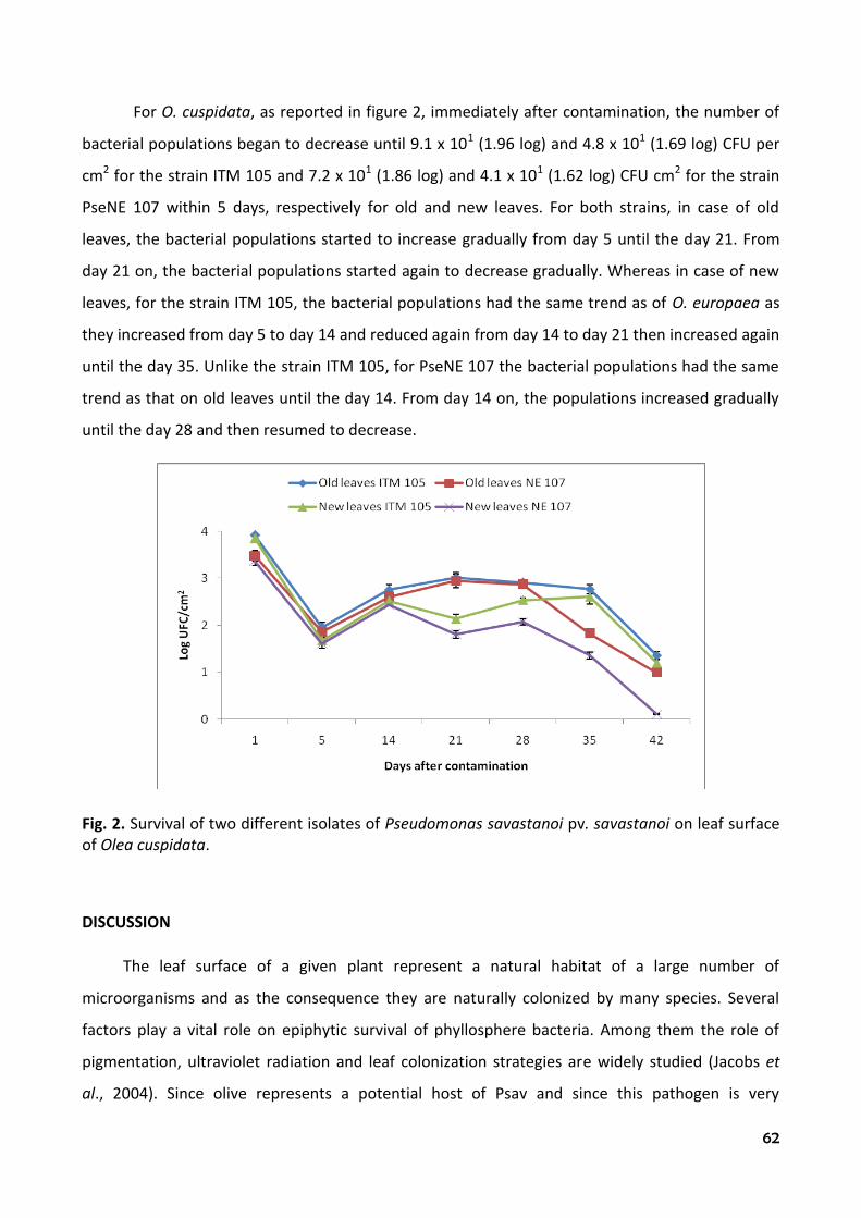
62
For O. cuspidata, as reported in figure 2, immediately after contamination, the number of
bacterial populations began to decrease until 9.1 x 101 (1.96 log) and 4.8 x 101 (1.69 log) CFU per
cm2 for the strain ITM 105 and 7.2 x 101 (1.86 log) and 4.1 x 101 (1.62 log) CFU cm2 for the strain
PseNE 107 within 5 days, respectively for old and new leaves. For both strains, in case of old
leaves, the bacterial populations started to increase gradually from day 5 until the day 21. From
day 21 on, the bacterial populations started again to decrease gradually. Whereas in case of new
leaves, for the strain ITM 105, the bacterial populations had the same trend as of O. europaea as
they increased from day 5 to day 14 and reduced again from day 14 to day 21 then increased again
until the day 35. Unlike the strain ITM 105, for PseNE 107 the bacterial populations had the same
trend as that on old leaves until the day 14. From day 14 on, the populations increased gradually
until the day 28 and then resumed to decrease.
Fig. 2. Survival of two different isolates of Pseudomonas savastanoi pv. savastanoi on leaf surface of Olea cuspidata.
DISCUSSION
The leaf surface of a given plant represent a natural habitat of a large number of
microorganisms and as the consequence they are naturally colonized by many species. Several
factors play a vital role on epiphytic survival of phyllosphere bacteria. Among them the role of
pigmentation, ultraviolet radiation and leaf colonization strategies are widely studied (Jacobs et
al., 2004). Since olive represents a potential host of Psav and since this pathogen is very

63
consistently present on the olive phylloplane respect to other microorganisms, also on the
asymptomatic plant phylloplane present in the open air, as demonstrated by Ercolani, 1978, we
focused to evaluate the survival capacity of this pathogen in a controlled environment in relation
to the different Olea species and leaf age.
Results obtained from both the experiments showed the ability of olive knot pathogen not
only to survive but to higher multiply on O. cuspidata respect to O. europaea. Both the strains of
Psav higher multiply on wild olive. In fact, beginning from very low CFU/cm2 remained from the
initial fall, the bacterium within 4 weeks reached a maximum peak. Nevertheless, bacterial survival
showed a further fall afterwards which has not been studied since this can happen only in
controlled environment while in the field other bacterial inocula can reach on the phylloplane
from different source adding or substituting to those previous and allowing a constant and
continue presence of the pathogen on the plant surface unlike to other pseudomonads (Ercolani,
1978).
The initial fall of bacterial population during the first five days could be probably due to the
fact that bacteria coming from natural (from infected tissues such as tubercle) or artificial
(substrate of culture) environments, suitable for their growth, do not posses a good capacity to
survive as epiphytes (Varvaro and Ferrulli, 1978). Only a few bacterial cell, probably due to the
modification caused by environment, are able to adjust and continue to live in a state of reduced
metabolism known as hypobiosis (Leben, 1974).
Italian isolate of Psav better multiplied on both olive species, respect to the isolate obtained
in Nepal with higher value expressed as CFU/cm2. This is probably because this isolate possesses a
high level of virulence attributable to the continue contact of the pathogen with the host from
centuries since olive is native of the Mediterranean region unlike European olive plants recently
introduced in Nepal.
The fact that the pathogen better multiplies on O. cuspidata, where the initial fall was
significantly lower respect to that observed on O. europaea, can be opined because of the capacity
of wild olive to release different solutes in leaf exudates and nutrients given their broad and
tender leaves. In fact, higher nutrient availability in the leaf exudates is found to be positively
correlated with the higher colonization of olive phylloplane by Psav (Balestra and Varvaro, 1997).
Higher multiplication of Psav on O. cuspidata phylloplane is very important since the presence of
this pathogen has not been reported in Nepal from areas where wild olive is present. For this

64
reason, strong control measures will be taken to avoid the diffusion of this pathogen from area
where infected European olive was found.
Only few significant differences were observed regarding the influence of leaf age on
multiplication of bacteria. Generally, higher multiplication was observed on old leaf surfaces than
the young ones.
REFERENCES
1. Armenise V. (1950). Cicli di accrescimento e differenziazione delle gemme in piante perenni nel
territorio di Bari. VII. L’accrescimento di Olea europaea L. negli anni 1948-1949. Nuovo Giornale
Botanico Italiano 57, 391-417.
2. Balestra G.M., Lamichhane J.R., Kshetri M.B., Mazzaglia A. and Varvaro L. (2009). First Report of
Pseudomonas savastanoi pv. savastanoi on olive in Nepal. Plant Pathology, 58, 393.
3. Balestra G.M. and Varvaro L. (1997). Influence of nitrogen fertilization on the colonization of
olive phylloplane by Pseudomonas syringae subsp. savastanoi. In: Pseudomonas syringae
pathovars and related pathogen, Edited by K. Rudolph.
4. Bartolucci P. and Dhakal B. (1999). (TCP/NEP/6713) Prospects for olive growing in Nepal, FAO
Technical documents. Online
5. Bradbury J.F. (1986). Guide to plant pathogenic bacteria. CAB International, Wallingford, U.K.
6. Crossman N.D. (2002). The impact of the European olive (Olea europaea L. subsp. europaea) on
grey box (Eucalyptus microcarpa Maiden) woodland in South Australia. Plant Protection Quarterly
17(4), 140-146.
7. Davis T.D. and Haissig B.E. (eds) 1994. Biology of Adventitious Root Formation. Plenum Press,
New York, London 342 pp.
8. Dye D.W., Bradbury J.F., Goto M., Hayward A.C., Lelliot R.A. and Schroth M.N. (1980).
International standards for naming pathovars of phytopathogenic bacteria and a list of pathovar
names and pathotype strains. Review of Plant Pathology 59, 153-168.
9. Ercolani G.L. (1978). Pseudomonas savastanoi and other bacteria colonizing the surface of olive
leaves in the field. Journal of General Microbiology 109, 245-257.
10. Gardan L., Bollet C., Abu-Ghorrah M.A., Grimont F. and Grimont P.A.D. (1992). DNA
relatedness among the pathovar strains of Pseudomonas syringae subsp. savastanoi Janse (1982)
and proposal of Pseudomonas savastanoi sp. International Journal of Systematic Bacteriology 42,
606-12.

65
11. Howard B.H. (1994). Manipulating rooting potential in stockplants before collecting cuttings.
In: Davis T.D. and B.E. Haissig (eds), Biology of Adventitious Root Formation. Plenum Press, New
York, London, 123-142.
12. Jacobs J.L., Carroll T.L. and Sundin G.W. (2005). The Role of Pigmentation, Ultraviolet Radiation
Tolerance, and Leaf Colonization Strategies in the Epiphytic Survival of Phyllosphere Bacteria.
Journal of Microbial Ecology 49, 104-113.
13. Lamichhane J.R., Balestra G.M. and Varvaro L. (2010). Phytobacteriological investigation on
Olea spp. in different districts of Nepal. Petria, 20 (2), 147-148.
14. Lavermicocca P., Lonigro S.L., Valerio F., Evidente A. and Visconti A. (2002). Reduction of olive
knot disease by a bacteriocin from Pseudomonas syringae pv. ciccaronei. Applied and
Environmental Microbiology. 68, 1403-1407.
15. Leben C. (1974). Survival of plant pathogenic bacteria. Ohio Agricultural Research and
Development Center, Special circular 100.
16. Marchi G., Mori B., Pollacci P., Mencuccini M. and Surico G. (2009). Systemic spread of
Pseudomonas savastanoi pv. savastanoi in olive explants. Plant Pathology 58, 152–158.
17. Negash L. (1995). Indigenous trees of Ethiopia, Biology, uses and propagation techniques. SLU
Reprocentralen, Umea, Sweden, 285 pp.
18. Noad T. and Birnie A. (1989). Trees of Kenya. 2nd edition. General Printers Ltd., Nairobi, 308
pp.
19. Parret A.H.A., K. Temmerman and De Mot R. (2005). Novel lectin-like bacteriocins of biocontrol
strain Pseudomonas fluorescens Pf-5. Applied and Environmental Microbiology 71, 5197-5207.
20. Ritchie G.A. (1994). Commercial application of adventitious rooting to forestry. In: Davis T.D.
and B.E. Haissig (eds), Biology of Adventitious Root Formation. Plenum Press, New York, London,
pp. 37-52.
21. Rodríguez-Moreno L., Jiménez A.J. and Ramos C. (2009). Endopathogenic lifestyle of
Pseudomonas savastanoi pv. savastanoi in olive knots. Microbial Biotechnology 2(4), 476-488.
22. Saad A.T. and Hanna L. (2002). Two new hosts of Pseudomonas savastanoi and variability in
strains isolated from different hosts. Phytopathology 92, S71.
23. Saad A.T. and Melkonian S. (1992). The distribution and host range of Pseudomonas syringae
sp. savastanoi in Lebanon. MPU/EPPO Conference on olive diseases, Sounion, 67.
24. Santangelo E., Balestra G.M., Varvaro L. and Soressi G.P. (1998). Ottenimento di piante di
pomodoro tolleranti sia a Fenthion che a Pseudomonas syringae pv. tomato a seguito di

66
rigenerazione in vitro, in presenza di Fenthion, di espianti cotiledonari eterozigoti per il gene Pto di
resistenza al batterio. Atti Giornate Fitopatologiche, 719-724.
25. Spennemann D.H.R. and Allen L.R. (2000). Feral olives (Olea europaea) as future woody weeds
in Australia: a review. Australian Journal of Experimental Agriculture 40(6), 889–901.
26. Quesada J.M., García A., Bertolini E., López M.M. and Penyalver R. (2007). Recovery of
Pseudomonas savastanoi pv. savastanoi from symptomless shoots of naturally infected olive trees.
International Microbiology 10, 77-84.
27. Quesada J.M., Penyalver R., Pérez-Panadés J., Salcedo C.I., Carbonell E.A. and López M.M.
(2010). Dissemination of Pseudomonas savastanoi pv. savastanoi populations and subsequent
appearance of olive knot disease. Plant Pathology 59, 262-269.
28. Varvaro L. and Ferrulli F. (1983). Sopravvivenza di Pseudomonas syringae pv. savastanoi
(Smith) Young et al. sulle foglie di due varietà di olivo (Olea europaea L.). Phytopathologia
Mediterranea 23, 1-4.
29. Varvaro L. and Surico G. (1978). Comportamento di diverse cultivars di olivo (Olea europaea L.)
alla inoculazione artificiale con Pseudomonas savastanoi pv. savastanoi (Smith) Stevens.
Phytopathologia Mediterranea 17(3), 174-177.
30. Varvaro L. and Surico G. (1987). Multiplication of wild types of Pseudomonas savastanoi pv.
savastanoi (Smith) Young et al. and their indolacetic deficient mutants in olive tissues.. In: Plant
pathogenic bacteria, (E. Civerolo et al. Editors), Martinus Nijhoff publishers, Dordrecht, 556-565.
31. Young J.M. (2004). Olive knot and its pathogens. Australasian Plant Pathology 33, 33-39.

67
CHAPTER 9. Reaction of two olive species to different Pseudomonas savastanoi pv. savastanoi strains
J. R. Lamichhane1, 2
1Dipartimento di Protezione delle Piante, Facoltà di Agraria, Università degli Studi della Tuscia,
01100, Viterbo, Italy;
2Central Horticulture Centre, Kirtipur, Kathmandu, Nepal

68
ABSTRACT
A native species of olive (Olea cuspidata Wall.) widespread in the Himalayan regions of
Nepal was tested for the first time for its susceptibility to Pseudomonas savastanoi pv. savastanoi,
the causal agent of olive knot disease. Five different strains of the bacterial pathogen isolated
from different geographic areas were used. European olive (O. europaea L.) was used as control.
Both Olea species were artificially inoculated by pricking plant stems of 3, 9 and 20 month of age.
O. europaea showed less susceptibility to P. savastanoi pv. savastanoi compare to O. cuspidata
although no difference statistically significant was found. Plant age did not influence significantly
the host-pathogen interaction. The bacterial strains showed differences statistically significant
among them in terms of bacterial populations inside the knot.
Key words: Olea europaea, Olea cuspidata, artificial inoculation, olive knot disease, Nepal
INTRODUCTION
Pseudomonas savastanoi pv. savastanoi (Gardan et al., 1992) (hereafter Psav) causes the
endemic disease of European olive (Olea europaea L.) known as olive knot or tubercle which
occurs in almost all the regions of the world where olive is grown, including the regions where this
crop has recently been introduced like Australia and Nepal (Balestra et al., 2009; Hall et al., 2004).
The bacterium survives epiphytically on asymptomatic olive plants (Bertolini et al., 2003; Ercolani,
1978; Marchi et al., 2009; Quesada et al., 2007; Penyalver et al., 2006) and enters into the plant
tissues once there are suitable conditions and multiplies rapidly affecting olive trees by producing
tumorous outgrowths on trunks, branches and occasionally on the leaves and fruits (Wilson,
1935). The pathogen can cause severe damage in olive groves, mainly when weather conditions
favour the survival of epiphytic populations of the bacterium and their entry into the bark. Psav
has epiphytic resident populations on olive twigs, leaves and drupes. Population sizes were
influenced by seasons, reaching a higher level in spring and fall, when wet weather conditions
occur (Ercolani, 1978). Tissues can be infected through leaf scars, wounds and fissures on stems
and twigs, caused by meteorological phenomena and insect miners, as well as by harvest and
pruning practices. Both olive yield and quality can be reduced as a consequence of bacterial
infections of the plant by the pathogen (Schroth, 1973).
The secreted phytohormones produced by Psav were thought to be the main factors
responsible for the disease caused by this plant pathogen for many years (Surico et al., 1985;
Surico and Iacobellis, 1992). Afterward, Psav mutants unable to produce IAA and/or cytokinins

69
multiplied in the host tissues as well as the parental strain confirmed that the phytohormones
alone are not responsible of the olive knot disease (Iacobellis et al., 1994). Some authors also
proved that the virulence of Psav is due to the capacity of this bacterium to produce hrp genes like
that of many other plant pathogenic bacteria (Lindgren, 1997; Sisto et al., 2004). More recently
other auxin-producing bacterial species were found to be actively involved in association with Psav
in knot development (Cimmino et al., 2006; Marchi et al., 2006; Ouzari et al., 2008).
Although European olive and its cultivar behaviour has largely been studied towards Psav
(Ercolani, 1978, 1991; Penyalver et al., 2006, Rodríguez-Moreno et al., 2008, Rodríguez-Moreno et
al., 2009; Quesada et al., 2010; Varvaro and Surico, 1978a, 1978b), no information is yet available
concerning the reaction of other olive species. In particular, we focused on African olive (Olea
cuspidata Wall.), which is widespread mostly in African, Asian and Oceanic regions (Crossman,
2002; Spennemann and Allen, 2000), to several isolates of Psav. O. cuspidata is widespread in
Himalayan regions of Nepal and represents an economically and ecologically very important crop
particularly in Dolpa, Bajura, Mugu and Humla districts (Bartolucci and Dhakal, 1999; Lamichhane
et al., 2010b). The objective of our study was to evaluate the susceptibility of O. cuspidata
comparing with that of O. europaea to determine the difference in their susceptibility to different
Psav isolates.
MATERIALS AND METHODS
Plant materials
In case of O. cuspidata, plants were obtained by directly sowing the seeds. Before sowing,
the seeds were put into a wide basin containing vermiculite, wetted and maintained at 4°C for 10
days since this species needs some chill hours for germination because of the seed dormancy.
After 10 days of chilling, the basin was kept in a greenhouse and maintained at 20°C for 2 months.
Afterwards, the plantlets were transplanted into the 2 litre plastic pots (15 cm wide and 30 cm
deep) containing soil, pit and sand (ratio 1:1:1). Whereas for O. europaea, two year old plants
obtained from rooted cuttings (vegetative propagation) were used. The plants were kept into the
same size plastic pots. Cultivar Leccino was used since this is well-known for its susceptibility to
Psav.
Bacterial cultures
In the experiments, five bacterial strains of Psav were used: one (PseNE 107) isolated in
Nepal (Balestra et. al., 2008), other Italian strains (ITM 105, strain 311) belong to Istituto Tossine e

70
micotossine da parassiti vegetali Bari (Marchi et al., 2009), two local isolates obtained in Viterbo
(VT) and Montefiascone (MF). The isolates were first grown for 48 h at 27±1°C in nutrient agar
containing 1% glycerol (NAG). Inoculum was prepared from 24 h cultures of Psav re-streaked on
the same medium and standardized turbidimetrically to a concentration of 108 CFU per ml of
sterile distilled water (SDW).
Inoculation techniques
Plants belonging both to O. europaea and O. cuspidata were inoculated at different stages of
development (3, 9 and 20 months of age) by pricking the stems with a 5-10 µl aliquot of bacterial
inoculums with a hypodermic needle containing bacterial suspension. Five plants per isolate (total
25 plants for each species) were inoculated at each stage of development. Each plant was
inoculated at two sites. Another set of plants (2 plants per strain per species) was inoculated with
5-10 µl of sterile distilled water (SDW) as negative control. Inoculated plants were kept in a
glasshouse where the temperature ranged from 20 to 24°C for the 120-day duration of the
experiment.
Disease observation
Inoculated plants belonging to both olive species were periodically controlled for the
observation of disease symptoms.
Statistical analysis
Data from each experiment were averaged, analyzed by analysis of variance (ANOVA) and
converted to CFU/g of knot weight. The average number of bacteria present into the every single
knot was determined.
RESULTS
Disease observation
Characteristic knot symptoms were observed on the inoculated plants of both olive species
within 5 weeks after inoculation. The values averaged from first, second and third artificial
inoculations on the disease appearance were reported in table 1. The symptoms were observed
before on O. cuspidata within the range of time between 3 and 4 weeks, whereas the knots
appeared lately on O. europaea (within 4 and 5 weeks) except those caused by Local Vt isolate.

71
Table 1. Appearance of olive knot symptoms on two olive species by strains of Pseudomonas savastanoi pv. savastanoi
Week after inoculation
Strains O. cuspidata O. europaea
ITM 105 3 4
311 4 5
PseNE 107 3 4
MF4 4 4
Local Vt 3 3
Sampling of knots and processing
After 6 months from the artificial inoculation the knots formed on the plants of both Olea
species were aseptically cut and weighed separately. Every single knot was separately fragmented
and homogenized by using a mixer (Ultra-Turrax T25), in presence of 20 ml of phosphate buffer
0.05 M, pH 6.6, for 2 min at 10,000 rpm. The suspensions obtained after mixing were then used
for ten-fold serial dilutions in phosphate buffer. Duplicate plating was carried out transferring an
aliquot of 0,1 ml in Petri dishes containing a specific medium PVF1 for P. savastanoi pv. savastanoi
(Surico and Marchi, 2002). Colony counts were performed after incubation of the dishes at 26±1°C
for 72 hours. The average number of bacteria present into every single knot was determined.
Calculation of bacterial populations present into the knots
The bacterial populations into each inoculation site (expressed as CFU/g of knot) obtained
separately in three different periods of time were reported in table 2. No difference statistically
significant was observed among the two olive species inoculated in the same period of time since
both of them were susceptible to the strains of bacterial isolates. A clear difference with values
statistically significant among the bacterial isolates were observed on plants inoculated at 3
months but not those inoculated at 9 and 20 months, except for the strain 311.

72
Table 2. Bacterial populations present in each gram of knot (log CFU/g) caused by different strains of Pseudomonas savastanoi pv. savastanoi on two Olea species of different age
3 months 9 months 20 months
Strains A B A B A B
ITM 105
7.47±0.17*
f
7.26±0.23
ef
7.16±0.31
b
7.14±0.26
b
6.45±0.16
bc
6.35±0.17
bc
311 4.95±0.08 a 5.03±0.14 a 5.29±0.26 a 5.35±0.29 a 4.89±0.20 a 4.69±0.11 a
NEP 6.69±0.13 cd 6.94±0.15 de 6.76±0.20 b 6.77±0.17 b 6.16±0.15 b 5.79±0.13 b
MF4 6.07±0.22 b 6.28±0.09 bc 6.78±0.77 b 6.69±0.27 b 6.40±0.47 bc 6.08±0.41 b
Local Vt 7.67±0.07 f 7.47±0.11 f 7.02±0.10 b 7.23±0.05 b 7.07±0.32 c 6.39±0.07 bc
A Olea cuspidata B Olea europaea
Means followed by the same letter are not significantly different at P = 0.05
* Standard error
On 3 month old plants, the bacterial populations of the local isolate Vt were the highest with
4.68 x 10⁷ (7.68 log) and 2.96 x 10⁷ (7.47 log) CFU/g of knot respectively on O. cuspidata and O.
europaea. In terms of bacterial populations per gram of knot, the isolate Vt was followed by the
strain ITM 105, NEP, MF and the strain 311. The latter was characterized by the lowest values of
bacterial populations with 8.92 x 10⁴ (4.95 log) and 1.08 x 10⁵ (5.03 log) CFU/g of knot respectively
on O. cuspidata and O. europaea. Same trend was observed also on the plants inoculated at 9 and
20 months where bacterial populations expressed as CFU/g remained unvaried (Tab. 2). Individual
values of bacterial populations obtained on plants inoculated at 3, 9 and 20 months showed that
there was no difference statistically significant since all the plants were infected and every
bacterial strain multiplied similarly in the host tissues (Tab. 2).
The values averaged from the three individual experiments on O. cuspidata showed that
there are difference statistically significant among the isolates in terms of bacterial populations
per gram of knot weight on the same olive species. In particular the isolate Local Vt, MF4 and 311
differed significantly among them with respective values of 1.8 x 10⁷, 2.6 x 10⁶ and 1.1 x 10⁵ (7.25
log, 6.42 log and 5.04 log) CFU/g of olive knot. Whereas no difference statistically significant was
found between the isolate ITM 105 and NEP (Fig. 1). The values obtained on O. europaea
confirmed the same trend since only isolate MF differed significantly with other four isolates (Fig.
2). Interestingly, rather than some difference statistically significant among the isolates on the

73
same species, no significant differences were found in bacterial populations of the same isolate
between the two Olea species (Figs. 1 and 2).
Fig. 1. Relation between average values of knot weight and bacterial populations inside the knots on Olea cuspidata
Fig. 2. Relation between average values of knot weight and bacterial populations inside the knots on Olea europaea
No correlation was found among the knot weight and their respected bacterial populations
on both olive species since knots with lower weight showed higher contents of bacterial
populations (Fig. 1). In fact, on O. cuspidata the local isolate Vt with average knot weight of 0.49 g

74
showed the highest bacterial populations of 1.8 x 10⁷ (7.25 log) CFU/g compared to the isolate ITM
105 which rather than a higher average knot weight (1.02 g) had a lower value with 1.07 x 10⁷
(7.02 log) CFU/g of bacterial populations (Fig. 1). The same trend was confirmed also on O.
europaea where no proportional increase in bacterial populations was found with higher knot
weight (Fig. 2).
DISCUSSION
Pseudomonas savastanoi pv. savastanoi strains inoculated artificially on two Olea species on
different stage of development showed that O. cuspidata reacts positively as O. europaea to Psav,
which is a new finding since this species native of Himalaya was unknown up to now for its
reaction to Psav. Unlike many wild plant species which are generally resistant to plant pathogens
and often used as source of resistance in plants breeding, O. cuspidata surprisingly gave positive
response to Psav. Values averaged from the three individual experiments showed that the
symptoms appearance was early on O. cuspidata by 1 week, except some isolates (MF4 and Local
Vt) which gave the same result on both Olea species. Nevertheless, the average knot weight was
higher on O. cuspidata for some strains (ITM 105, PseNE 107 and 311) and lower for other (MF4
and Local Vt). The average values of bacterial population expressed as CFU/g were higher for all
strains on O. cuspidata (Figs. 1 and 2) than on O. europaea although with difference not
statistically significant.
Preliminary study by using two different strains of Psav (ITM 105 and PseNE 107) on one
year old plants of O. europaea and O. cuspidata showed that the latter is more susceptible to the
bacterial pathogen both in terms of symptom appearance, knot weights and values of bacterial
populations (Lamichhane et al., 2010a). Our results are in accordance with the previous study,
since Psav strains previously used confirmed a clear difference both in terms of symptoms
appearance, knot weight and bacterial populations per gram of knot. Nevertheless, the new
strains of Psav used in this study did not give a clear difference between the two Olea species in
terms of knot weight and symptoms appearance with higher values on O. cuspidata. Less
susceptibility of O. europaea compare to that of O. cuspidata to Psav is probably due to the
continuous contact of P. savastanoi pv. savastanoi from centuries with O. europaea that led this
species to develop some kind of resistance compared to O. cuspidata, which had never been in
contact with Psav. Plant age did not influence significantly host-pathogen interaction since the

75
strains caused knot symptoms similarly on plants of different age with similar Psav average
populations.
The different time of symptom appearance and the different knot weight caused by the
strains of Psav on two different species was probably due to the different reactivity showed by the
two Olea species to artificial inoculations with Psav strains. The same hypothesis was previously
confirmed by Varvaro and Surico (1978a) on three cultivars of O. europaea. The lack of correlation
between the knot weight and their respective Psav populations confirm that knot weight is
independent by the number of Psav populations inside the infected tissues but depend probably
on the concentration of IAA produced by auxin-producing bacteria associated to Psav-induced
olive knots as described by previous authors (Cimmino et al., 2006; Marchi et al., 2006; Ouzari et
al., 2008). Hence, lower values of Psav population into the knot of bigger size might be because of
the higher population of other auxin-producing bacteria which probably limit Psav multiplication
by producing antibiotic compounds and competing for space and nutrients (Cimmino et al., 2006).
The overall findings reported in this study lead us to conclude that the absence of olive knot
symptoms on O. cuspidata and the consequent lack of Psav colonies from the leaves processed
during previous study (Lamichhane et al., 2010b) was not because of the resistance of O.
cuspidtata but due to the fact that the causal agent of olive knot disease is not present in the
Himalayan regions where O. cuspidata is widespread. Since Psav was already reported from Nepal
on O. europaea (Balestra et al., 2009) and given a high susceptibility of O. cuspidata to Psav, an
immediate implementation of control strategies based on the good agronomic and cultural
practices are strongly recommended to avoid the contamination and consequent diffusion of the
pathogen on O. cuspidata, given its wide economical and ecological importance in Nepal. In
particular, the infected materials from O. europaea must be collected and destroyed to avoid the
use of infected materials for vegetative propagation. If possible, it is to be hoped to spray copper
compounds on olive plants.
REFERENCES
1. Balestra G.M., Lamichhane J.R., Kshetri M.B., Mazzaglia A. and Varvaro L. (2009). First Report of
Pseudomonas savastanoi pv. savastanoi on olive in Nepal. Plant Pathology, 58, 393.
2. Bartolucci P. and Dhakal B. (1999). (TCP/NEP/6713) Prospects for olive growing in Nepal, FAO
Technical documents, Online.

76
3. Bertolini E., Penyalver R., García A., Olmos A., Quesada J.M., Cambra M. and López M.M. (2003).
Highly sensitive detection of Pseudomonas savastanoi pv. savastanoi in asymptomatic olive plants
by nested-PCR in a single closed tube. Journal of Microbiological Methods 52, 261-266.
4. Cimmino A., Andolfi A., Marchi G. and Surico G. (2006). Phytohormone production by strains of
Pantoea agglomerans from knots on olive plants caused by Pseudomonas savastanoi pv.
savastanoi. Phytopathologia Mediterranea 45, 247-252.
5. Crossman N.D. (2002). The impact of the European olive (Olea europaea L. subsp. europaea) on
grey box (Eucalyptus microcarpa Maiden) woodland in South Australia. Plant Protection Quarterly
17(4), 140-146.
6. Ercolani G.L. (1978). Pseudomonas savastanoi and other bacteria colonizing the surface of olive
leaves in the field. Journal of General Microbiology 109, 245-57.
7. Ercolani G.L. (1991) Distribution of epiphytic bacteria on olive leaves and the influence of leaf
age and sampling time. Microbial Ecology 21, 35-48.
8. Gardan L., Bollet C., Abu-Ghorrah M.A., Grimont F. and Grimont P.A.D. (1992). DNA relatedness
among the pathovar strains of Pseudomonas syringae subsp. savastanoi Janse (1982) and proposal
of Pseudomonas savastanoi sp. nov. International Journal of Systematic Bacteriology 42, 606-612.
9. Hall B.H., Cother E.J., Whattam M., Noble D., Luck J. and Cartwright D. (2004). First report of
olive knot caused by Pseudomonas savastanoi pv. savastanoi on olives (Olea europaea) in
Australia. Australasian Plant Pathology 33, 433-436.
10. Iacobellis N.S., Sisto A., Surico G., Evidente A. and Di Maio E. (1994). Pathogenicity of
Pseudomonas syringae subsp. savastanoi mutants defective in phytohormone production. Journal
of Phytopathology 140, 238-48.
11. Lamichhane J.R., Balestra G.M. and Varvaro L. (2010a). Comparison between two different
olive species on their susceptibility towards Pseudomonas savastanoi pv. savastanoi. The 12th
International Conference on Plant Pathogenic Bacteria, La Réunion Island 7-11 June, p. 103.
12. Lamichhane J.R., Balestra G.M. and Varvaro L. (2010b). Phytobacteriological investigation on
Olea spp. in different districts of Nepal. Petria, 20 (2), 147-148.
13. Lindgren P.B. (1997). The role of hrp genes during plant-bacterial interactions. Annual Review
of Phytopathology 35, 129-152.
14. Marchi G., Mori B., Pollacci P., Mencuccini M. and Surico G. (2009). Systemic spread of
Pseudomonas savastanoi pv. savastanoi in olive explants. Plant Pathology 58, 152-8.
77
15. Marchi G., A. Sisto, A. Cimmino, A. Andolfi, A. Evidente and Surico G., 2006. Interaction
between Pseudomonas savastanoi and Pantoea agglomerans in the olive knots. Plant Pathology
55, 614-624.
16. Ouzari H., Khsairi A., Raddadi N., Jaoua L., Hassen A., Zarrouk M., Daffonchio D. and Boudabous
A. (2008). Diversity of auxin-producing bacteria associated to Pseudomonas savastanoi-induced
olive knots. Journal of Basic Microbiology, 48, 1-8.
17. Penyalver R., García A., Ferrer A., Bertolini E., Quesada J.M., Salcedo C.I., Piquer J., Pérez-
Panadés J., Carbonell E.A., del Río C., Caballero J.M. and López M.M. (2006). Factors affecting
Pseudomonas savastanoi pv. savastanoi plant inoculations and their use for evaluation of olive
cultivar susceptibility. Phytopathology 96, 313-9.
18. Quesada J.M., García A., Bertolini E., López M.M. and Penyalver R. (2007). Recovery of
Pseudomonas savastanoi pv. savastanoi from symptomless shoots of naturally infected olive trees.
International Microbiology 10, 77-84.
19. Quesada J.M., Penyalver R., Pérez-Panadés J., Salcedo C.I., Carbonell E.A. and López M.M.
(2010). Dissemination of Pseudomonas savastanoi pv. savastanoi populations and subsequent
appearance of olive knot disease. Plant Pathology 59, 262-269.
20. Rodríguez-Moreno L., Barceló-Munõz A. and Ramos C. (2008). In vitro analysis of the
interaction of Pseudomonas savastanoi pvs. savastanoi and nerii with micropropagated olive
plants. Phytopathology 98, 815-22.
21. Rodríguez-Moreno L., Jiménez A.J. and Ramos C. (2009). Endopathogenic lifestyle of
Pseudomonas savastanoi pvs. savastanoi in olive knots. Microbial Biotechnology 2, 476-88.
22. Schroth M.N. (1973). Quantitative assessment of the effect of the olive knot disease on olive
yield and quality. Phytopathology 63, 1064-5.
23. Sisto A., Cipriani M.G. and Morea M. (2004). Knot formation caused by Pseudomonas syringae
subsp. savastanoi on olive plants is hrp dependent. Phytopathology 94, 484-9.
24. Spennemann D.H.R. and Allen L.R. (2000). Feral olives (Olea europaea) as future woody weeds
in Australia: a review. Australian Journal of Experimental Agriculture 40(6), 889-901.
25. Surico G. and Iacobellis N.S. (1992). Phytohormones and olive knot disease. Pages 209-229 in:
Molecular Signals in Plant-Microbe Communications. D. P. S. Verma, ed. CRC Press, Boca Raton, FL.
26. Surico G., Iacobellis N.S. and Sisto S. (1985). Studies on the role of indole- 3-acetic acid and
cytokinins in the formation of knots on olive and oleander plants by Pseudomonas syringae pv.
savastanoi. Physiological Plant Pathology 26, 309-20.

78
27. Surico G. and Marchi G. (2002). Olive knot disease: new insights into the ecology, physiology
and epidemiology of Pseudomonas savastanoi pv. savastanoi. Sixth International Conference on
Pseudomonas syringae pathovars and related pathogens, Maratea, Italy, September 15-19.
28. Surico G., Sparapano L., Lerario P., Durbin R.D. and Iacobellis N. (1976). Studies on growth
promoting substances produced by Pseudomonas savastanoi. Proceedings of IV MPU Congress,
Zadar, 449-458.
29. Varvaro L. and Surico G. (1978a). Comportamento di diverse cultivars d’Olivo (Olea europaea
L.) alla inoculazione artificiale con Pseudomonas savastanoi (E. F. Smith) Stevens. Phytopathologia
Mediterranea 17, 174-177.
30. Varvaro L. and Surico G. (1978b). moltiplicazione di Pseudomonas savastanoi (E. F. Smith)
Stevens nei tessuti dell’Olivo (Olea europaea L.). Phytopathologia Mediterranea 17(3), 179-186.
31. Vivian A. and Mansfield J. (1993). A proposal for a uniform genetic nomenclature for avirulence
genes in phytopathogenic pseudomonads. Molecular Plant-Microbe Interactions 6, 9-10.
32. Wilson E.E. (1935). The olive knot disease: its inception, development and control. Hilgardia 9,
233-64.

79
CHAPTER 10. In vitro antagonistic action of the bacterial species from Olea spp. phylloplane against Pseudomonas savastanoi pv. savastanoi J. R. Lamichhane1, 2
1Dipartimento di Protezione delle Piante, Facoltà di Agraria, Università degli Studi della Tuscia,
01100, Viterbo, Italy;
2Central Horticulture Centre, Kirtipur, Kathmandu, Nepal

80
ABSTRACT
Over three growing seasons, more than 250 bacterial strains were isolated from the
phylloplane of two asymptomatic species of olive plants in Nepal. Eighty six representative strains
of them were screened for their antagonistic behavior against five different strains of
Pseudomonas savastanoi pv. savastanoi, the causal agent of olive knot disease. None of the
bacterial isolates exhibited in vitro antagonism against the tested phytopathogen.
Key words: Antagonism, phylloplane isolates, Nepal
INTRODUCTION
Aerial leaf surfaces are habitat for epiphytic bacteria, which are able to colonize plant leaf
surfaces and survive for a long time even under difficult physical conditions (temperature, rainfall).
Bacteria are generally the predominant initial inhabitants of the newly expanded phylloplane,
while yeasts and filamentous fungi dominate after in the growing season (Kinkel et al., 1987). The
common phylloplane resident bacteria including plant pathogens (PPB) live epiphytically on host
and nonhost plant species (Beattie and Lindow, 1995; Blakeman and Fokkema, 1982). Several
studies provided information regarding antagonism between the microorganisms on the leaf
surface: in particular, the role of some saprophytic bacteria from plant phylloplane in controlling
the growth and multiplication of pathogenic bacteria, known as biological control. Hence, the
latter maybe an alternative to chemicals in the control of some pathogenic bacteria which provide
many economic and ecologic advantages.
Bacterial knot of olive (Olea europaea L.) caused by Pseudomonas savastanoi pv. savastanoi
(Psav) is an endemic disease of great economic importance which has been reported in almost all
olive cultivated regions. Besides olive, the pathogen causes a similar disease on many other plant
species (Bradbury 1986; Saad and Hanna, 2002). The pathogen causes outgrowths (tumorous
growth or knots) on the stems, branches and occasionally on the leaves and fruits of host plants.
Besides from knots, Psav has been isolated from the phylloplane of diseased and healthy olive
leaves and stems where the pathogen has a resident epiphytic phase. Psav enters into the plant
tissues and causes the disease only by means of wounds created by cultural practices and
meteorological phenomena (Ercolani, 1991; Quesada et al., 2007).
Biological control of several plant pathogens have widely been studied in the last decades.
Nevertheless, only little is known on biocontrol of P. savastanoi pv. savastanoi by using

81
fluorescent pseudomonas (Lavermicocca et al., 2002, Zadeh et al., 2008). Since the effectiveness
of a bacterial strain as a biocontrol agent depend on the ecological similarity between the
antagonist and the target pathogen in the phyllosphere of the host plant (Völksch and May, 2001)
we focused to the saprophytic bacteria from olive plants in searching those antagonist against
Psav. The objective of this study was to isolate the bacterial species from the phylloplane of field
grown and naturally present olive species in Nepal and to assess their potential antagonistic
activities against Psav by using in vitro screening methods.
MATERIALS AND METHODS
Plant material and field
Two species of olive plants, one field grown (Olea europaea L.) and another native species of
Nepal (O. cuspidata Wall.) were used in this study. Twenty eight olive cultivars belong to O.
europaea from three different districts (Bajura, Dolpa and Kathmandu) situated respectively at
1500, 2500 and 1300m ASL and O. cuspidata from the same districts were used in this study. The
cultivars belong to O. europaea were: Anghiari, Ascolana, Bosana, Bourbon, Canino, Carboncella,
Carolea, Cassanese, Cipressino, Coratina, Corona, Femminella, Frantoio, Frattese, Itrana, Leccino,
Leoncino, Marina, Maurino, Moraiolo, Nocellara del Belice, Pendolino, Piantone, Rajo, Rasara,
Rosciola, Taggiasca and Valle Corsana.
Sampling of olive leaves
Leaf samples were randomly collected from all the asymptomatic plants during a 3-year
period (April 2007- April 2009) in Bajura and only for one year period (April 2009) in Dolpa and
Kathmandu districts. Sampling was made early in the morning. For O. europaea, 40 leaves (both
young and old leaves) from each plant and five plants for each variety for a total of 200 leaves per
variety were aseptically collected in sterile lab bags and bulked. For O. cuspidata, the same
procedure was used by collecting 10 samples, each represented by 200 leaves.
Isolation of bacteria from plant material
Each bulk sample was aseptically divided in 10 groups (20 leaves per replicate), placed in a
sterile 250ml conical flask with 40 ml of phosphate buffer (0.1M, pH 6.8) and shaken in the dark by
using an orbital stirrer at 26±1 °C and 150 rpm for 2h. Previous studies indicated that over 90% of
the phylloplane bacteria passed into suspension and that there was no error due to bacteria
multiplying in the washing fluid (Crosse, 1959; Ercolani, 1978). These results were obtained by
previous authors by plating 10-fold serial dilutions of homogenates of washed and unwashed

82
leaves. Furthermore, Ercolani (1976) showed that virtually no bacteria passed into suspension
from insides the leaves which allow us to make recovery of only phylloplane bacteria. After
washing, the solution was serially diluted in phosphate buffer and spread plated in duplicate onto
nutrient agar medium supplemented by 5% (v/v) of sucrose (NAS), amended by 800 µl/l of
cycloheximide (previously prepared as stock solution by diluting 1g in 10 ml of ethanol) to prevent
fungal contamination. One hundred µl of diluted solution was used for plating. After 48-72 h of
incubation at 26±1°C, single colonies were removed with a sterile needle and transferred to fresh
NAS medium. Examination by means of a stereomicroscope of the developed bacterial colonies,
Gram stain and fluorescent tests were carried out in order to classify the isolates into groups for
further identification. For in vitro biocontrol assay 45, 23 and 18 representative isolates, including
both non-pseudomonads and fluorescent pseudomonads, respectively from Bajura, Dolpa and
Kathmandu, were used. Although fluorescent pseudomonads are known to be the most efficient
antagonists (Kearns and Hale, 1996; Patil et al., 1998; Zadeh et al., 2008), we evaluated all
phylloplane bacteria.
Identification of bacterial isolates
Isolates were identified at genus level by using the diagnostic tables of Cowan and Steel
(1965) by which they were divided in groups to obtain only those effectively different, since
bacteria colonizing olive phylloplane are frequently represented by only few species (Ercolani,
1978). Later, the isolates were identified at species level by molecular analysis by sequencing the
16S rDNA region of each representative isolate for each bacterial species. A 1500-bp region of the
16S rDNA was amplified by using the non specific primers NOC 1F and NOC 3R and sequenced. A
BlastN search was performed for comparing their similarity with the strains present in the
database (GenBank, EMBL and DDBJ).
Strains of bacterial pathogen
Five strains of P. savastanoi pv. savastanoi with different geographical origins were used.
PseNE 107 isolated from olive in Nepal (Balestra et. al., 2008), Italian strains ITM 105 and 311,
belong to Istituto Tossine e micotossine da parassiti vegetali Bari (Marchi et al., 2009) and two
local isolates obtained in Viterbo (VT) and Montefiascone (MF), were used in the tests.
Detection of antagonistic activity
Conventional deferred antagonism assays were performed as described by Parret et al.
(2003): agar diffusion method relying on detection of halo formation in an overlay of indicator

83
cells due to growth inhibition by chloroform-killed producer cells. The growth inhibition was
quantified by measuring IZR (inhibition zone radius) from the edge of the producer colony.
RESULTS
Molecular identification as described above showed that the phylloplane isolates obtained
from olea species were Bacillus asahii, Bacillus cereus, Bacillus circulans, Bacillus megaterium,
Bacillus pumilus, Bacillus silvestris, Bacillus simplex, Bacillus subtilis, Brenneria serratia,
Microbacterium testaceum, Micrococcus luteus, Pantoea agglomerans, Pseudomonas fluorescens,
Pseudomonas putida and Xanthomonas campestris group.
None of the bacterial isolates from phylloplane mentioned above inhibited growth of Psav
strains in vitro.
DISCUSSION
Although related phytopathogenic and non-pathogenic Pseudomonas bacteria have been
reported to inhibit P. savastanoi pv. savastanoi strains (Lavermicocca et al., 2002; Zadeh et al.,
2008), in our case no growth inhibition of Psav was observed by the phylloplane bacteria isolated
from olive plants. The absence of the possible bacterial antagonist of Psav on olive phylloplane is
probably due to the fact that all the leaf samples have been collected from the asymptomatic olive
plants where Psav was not present (Lamichhane et al., 2010). Previous study demonstrated that
the presence and the effectiveness of a given antagonist from phyllosphere of the infected plants
depend on the ecological similarity between the antagonist and the target pathogen (May et al.,
1996; Völksch and May, 2001). Hence, the lack of olive knot pathogen on olive phylloplane could
be related to the consequent absence of the antagonist. For this reason, the study of phylloplane
bacteria of the infected plants together with those of healthy one will give a more precise idea on
the possible presence of antagonist in relation to that of the pathogen and their effectiveness as
biocontrol agents.
Antagonistic bacteria of P. savastanoi pv. savastanoi reported until now are only few and
they all are isolates from plant rhizoplane (Zadeh et al., 2008). However, we attempted to isolate
the antagonist of Psav for the first time but with no satisfactory result.

84
REFERENCES
1. Beattie G.A. and Lindow S.E. (1995). The secret life of foliar bacterial pathogens on leaves.
Annual Review of Phytopathology 33, 145-172.
2. Blakeman J.P. and Fokkema N.J. (1982). Potential for biological control of plant diseases on the
phylloplane. Annual Review of Phytopathology 20, 167-192.
3. Bradbury J.F. (1986). Guide to plant pathogenic bacteria. CAB International, Wallingford, U.K.
4. Ercolani G.L. (1991) Distribution of epiphytic bacteria on olive leaves and the influence of leaf
age and sampling time. Microbial Ecology 21, 35-48.
5. Kearns L.P. and Hale C.N. (1996). Partial characterization of an inhibitory strain of Erwinia
herbicola with potential as a biocontrol agent for Erwinia amylovora, the fire blight pathogen.
Journal of Applied Bacteriology, 81, 369–374.
6. Kinkel L.L., J.H. Andrews, F.M. Berbee and Nordheim E.V. (1987). Leaves as islands for microbes.
Oecologia, 71,405–408.
7. Lamichhane J.R., Balestra G.M. and Varvaro L. (2010). Phytobacteriological investigation on Olea
spp. in different districts of Nepal. Petria, 20 (2), 147-148.
8. Lavermicocca P., Lonigro S.L., Valerio F., Evidente A. and Visconti A. (2002). Reduction of olive
knot disease by a bacteriocin from Pseudomonas syringae pv. ciccaronei. Applied and
Environmental Microbiology. 68, 1403-1407.
9. May R., Völksch B. and Kampmann G. (1997). Antagonistic Activities of Epiphytic Bacteria from
Soybean Leaves against Pseudomonas syringae pv. glycinea in vitro and in planta. Microbial
Ecology 34, 118–124.
10. Patil R., K. Jagadeesh, P. Krishnaraj and Kulkarni J. (1998). Bacterization of groundnut with
Pseudomonas fluorescens for the control of collar rot caused by Sclerotium rolfsii Sacc. Karnataka
Journal of Agricultural Sciences, 11(2), 423–425.
11. Quesada J.M., García A., Bertolini E., López M.M. and Penyalver R. (2007). Recovery of
Pseudomonas savastanoi pv. savastanoi from symptomless shoots of naturally infected olive trees.
International Microbiology 10, 77-84.
12. Rokni Zadeh H., Khavazi K., Asgharzadeh A., Hosseinimazinani M. and De Mot R. (2008).
Biocontrol of Pseudomonas savastanoi, causative agent of olive knot disease: antagonistic
potential of non-pathogenic rhizosphere isolates of fluorescent pseudomonas. Applied Biological
Science 73(1), 199-203.

85
13. Saad A.T. and Hanna L. (2002). Two new hosts of Pseudomonas savastanoi and variability in
strains isolated from different hosts. Phytopathology 92, S71.
14. Völksch B. and May R. (2001). Biological control of Pseudomonas syringae pv. glycinea by
epiphytic bacteria under field conditions. Microbial Ecology 41, 1284-1292.

86
CHAPTER 11. GENERAL DISCUSSION

87
Olive knot disease was observed in Nepal only on the plants present in the private orchard of
Makwanpur where O. europaea was introduced two decades ago unlike those introduced in
Bajura, Dolpa and Kathmandu three years ago. This finding might be linked to the fact that
Pseudomonas savastanoi pv. savastanoi arrived together with the plant materials shipped by
private growers from other countries given that no necessary control measures were performed
during plant’s shipment.
Besides few studies relative to the bacterial ecology of European olive phylloplane present in
the Mediterranean ecosystems, no studies were made in other regions of the world neither on
cultivated olive nor on other species belonging to Olea. The present study has shown the
difference among the bacterial ecology of the phylloplane of both cultivated and naturally present
olive species in several Himalayan regions of Nepal characterized by different climatic and
geographic conditions. The lower colonization of olive phylloplane respect to the values previously
found by some authors can be explained by two possible reasons: a) our study was carried out in
an environment which is new for european olive which has recently been introduced in these
regions, so colonization of a new species probably takes longer time respect to that of the
Mediterranean ecosystem which is native of Olea species with thousand years of interactions with
bacterial phylloplane community; b) drought stress could have influenced negatively the growth
and multiplication of epiphytic bacteria, given that average values of rainfall are very low in these
Districts in the spring, since almost all rainfall occurs during the monsoon season between July and
August. Widely different climatic and geographic conditions of the three Districts can be possible
reason of the different colonization of olive phyllosphere. Moreover, high altitudes and the
presence of constant afternoon strong wind blows which characterize Bajura and Dolpa Districts
may have had negatively effect on bacterial epiphytic survival reducing the moisture of leaf
surfaces and relative humidity which favours epiphytic bacterial multiplication. The wind can even
transport bacteria from this area to the lower parts of that region, as bacterial immigration of
consistent levels has been demonstrated already which probably is favoured also through wind.
Moreover, lower bacterial epiphytic populations can be explained since when the prevailing high
temperature can enhance bacterial growth, often water is scarce in Bajura and Dolpa Districts,
unlike in Kathmandu District, and when water is available, temperatures are not always
favourable. Regarding bacterial communities of Olea phylloplane, the dominant bacterial
colonizers were P. agglomerans and X. campestris group followed by different endospore-forming
Bacillus spp., the latter because of their resistant structures are frequently present and isolated

88
from plant phylloplane also from difficult environments. The abundance of P. agglomerans can be
explained easily since this species are considered to be ubiquitous in the phylloplane of various
crop plants together with Pseudomonas. No detection of P. savastanoi pv. savastanoi on Olea
phylloplane is due to the non presence of this bacterial pathogen in these Himalayan Districts,
except in Makwanpur where olive has been introduced before. Bacterial species identified from
Olea phylloplane in Nepal are significantly different respect to those found in south-east Italy.
Our results showed the ability of olive knot pathogen not only to survive but to higher
multiply on O. cuspidata respect to O. europaea. In fact, beginning from very low CFU/cm2 the
bacterium remained from the initial fall and within 4 weeks its population reached a maximum
peak. The initial fall of bacterial population during the first five days is probably for the fact that
bacteria coming from natural (from infected tissues such as tubercle) or artificial (substrate of
culture) environments, suitable for their growth, do not posses a good capacity to survive as
epiphytes. Italian isolate of Psav better multiplied on both olive species, respect to the isolate
obtained in Nepal with higher value expressed as CFU/cm2. This is probably because this isolate
possesses a high level of virulence attributable to the higher capacity of the pathogen in producing
auxin and cytokinins. Only few significant differences were observed regarding the influence of
leaf age on multiplication of bacteria. Generally, higher multiplication was observed on old leaf
surfaces than the young one, which probably be given by higher nutrient contents.
Pseudomonas savastanoi pv. savastanoi strains artificially inoculated on two Olea species on
different stage of development showed that O. cuspidata reacts positively as O. europaea to Psav,
which is a new finding since this species native of Himalaya was unknown up to now for its
reaction to Psav. Less susceptibility of O. europaea compare to that of O. cuspidata to Psav is
probably due to the continuous contact of P. savastanoi pv. savastanoi from centuries with O.
europaea that led this species to develop some sort of resistance compared to O. cuspidata, which
had never been in contact with Psav. The different time of symptom appearance and the different
knot weight caused by the strains of Psav on two different species was due to the different
reactivity showed by two Olea species to artificial inoculations. The lack of correlation between
the knot weight and their respective bacterial populations confirms that knot weight is
independent by the number of bacterial populations inside the infected tissues but depend on the
concentration of growth hormone (IAA and Cytokinin) produced by the bacterial pathogen inside
the infected tissues. Hence higher values of bacterial populations inside the knot, in particular
those of the strain Local Vt and ITM 105, were due to the higher virulence of these strains given

89
probably by the higher concentration of plants hormones produced by them. In addition, plants
age did not influence significantly host-pathogen interaction since all the strains reacted similarly
on plants of different age with similar values.
Although related phytopathogenic and non-pathogenic Pseudomonas bacteria have been
reported to inhibit P. savastanoi pv. savastanoi strains, no growth inhibition of Psav was observed
by our olive phylloplane bacteria. The absence of the possible bacterial antagonist against Psav on
olive phylloplane is probably due to the fact that all the leaf samples have been collected from the
asymptomatic olive plants, where Psav was not present. Previous study demonstrated that the
presence and the effectiveness of a given antagonist from phyllosphere of the infected plants
depend on the ecological similarity between the antagonist and the target pathogen. Hence, the
lack of olive knot pathogen on olive phylloplane could be related to the consequent absence of the
antagonist. For this reason, the study of phylloplane bacteria of the infected plants together with
those of healthy one will give a more precise idea on the possible presence of antagonist in
relation to that of the pathogen and their effectiveness as biocontrol agents. In addition,
antagonist bacteria of P. savastanoi pv. savastanoi reported until now are very few and they all
have been isolated from plant rhizoplane. However, we attempted to isolate the antagonist of
Psav for the first time but with no satisfactory result.

90
CHAPTER 12. CONCLUSIONS

91
The finding of Pseudomonas savastanoi pv. savastanoi in Nepal must be considered seriously
and seeks immediate implementation of control strategies since this pathogen is not present in
other areas, except than in Makwanpur, where Olea spp. are present. Only the use of the
adequate agronomic and cultural practices can be the effective way to avoid the contamination
and consequent diffusion of Psav to the other areas where wild olive species, in particular O.
cuspidata, are present since this species is found to be very susceptible to Psav.
Bacterial phylloplane colonizers of O. europaea and O. cuspidata were almost the same,
although in different proportions. Only some of them are are similar as those described by some
previous authors on O. europaea from the Mediterranean areas. Lower average bacterial
phylloplane populations respect to those found in Mediterranean climate might probably be due
to the less suitable climatic conditions in Nepal.
Olea cuspidata, an unknown olive species up to now for its reaction to Psav showed to be
highly suitable for the survival and multiplication of Psav, which is a new finding. In fact, Psav
better multiply on O. cuspidata respect to O. europaea. Higher bacterial multiplication on old
leaves compare to that on new ones is associated to the higher nutrient contents of the old leaves.
Psav not only better survive and multiply epiphytically on O. cuspidata but also into the plant
tissues since both the symptom appearance, average knot weight and the bacterial populations
inside the knots are higher on this species respect to that of O. europaea during the artificial
inoculations. Higher susceptibility of O. cuspidata to Psav is probably due to the fact that this olive
species has never been in contact with Psav and the plant did not develop the resistance, unlike O.
europaea which has continuously been in contact with Psav with consequent evolution from
centuries.
In nature, the survival and multiplication of a pathogenic bacteria are often influenced by
other saprophytic and possible antagonistic species, since ecological similarity between different
bacterial species on phylloplane has been demonstrated. The lack in finding from Olea phylloplane
of possible antagonists against Psav can be related to the absence of the bacterial pathogen on
Olea phylloplane.

92
CHAPTER 13. PHOTOGRAPHS

93
Fig. 1. Surveyed Districts and reported olive species in Nepal
Fig. 2. European olive planted within the FAO project in Kathmandu District
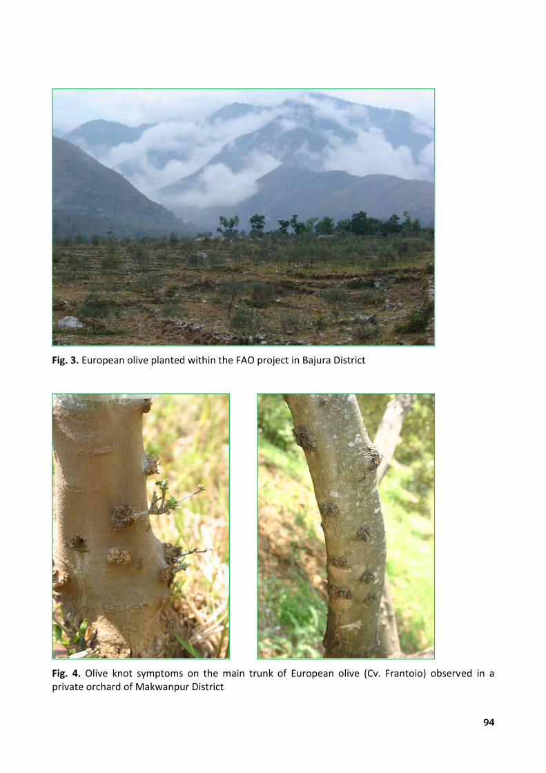
94
Fig. 3. European olive planted within the FAO project in Bajura District
Fig. 4. Olive knot symptoms on the main trunk of European olive (Cv. Frantoio) observed in a private orchard of Makwanpur District

95
Fig. 5. Natural forests of wild olive (Olea cuspidata) present in Bajura District (a)
Fig. 6. Natural forests of wild olive (Olea cuspidata) present in Bajura District (b)

96
Fig. 7. Secular plant of wild olive (Olea cuspidata) present in Dolpa District
Fig. 8. Olive fruits of Olea cuspidata (left) and Olea europaea (right)

97
Fig. 9. Production of wild olive (Olea cuspidata) by seed in the greenhouse
Fig. 10. Olive knot symptoms developed on 9 month old plantlets of artificially inoculated wild olive (Olea cuspidata)

98
Fig. 11. Olive knot symptoms developed on 20 month old plantlets of artificially inoculated wild olive (Olea cuspidata)
Fig. 12. Olive knot symptoms developed on 20 month old plantlets of artificially inoculated european olive (Olea europaea cv. Frantoio)

99
ACKNOWLEDGEMENTS
Many thanks to my mom, Shanti, for her continuing love in support of my study All the tears of joy you shed for me every time you saw me or heard my voice have given me a lot of energy.
Heartily thanks to my dad Rewati P. Lamichhane, who had a dream to see his child receiving
the PhD. A special thank to my brother Uttam for letting me give several chances. I never would have
achieved this goal without your support. Thanks to all my sisters. No one has ever been received more love and unconditional support
than I have been by you all. I am grateful to all the field workers of Kolti (Bajura), Juphal (Dolpa) and Kirtipur
(Kathmandu) Districts for their help during the field surveys, sample collection and shipments. I am thankful to the colleagues from Department of Plant Protection, University of Tuscia, in
particular to Dr. Antonio Rossetti, Dr. Maria Pia Aleandri, Dr. Mariagrazia Antonelli, Dr. Diana Martignoni and Dr. Angelo Mazzaglia. I have shared with you a superb Doctoral period.
Many thanks to Dr. Alfredo Fabi for technical support in laboratory activities.
I am really grateful to Prof. Andrea Vannini, the coordinator of the FAO project (GCP/NEP/056/ITA), for involving me within the project during which I got chance to survey and study olive fields in Nepal.
A particular thanks to Dr. Giorgio M. Balestra, a hard working researcher, for the continuous
support. Finally, let me thank my Supervisor Prof. Leonardo Varvaro, a big hearted person, for
offering me continuous support, suggestions and encouragements. You have not only been my supervisor at work but also an excellent guardian.