trypsin-like proteases and their role in muco-obstructive
TRANSCRIPT

International Journal of
Molecular Sciences
Review
Trypsin-Like Proteases and Their Role in Muco-ObstructiveLung Diseases
Emma L. Carroll 1,†, Mariarca Bailo 2,†, James A. Reihill 1 , Anne Crilly 2 , John C. Lockhart 2, Gary J. Litherland 2,Fionnuala T. Lundy 3 , Lorcan P. McGarvey 3, Mark A. Hollywood 4 and S. Lorraine Martin 1,*
�����������������
Citation: Carroll, E.L.; Bailo, M.;
Reihill, J.A.; Crilly, A.; Lockhart, J.C.;
Litherland, G.J.; Lundy, F.T.;
McGarvey, L.P.; Hollywood, M.A.;
Martin, S.L. Trypsin-Like Proteases
and Their Role in Muco-Obstructive
Lung Diseases. Int. J. Mol. Sci. 2021,
22, 5817. https://doi.org/10.3390/
ijms22115817
Academic Editor: Daniel Taillandier
Received: 7 May 2021
Accepted: 26 May 2021
Published: 29 May 2021
Publisher’s Note: MDPI stays neutral
with regard to jurisdictional claims in
published maps and institutional affil-
iations.
Copyright: © 2021 by the authors.
Licensee MDPI, Basel, Switzerland.
This article is an open access article
distributed under the terms and
conditions of the Creative Commons
Attribution (CC BY) license (https://
creativecommons.org/licenses/by/
4.0/).
1 School of Pharmacy, Queen’s University, Belfast BT9 7BL, UK; [email protected] (E.L.C.);[email protected] (J.A.R.)
2 Institute for Biomedical and Environmental Health Research, School of Health and Life Sciences,University of the West of Scotland, Paisley PA1 2BE, UK; [email protected] (M.B.);[email protected] (A.C.); [email protected] (J.C.L.); [email protected] (G.J.L.)
3 Wellcome-Wolfson Institute for Experimental Medicine, School of Medicine, Dentistry andBiomedical Sciences, Queen’s University, Belfast BT9 7BL, UK; [email protected] (F.T.L.);[email protected] (L.P.M.)
4 Smooth Muscle Research Centre, Dundalk Institute of Technology, A91 HRK2 Dundalk, Ireland;[email protected]
* Correspondence: [email protected]† These authors contributed equally to this work.
Abstract: Trypsin-like proteases (TLPs) belong to a family of serine enzymes with primary substratespecificities for the basic residues, lysine and arginine, in the P1 position. Whilst initially perceivedas soluble enzymes that are extracellularly secreted, a number of novel TLPs that are anchored inthe cell membrane have since been discovered. Muco-obstructive lung diseases (MucOLDs) arecharacterised by the accumulation of hyper-concentrated mucus in the small airways, leading topersistent inflammation, infection and dysregulated protease activity. Although neutrophilic serineproteases, particularly neutrophil elastase, have been implicated in the propagation of inflammationand local tissue destruction, it is likely that the serine TLPs also contribute to various disease-relevantprocesses given the roles that a number of these enzymes play in the activation of both the epithelialsodium channel (ENaC) and protease-activated receptor 2 (PAR2). More recently, significant attentionhas focused on the activation of viruses such as SARS-CoV-2 by host TLPs. The purpose of this reviewwas to highlight key TLPs linked to the activation of ENaC and PAR2 and their association withairway dehydration and inflammatory signalling pathways, respectively. The role of TLPs in viralinfectivity will also be discussed in the context of the inhibition of TLP activities and the potential ofthese proteases as therapeutic targets.
Keywords: trypsin-like proteases; ENaC; PAR2; airway dehydration; inflammation; virus activation;influenza; SARS-CoV-2; COVID-19; serpins; protease inhibitors
1. Introduction
At the turn of the 21st century, the field of degradomics emerged as a discipline thatemploys genomic and proteomic approaches to elucidate protease and protease-substraterepertoires, or “degradomes”, on an organism-wide scale [1]. This has led to an enormousvolume of omics-driven research which has required the initial simplistic view of proteasesas nonspecific protein-degrading enzymes to be overwritten. Proteases have been unveiledas key components of regulatory mechanisms in health and disease, with profoundlydiverse substrates and biological effects [2]. In the healthy lung, proteases are tightlyregulated and are responsible for maintaining homeostasis and managing processes such asregeneration and repair [3]. Conversely, the dysregulation of proteolytic activity is apparentin a broad range of chronic muco-obstructive lung diseases (MucOLDs) which include
Int. J. Mol. Sci. 2021, 22, 5817. https://doi.org/10.3390/ijms22115817 https://www.mdpi.com/journal/ijms

Int. J. Mol. Sci. 2021, 22, 5817 2 of 22
cystic fibrosis (CF), chronic obstructive pulmonary disease (COPD), non-CF bronchiectasis,asthma and primary ciliary dyskinesia [4,5].
In studies investigating the role of proteases in chronic lung disease, significantattention has been given to the serine, cysteine and matrix metallo- (MMPs) proteasegroups. In particular, extensive findings regarding the role of neutrophil serine proteases(NSPs) in pulmonary disease have been reported [6]. More specifically, neutrophil elastase(NE) is well established as playing a role in multiple aspects of infection and inflammationin the lung and has been identified both as a biomarker of infection and a therapeutictarget [7,8]. Although the role of the TLPs in chronic airways disease is less well established,TLPs have been identified as likely contributors to numerous disease-relevant processes. Ofnote, TLPs are implicated in the activation of the epithelial sodium channel (ENaC), which,when dysregulated in MucOLDs, results in airways dehydration and impaired mucociliaryclearance (MCC) mechanisms [9,10]. TLPs are also key regulators of protease-activatedreceptor 2 (PAR2), which is involved in the stimulation of a number of inflammatorysignalling pathways [11]. Furthermore, host TLPs have been implicated in the activation ofvarious viruses including influenza and coronaviruses [12].
This review provides an overview of serine TLPs before focusing on their role in theactivation of ENaC, PAR2 and viruses. Their inhibition and potential as therapeutic targetsare also discussed.
2. Serine Trypsin-Like Proteases (TLPs)
Proteolytic enzymes are commonly classified according to the reactive amino acidresidue which acts as the nucleophile within the catalytic site (i.e., serine, cysteine, aspartyl,threonine or glutamyl proteases), or is based on the cofactor necessary for catalytic activity(metalloproteases) [13]. Serine proteases employ a classical catalytic triad mechanism whichrelies on the coordination of an Asp, His and Ser residue within the active site. Together, theyexecute a charge-relay that results in the covalent catalysis of the substrate; the nucleophilicSer, responsible for initiating catalysis, is generated as a result of deprotonation by His whichacts as a general acid–base, orientated by the proton-withdrawing Asp [14]. The MEROPsclassification system further stratifies the grouping of proteases into clans on the basis of thecatalytic mechanism and families according to common ancestry [14,15]. As such, serineproteases have been divided into 13 clans and 40 families and are the most abundantproteolytic enzymes known, representing ~2% of identified genes in vertebrates [14,15].
Serine proteases belonging to clan PA (proteases of mixed nucleophile, superfamily(A)) are one of the most extensively studied groups of enzymes to date [14]. Over two thirdsof this clan encompass the S1 family of serine proteases, which is further composed of twodistinct subfamilies, S1A and S1B [16,17]. The S1B proteases are ubiquitously expressedintracellular enzymes, whereas S1A proteases modulate an array of cellular processes viaselective cleavage of specific substrates in the extracellular environment [17]. TLPs aremembers of the S1A subfamily that possess primary substrate specificities for the basicresidues lysine and arginine in the P1 position [14]. These enzymes play key roles innumerous biological systems such as digestion, blood coagulation, wound healing andimmunity. Until the turn of the millennium, TLPs were predominantly considered solubleenzymes that are secreted extracellularly, as is the case for well characterized TLPs such astrypsin, plasmin or urokinase [18]. Since then, a number of novel TLPs that are anchored inthe cell membrane, either via a glycosylphosphatidylinositol (GPI) linkage (e.g., prostasin)or by a transmembrane domain at the amino (N)- or carboxyl (C)-terminus, have beenidentified [18]. Important examples of airway TLPs, both secreted and membrane-bound(Figure 1), are described herein.

Int. J. Mol. Sci. 2021, 22, 5817 3 of 22Int. J. Mol. Sci. 2021, 22, x FOR PEER REVIEW 3 of 22
Figure 1. Airway trypsin-like proteases (TLPs). β-tryptase is released from mast cells in the airway epithelium in its active heterotrimeric form. Prostasin is a glycophosphatidylinositol (GPI)-an-chored protein that contains a serine protease catalytic domain. The type II transmembrane serine proteases (TTSPs) are illustrated with their conserved serine protease domain linked to their re-spective extracellular domains (SEA, sea urchin sperm protein, enterokinase and agrin; LDLA, low-density lipoprotein receptor A; group A scavenger receptor; CUB C1s/C1r, urchin embryonic growth factor and bone morphogenetic protein-1) and transmembrane domains. Amino and car-boxy termini are indicated by N and C, respectively.
2.1. Tryptase Mast cells express and store α-, β- and γ-tryptases [19]. The β-isoenzymes are the
dominant form stored in mast cell secretory granules and its monomers assemble into an active tetramer with a molecular mass of 134 kDa. Consequently, these are the main iso-enzymes that are released during mast cell degranulation [19]. The unique tetrameric ar-chitecture of β-tryptases affords the ability of this enzyme to undertake biochemical and biological roles that deviate from those typical of trypsin and most other TLPs [19,20]. Mast cell tryptase has been implicated as the driving force behind a range of chronic air-way inflammation and remodelling processes, such as airway smooth muscle and epithe-lial cell hyperplasia and angiogenesis, with involvement in the development of allergic asthma and the pathogenesis of COPD [19,21]. Conformational restrictions, resulting from the small size of the enzyme active site, help explain why tryptase is notoriously resistant to proteinaceous inactivators of TLPs such as aprotinin [22].
2.2. Prostasin Prostasin is a 40 kDa protein originally isolated from seminal fluid in its secreted
form [23]. Membrane association of prostasin is mediated by a GPI anchor at the C-termi-nus linked to a single serine protease domain [24]. Enzyme activation is initiated upon cleavage of the pro-protein to produce a light chain and a heavy chain that are disulphide-linked [25]. An unusual feature of prostasin, when compared to other serine proteases, is a high level of sensitivity to monovalent and divalent cations, which may relate to its role in sodium ion channel regulation in lung tissue [26].
2.3. Human Airways Trypsin-Like Protease (HAT) The 46 kDa human airways trypsin-like protease (HAT), also referred to as
TMPRSS11D (transmembrane serine protease 11D) or serine 11D, is a member of the type II transmembrane serine protease (TTSP) family of cell surface proteolytic enzymes. A common domain structure composed of a short N-terminal cytoplasmic domain, a trans-membrane domain, a variable stem region, and a C-terminal serine protease domain is
Figure 1. Airway trypsin-like proteases (TLPs). β-tryptase is released from mast cells in the airwayepithelium in its active heterotrimeric form. Prostasin is a glycophosphatidylinositol (GPI)-anchoredprotein that contains a serine protease catalytic domain. The type II transmembrane serine proteases(TTSPs) are illustrated with their conserved serine protease domain linked to their respective extracel-lular domains (SEA, sea urchin sperm protein, enterokinase and agrin; LDLA, low-density lipoproteinreceptor A; group A scavenger receptor; CUB C1s/C1r, urchin embryonic growth factor and bonemorphogenetic protein-1) and transmembrane domains. Amino and carboxy termini are indicatedby N and C, respectively.
2.1. Tryptase
Mast cells express and store α-, β- and γ-tryptases [19]. The β-isoenzymes are thedominant form stored in mast cell secretory granules and its monomers assemble intoan active tetramer with a molecular mass of 134 kDa. Consequently, these are the mainisoenzymes that are released during mast cell degranulation [19]. The unique tetramericarchitecture of β-tryptases affords the ability of this enzyme to undertake biochemical andbiological roles that deviate from those typical of trypsin and most other TLPs [19,20]. Mastcell tryptase has been implicated as the driving force behind a range of chronic airwayinflammation and remodelling processes, such as airway smooth muscle and epithelial cellhyperplasia and angiogenesis, with involvement in the development of allergic asthmaand the pathogenesis of COPD [19,21]. Conformational restrictions, resulting from thesmall size of the enzyme active site, help explain why tryptase is notoriously resistant toproteinaceous inactivators of TLPs such as aprotinin [22].
2.2. Prostasin
Prostasin is a 40 kDa protein originally isolated from seminal fluid in its secretedform [23]. Membrane association of prostasin is mediated by a GPI anchor at the C-terminus linked to a single serine protease domain [24]. Enzyme activation is initiatedupon cleavage of the pro-protein to produce a light chain and a heavy chain that aredisulphide-linked [25]. An unusual feature of prostasin, when compared to other serineproteases, is a high level of sensitivity to monovalent and divalent cations, which mayrelate to its role in sodium ion channel regulation in lung tissue [26].
2.3. Human Airways Trypsin-Like Protease (HAT)
The 46 kDa human airways trypsin-like protease (HAT), also referred to as TM-PRSS11D (transmembrane serine protease 11D) or serine 11D, is a member of the typeII transmembrane serine protease (TTSP) family of cell surface proteolytic enzymes. Acommon domain structure composed of a short N-terminal cytoplasmic domain, a trans-membrane domain, a variable stem region, and a C-terminal serine protease domain isshared by all members of the TTSP family [18]. HAT exhibits a mosaic-like structure with a

Int. J. Mol. Sci. 2021, 22, 5817 4 of 22
single SEA (sea urchin sperm protein, enterokinase, and agrin) domain in the stem regionand a C-terminal serine protease domain [18,27]. Upon proteolytic cleavage of the HATzymogen, the soluble mature protein is shed from the cell surface in its active form [27].The active protease was initially discovered in the mucoid sputum of chronic respira-tory diseases patients, and its presence throughout the respiratory tree was subsequentlyconfirmed [28,29].
2.4. Transmembrane Serine Protease 2 (TMPRSS2)
TMPRSS2 is a TTSP which exists as a full-length inactive zymogen of 70 kDa andundergoes autoactivation to produce a shorter secreted product of 32 kDa [30]. The serineprotease domain of TMPRSS2 is linked to a group A scavenger receptor domain that ispreceded by a single LDLA (low-density lipoprotein receptor class A) domain in its stemregion [18]. In the respiratory system, TMPRSS2 is mainly expressed in bronchial epithelialcells [31]; however, as homozygous TMPRSS2-null mice are asymptomatic, its role in vivoremains unclear. TMPRSS2 may therefore play a specialised but nonvital role that is evidentonly in the context of systemic distress and disease [32].
2.5. Matriptase
Matriptase is a member of the TTSP family and has a molecular mass of 95 kDa [32].Structurally, matriptase possesses a SEA domain, two CUB (C1s/C1r, urchin embryonicgrowth factor and bone morphogenetic protein 1) domains, and four LDLA domains inits stem region [18]. The activity of matriptase is regulated by two coupled mechanisms:“autoactivation”, driven by the intrinsic proteolytic activity of the zymogen [33], andinhibition, in which the protease is promptly bound and inactivated by its endogenousinhibitor, hepatocyte growth factor activator inhibitor type 1 (HAI-1) [34,35]. The endproduct of the activation of matriptase is an inactive complex [36] which is shed fromthe site of activation, the basolateral membrane, or secreted in the lumen of the apicalmembrane in polarized epithelial cells [37]. Importantly, matriptase is an activator ofprostasin, and the expression of both is co-localised throughout the respiratory tract [38].
3. TLP Activation of the Epithelial Sodium Channel (ENaC)
ENaC is found on the apical plasma membrane of the airways and constitutes therate-limiting step of Na+ absorption from the airway lumen into the blood. This absorptionof Na+ is followed by the paracellular movement of H2O in the same direction [39,40].A number of serine TLPs are known to increase channel activity, thus influencing thehydration status of the airways [41].
3.1. Structure and Function of ENaC
As a member of the ENaC/degenerin superfamily, the amiloride-sensitive ENaCshares 15–20% of sequence identity with the acid-sensing ion channels (ASICs) familyof proteins [42]. Knowledge of the sequences of ASICs and ENaC subunits led to therealisation that these channels, along with their homologs in invertebrates, possess acommon subunit architecture comprising a large, highly organised extracellular segment,two hydrophobic α-helical segments (which, alongside other sub-units, serve to stabilisethe position of the channel in the membrane to form a pore) and two short amino (N) andcarboxy (C) terminal segments that reside in the cytoplasm [43,44]. The resolution of thecrystal structure of chicken ASIC1 and further co-crystallisation studies with ASIC toxinspsalmotoxin (PcTx1) or mamba intestinal toxin (MIT-toxin) validated and also greatlyenhanced the understanding of previous sequence-dependent findings [45]. Moreover,the ASIC1 structure shed light on previous discrepancies regarding subunit stoichiometrywhen it was revealed to be a channel composed of three subunits [45]. As it seemed likelythat this trimeric configuration would be conserved within the ENaC/degenerin family,researchers were then able to predict ENaC channel properties using ASIC1 models. This

Int. J. Mol. Sci. 2021, 22, 5817 5 of 22
was particularly important given the fact that the 3-D crystal structure of this Na+ channelremained undetermined until fairly recently [46].
ASICs can function as homotrimers, whereas ENaC requires a heterotrimer conformation—αβγ or δβγ [44]. An αβγ conformation is typically found on the apical plasma membraneof the airway [47]. The shape of each subunit is thought to resemble the anatomy of a handclenching a small ball. This analogy accounts for the reason why the extracellular domainsare commonly referred to as palm, β-ball, thumb, finger and knuckle [46]. The extracellulardomains represent ~70% of the sequence of subunits, with the highest homology betweenASICs and ENaC found in the palm and β-ball regions [44,48]. In contrast, the fingerdomain is the least conserved domain in the ENAC/degenerin family, as evidenced by anASIC1 subunit that shares only 8% of homology with the α subunit of ENaC in the fingerdomain [48,49].
3.2. TLPs Contribute to the Proteolytic Activation of ENaC
ENaC exists in a constitutively active state and does not rely on an activating factor,unlike other members of the ENaC/degenerin family such as ASICs that depend on protonbinding as a prerequisite for transient activation [43–45]. Indeed, a unique feature ofENaC regulation is the influence that proteases have on channel activity. Upon traffickingthrough the trans-Golgi network (TGN), newly synthesized ENaC subunits are susceptibleto proteolytic processing by the serine proprotein convertase, furin. As an endopeptidase,furin cleaves the α-subunit of ENaC at two specific sites flanking an inhibitory tract ofamino acids within the finger domain. The release of this peptide results in the partialactivation of the channel [50,51]. The cleavage of γ-ENaC by furin at a single site canfurther prime ENaC for the subsequent full activation at the cell membrane, which requiresanother proteolytic cleavage event distal to the furin cleavage site to remove the inhibitoryfragment within γ-ENaC [52,53] (Figure 2). It has been postulated that the complexity ofprotease regulation coincides with the ability of ENaC to evolve into a constitutively activechannel in order to facilitate the bulk movement of Na+ across an epithelial layer [53].
Serine proteases represent the major class of enzymes involved in ENaC cleavage. Inparticular, a number of membrane-bound or extracellular soluble TLPs have been identifiedas putative channel-activating proteases (CAPs). These activities are particularly importantregulators of ENaC function given that a subpopulation of ENaC appears able to movedirectly to the plasma membrane, bypassing furin processing in the TGN [54]. Thesechannels are termed “near silent” as they exhibit minimal activity. Patch clamp studieshave revealed the open probability of these near-silent channels, activity of which increases>50-fold after the addition of exogenous trypsin to levels comparable to basally activeENaC [54].

Int. J. Mol. Sci. 2021, 22, 5817 6 of 22Int. J. Mol. Sci. 2021, 22, x FOR PEER REVIEW 6 of 22
Figure 2. ENaC is a heterotrimeric structure composed of α, β and γ subunits at the apical surface of the airway epithelium. Newly synthesised ENaC is trafficked intracellularly through the trans-Golgi network (TGN) where it is subject to cleavage by furin at two specific sites in the α subunit, which results in the release of a peptide inhibitory tract. Partial cleavage of one site in the γ subu-nit by furin also takes place. The γ subunit requires further processing at the cell surface by chan-nel activating proteases (CAPs), some of which are TLPs, at a site distal to the furin cleavage site in order to fully activate ENaC through the release of a second inhibitory fragment. Alternatively, a subpopulation of channels known as “silent” ENaC are able to bypass furin processing in the TGN and move directly to the cell membrane where they show minimal activity until they are cleaved by soluble or membrane-bound CAPs.
3.3. Over-Activation of ENaC Leads to Airways Dehydration and Impaired Mucociliary Clear-ance Mechanisms in MucOLDs
MucOLDs are characterised by the presence of hyperconcentrated airway mucus and the formation and adhesion of mucus plaques and plugs at airway surfaces [58,59]. This creates an apt milieu for microbial growth, airflow obstruction, persistent inflammation and infection [60,61]. As a result, patients with MucOLDs experience frequent acute exac-erbations, a progressive decline in lung function, and poor quality of life [62,63].
One of the main drivers of MucOLDs is the lack of effective MCC mechanisms within the lungs [64]. The mucus lining of the airway epithelium plays an essential role in pre-venting water loss and maintaining adequate airway hydration [65]. It is also part of the innate immune response and acts as a physical barrier to inhaled insults, performing the critical task of trapping and eliminating these foreign substances by means of ciliary transport or cough [60,66,67]. MCC mechanisms often become compromised as a conse-quence of aberrant processes involved in mucus secretion, ciliary function and airway surface hydration [68].
Growing evidence implicates the hydration status of the airways as a principal de-terminant affecting MCC. Indeed, patients with pseudohypoaldosteronism were shown to have a marked increase in airway hydration and MCC rates due to loss-of-function mutations in ENaC [50]. Another study utilising the βENaC overexpressing transgenic murine model found that depletion of the airway surface liquid (ASL) volume in these
Figure 2. ENaC is a heterotrimeric structure composed of α, β and γ subunits at the apical surface ofthe airway epithelium. Newly synthesised ENaC is trafficked intracellularly through the trans-Golginetwork (TGN) where it is subject to cleavage by furin at two specific sites in the α subunit, whichresults in the release of a peptide inhibitory tract. Partial cleavage of one site in the γ subunit by furinalso takes place. The γ subunit requires further processing at the cell surface by channel activatingproteases (CAPs), some of which are TLPs, at a site distal to the furin cleavage site in order to fullyactivate ENaC through the release of a second inhibitory fragment. Alternatively, a subpopulationof channels known as “silent” ENaC are able to bypass furin processing in the TGN and movedirectly to the cell membrane where they show minimal activity until they are cleaved by soluble ormembrane-bound CAPs.
To identify the specific proteases involved in ENaC regulation, Vallet and colleaguescarried out a screen of Xenopus A6 cell complementary DNA libraries, and they reportedthe identification of a novel membrane-bound serine protease capable of inducing a two-to three-fold increase in Na+ current, when co-expressed with ENaC in oocytes [55].The murine homolog of this amphibian serine protease is channel-activating protease 1(mCAP1), and the mammalian ortholog of CAP1 is prostasin [56]. GPI-anchored prostasin isnot always membrane-bound; prostasin can also be cleaved by GPI-specific phospholipaseC and secreted extracellularly.
An additional membrane-bound channel-activating protease, matriptase or mCAP3,was later elucidated by homology cloning in mice [57]. Similar to mCAP1, mCAP3 wasfound to activate ENaC through an increase in the open probability (Po) of the channelwith no significant change in channel number (N) observed, in co-expression studies inXenopus oocytes [57].
3.3. Over-Activation of ENaC Leads to Airways Dehydration and Impaired Mucociliary ClearanceMechanisms in MucOLDs
MucOLDs are characterised by the presence of hyperconcentrated airway mucus andthe formation and adhesion of mucus plaques and plugs at airway surfaces [58,59]. Thiscreates an apt milieu for microbial growth, airflow obstruction, persistent inflammationand infection [60,61]. As a result, patients with MucOLDs experience frequent acuteexacerbations, a progressive decline in lung function, and poor quality of life [62,63].

Int. J. Mol. Sci. 2021, 22, 5817 7 of 22
One of the main drivers of MucOLDs is the lack of effective MCC mechanisms withinthe lungs [64]. The mucus lining of the airway epithelium plays an essential role in pre-venting water loss and maintaining adequate airway hydration [65]. It is also part of theinnate immune response and acts as a physical barrier to inhaled insults, performing thecritical task of trapping and eliminating these foreign substances by means of ciliary trans-port or cough [60,66,67]. MCC mechanisms often become compromised as a consequenceof aberrant processes involved in mucus secretion, ciliary function and airway surfacehydration [68].
Growing evidence implicates the hydration status of the airways as a principal de-terminant affecting MCC. Indeed, patients with pseudohypoaldosteronism were shownto have a marked increase in airway hydration and MCC rates due to loss-of-functionmutations in ENaC [50]. Another study utilising the βENaC overexpressing transgenicmurine model found that depletion of the airway surface liquid (ASL) volume in thesemice led to decreased MCC, mucus adhesion and a spontaneous mortality of ~60%, after30 days, due to mucus obstruction [51]. Conversely, cilia dysfunction in various murinemodels resulted in little-to-no obstructive disease traits [52].
An imbalance between CAPs and their natural inhibitors leads to an increased pro-teolytic activation of ENaC and subsequent ASL volume depletion in CF (Figure 3) [52].Prostasin has been reported at excessive levels in CF and has been suggested as a majorregulator of basal ENaC activity in airway epithelial cells, as the knockdown of enzyme ex-pression using siRNA in CF cells led to a 75% reduction in Na+ transport via ENaC [69,70].Significantly elevated levels of both cell-attached and soluble tryptic activity have also beenreported in CuFi-1 (CF Phe508del cell line) compared with NuLi-1 (non-CF) controls [71].Similarly, COPD airway epithelial cells were found to secrete ~40% more TLP activitythan healthy cell controls, which may contribute to an enhanced ENaC activity in thisdisease [72].
Int. J. Mol. Sci. 2021, 22, x FOR PEER REVIEW 7 of 22
mice led to decreased MCC, mucus adhesion and a spontaneous mortality of ~60%, after 30 days, due to mucus obstruction [51]. Conversely, cilia dysfunction in various murine models resulted in little-to-no obstructive disease traits [52].
An imbalance between CAPs and their natural inhibitors leads to an increased pro-teolytic activation of ENaC and subsequent ASL volume depletion in CF (Figure 3) [52]. Prostasin has been reported at excessive levels in CF and has been suggested as a major regulator of basal ENaC activity in airway epithelial cells, as the knockdown of enzyme expression using siRNA in CF cells led to a 75% reduction in Na+ transport via ENaC [69,70]. Significantly elevated levels of both cell-attached and soluble tryptic activity have also been reported in CuFi-1 (CF Phe508del cell line) compared with NuLi-1 (non-CF) controls [71]. Similarly, COPD airway epithelial cells were found to secrete ~40% more TLP activity than healthy cell controls, which may contribute to an enhanced ENaC activ-ity in this disease [72].
Figure 3. Excessive proteolytic cleavage of ENaC α and γ subunits by trypsin-like proteases (TLPs) and other serine enzymes increases channel activity, leading to the hyperabsorption of Na+, airway surface liquid (ASL) dehydration through increased absorption of water, and impaired mucocili-ary clearance (MCC).
Optimal ASL hydration is also regulated by ENaC-dependent sodium and fluid ab-sorption working in tandem with cystic fibrosis transmembrane conductance regulator (CFTR) chloride secretion across the apical membrane of epithelial cells. Studies carried out to unravel the complexity of CFTR and ENaC interactions have discovered that the absence of CFTR leads to the activation of ENaC by cAMP/PKC (cyclic adenosine mono-phosphate/protein kinase C), whereas CFTR expression causes ENaC inhibition by cAMP/PKA (protein kinase A) [73,74]. Hence, the loss of CFTR function in CF results in the hyperactivity of ENaC, which leads to airways dehydration and as a consequence mu-cus stasis and pathogen colonisation [75]. Similarly, in COPD, cigarette smoke is known to decrease CFTR function, resulting in an elevation of ENaC activity [76]. This is further supported by the observed positive correlation between lung function and CFTR protein levels and a negative correlation between lung function and α- and β-ENaC protein levels in the lung tissue of COPD patients [77]. Thus, inhibition of ENaC is viewed as a viable treatment strategy for chronic lung diseases.
3.4. TLPs and Mucus Hypersecretion A macromolecular gel-forming mucin known as MUC5AC is a major component of
the mucus layer of human airways [78]. MUC5AC expression is upregulated in chronic
Figure 3. Excessive proteolytic cleavage of ENaC α and γ subunits by trypsin-like proteases (TLPs)and other serine enzymes increases channel activity, leading to the hyperabsorption of Na+, airwaysurface liquid (ASL) dehydration through increased absorption of water, and impaired mucociliaryclearance (MCC).
Optimal ASL hydration is also regulated by ENaC-dependent sodium and fluid ab-sorption working in tandem with cystic fibrosis transmembrane conductance regulator(CFTR) chloride secretion across the apical membrane of epithelial cells. Studies carried outto unravel the complexity of CFTR and ENaC interactions have discovered that the absenceof CFTR leads to the activation of ENaC by cAMP/PKC (cyclic adenosine monophos-

Int. J. Mol. Sci. 2021, 22, 5817 8 of 22
phate/protein kinase C), whereas CFTR expression causes ENaC inhibition by cAMP/PKA(protein kinase A) [73,74]. Hence, the loss of CFTR function in CF results in the hyperac-tivity of ENaC, which leads to airways dehydration and as a consequence mucus stasisand pathogen colonisation [75]. Similarly, in COPD, cigarette smoke is known to decreaseCFTR function, resulting in an elevation of ENaC activity [76]. This is further supported bythe observed positive correlation between lung function and CFTR protein levels and anegative correlation between lung function and α- and β-ENaC protein levels in the lungtissue of COPD patients [77]. Thus, inhibition of ENaC is viewed as a viable treatmentstrategy for chronic lung diseases.
3.4. TLPs and Mucus Hypersecretion
A macromolecular gel-forming mucin known as MUC5AC is a major component ofthe mucus layer of human airways [78]. MUC5AC expression is upregulated in chronicobstructive airway diseases such as COPD, CF and asthma, and this contributes to mucushypersecretion in patients with these diseases [79,80]. The inhibition of HAT activity mayoffer a potential therapeutic strategy in the prevention of excessive mucus production asthe treatment of airway epithelial cells with HAT was found to enhance MUC5AC geneexpression and mucus production in vitro [81].
4. Protease-Activated Receptor 2 (PAR2)
Serine proteases have also been implicated in the regulation of various pro-inflammatorysignalling pathways through their role in the activation of protease-activated receptors(PARs). As TLP activity has been associated with airway inflammation, PAR2 may not onlyrepresent a physiological substrate but also an intriguing pharmacological target in chronicconditions including asthma and COPD.
4.1. Structure and Mechanism of Activation of PAR2
Protease-activated receptor 2 (PAR2) is a member of the G-protein-coupled receptors(GPCRs) super family, along with PAR1, PAR3 and PAR4 [82]. While thrombin activatesPAR1 [83], PAR3 [84] and PAR4 [85], PAR2 is distinct in its activation by trypsin andTLPs [86,87]. PAR2 consists of a central core domain composed of seven transmembrane(TM) helices connected by three intracellular loops (ICL1-3) and three extracellular loops(ECL1-3). Extracellularly, the N-terminus contains a signal peptide and a pro-domain,whereas the C-terminus is located intracellularly [88]. PARs differ from other GPCRs asthey are not activated by the binding of a soluble ligand in vivo but, instead, througha protease-mediated cleavage event, commonly orchestrated by serine proteases. Thecleavage at Arg36 (human sequence) of the N terminus removes the pro-domain peptide,unmasking a new N-terminus epitope (SLIGKV in humans) which acts as a tethered ligand(TL) [83,87,89]. The TL then binds to the ECL-2, causing a conformational change whichtriggers intracellular signalling [90] (Figure 4).
Int. J. Mol. Sci. 2021, 22, x FOR PEER REVIEW 8 of 22
obstructive airway diseases such as COPD, CF and asthma, and this contributes to mucus hypersecretion in patients with these diseases [79,80]. The inhibition of HAT activity may offer a potential therapeutic strategy in the prevention of excessive mucus production as the treatment of airway epithelial cells with HAT was found to enhance MUC5AC gene expression and mucus production in vitro [81].
4. Protease-Activated Receptor 2 (PAR2) Serine proteases have also been implicated in the regulation of various pro-inflam-
matory signalling pathways through their role in the activation of protease-activated re-ceptors (PARs). As TLP activity has been associated with airway inflammation, PAR2 may not only represent a physiological substrate but also an intriguing pharmacological target in chronic conditions including asthma and COPD.
4.1. Structure and Mechanism of Activation of PAR2 Protease-activated receptor 2 (PAR2) is a member of the G-protein-coupled receptors
(GPCRs) super family, along with PAR1, PAR3 and PAR4 [82]. While thrombin activates PAR1 [83], PAR3 [84] and PAR4 [85], PAR2 is distinct in its activation by trypsin and TLPs [86,87]. PAR2 consists of a central core domain composed of seven transmembrane (TM) helices connected by three intracellular loops (ICL1-3) and three extracellular loops (ECL1-3). Extracellularly, the N-terminus contains a signal peptide and a pro-domain, whereas the C-terminus is located intracellularly [88]. PARs differ from other GPCRs as they are not activated by the binding of a soluble ligand in vivo but, instead, through a protease-mediated cleavage event, commonly orchestrated by serine proteases. The cleav-age at Arg36 (human sequence) of the N terminus removes the pro-domain peptide, un-masking a new N-terminus epitope (SLIGKV in humans) which acts as a tethered ligand (TL) [83,87,89]. The TL then binds to the ECL-2, causing a conformational change which triggers intracellular signalling [90] (Figure 4).
Figure 4. Proteolytic processing of the N-terminus of PAR2 at Arg 36 by TLPs generates a tethered ligand (SLIGKV) that triggers intracellular G-protein signalling through binding to the extracellu-lar loop 2 (ECL-2).
PAR2 induces different transduction pathways depending on the activating protease. The cleavage of PAR2 by proteases including trypsin [91] and mast cell tryptase [92] trig-gers common signalling pathways, via G protein α-subtypes, Gq, Gs or G12/13, referred to as “canonical” activation [93] (Figure 5A). Canonical PAR2 activation leads to the hydrol-ysis of phosphatidylinositol 4,5-bisphosphate (PIP2) and initiates the Ca2+/inositol 1,4,5-trisphosphate (IP3)/PKC signalling pathway [94], with the subsequent activation of NFκB [95]. When TLPs cleave PAR2, activation of Gαq occurs, followed by calcium mobilisation, mitogen-activated protein kinase (MAPK) activation and associated inflammatory re-sponses [96]. For instance, in airway epithelial cells, the activation of PAR2 results in the secretion of pro-inflammatory mediators, including interleukin (IL)-6 and IL-8, which par-ticipate in the development and progression of inflammation in chronic pulmonary dis-eases [97].
Figure 4. Proteolytic processing of the N-terminus of PAR2 at Arg 36 by TLPs generates a tetheredligand (SLIGKV) that triggers intracellular G-protein signalling through binding to the extracellularloop 2 (ECL-2).
PAR2 induces different transduction pathways depending on the activating protease.The cleavage of PAR2 by proteases including trypsin [91] and mast cell tryptase [92] trig-

Int. J. Mol. Sci. 2021, 22, 5817 9 of 22
gers common signalling pathways, via G protein α-subtypes, Gq, Gs or G12/13, referredto as “canonical” activation [93] (Figure 5A). Canonical PAR2 activation leads to the hy-drolysis of phosphatidylinositol 4,5-bisphosphate (PIP2) and initiates the Ca2+/inositol1,4,5-trisphosphate (IP3)/PKC signalling pathway [94], with the subsequent activation ofNFκB [95]. When TLPs cleave PAR2, activation of Gαq occurs, followed by calcium mobili-sation, mitogen-activated protein kinase (MAPK) activation and associated inflammatoryresponses [96]. For instance, in airway epithelial cells, the activation of PAR2 results inthe secretion of pro-inflammatory mediators, including interleukin (IL)-6 and IL-8, whichparticipate in the development and progression of inflammation in chronic pulmonarydiseases [97].
Int. J. Mol. Sci. 2021, 22, x FOR PEER REVIEW 9 of 22
Other proteases such as NE and proteinase 3 (PR-3) [98] cleave PAR2 at a “noncanon-ical” site, unmasking a distinct TL which activates alternative sets of signalling pathways [99,100]. This phenomenon of different upstream effectors leading to the activation of dif-ferent sets of pathways has been defined as “biased signalling” [93]. For example, human NE has the ability to disarm PAR2 by removing the TL, making the receptor unresponsive to any further TLP activity [98]. NE, however, can activate the extracellular signal-regu-lated kinase (ERK) 1/2 pathway independent of calcium increase and β-arrestin interac-tions, which suggests that this protease activates MAPK through G12/13 [93] (Figure 5B). Mechanisms of desensitization and termination pathways are used to set the duration of PAR2 signalling. PAR2 is not constitutively internalised; rather, β -arrestin-1 and -2 bind to the receptor after its activation to drive its internalisation in endocytic vesicles [101]. PAR2 then uncouples from the G protein complex and is carried toward the internalisa-tion machinery, thus terminating G-protein signalling [102,103]. Post internalisation, PAR2 induces ERK 1/2 signalling through β-arrestin in the cytoplasm. Intracellular accu-mulation and de novo PAR2 synthesis restore receptor reserves to allow recycling back to the plasma membrane [94]. Alternatively, mono-ubiquitination of PAR2 can mediate its lysosomal degradation [101,104].
Figure 5. Classical (canonical) and biased (noncanonical) activation of PAR2. (A) Classical: Activa-tion of PAR2, through the proteolytic removal of the N-terminal pro-peptide domain by trypsin and TLPs, unmasks the tethered ligand in order to trigger signalling via Gαq, and calcium flux leads to associated inflammatory responses. (B) Biased: Proteases cleaving downstream of the acti-vation site (e.g., NE and PR-3) disarm the receptor to truncate the tethered ligand, rendering it unavailable for further activation, although the MAPK pathway can still be activated independent of calcium increase and β-arrestin interactions.
4.2. Regulation of PAR2 by TLPs.
Figure 5. Classical (canonical) and biased (noncanonical) activation of PAR2. (A) Classical: Activationof PAR2, through the proteolytic removal of the N-terminal pro-peptide domain by trypsin andTLPs, unmasks the tethered ligand in order to trigger signalling via Gαq, and calcium flux leads toassociated inflammatory responses. (B) Biased: Proteases cleaving downstream of the activationsite (e.g., NE and PR-3) disarm the receptor to truncate the tethered ligand, rendering it unavailablefor further activation, although the MAPK pathway can still be activated independent of calciumincrease and β-arrestin interactions.
Other proteases such as NE and proteinase 3 (PR-3) [98] cleave PAR2 at a “non-canonical” site, unmasking a distinct TL which activates alternative sets of signallingpathways [99,100]. This phenomenon of different upstream effectors leading to the ac-tivation of different sets of pathways has been defined as “biased signalling” [93]. Forexample, human NE has the ability to disarm PAR2 by removing the TL, making thereceptor unresponsive to any further TLP activity [98]. NE, however, can activate theextracellular signal-regulated kinase (ERK) 1/2 pathway independent of calcium increaseand β-arrestin interactions, which suggests that this protease activates MAPK throughG12/13 [93] (Figure 5B). Mechanisms of desensitization and termination pathways are used

Int. J. Mol. Sci. 2021, 22, 5817 10 of 22
to set the duration of PAR2 signalling. PAR2 is not constitutively internalised; rather, β-arrestin-1 and -2 bind to the receptor after its activation to drive its internalisation inendocytic vesicles [101]. PAR2 then uncouples from the G protein complex and is carriedtoward the internalisation machinery, thus terminating G-protein signalling [102,103]. Postinternalisation, PAR2 induces ERK 1/2 signalling through β-arrestin in the cytoplasm.Intracellular accumulation and de novo PAR2 synthesis restore receptor reserves to allowrecycling back to the plasma membrane [94]. Alternatively, mono-ubiquitination of PAR2can mediate its lysosomal degradation [101,104].
4.2. Regulation of PAR2 by TLPs
Similar to ENaC, various TLPs are associated with the hydrolysis and activation ofPAR2. The first protease known to activate PAR2 was trypsin [86,87]. Trypsin has beenextensively reported in the digestive process; however, there is little evidence of enzymeinvolvement in the progression and development of chronic airway diseases, despite thefact that the dysfunction of endogenous trypsin inhibitors has long been implicated in thedecline of airway function [105]. In experiments carried out on mouse, rat, guinea-pig andhuman airways, trypsin co-localises with PAR2 in the epithelium and activates the receptor,resulting in broncho-relaxation [106].
Tryptase has also been reported to activate human PAR2 [92] through which it caninitiate a proliferative and inflammatory response in different systems, including theintestines and skin. Although the role of tryptase as a mediator in allergic diseases is welldocumented [107], it is also associated with chronic airways disease. Tryptase and PAR2have been shown to promote the hyper-responsiveness [108] and hyper-proliferation ofsmooth muscle cells in airways of asthmatic subjects [109]. Tryptase is also involved inthe proliferation of fibroblasts in patients with chronic disease such as asthma, COPD andpulmonary fibrosis [110]. The role of tryptase and PAR2 in the migration and subsequentremodelling of human lung fibroblasts has recently been reported [111], and it establishedthat a PAR2 blockade reversed these effects.
The matriptase activation of PAR2 was discovered in human endothelial cells. PAR2was found to be responsible for the increased secretion of pro-inflammatory cytokines,suggesting a role in the pathogenesis of atherosclerosis [112]. In the mouse embryo,matriptase drives the closure of neural tubes through PAR2 [113]. A more recent study alsoreported the role of the matriptase-PAR2 signalling in the morphogenesis and homeostasisof epithelial tissues [114]. Furthermore, the discovery that matriptase is involved in thepathogenesis of idiopathic pulmonary fibrosis (IPF) [115] led Bardou et al. to propose amechanism by which matriptase overexpression in IPF drives fibro-proliferative pathwaysignalling through the activation of PAR2, which was supported by both a human and anexperimental mouse model [116].
Given the fact that matriptase is a physiological activator of prostasin and that theyco-localise in the epithelium of several tissues, including the respiratory tract [38], prostasin-PAR2 signalling has also been investigated. Evidence that PAR2 is a downstream effectorof prostasin in vivo was suggested by Frateschi et al. (2011) after inflammatory responsesin the skin mediated via PAR2 were completely reversed in PAR2 knockout murine modelsof inflammation and ichthyosis [117].
HAT has also been shown to activate PAR2. Similar to tryptase, the first reportsof HAT–PAR2 signalling showed a role in fibroblast proliferation in human bronchialairways [118]. The HAT activation of PAR2 has also been associated with high levels ofMUC5AC in human airway epithelial cells (hAECs), suggesting a role for the receptor inairway disease with the hypersecretion of mucus [119].
4.3. Role of PAR2 in Inflammation
In hAEC studies, PAR2 expression has been linked to high levels of IL-8 [120] and,in the airways, has also been identified on bronchial epithelial and smooth muscle cells,where it is able to trigger inflammatory signalling [11,121]. Moreover, PAR2 drives fibro-

Int. J. Mol. Sci. 2021, 22, 5817 11 of 22
proliferative processes in the development of inflammatory pulmonary disease, includingpulmonary fibrosis, asthma and bronchitis [97]. Increased expressions of PAR2 have beenfound on the bronchial epithelium of COPD [122] and asthmatic patients [123]. In thesecells, PAR2 activation induces the release of IL-6, IL-8 and prostaglandin E2 [121]. PAR2has been also linked to the progression of lung fibrosis through the production of IL-8 [124].
Although PAR2 appears to play a role in airways inflammation, a number of studieshave suggested that its activation may be associated with broncho-protection, throughthe generation of the anti-inflammatory prostaglandin E2 [106,125]. Indeed, multiplestudies have also shown a protective anti-inflammatory role for PAR2 in vivo. For example,the stimulation of PAR2 in a murine model of allergic inflammation ameliorated airwayeosinophilia, showing a broncho-dilatory effect [126]. Broncho-relaxation as a result ofPAR2 activation was also observed in the airways of LPS-treated rats [127] and in guineapigs with a histamine-induced bronchoconstriction [128]. The duality of roles for PAR2may be due to different factors and will require further clarification. Diverse proteasescleaving the receptor, differences in localisation and site of action of PAR2 in the airway,in addition to variations between species, all need to be considered and could explain thefunctional disparities reported.
5. Proteolytic Regulation of Virus Cell Entry
In addition to the role TLPs play in airways hydration and inflammatory processes,TLPs have also been found to be critical determinants of viral infectivity. Within theairways, viral infections are associated with the pathogenesis of exacerbation events inCF [129], asthma [130] and COPD [131].
The exploitation of host proteases is a common mechanism mediated by virusesthat assists the cleavage of viral protein necessary for replication and infectivity. Thepro-protein convertase furin is predominantly associated with the activation of viruses,including respiratory syncytial virus (RSV), human immunodeficiency virus (HIV), humanpapilloma virus (HPV), zika virus (ZIKV), Ebola (EBOV), Marburg (MBGV), influenzavirus and coronavirus [132]. Certain viruses including influenza and coronavirus use TLPs,along with cathepsins and other proprotein-convertases, to assist viral processing.
5.1. Activation of Influenza by TLPs
Influenza is part of a family of enveloped viruses containing a single-stranded RNA.They are divided into three strains, two of which are responsible for human infectionthrough the processing of its surface protein hemagglutinin (HA) by serine proteases [133].It is estimated that the global average of respiratory deaths associated with influenza eachyear is 389,000, which corresponds to ~2% of all annual respiratory deaths [134].
Amongst the TLPs, HAT and TMPRSS2 have been reported to cleave the human HAfusion protein [135,136]. Some years later, the secreted domain of matriptase was describedas an additional activator of HA [137], with a crucial role in multicycle replication in thehuman respiratory epithelium [138]. Recently, Harbig et al. identified an orthologue ofprostasin as another potential cleaving protease of influenza in murine lung [139].
5.2. Activation of Coronavirus by TLPs
Coronaviruses are a group of viruses composed of a single-stranded RNA genome en-closed in an envelope. Although human infection can cause mild respiratory disease [140],certain virus forms such as severe acute respiratory syndrome coronavirus (SARS-CoV)and the Middle East respiratory syndrome coronavirus (MERS-CoV) can lead to life-threatening disease, particularly in more susceptible individuals [141]. A new strain ofcoronavirus, SARS-coronavirus 2 (SARS-CoV-2), emerged in China in 2019 before quicklygaining pandemic status. The associated disease has been named coronavirus disease 19(COVID-19) [142] and is particularly associated with a range of respiratory symptoms froma persistent cough through to pneumonia requiring ventilation, leading to an increasedrisk of sepsis and fatality. Those recovering from COVID-19 can experience long-lasting
Int. J. Mol. Sci. 2021, 22, 5817 12 of 22
consequences that include chronic cough, bronchiectasis, interstitial lung disease suchas ARDS (acute respiratory distress syndrome) as a result of inflammatory damage, andpulmonary vascular disease [143].
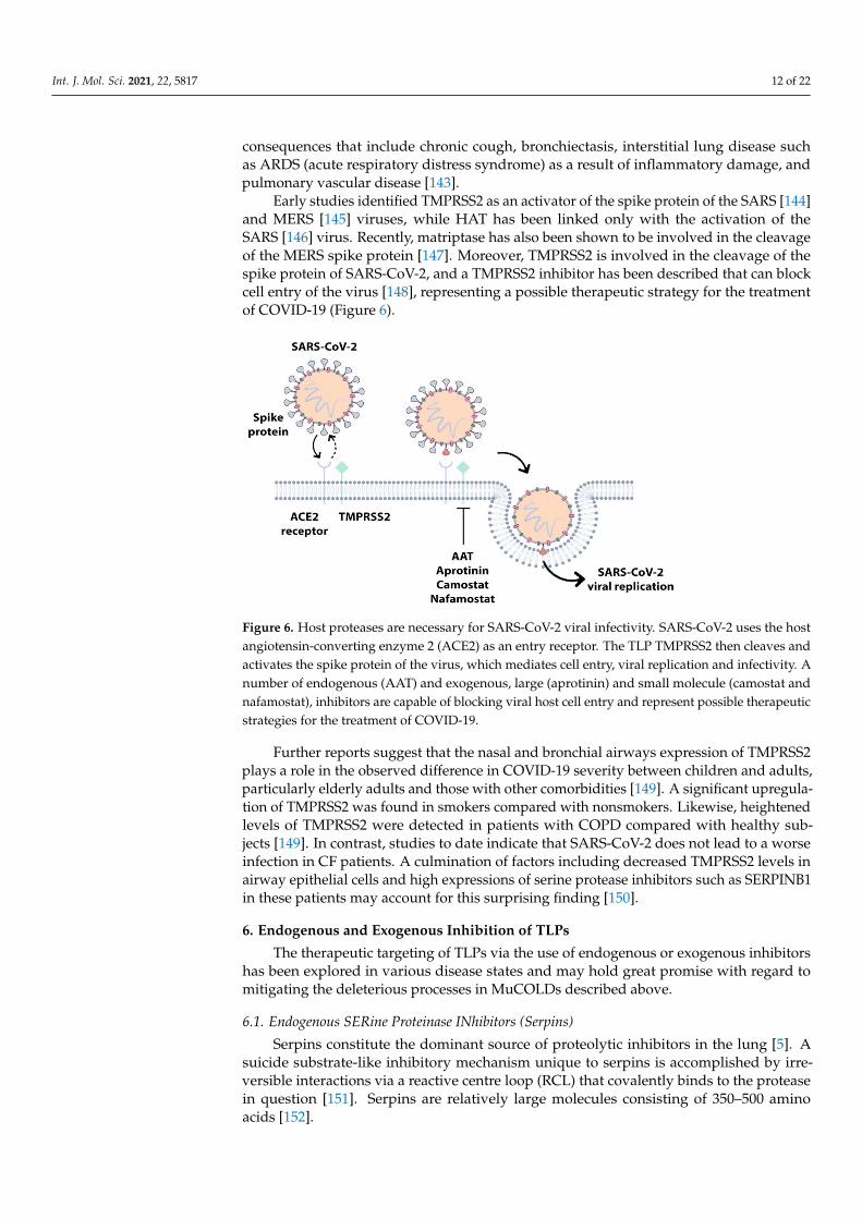
Early studies identified TMPRSS2 as an activator of the spike protein of the SARS [144]and MERS [145] viruses, while HAT has been linked only with the activation of theSARS [146] virus. Recently, matriptase has also been shown to be involved in the cleavageof the MERS spike protein [147]. Moreover, TMPRSS2 is involved in the cleavage of thespike protein of SARS-CoV-2, and a TMPRSS2 inhibitor has been described that can blockcell entry of the virus [148], representing a possible therapeutic strategy for the treatmentof COVID-19 (Figure 6).
Int. J. Mol. Sci. 2021, 22, x FOR PEER REVIEW 12 of 22
cell entry of the virus [148], representing a possible therapeutic strategy for the treatment of COVID-19 (Figure 6).
Further reports suggest that the nasal and bronchial airways expression of TMPRSS2 plays a role in the observed difference in COVID-19 severity between children and adults, particularly elderly adults and those with other comorbidities [149]. A significant upreg-ulation of TMPRSS2 was found in smokers compared with nonsmokers. Likewise, height-ened levels of TMPRSS2 were detected in patients with COPD compared with healthy subjects [149]. In contrast, studies to date indicate that SARS-CoV-2 does not lead to a worse infection in CF patients. A culmination of factors including decreased TMPRSS2 levels in airway epithelial cells and high expressions of serine protease inhibitors such as SERPINB1 in these patients may account for this surprising finding [150].
Figure 6. Host proteases are necessary for SARS-CoV-2 viral infectivity. SARS-CoV-2 uses the host angiotensin-converting enzyme 2 (ACE2) as an entry receptor. The TLP TMPRSS2 then cleaves and activates the spike protein of the virus, which mediates cell entry, viral replication and infec-tivity. A number of endogenous (AAT) and exogenous, large (aprotinin) and small molecule (camostat and nafamostat), inhibitors are capable of blocking viral host cell entry and represent possible therapeutic strategies for the treatment of COVID-19.
6. Endogenous and Exogenous Inhibition of TLPs The therapeutic targeting of TLPs via the use of endogenous or exogenous inhibitors
has been explored in various disease states and may hold great promise with regard to mitigating the deleterious processes in MuCOLDs described above.
6.1. Endogenous SERine Proteinase INhibitors (Serpins) Serpins constitute the dominant source of proteolytic inhibitors in the lung [5]. A su-
icide substrate-like inhibitory mechanism unique to serpins is accomplished by irreversi-ble interactions via a reactive centre loop (RCL) that covalently binds to the protease in question [151]. Serpins are relatively large molecules consisting of 350–500 amino acids [152].
As the archetype member of the SERPIN supergene family, the plasma α1-antitrypsin (AAT) (SERPINA1) protein was identified as an inhibitor of trypsin and named accord-ingly [105,153]. It was subsequently found that the native protein is most active against human NE, and AAT deficiency is a well-known instigator of COPD [105]. Additional AAT protease inhibitory roles came to light in later years, with the discovery that AAT also neutralizes cell-surface protease function. One such example is the finding that AAT inactivates the catalytic domain of matriptase in vitro [154]. Further reports demonstrated
Figure 6. Host proteases are necessary for SARS-CoV-2 viral infectivity. SARS-CoV-2 uses the hostangiotensin-converting enzyme 2 (ACE2) as an entry receptor. The TLP TMPRSS2 then cleaves andactivates the spike protein of the virus, which mediates cell entry, viral replication and infectivity. Anumber of endogenous (AAT) and exogenous, large (aprotinin) and small molecule (camostat andnafamostat), inhibitors are capable of blocking viral host cell entry and represent possible therapeuticstrategies for the treatment of COVID-19.
Further reports suggest that the nasal and bronchial airways expression of TMPRSS2plays a role in the observed difference in COVID-19 severity between children and adults,particularly elderly adults and those with other comorbidities [149]. A significant upregula-tion of TMPRSS2 was found in smokers compared with nonsmokers. Likewise, heightenedlevels of TMPRSS2 were detected in patients with COPD compared with healthy sub-jects [149]. In contrast, studies to date indicate that SARS-CoV-2 does not lead to a worseinfection in CF patients. A culmination of factors including decreased TMPRSS2 levels inairway epithelial cells and high expressions of serine protease inhibitors such as SERPINB1in these patients may account for this surprising finding [150].
6. Endogenous and Exogenous Inhibition of TLPs
The therapeutic targeting of TLPs via the use of endogenous or exogenous inhibitorshas been explored in various disease states and may hold great promise with regard tomitigating the deleterious processes in MuCOLDs described above.
6.1. Endogenous SERine Proteinase INhibitors (Serpins)
Serpins constitute the dominant source of proteolytic inhibitors in the lung [5]. Asuicide substrate-like inhibitory mechanism unique to serpins is accomplished by irre-versible interactions via a reactive centre loop (RCL) that covalently binds to the proteasein question [151]. Serpins are relatively large molecules consisting of 350–500 aminoacids [152].

Int. J. Mol. Sci. 2021, 22, 5817 13 of 22
As the archetype member of the SERPIN supergene family, the plasma α1-antitrypsin(AAT) (SERPINA1) protein was identified as an inhibitor of trypsin and named accord-ingly [105,153]. It was subsequently found that the native protein is most active againsthuman NE, and AAT deficiency is a well-known instigator of COPD [105]. AdditionalAAT protease inhibitory roles came to light in later years, with the discovery that AATalso neutralizes cell-surface protease function. One such example is the finding that AATinactivates the catalytic domain of matriptase in vitro [154]. Further reports demonstratedthat AAT reduces ENaC activity both in vitro and in vivo [155]; thus, AAT may be a rele-vant therapeutic option in the management of impaired MCC in chronic airways disease.Another recent study detailed the novel inhibition of TMPRSS2 by AAT and suggested arole for AAT in the derailing of the SARS-CoV-2 cell cycle (Figure 6) [156]. AAT has alsobeen found to have anti-inflammatory effects separate to its protease inhibitory role [157].For example, AAT is known to interact with high-density lipoprotein (HDL) [158], and AATcomplexed with HDL exhibited superior protection against elastase-induced pulmonaryemphysema in a mouse model, when compared to HDL or AAT alone [159]. In this study,AAT-HDL complexes achieved a reduction in the concentration of pro-inflammatory cy-tokines (monocyte chemoattractant protein-1 (MCP-1), interleukin-1 beta (IL-1β), tumournecrosis factor alpha (TNF-α)), MMP-2 and MMP-9 activity, and a decline in neutrophilinfiltration [159].
In addition to regulating urokinase, tissue plasminogen activators and intracellularfurin activity [160], plasminogen activator inhibitor 1 (PAI-1), encoded by the gene SER-PINE1, is also able to target cell-surface proteases. For example, human tryptase, HAT, andTMPRSS2 activities are all suppressed in the presence of PAI-1 [161]. Localized delivery ofPAI-1 to the respiratory tract may therefore offer anti-inflammatory and anti-viral benefits.
Another serpin capable of modulating TLP activity is protease nexin-1 (PN-1, SER-PINE2). PN-1 is the cognate serpin for prostasin and has been shown to prevent theprostasin-induced absorption of Na+ on the airway surface by forming an inhibitory com-plex [162]. Other studies have identified that PN-1 also inhibits matriptase on the airwayepithelia [41,162], which may have a knock-on effect on prostasin and ENaC activities.
6.2. Endogenous Kunitz-Type Inhibitors: HAI-1 and HAI-2
Kunitz-type inhibitors are ubiquitously expressed biological regulators of proteolysis.These small globular proteins possess Kunitz-type domains composed of alpha and betafolds with stabilising disulphide bridges [163]. In contrast to serpins, Kunitz-type pro-teins bind reversibly within the protease-binding loop due to gradual cleavage by targetproteases [163].
The presence of two Kunitz domains in the structures of the closely related Kunitz-type transmembrane serine protease inhibitors HAI-1 and HAI-2 aids inactivation ofvarious TLPs including matriptase, trypsin and prostasin [164,165]. HAI-1 has also beenreported as an inhibitor of HAT activation and proteolytic function [166]. The knockout ofeither of these endogenous inhibitors in mice induces severe developmental defects andlethality [167].
6.3. Exogenous Large- and Small-Molecule Inhibitors of TLPs
A small polypeptide consisting of three Kunitz domains was extracted from bovinelung in 1936 [168] and became known as aprotinin (also referred to as bovine pancreatictrypsin inhibitor, BPTI or Trasylol). This recombinant proteinaceous inhibitor, which is 58amino acids long, efficiently inhibits the majority of TLPs and was deemed as relatively welltolerated in animals and humans [169]. Trasylol was administered intravenously for manyyears to reduce blood loss and transfusion requirements in a number of surgical procedures,including open heart surgery. However, the anaphylactic potential of this nonhumanprotein became evident upon re-exposure to the drug [170], with an increased number ofdeaths reported when compared to treatment with standard antifibrinolytics [171]. Thiseffect led to the temporary withdrawal of Trasylol from the worldwide market in 2007 [172].

Int. J. Mol. Sci. 2021, 22, 5817 14 of 22
The suspension of sale was lifted in 2012 and aprotinin is now indicated prophylactically ona risk–benefit basis to reduce perioperative blood loss and the need for blood transfusionin patients undergoing cardiopulmonary bypass [173]. A growing interest has emerged inthe use of a low dose of aerosolised aprotinin to control viral replication in diseases such asinfluenza and SARS-CoV-2 (Figure 6) [174,175].
Camostat is a synthetic, low-molecular-weight, broad-spectrum inhibitor of TLPs, firstapproved in Japan in 2006 for the treatment of chronic pancreatitis and postoperative refluxesophagitis [176]. Similar to aprotinin, camostat has shown promise in the reduction ofinfluenza viral replication, and the repurposing of this drug for COVID-19 treatment iscurrently underway given its potent inhibitory action against the virus-activating host cellprotease TMPRSS2 (Figure 6) [177–179].
The affinities of aprotinin and camostat for matriptase and prostasin is associated witha reduction in protease-mediated ENaC current at the apical surface of airway epithelialcells [41,55,180]. Furthermore, the topical airway administration of camostat success-fully attenuated the ENaC activity and augmented mucus clearance rates in vivo [180].Likewise, aprotinin and camostat are both capable of blocking matriptase-induced PAR2cleavage [181].
Structurally related to camostat, nafamostat is a small molecule that has been ap-proved as a mucolytic therapy, reducing ENaC activity in CF disease due to its potentinhibition against CAPs [69]. Studies have revealed that nafamostat also has the ability toinhibit SARS-CoV-2 spike protein-mediated entry into host cells, which relies on TMPRSS2cleavage, with around a 15-fold higher efficiency than camostat (Figure 6) [182]. Thishas prompted the entry of nafamostat into clinical trials to test its effectiveness againstpneumonia in COVID-19 patients [179].
Interestingly, based on the preference of TLPs for an arginine or lysine residue inthe P1 position, a number of rationally designed novel compounds have been developed.For example, QUB-TL1 is a synthetic molecule consisting of an arginine-derived diphenylphosphonate moiety that reacts with the active site serine residues in order to irreversiblybind to TLPs [71]. QUB-TL1 has demonstrated the inhibition of extracellularly located furin,matriptase and prostasin in CF airway epithelial cells [71]. Improvements in ASL heightand MCC were seen in CF epithelial cells after treatment with QUB-TL1, thus offering arole for such molecules in CF and potentially other MucOLDs. As a research tool, giventhe limitations on the use of peptide-based fluorogenic substrates to determine specificactivities from within complex biological samples due to their susceptibility to cleavage byother classes of proteases, QUB-TL1 has been used to tease out a QUB-TL1-sensitive poolof activity from supernatant (sol), processed from purulent CF sputum [183]. This studyfound TLP activity to be inversely correlated with the percent predicted FEV1 (r = −0.4,p = 0.03) with a high TLP activity associated with a significantly reduced survival (p = 0.04)[hazard ratio (HR) of 7.21 (per log unit TLP activity (p = 0.03))]. In contrast, NE displayedno significant associations with lung function or patient survival. This is the first study tohighlight a potential role for TLP activity as a novel noninvasive biomarker for long-termrisk in CF lung disease.
7. Conclusions
To conclude, airway TLPs are an important, yet largely overlooked sub-group of serineproteases. Given the key roles that have already been identified for these enzymes in theregulation of numerous processes involved in airways dehydration, inflammation and viralinfection, it seems prudent that further work is conducted to dissect out the contributionmade by individual activities. To date, inhibitors, both large and small, have exerted abroad-spectrum activity but have a heightened risk of off-target effects. The developmentof a more selective approach to inhibition may allow the consideration of the TLPs aspotential therapeutic candidates within the context of muco-obstructive lung diseases.

Int. J. Mol. Sci. 2021, 22, 5817 15 of 22
Author Contributions: Conceptualization, E.L.C., M.B., S.L.M., A.C., J.C.L., G.J.L. and J.A.R.;writing—original draft preparation, E.L.C. and M.B.; writing—review and editing, S.L.M., A.C.,J.C.L., G.J.L., J.A.R., F.T.L., L.P.M. and M.A.H.; supervision, S.L.M., A.C., J.C.L., G.J.L., F.T.L. andL.P.M.; funding acquisition, S.L.M., M.A.H., J.C.L., G.J.L., A.C., F.T.L. and L.P.M. All authors haveread and agreed to the published version of the manuscript.
Funding: This study was supported by the Borders and Regions Airways Training Hub project(BREATH; INT-VA/045) funded by the European Union (EU), under the INTERREG VA Programme,managed by the Special EU Programmes Body (to S.L.M, M.A.H., J.C.L., L.P.M., F.T.L, G.J.L. and A.C.).E.C., M.B. and J.A.R. are directly funded by BREATH; E.C. and M.B. by BREATH PhD studentships.
Conflicts of Interest: The authors declare no conflict of interest.
References1. López-Otín, C.; Overall, C.M. Protease degradomics: A new challenge for proteomics. Nat. Rev. Mol. Cell Biol. 2002, 3, 509–519.
[CrossRef]2. Doucet, A.; Overall, C.M. Protease proteomics: Revealing protease in vivo functions using systems biology approaches. Mol. Asp.
Med. 2008, 29, 339–358. [CrossRef]3. Greene, C.M.; McElvaney, N.G. Proteases and antiproteases in chronic neutrophilic lung disease—Relevance to drug discovery.
Br. J. Pharmacol. 2009, 158, 1048–1058. [CrossRef]4. Tetley, T.D. New perspectives on basic mechanisms in lung disease. 6. Proteinase imbalance: Its role in lung disease. Thorax 1993,
48, 560–565. [CrossRef]5. Taggart, C.; Mall, M.A.; Lalmanach, G.; Cataldo, D.; Ludwig, A.; Janciauskiene, S.; Heath, N.; Meiners, S.; Overall, C.M.; Schultz,
C.; et al. Protean proteases: At the cutting edge of lung diseases. Eur. Respir. J. 2017, 49, 1501200. [CrossRef] [PubMed]6. Chakraborti, S.; Sarkar, J.; Pramanik, P.K.; Chakraborti, T. Role of Proteases in Lung Disease: A Brief Overview. In Proteases in
Human Diseases; Springer Science and Business Media: Berlin/Heidelberg, Germany, 2017; pp. 333–374.7. Sallenave, J.-M. Editorial: Neutrophil elastase and the lung: Is it degradation, repair, emphysema, or fibrosis? What tilts it left or
right? J. Leukoc. Biol. 2015, 98, 137–139. [CrossRef] [PubMed]8. Polverino, E.; Rosales-Mayor, E.; Dale, G.E.; Dembowsky, K.; Torres, A. The role of neutrophil elastase inhibitors in lung diseases.
Chest 2017, 152, 249–262. [CrossRef] [PubMed]9. Åstrand, A.B.M.; Hemmerling, M.; Root, J.; Wingren, C.; Pesic, J.; Johansson, E.; Garland, A.L.; Ghosh, A.; Tarran, R. Linking
increased airway hydration, ciliary beating, and mucociliary clearance through ENaC inhibition. Am. J. Physiol. Cell. Mol. Physiol.2015, 308, L22–L32. [CrossRef]
10. Mall, M.A.; Danahay, H.; Boucher, R.C. Emerging concepts and therapies for mucoobstructive lung disease. Ann. Am. Thorac. Soc.2018, 15, S216–S226. [CrossRef]
11. Cocks, T.; Moffatt, J. Protease-activated Receptor-2 (PAR2) in the Airways. Pulm. Pharmacol. Ther. 2001, 14, 183–191. [CrossRef]12. Laporte, M.; Naesens, L. Airway proteases: An emerging drug target for influenza and other respiratory virus infections. Curr.
Opin. Virol. 2017, 24, 16–24. [CrossRef] [PubMed]13. López-Otín, C.; Bond, J.S. Proteases: Multifunctional enzymes in life and disease*. J. Biol. Chem. 2008, 283, 30433–30437. [CrossRef]14. Di Cera, E. Serine proteases. IUBMB Life 2009, 61, 510–515. [CrossRef]15. Rawlings, N.D.; Barrett, A.J.; Bateman, A. MEROPS: The peptidase database. Nucleic Acids Res. 2009, 38, D227–D233. [CrossRef]16. Laskar, A.; Rodger, E.J.; Chatterjee, A.; Mandal, C. Modeling and structural analysis of PA clan serine proteases. BMC Res. Notes
2012, 5, 256. [CrossRef]17. Page, M.J.; Di Cera, E. Serine peptidases: Classification, structure and function. Cell. Mol. Life Sci. 2008, 65, 1220–1236. [CrossRef]
[PubMed]18. Böttcher-Friebertshäuser, E. Membrane-anchored serine proteases: Host cell factors in proteolytic activation of viral glycoproteins.
In Activation of Viruses by Host Proteases; Springer: Berlin/Heidelberg, Germany, 2018; pp. 153–203.19. Sommerhoff, C.P. Mast cell tryptases and airway remodeling. Am. J. Respir. Crit. Care Med. 2001, 164, S52–S58. [CrossRef]
[PubMed]20. Hallgren, J.; Pejler, G. Biology of mast cell tryptase. FEBS J. 2006, 273, 1871–1895. [CrossRef] [PubMed]21. Ferguson, T.; McCafferty, D.; Robb, C.; Moffitt, K.; Walker, B. The development of a novel immunoassay for the quantification of
active tryptase. Monit. Airw. Dis. 2017, 50, PA4017. [CrossRef]22. Nimishakavi, S.; Raymond, W.W.; Gruenert, D.C.; Caughey, G.H. Divergent inhibitor susceptibility among airway lumen-
accessible tryptic proteases. PLoS ONE 2015, 10, e0141169. [CrossRef]23. Yu, J.; Chao, L.; Chao, J. Prostasin is a novel human serine proteinase from seminal fluid. Purification, tissue distribution, and
localization in prostate gland. J. Biol. Chem. 1994, 269, 18843–18848. [CrossRef]24. Chen, L.-M.; Skinner, M.L.; Kauffman, S.W.; Chao, J.; Chao, L.; Thaler, C.D.; Chai, K.X. Prostasin is a glycosylphosphatidylinositol-
anchored active serine protease. J. Biol. Chem. 2001, 276, 21434–21442. [CrossRef]

Int. J. Mol. Sci. 2021, 22, 5817 16 of 22
25. Rickert, K.W.; Kelley, P.; Byrne, N.J.; Diehl, R.E.; Hall, D.L.; Montalvo, A.M.; Reid, J.C.; Shipman, J.M.; Thomas, B.W.; Munshi, S.K.;et al. Structure of human prostasin, a target for the regulation of hypertension. J. Biol. Chem. 2008, 283, 34864–34872. [CrossRef][PubMed]
26. Shipway, A.; Danahay, H.; Williams, J.A.; Tully, D.C.; Backes, B.J.; Harris, J.L. Biochemical characterization of prostasin, a channelactivating protease. Biochem. Biophys. Res. Commun. 2004, 324, 953–963. [CrossRef]
27. Menou, A.; Duitman, J.; Flajolet, P.; Sallenave, J.-M.; Mailleux, A.A.; Crestani, B. Human airway trypsin-like protease, a serineprotease involved in respiratory diseases. Am. J. Physiol. Cell. Mol. Physiol. 2017, 312, L657–L668. [CrossRef]
28. Yasuoka, S.; Ohnishi, T.; Kawano, S.; Tsuchihashi, S.; Ogawara, M.; Masuda, K.; Yamaoka, K.; Takahashi, M.; Sano, T. Purification,characterization, and localization of a novel trypsin-like protease found in the human airway. Am. J. Respir. Cell Mol. Biol. 1997,16, 300–308. [CrossRef]
29. Takahashi, M.; Sano, T.; Yamaoka, K.; Kamimura, T.; Umemoto, N.; Nishitani, H.; Yasuoka, S. Localization of human airwaytrypsin-like protease in the airway: An immunohistochemical study. Histochem. Cell Biol. 2001, 115, 181–187. [CrossRef]
30. Bertram, S.; Glowacka, I.; Blazejewska, P.; Soilleux, E.; Allen, P.; Danisch, S.; Steffen, I.; Choi, S.Y.; Park, Y.; Schneider, H.; et al.TMPRSS2 and TMPRSS4 facilitate tryp-sin-independent spread of influenza virus in Caco-2 cells. J. Virol. 2010, 84, 10016–10025.[CrossRef] [PubMed]
31. Stopsack, K.H.; Mucci, L.A.; Antonarakis, E.S.; Nelson, P.S.; Kantoff, P.W. TMPRSS2 and COVID-19: Serendipity or opportunityfor intervention? Cancer Discov. 2020, 10, 779–782. [CrossRef]
32. Kim, T.S.; Heinlein, C.; Hackman, R.C.; Nelson, P.S. Phenotypic analysis of mice lacking the Tmprss2-encoded protease. Mol. Cell.Biol. 2006, 26, 965–975. [CrossRef]
33. Oberst, M.D.; Williams, C.A.; Dickson, R.B.; Johnson, M.D.; Lin, C.-Y. The activation of matriptase requires its noncatalyticdomains, serine protease domain, and its cognate inhibitor. J. Biol. Chem. 2003, 278, 26773–26779. [CrossRef]
34. Lee, M.-S.; Kiyomiya, K.-I.; Benaud, C.; Dickson, R.B.; Lin, C.-Y. Simultaneous activation and hepatocyte growth factor activatorinhibitor 1-mediated inhibition of matriptase induced at activation foci in human mammary epithelial cells. Am. J. Physiol. Physiol.2005, 288, C932–C941. [CrossRef] [PubMed]
35. Oberst, M.D.; Chen, L.-Y.L.; Kiyomiya, K.-I.; Williams, C.A.; Lee, M.-S.; Johnson, M.D.; Dickson, R.B.; Lin, C.-Y. HAI-1 regulatesactivation and expression of matriptase, a membrane-bound serine protease. Am. J. Physiol. Physiol. 2005, 289, C462–C470.[CrossRef] [PubMed]
36. Tseng, I.-C.; Xu, H.; Chou, F.-P.; Li, G.; Vazzano, A.P.; Kao, J.P.; Johnson, M.D.; Lin, C.-Y. Matriptase activation, an early cellularresponse to acidosis. J. Biol. Chem. 2010, 285, 3261–3270. [CrossRef] [PubMed]
37. Tseng, C.-C.; Jia, B.; Barndt, R.; Gu, Y.; Chen, C.-Y.; Tseng, I.-C.; Su, S.-F.; Wang, J.-K.; Johnson, M.D.; Lin, C.-Y. Matriptaseshedding is closely coupled with matriptase zymogen activation and requires de novo proteolytic cleavage likely involving itsown activity. PLoS ONE 2017, 12, e0183507. [CrossRef]
38. List, K.; Hobson, J.P.; Molinolo, A.; Bugge, T.H. Co-localization of the channel activating protease prostasin/(CAP1/PRSS8) withits candidate activator, matriptase. J. Cell. Physiol. 2007, 213, 237–245. [CrossRef] [PubMed]
39. Garty, H.; Palmer, L.G. Epithelial sodium channels: Function, structure, and regulation. Physiol. Rev. 1997, 77, 359–396. [CrossRef]40. Boucher, R.C. Human airway ion transport. Part one. Am. J. Respir. Crit. Care Med. 1994, 150, 271–281. [CrossRef] [PubMed]41. Gaillard, E.A.; Kota, P.; Gentzsch, M.; Dokholyan, N.; Stutts, M.J.; Tarran, R. Regulation of the epithelial Na+ channel and airway
surface liquid volume by serine proteases. Eur. J. Physiol. 2010, 460, 1–17. [CrossRef]42. Kellenberger, S.; Schild, L. Epithelial sodium channel/degenerin family of ion channels: A variety of functions for a shared
structure. Physiol. Rev. 2002, 82, 735–767. [CrossRef]43. Hanukoglu, I.; Hanukoglu, A. Epithelial sodium channel (ENaC) family: Phylogeny, structure–function, tissue distribution, and
associated inherited diseases. Gene 2016, 579, 95–132. [CrossRef] [PubMed]44. Hanukoglu, I. ASIC and ENaC type sodium channels: Conformational states and the structures of the ion selectivity filters. FEBS
J. 2016, 284, 525–545. [CrossRef]45. Boscardin, E.; Alijevic, O.; Hummler, E.; Frateschi, S.; Kellenberger, S. The function and regulation of acid-sensing ion channels
(ASICs) and the epithelial Na+channel (ENaC): IUPHAR Review 19. Br. J. Pharmacol. 2016, 173, 2671–2701. [CrossRef]46. Noreng, S.; Bharadwaj, A.; Posert, R.; Yoshioka, C.; Baconguis, I. Structure of the human epithelial sodium channel by cryo-electron
microscopy. eLife 2018, 7. [CrossRef] [PubMed]47. Bhalla, V.; Hallows, K.R. Mechanisms of ENaC regulation and clinical implications. J. Am. Soc. Nephrol. 2008, 19, 1845–1854.
[CrossRef]48. Kashlan, O.B.; Kleyman, T.R. ENaC structure and function in the wake of a resolved structure of a family member. Am. J. Physiol.
Physiol. 2011, 301, F684–F696. [CrossRef] [PubMed]49. Blobner, B.M.; Wang, X.P.; Kashlan, O.B. Conserved cysteines in the finger domain of the epithelial Na+ channel alpha and
gamma subunits are proximal to the dynamic finger-thumb domain interface. J. Biol. Chem. 2018, 293, 4928–4939. [CrossRef]50. Kleyman, T.R.; Carattino, M.D.; Hughey, R.P. ENaC at the cutting edge: Regulation of epithelial sodium channels by proteases. J.
Biol. Chem. 2009, 284, 20447–20451. [CrossRef] [PubMed]51. Ray, E.C.; Kleyman, T.R. Cutting it out: ENaC processing in the human nephron. J. Am. Soc. Nephrol. 2014, 26, 1–3. [CrossRef]
[PubMed]

Int. J. Mol. Sci. 2021, 22, 5817 17 of 22
52. Thibodeau, P.H.; Butterworth, M.B. Proteases, cystic fibrosis and the epithelial sodium channel (ENaC). Cell Tissue Res. 2012, 351,309–323. [CrossRef]
53. Kleyman, T.R.; Kashlan, O.B.; Hughey, R.P. Epithelial Na+ channel regulation by extracellular and intracellular fac-tors_Kleymanetal. Annu. Rev. Physiol. 2018, 80, 263–281. [CrossRef]
54. Caldwell, R.A.; Boucher, R.; Stutts, M. Serine protease activation of near-silent epithelial Na+ channels. AJP Cell Physiol. 2003, 286,190C–194C. [CrossRef]
55. Vallet, V.; Chraibi, A.; Gaeggeler, H.-P.; Horisberger, J.-D.; Rossier, B.C. An epithelial serine protease activates the amiloride-sensitive sodium channel. Nat. Cell Biol. 1997, 389, 607–610. [CrossRef]
56. Vuagniaux, G.; Vallet, V.; Jaeger, N.F.; Pfister, C.; Bens, M.; Farman, N.; Courtois-Coutry, N.; Vandewalle, A.; Rossier, B.C.;Hummler, E. Activation of the amiloride-sensitive epithelial sodium channel by the serine protease mCAP1 expressed in a mousecortical collecting duct cell line. J. Am. Soc. Nephrol. 2000, 11, 828–834. [CrossRef]
57. Vuagniaux, G.; Vallet, V.; Jaeger, N.F.; Hummler, E.; Rossier, B.C. Synergistic activation of ENaC by three membrane-boundchannel-activating serine proteases (mCAP1, mCAP2, and mCAP3) and serum- and glucocorticoid-regulated kinase (Sgk1) inXenopus Oocytes. J. Gen. Physiol. 2002, 20, 191–201. [CrossRef] [PubMed]
58. Boucher, R.C. Muco-obstructive lung diseases. N. Engl. J. Med. 2019, 380, 1941–1953. [CrossRef] [PubMed]59. Livraghi-Butrico, A.; Grubb, B.R.; Wilkinson, K.J.; Volmer, A.S.; Burns, K.A.; Evans, C.M.; O’Neal, W.K.; Boucher, R.C. Contribution
of mucus concentration and secreted mucins Muc5ac and Muc5b to the pathogenesis of muco-obstructive lung disease. MucosalImmunol. 2017, 10, 395–407. [CrossRef]
60. Randell, S.H.; Boucher, R.C. Effective mucus clearance is essential for respiratory health. Am. J. Respir. Cell Mol. Biol. 2006, 35,20–28. [CrossRef] [PubMed]
61. Fahy, J.V.; Dickey, B.F. Airway mucus function and dysfunction. N. Engl. J. Med. 2010, 363, 2233–2247. [CrossRef] [PubMed]62. Kim, V.; Ramos, F.; Krahnke, J. Clinical issues of mucus accumulation in COPD. Int. J. Chronic Obstr. Pulm. Dis. 2014, 9, 139–150.
[CrossRef]63. Mall, M. Unplugging mucus in cystic fibrosis and chronic obstructive pulmonary disease. Ann. Am. Thorac. Soc. 2016, 13.64. Munkholm, M.; Mortensen, J. Mucociliary clearance: Pathophysiological aspects. Clin. Physiol. Funct. Imaging 2014, 34, 171–177.
[CrossRef]65. Ma, J.; Rubin, B.K.; Voynow, J.A. Mucins, mucus, and goblet cells. Chest 2018, 154, 169–176. [CrossRef]66. Thornton, D.J.; Rousseau, K.; McGuckin, M.A. Structure and function of the polymeric mucins in airways mucus. Annu. Rev.
Physiol. 2008, 70, 459–486. [CrossRef] [PubMed]67. Voynow, J.A.; Rubin, B.K. Mucins, mucus, and sputum. Chest 2009, 135, 505–512. [CrossRef] [PubMed]68. Whitsett, J.; Alenghat, T. Respiratory epithelial cells orchestrate pulmonary innate immunity. Nat. Immunol. 2015, 16, 27–35.
[CrossRef]69. Nimishakavi, S.; Besprozvannaya, M.; Raymond, W.W.; Craik, C.S.; Gruenert, D.C.; Caughey, G.H. Activity and inhibition of
prostasin and matriptase on apical and basolateral surfaces of human airway epithelial cells. Am. J. Physiol. Cell. Mol. Physiol.2012, 303, L97–L106. [CrossRef] [PubMed]
70. Tong, Z.; Illek, B.; Bhagwandin, V.J.; Verghese, G.M.; Caughey, G.H. Prostasin, a membrane-anchored serine peptidase, regulatessodium currents in JME/CF15 cells, a cystic fibrosis airway epithelial cell line. Am. J. Physiol. Cell. Mol. Physiol. 2004, 287,L928–L935. [CrossRef] [PubMed]
71. Reihill, J.A.; Walker, B.; Hamilton, R.A.; Ferguson, T.E.G.; Elborn, J.S.; Stutts, M.J.; Harvey, B.; Saint-Criq, V.; Hendrick, S.M.;Martin, S.L. Inhibition of protease–epithelial sodium channel signaling improves mucociliary function in cystic fibrosis airways.Am. J. Respir. Crit. Care Med. 2016, 194, 701–710. [CrossRef] [PubMed]
72. Martin, L.; Reihill, J.; Douglas, L.; Ferguson, T.; Walker, B. Detection and inhibition of ENaC-activating proteases associated withairway dehydration in COPD. Eur. Respir. J. 2017, 50, PA4933.
73. Kunzelmann, K.; Kiser, G.; Schreiber, R.; Riordan, J. Inhibition of epithelial Na+ currents by intracellular domains of the cysticfibrosis transmembrane conductance regulator. FEBS Lett. 1997, 400, 341–344. [CrossRef]
74. Boucher, R.C.; Stutts, M.J.; Knowles, M.R.; Cantley, L.; Gatzy, J.T. Na+ transport in cystic fibrosis respiratory epithelia. Abnormalbasal rate and response to adenylate cyclase activation. J. Clin. Investig. 1986, 78, 1245–1252. [CrossRef]
75. Hobbs, C.A.; Da Tan, C.; Tarran, R. Does epithelial sodium channel hyperactivity contribute to cystic fibrosis lung disease? J.Physiol. 2013, 591, 4377–4387. [CrossRef]
76. Ghosh, A.; Boucher, R.C.; Tarran, R. Airway hydration and COPD. Cell. Mol. Life Sci. 2015, 72, 3637–3652. [CrossRef] [PubMed]77. Seys, L.J.M.; Verhamme, F.M.; Dupont, L.L.; Desauter, E.; Duerr, J.; Agircan, A.S.; Conickx, G.; Joos, G.F.; Brusselle, G.G.; Mall,
M.A.; et al. Airway surface dehydration aggravates cigarette smoke-induced hallmarks of COPD in mice. PLoS ONE 2015, 10,e0129897. [CrossRef] [PubMed]
78. Wang, G.; Xu, Z.; Wang, R.; Al-Hijji, M.; Salit, J.; Strulovici-Barel, Y.; Tilley, A.; Mezey, J.G.; Crystal, R.G. Genes associated withMUC5AC expression in small airway epithelium of human smokers and non-smokers. BMC Med. Genom. 2012, 5, 21. [CrossRef][PubMed]
79. Turner, J.; Jones, C.E. Regulation of mucin expression in respiratory diseases. Biochem. Soc. Trans. 2009, 37, 877–881. [CrossRef]80. Thai, P.; Loukoianov, A.; Wachi, S.; Wu, R. Regulation of airway mucin gene expression. Annu. Rev. Physiol. 2008, 70, 405–429.
[CrossRef]

Int. J. Mol. Sci. 2021, 22, 5817 18 of 22
81. Chokki, M.; Yamamura, S.; Eguchi, H.; Masegi, T.; Horiuchi, H.; Tanabe, H.; Kamimura, T.; Yasuoka, S. Human airway trypsin-likeprotease increases mucin gene expression in airway epithelial cells. Am. J. Respir. Cell Mol. Biol. 2004, 30, 470–478. [CrossRef][PubMed]
82. Hollenberg, M.D.; Compton, S.J. International union of pharmacology. XXVIII. Proteinase-activated receptors. Pharmacol. Rev.2002, 54, 203–217. [CrossRef]
83. Vu, T.-K.H.; Hung, D.T.; Wheaton, V.I.; Coughlin, S.R. Molecular cloning of a functional thrombin receptor reveals a novelproteolytic mechanism of receptor activation. Cell 1991, 64, 1057–1068. [CrossRef]
84. Ishihara, H.; Connolly, A.J.; Zeng, D.; Kahn, M.L.; Zheng, Y.W.; Timmons, C.; Tram, T.; Coughlin, S.R. Protease-activated receptor3 is a second thrombin receptor in humans. Nat. Cell Biol. 1997, 386, 502–506. [CrossRef] [PubMed]
85. Xu, W.F.; Andersen, H.; Whitmore, T.E.; Presnell, S.R.; Yee, D.P.; Ching, A.; Gilbert, T.; Davie, E.W.; Foster, D.C. Cloning andcharacterization of human prote-ase-activated receptor 4. Proc. Natl. Acad. Sci. USA 1998, 95, 6642–6646. [CrossRef]
86. Nystedt, S.; Emilsson, K.; Wahlestedt, C.; Sundelin, J. Molecular cloning of a potential proteinase activated receptor. Proc. Natl.Acad. Sci. USA 1994, 91, 9208–9212. [CrossRef] [PubMed]
87. Nystedt, S.; Emilsson, K.; Larsson, A.-K.; Strömbeck, B.; Sundelin, J. Molecular cloning and functional expression of the geneencoding the human proteinase-activated receptor 2. J. Biol. Inorg. Chem. 1995, 232, 84–89. [CrossRef] [PubMed]
88. Hamm, H.E. How activated receptors couple to G proteins. Proc. Natl. Acad. Sci. USA 2001, 98, 4819–4821. [CrossRef]89. Ramachandran, R.; Mihara, K.; Mathur, M.; Rochdi, M.D.; Bouvier, M.; Defea, K.; Hollenberg, M.D. Agonist-biased signaling via
proteinase activated receptor-2: Differential activation of calcium and mitogen-activated protein kinase pathways. Mol Pharmacol.2009, 76, 791–801. [CrossRef]
90. Adams, M.N.; Ramachandran, R.; Yau, M.-K.; Suen, J.Y.; Fairlie, D.P.; Hollenberg, M.D.; Hooper, J.D. Structure, function andpathophysiology of protease activated receptors. Pharmacol. Ther. 2011, 130, 248–282. [CrossRef]
91. Nystedt, S.; Larsson, A.-K.; Åberg, H.; Sundelin, J. The mouse proteinase-activated receptor-2 cDNA and gene. J. Biol. Chem. 1995,270, 5950–5955. [CrossRef]
92. Molino, M.; Barnathan, E.S.; Numerof, R.; Clark, J.; Dreyer, M.; Cumashi, A.; Hoxie, J.A.; Schechter, N.; Woolkalis, M.; Brass, L.F.Interactions of mast cell tryptase with thrombin receptors and PAR-2. J. Biol. Chem. 1997, 272, 4043–4049. [CrossRef]
93. Hollenberg, M.D.; Mihara, K.; Polley, D.; Suen, J.Y.; Han, A.; Fairlie, D.P.; Ramachandran, R. Biased signalling and proteinase-activated receptors (PARs): Targeting inflammatory disease. Br. J. Pharmacol. 2013, 171, 1180–1194. [CrossRef]
94. Bühm, S.K.; Khitin, L.M.; Grady, E.F.; Aponte, G.; Payan, D.G.; Bunnett, N.W. Mechanisms of desensitization and resensitizationof proteinase-activated receptor-2. J. Biol. Chem. 1996, 271, 22003–22016. [CrossRef]
95. Macfarlane, S.R.; Sloss, C.M.; Cameron, P.; Kanke, T.; McKenzie, R.C.; Plevin, R. The role of intracellular Ca2+ in the regulation ofproteinase-activated receptor-2 mediated nuclear factor kappa B signalling in keratinocytes. Br. J. Pharmacol. 2005, 145, 535–544.[CrossRef]
96. Santulli, R.J.; Derian, C.K.; Darrow, A.L.; Tomko, K.A.; Eckardt, A.J.; Seiberg, M.; Scarborough, R.M.; Andrade-Gordon, P.Evidence for the presence of a protease-activated receptor distinct from the thrombin receptor in human keratinocytes. Proc. Natl.Acad. Sci. USA 1995, 92, 9151–9155. [CrossRef] [PubMed]
97. Sokolova, E.; Reiser, G. A novel therapeutic target in various lung diseases: Airway proteases and protease-activated receptors.Pharmacol. Ther. 2007, 115, 70–83. [CrossRef] [PubMed]
98. Ramachandran, R.; Mihara, K.; Chung, H.; Renaux, B.; Lau, C.S.; Muruve, D.A.; DeFea, K.A.; Bouvier, M.; Hollenberg, M.D.Neutrophil elastase acts as a biased agonist for proteinase-activated receptor-2 (PAR2). J. Biol. Chem. 2011, 286, 24638–24648.[CrossRef]
99. Gieseler, F.; Ungefroren, H.; Settmacher, U.; Hollenberg, M.D.; Kaufmann, R. Proteinase-activated receptors (PARs)—Focuson re-ceptor-receptor-interactions and their physiological and pathophysiological impact. Cell Commun. Signal. 2013, 11, 86.[CrossRef]
100. Zhao, P.; Metcalf, M.; Bunnett, N.W. Corrigendum: Biased Signaling of protease-activated receptors. Front. Endocrinol. 2015, 5.[CrossRef]
101. Soh, U.J.K.; Dores, M.R.; Chen, B.; Trejo, J. Signal transduction by protease-activated receptors. Br. J. Pharmacol. 2010, 160, 191–203.[CrossRef] [PubMed]
102. Stalheim, L.; Ding, Y.; Gullapalli, A.; Paing, M.M.; Wolfe, B.L.; Morris, D.R.; Trejo, J. Multiple independent functions of arrestinsin the regulation of protease-activated receptor-2 signaling and trafficking. Mol. Pharmacol. 2004, 67, 78–87. [CrossRef] [PubMed]
103. Defea, K. Beta-arrestins and heterotrimeric G-proteins: Collaborators and competitors in signal transduction. Br. J. Pharmacol.2008, 153, S298–S309. [CrossRef]
104. Jacob, C.; Cottrell, G.S.; Gehringer, D.; Schmidlin, F.; Grady, E.F.; Bunnett, N.W. c-Cbl Mediates ubiquitination, degradation, anddown-regulation of human protease-activated receptor 2. J. Biol. Chem. 2005, 280, 16076–16087. [CrossRef] [PubMed]
105. Stockley, R.A. The multiple facets of alpha-1-antitrypsin. Ann. Transl. Med. 2015, 3, 130. [PubMed]106. Cocks, T.M.; Fong, B.; Chow, J.M.; Anderson, G.P.; Frauman, A.G.; Goldie, R.G.; Henry, P.J.; Carr, M.J.; Hamilton, J.R.; Moffatt, J.D.
A protective role for protease-activated receptors in the airways. Nat. Cell Biol. 1999, 398, 156–160. [CrossRef]107. Theoharides, T.C.; Alysandratos, K.-D.; Angelidou, A.; Delivanis, D.-A.; Sismanopoulos, N.; Zhang, B.; Asadi, S.; Vasiadi, M.;
Weng, Z.; Miniati, A.; et al. Mast cells and inflammation. Biochim. Biophys. Acta Mol. Basis Dis. 2012, 1822, 21–33. [CrossRef]

Int. J. Mol. Sci. 2021, 22, 5817 19 of 22
108. Barrios, V.E.; Jarosinski, M.A.; Wright, C.D. Proteinase-activated receptor-2 mediates hyperresponsiveness in isolated guinea pigbronchi. Biochem. Pharmacol. 2003, 66, 519–525. [CrossRef]
109. Berger, P.; Perng, D.W.; Thabrew, H.; Compton, S.J.; Cairns, J.A.; McEuen, A.R.; Marthan, R.; Tunon De Lara, J.M.; Walls, A.F.Tryptase and agonists of PAR-2 induce the pro-liferation of human airway smooth muscle cells. J. Appl. Physiol. 2001, 91,1372–1379. [CrossRef] [PubMed]
110. Akers, I.A.; Parsons, M.; Hill, M.R.; Hollenberg, M.D.; Sanjar, S.; Laurent, G.J.; McAnulty, R. Mast cell tryptase stimulates humanlung fibroblast proliferation via protease-activated receptor-2. Am. J. Physiol. Cell. Mol. Physiol. 2000, 278, L193–L201. [CrossRef]
111. Bagher, M.; Larsson-Callerfelt, A.-K.; Rosmark, O.; Hallgren, O.; Bjermer, L.; Westergren-Thorsson, G. Mast cells and mast celltryptase enhance migration of human lung fibroblasts through protease-activated receptor 2. Cell Commun. Signal. 2018, 16, 59.[CrossRef]
112. Seitz, I.; Hess, S.; Schulz, H.; Eckl, R.; Busch, G.; Montens, H.P.; Brandl, R.; Seidl, S.; Schömig, A.; Ott, I. Membrane-type serineprotease-1/matriptase induces interleukin-6 and -8 in endothelial cells by activation of protease-activated receptor-2: Potentialimplications in atherosclerosis. Arterioscler. Thromb. Vasc. Biol. 2007, 27, 769–775. [CrossRef]
113. Camerer, E.; Barker, A.; Duong, D.N.; Ganesan, R.; Kataoka, H.; Cornelissen, I.; Darragh, M.R.; Hussain, A.; Zheng, Y.-W.;Srinivasan, Y.; et al. Local protease signaling contributes to neural tube closure in the mouse embryo. Dev. Cell 2010, 18, 25–38.[CrossRef] [PubMed]
114. Schepis, A.; Barker, A.; Srinivasan, Y.; Balouch, E.; Zheng, Y.; Lam, I.; Clay, H.; Hsiao, C.-D.; Coughlin, S.R. Protease signalingregulates apical cell extrusion, cell contacts, and proliferation in epithelia. J. Cell Biol. 2018, 217, 1097–1112. [CrossRef] [PubMed]
115. King, T.E.J.; Pardo, A.; Selman, M. Idiopathic pulmonary fibrosis. Lancet 2011, 378, 1949–1961. [CrossRef]116. Bardou, O.; Menou, A.; François, C.; Duitman, J.W.; Von Der Thüsen, J.H.; Borie, R.; Sales, K.U.; Mutze, K.; Castier, Y.; Sage, E.;
et al. Membrane-anchored serine protease matriptase is a trigger of pulmonary fibrogenesis. Am. J. Respir. Crit. Care Med. 2016,193, 847–860. [CrossRef]
117. Frateschi, S.; Camerer, E.; Crisante, G.; Rieser, S.; Membrez, M.; Charles, R.-P.; Beermann, F.; Stehle, J.-C.; Breiden, B.; Sandhoff, K.;et al. PAR2 absence completely rescues inflammation and ichthyosis caused by altered CAP1/Prss8 expression in mouse skin.Nat. Commun. 2011, 2, 161. [CrossRef]
118. Matsushima, R.; Takahashi, A.; Nakaya, Y.; Maezawa, H.; Miki, M.; Nakamura, Y.; Ohgushi, F.; Yasuoka, S. Human airwaytrypsin-like protease stim-ulates human bronchial fibroblast proliferation in a protease-activated receptor-2-dependent pathway.Am. J. Physiol. Lung. Cell. Mol. Physiol. 2006, 290, L385–L395. [CrossRef]
119. Liu, C.; Li, Q.; Zhou, X.; Kolosov, V.P.; Perelman, J.M. Human airway trypsin-like protease induces mucin5AC hypersecretion viaa protease-activated receptor 2-mediated pathway in human airway epithelial cells. Arch. Biochem. Biophys. 2013, 535, 234–240.[CrossRef] [PubMed]
120. Miki, M.; Yasuoka, S.; Tsutsumi, R.; Nakamura, Y.; Hajime, M.; Takeuchi, Y.; Miki, K.; Kitada, S.; Maekura, R.; Maeda, H. Humanairway trypsin-like protease enhances interleukin-8 synthesis in bronchial epithelial cells by activating protease-activated receptor2. Arch. Biochem. Biophys. 2019, 664, 167–173. [CrossRef]
121. Asokananthan, N.; Graham, P.T.; Fink, J.; Knight, D.A.; Bakker, A.J.; McWilliam, A.S.; Thompson, P.J.; Stewart, G.A. Activation ofprotease-activated receptor (PAR)-1, PAR-2, and PAR-4 stimulates IL-6, IL-8, and prostaglandin E2release from human respiratoryepithelial cells. J. Immunol. 2002, 168, 3577–3585. [CrossRef]
122. Miotto, D.; Hollenberg, M.D.; Bunnett, N.W.; Papi, A.; Braccioni, F.; Boschetto, P.; Rea, F.; Zuin, A.; Geppetti, P.; Saetta, M.; et al.Expression of protease activated receptor-2 (PAR-2) in central airways of smokers and non-smokers. Thorax 2002, 57, 146–151.[CrossRef]
123. Knight, D.A.; Lim, S.; Scaffidi, A.K.; Roche, N.; Chung, K.F.; Stewart, G.A.; Thompson, P.J. Protease-activated receptors in humanairways: Up-regulation of PAR-2 in respiratory epithelium from patients with asthma. J. Allergy Clin. Immunol. 2001, 108, 797–803.[CrossRef]
124. Moriyuki, K.; Nagataki, M.; Sekiguchi, F.; Nishikawa, H.; Kawabata, A. Signal transduction for formation/release of interleukin-8caused by a PAR2-activating peptide in human lung epithelial cells. Regul. Pept. 2008, 145, 42–48. [CrossRef]
125. Henry, P.J. The protease-activated receptor2 (PAR2)-prostaglandin E2-prostanoid EP receptor axis: A potential bronchoprotec-tiveunit in the respiratory tract? Eur. J. Pharmacol. 2006, 533, 156–170. [CrossRef]
126. De Campo, B.A.; Henry, P.J. Stimulation of protease-activated receptor-2 inhibits airway eosinophilia, hyperresponsiveness andbronchoconstriction in a murine model of allergic inflammation. Br. J. Pharmacol. 2005, 144, 1100–1108. [CrossRef]
127. Morello, S.; Vellecco, V.; Roviezzo, F.; Maffia, P.; Cuzzocrea, S.; Cirino, G.; Cicala, C. A protective role for proteinase activatedreceptor 2 in airways of lipopolysaccharide-treated rats. Biochem. Pharmacol. 2005, 71, 223–230. [CrossRef] [PubMed]
128. Hagras, M.M.; Kamel, F. Effect of protease-activated receptor-2-activating peptide on guinea pig airway resistance and isolatedtracheal strips. J. Microsc. Ultrastruct. 2020, 8, 7–13. [CrossRef]
129. Wark, P.; Tooze, M.; Cheese, L.; Whitehead, B.; Gibson, P.G.; Wark, K.F.; McDonald, V.M. Viral infections trigger exacerbations ofcystic fibrosis in adults and children. Eur. Respir. J. 2012, 40, 510–512. [CrossRef] [PubMed]
130. Jackson, D.J.; Johnston, S. The role of viruses in acute exacerbations of asthma. J. Allergy Clin. Immunol. 2010, 125, 1178–1187.[CrossRef] [PubMed]
131. Linden, D.; Guo-Parke, H.; Coyle, P.V.; Fairley, D.; McAuley, D.F.; Taggart, C.C.; Kidney, J. Respiratory viral infection: A potential“missing link” in the pathogenesis of COPD. Eur. Respir. Rev. 2019, 28, 180063. [CrossRef]

Int. J. Mol. Sci. 2021, 22, 5817 20 of 22
132. Izaguirre, G. The proteolytic regulation of virus cell entry by furin and other proprotein convertases. Viruses 2019, 11, 837.[CrossRef] [PubMed]
133. Hutchinson, E.C. Influenza Virus. Trends Microbiol. 2018, 26, 809–810. [CrossRef] [PubMed]134. Paget, J.; Spreeuwenberg, P.; Charu, V.; Taylor, R.J.; Iuliano, A.D.; Bresee, J.; Simonsen, L.; Viboud, C.; Global Seasonal Influenza-
Associated Mortality Collaborator Network and GLaMOR Collaborating Teams. Global mortality associated with seasonalinfluenza epidemics: New burden estimates and predictors from the GLaMOR Project. J. Glob. Health 2019, 9, 020421. [CrossRef]
135. Böttcher, E.; Matrosovich, T.; Beyerle, M.; Klenk, H.-D.; Garten, W.; Matrosovich, M. Proteolytic activation of influenza viruses byserine proteases TMPRSS2 and HAT from human airway epithelium. J. Virol. 2006, 80, 9896–9898. [CrossRef] [PubMed]
136. Böttcher-Friebertshäuser, E.; Stein, D.A.; Klenk, H.-D.; Garten, W. Inhibition of influenza virus infection in human airway cellcultures by an antisense peptide-conjugated morpholino oligomer targeting the hemagglutinin-activating protease TMPRSS2. J.Virol. 2010, 85, 1554–1562. [CrossRef] [PubMed]
137. Hamilton, B.S.; Gludish, D.W.J.; Whittaker, G.R. Cleavage activation of the human-adapted influenza virus subtypes by matriptasereveals both subtype and strain specificities. J. Virol. 2012, 86, 10579–10586. [CrossRef]
138. Beaulieu, A.; Gravel, É.; Cloutier, A.; Marois, I.; Colombo, É.; Désilets, A.; Verreault, C.; LeDuc, R.; Marsault, É.; Richter, M.V.Matriptase proteolytically activates influenza virus and promotes multicycle replication in the human airway epithelium. J. Virol.2013, 87, 4237–4251. [CrossRef]
139. Harbig, A.; Mernberger, M.; Bittel, L.; Pleschka, S.; Schughart, K.; Steinmetzer, T.; Stiewe, T.; Nist, A.; Böttcher-Friebertshäuser, E.Transcriptome profiling and protease inhibition experiments identify proteases that activate H3N2 influenza A and influenza Bviruses in murine airways. J. Biol. Chem. 2020, 295, 11388–11407. [CrossRef]
140. Corman, V.M.; Lienau, J.; Witzenrath, M. Coronaviruses as the cause of respiratory infections. Internist 2019, 60, 1136–1145.[CrossRef] [PubMed]
141. Fehr, A.R.; Channappanavar, R.; Perlman, S. Middle east respiratory syndrome: Emergence of a pathogenic human coronavirus.Annu. Rev. Med. 2017, 68, 387–399. [CrossRef] [PubMed]
142. Chan, J.F.-W.; Yuan, S.; Kok, K.-H.; To, K.K.-W.; Chu, H.; Yang, J.; Xing, F.; Liu, J.; Yip, C.C.-Y.; Poon, R.W.-S.; et al. A familialcluster of pneumonia associated with the 2019 novel coronavirus indicating person-to-person transmission: A study of a familycluster. Lancet 2020, 395, 514–523. [CrossRef]
143. Fraser, E. Long term respiratory complications of covid-19. BMJ 2020, 370. [CrossRef]144. Matsuyama, S.; Nagata, N.; Shirato, K.; Kawase, M.; Takeda, M.; Taguchi, F. Efficient activation of the severe acute respiratory
syndrome coronavirus spike protein by the transmembrane protease TMPRSS2. J. Virol. 2010, 84, 12658–12664. [CrossRef]145. Shirato, K.; Kawase, M.; Matsuyama, S. Middle east respiratory syndrome coronavirus infection mediated by the transmembrane
serine protease TMPRSS2. J. Virol. 2013, 87, 12552–12561. [CrossRef]146. Bertram, S.; Glowacka, I.; Müller, M.A.; Lavender, H.; Gnirss, K.; Nehlmeier, I.; Niemeyer, D.; He, Y.; Simmons, G.; Drosten, C.;
et al. Cleavage and activation of the severe acute respiratory syndrome coronavirus spike protein by human airway trypsin-likeprotease. J. Virol. 2011, 85, 13363–13372. [CrossRef] [PubMed]
147. Whittaker, G.R.; Millet, J.K. Biochemical characterization of Middle East respiratory syndrome coronavirus spike proteinproteo-lytic processing. Methods Mol. Biol. 2020, 2099, 21–37.
148. Hoffmann, M.; Kleine-Weber, H.; Schroeder, S.; Kruger, N.; Herrler, T.; Erichsen, S.; Schiergens, T.S.; Herrler, G.; Wu, N.H.; Nitsche,A.; et al. SARS-CoV-2 cell entry depends on ACE2 and TMPRSS2 and Is blocked by a clinically proven protease inhibitor. Cell2020, 181, 271–280. [CrossRef] [PubMed]
149. Sharif-Askari, N.S.; Sharif-Askari, F.S.; Alabed, M.; Temsah, M.-H.; Al Heialy, S.; Hamid, Q.; Halwani, R. Airways expression ofSARS-CoV-2 receptor, ACE2, and TMPRSS2 Is lower in children than adults and increases with smoking and COPD. Mol. Ther.Methods Clin. Dev. 2020, 18, 1–6. [CrossRef]
150. Stanton, B.A.; Hampton, T.H.; Ashare, A. SARS-CoV-2 (COVID-19) and cystic fibrosis. Am. J. Physiol. Lung. Cell Mol. Physiol.2020, 319, L408–L415. [CrossRef] [PubMed]
151. Gettins, P.G.W. Serpin structure, mechanism, and function. Chem. Rev. 2002, 102, 4751–4804. [CrossRef]152. Law, R.H.P.; Zhang, Q.; McGowan, S.; Buckle, A.M.; Silverman, G.; Wong, W.; Rosado, C.J.; Langendorf, C.G.; Pike, R.N.; Bird, P.;
et al. An overview of the serpin superfamily. Genome Biol. 2006, 7, 216. [CrossRef]153. Janciauskiene, S.M.; Bals, R.; Koczulla, R.; Vogelmeier, C.; Köhnlein, T.; Welte, T. The discovery of α1-antitrypsin and its role in
health and disease. Respir. Med. 2011, 105, 1129–1139. [CrossRef]154. Janciauskiene, S.M.; Nita, I.; Subramaniyam, D.; Li, Q.; Lancaster, J.R., Jr.; Matalon, S. Alpha1-antitrypsin inhibits the activity of
the matriptase catalytic domain in vitro. Am. J. Respir. Cell Mol. Biol. 2008, 39, 631–637. [CrossRef]155. Lazrak, A.; Nita, I.; Subramaniyam, D.; Wei, S.; Song, W.; Ji, H.L.; Janciauskiene, S.; Matalon, S. Alpha(1)-antitrypsin inhibits
epithelial Na+ transport in vitro and in vivo. Am. J. Respir. Cell Mol. Biol. 2009, 41, 261–270. [CrossRef]156. Azouz, N.P.; Klingler, A.M.; Callahan, V.; Akhrymuk, I.V.; Elez, K.; Raich, L.; Henry, B.; Benoit, J.; Benoit, S.; Noé, F.; et al. Alpha 1
antitrypsin is an Inhibitor of the SARS-CoV-2-priming protease TMPRSS2. bioRxiv. 2020. [CrossRef]157. Janciauskiene, S.; Wrenger, S.; Immenschuh, S.; Olejnicka, B.; Greulich, T.; Welte, T.; Chorostowska-Wynimko, J. The multifaceted
effects of al-pha1-antitrypsin on neutrophil functions. Front. Pharmacol. 2018, 9, 341. [CrossRef]

Int. J. Mol. Sci. 2021, 22, 5817 21 of 22
158. Moreno, J.A.; Ortega-Gomez, A.; Rubio-Navarro, A.; Louedec, L.; Ho-Tin-Noe, B.; Caligiuri, G.; Nicoletti, A.; Levoye, A.; Plantier,L.; Meilhac, O. High-density lipoproteins po-tentiate alpha1-antitrypsin therapy in elastase-induced pulmonary emphysema. Am.J. Respir. Cell Mol. Biol. 2014, 51, 536–549. [CrossRef]
159. Houghton, A.M.; Rzymkiewicz, D.M.; Ji, H.; Gregory, A.D.; Egea, E.; E Metz, H.; Stolz, D.B.; Land, S.R.; Marconcini, L.; Kliment,C.R.; et al. Neutrophil elastase–mediated degradation of IRS-1 accelerates lung tumor growth. Nat. Med. 2010, 16, 219–223.[CrossRef] [PubMed]
160. Bernot, D.; Stalin, J.; Stocker, P.; Bonardo, B.; Scroyen, I.; Alessi, M.-C.; Peiretti, F. Plasminogen activator inhibitor 1 is anintracellular inhibitor of furin proprotein convertase. J. Cell Sci. 2011, 124, 1224–1230. [CrossRef]
161. Dittmann, M.; Hoffmann, H.H.; Scull, M.A.; Gilmore, R.H.; Bell, K.L.; Ciancanelli, M.; Wilson, S.; Crotta, S.; Yu, Y.; Flatley, B.;et al. A serpin shapes the extracellular environment to prevent influenza A virus maturation. Cell 2015, 160, 631–643. [CrossRef][PubMed]
162. Myerburg, M.M.; McKenna, E.E.; Luke, C.J.; Frizzell, R.A.; Kleyman, T.R.; Pilewski, J.M. Prostasin expression is regulated byairway surface liquid volume and is increased in cystic fibrosis. Am. J. Physiol. Cell. Mol. Physiol. 2008, 294, L932–L941. [CrossRef]
163. Cohen, I.; Coban, M.; Shahar, A.; Sankaran, B.; Hockla, A.; Lacham, S.; Caulfield, T.R.; Radisky, E.S.; Papo, N. Disulfideengineering of human Kunitz-type serine protease inhibitors enhances proteolytic stability and target affinity toward mesotrypsin.J. Biol. Chem. 2019, 294, 5105–5120. [CrossRef] [PubMed]
164. Szabo, R.; Hobson, J.P.; List, K.; Molinolo, A.; Lin, C.Y.; Bugge, T.H. Potent inhibition and global co-localization implicate thetrans-membrane Kunitz-type serine protease inhibitor hepatocyte growth factor activator inhibitor-2 in the regulation of epithelialmatriptase activity. J. Biol. Chem. 2008, 283, 29495–29504. [CrossRef] [PubMed]
165. Liu, M.; Yuan, C.; Jensen, J.K.; Zhao, B.; Jiang, Y.; Jiang, L.; Huang, M. The crystal structure of a multidomain protease inhibitor(HAI-1) reveals the mechanism of its auto-inhibition. J. Biol. Chem. 2017, 292, 8412–8423. [CrossRef]
166. Kato, M.; Hashimoto, T.; Shimomura, T.; Kataoka, H.; Ohi, H.; Kitamura, N. Hepatocyte growth factor activator inhibitor type1 inhibits protease activity and proteolytic activation of human airway trypsin-like protease. J. Biochem. 2011, 151, 179–187.[CrossRef]
167. Szabo, R.; Molinolo, A.; List, K.; Bugge, T.H. Matriptase inhibition by hepatocyte growth factor activator inhibitor-1 is essentialfor placental development. Oncogene 2006, 26, 1546–1556. [CrossRef]
168. Kunitz, M.; Northrop, J.H. Isolation from beef pancreas of crystalline trypsinogen trypsin, a trypsin inhibitor, and an inhibi-tor-trypsin compound. J. Gen. Physiol. 1936, 19, 991–1007. [CrossRef]
169. Ascenzi, P.; Bocedi, A.; Bolognesi, M.; Spallarossa, A.; Coletta, M.; Cristofaro, R.; Menegatti, E. The bovine basic pancreatic trypsininhibitor (kunitz inhibitor): A milestone protein. Curr. Protein Pept. Sci. 2003, 4, 231–251. [CrossRef] [PubMed]
170. Dietrich, W.; Ebell, A.; Busley, R.; Boulesteix, A.-L. Aprotinin and anaphylaxis: Analysis of 12,403 exposures to aprotinin incardiac surgery. Ann. Thorac. Surg. 2007, 84, 1144–1150. [CrossRef] [PubMed]
171. Fergusson, D.A.; Hebert, P.C.; Mazer, C.D.; Fremes, S.; MacAdams, C.; Murkin, J.M.; Teoh, K.; Duke, P.C.; Arellano, R.; Blajchman,M.A.; et al. A comparison of aprotinin and lysine ana-logues in high-risk cardiac surgery. N. Engl. J. Med. 2008, 358, 2319–2331.[CrossRef]
172. Hébert, P.C.; Fergusson, D.A.; Hutton, B.; Mazer, C.D.; Fremes, S.; Blajchman, M.; MacAdams, C.; Wells, G.; Robblee, J.; Bussières,J.; et al. Regulatory decisions pertaining to aprotinin may be putting patients at risk. Can. Med Assoc. J. 2014, 186, 1379–1386.[CrossRef] [PubMed]
173. Howell, N.; Senanayake, E.; Freemantle, N.; Pagano, D. Putting the record straight on aprotinin as safe and effective: Results froma mixed treatment meta-analysis of trials of aprotinin. J. Thorac. Cardiovasc. Surg. 2013, 145, 234–240. [CrossRef]
174. Zhirnov, O.; Klenk, H.; Wright, P. Aprotinin and similar protease inhibitors as drugs against influenza. Antivir. Res. 2011, 92,27–36. [CrossRef] [PubMed]
175. Bojkova, D.; Bechtel, M.; McLaughlin, K.M.; McGreig, J.E.; Klann, K.; Bellinghausen, C.; Rohde, G.; Konigk, D.; Braubach, P.;Ciesek, S.; et al. Aprotinin inhibits SARS-CoV-2 replication. Cells 2020, 9, 2377. [CrossRef]
176. De Savi, C.; Hughes, D.L.; Kvaerno, L. Quest for a COVID-19 cure by repurposing small-molecule drugs: Mechanism of action,clinical development, synthesis at scale, and outlook for supply. Org. Process. Res. Dev. 2020, 24, 940–976. [CrossRef]
177. Yamaya, M.; Shimotai, Y.; Hatachi, Y.; Kalonji, N.L.; Tando, Y.; Kitajima, Y.; Matsuo, K.; Kubo, H.; Nagatomi, R.; Hongo, S.; et al.The serine protease inhibitor camostat inhibits influenza virus replication and cytokine production in primary cultures of humantracheal epithelial cells. Pulm. Pharmacol. Ther. 2015, 33, 66–74. [CrossRef]
178. Hoffmann, M.; Hofmann-Winkler, H.; Smith, J.C.; Kruger, N.; Sorensen, L.K.; Sogaard, O.S.; Hasselstrøm, J.B.; Winkler, M.;Hempel, T.; Raich, L.; et al. Camostat mesylate inhibits SARS-CoV-2 activation by TMPRSS2-related proteases and its metaboliteGBPA exerts antiviral activity. bioRxiv 2020, 5, 103255.
179. Idda, M.L.; Soru, D.; Floris, M. Overview of the First 6 months of clinical trials for COVID-19 pharmacotherapy: The most studieddrugs. Front. Public Heal. 2020, 8. [CrossRef]
180. Coote, K.; Atherton-Watson, H.C.; Sugar, R.; Young, A.; MacKenzie-Beevor, A.; Gosling, M.; Bhalay, G.; Bloomfield, G.; Dunstan,A.; Bridges, R.J. Camostat attenuates airway epi-thelial sodium channel function in vivo through the inhibition of a channel-activating protease. J. Pharmacol. Exp. Ther. 2009, 329, 764–774. [CrossRef]
181. Rothmeier, A.S.; Liu, E.; Chakrabarty, S.; Disse, J.; Mueller, B.M.; Østergaard, H.; Ruf, W. Identification of the integrin-binding siteon coagulation factor VIIa required for proangiogenic PAR2 signaling. Blood 2018, 131, 674–685. [CrossRef]

Int. J. Mol. Sci. 2021, 22, 5817 22 of 22
182. Hoffmann, M.; Schroeder, S.; Kleine-Weber, H.; Muller, M.A.; Drosten, C.; Pohlmann, S. Nafamostat mesylate blocks activation ofSARS-CoV-2: New treatment option for COVID-19. Antimicrob. Agents Chemother. 2020, 64, e00754-20. [CrossRef]
183. Rickert-Zacharias, V.; Schultz, C.; Mall, M.A. A protease inhibitor tackles epithelial sodium channels in cystic fibrosis. Am. J.Respir. Crit. Care Med. 2016, 194, 650–652. [CrossRef] [PubMed]