trypanosoma evansi and surra: a review and perspectives on transmission, epidemiology and
TRANSCRIPT

Hindawi Publishing CorporationBioMed Research InternationalVolume 2013, Article ID 321237, 20 pageshttp://dx.doi.org/10.1155/2013/321237
Review ArticleTrypanosoma evansi and Surra: A Review andPerspectives on Transmission, Epidemiology and Control,Impact, and Zoonotic Aspects
Marc Desquesnes,1,2 Alan Dargantes,3 De-Hua Lai,4 Zhao-Rong Lun,4
Philippe Holzmuller,1 and Sathaporn Jittapalapong2
1 CIRAD, UMR-InterTryp, 34398 Montpellier, France2 Faculty of Veterinary Medicine, Kasetsart University, Chatuchak, Bangkok 10900, Thailand3 College of Veterinary Medicine, Central Mindanao University, Mindanao, University Town, Musuan, Maramag, Philippines4Center for Parasitic Organisms, State Key Laboratory of Biocontrol, School of Life Sciences, Sun Yat-Sen University,Guangzhou 510275, China
Correspondence should be addressed to Sathaporn Jittapalapong; [email protected]
Received 30 April 2013; Accepted 29 July 2013
Academic Editor: Jude M. Przyborski
Copyright © 2013 Marc Desquesnes et al. This is an open access article distributed under the Creative Commons AttributionLicense, which permits unrestricted use, distribution, and reproduction in any medium, provided the original work is properlycited.
This paper reviews the transmission modes of Trypanosoma evansi. Its worldwide distribution is attributed to mechanicaltransmission. While the role of tabanids is clear, we raise questions on the relative role of Haematobia sp. and the possible roleof Stomoxys sp. in delayed transmission. A review of the available trypanocidal drugs and their efficacy in various host species isuseful for understanding how they interact in disease epidemiology, which is complex. Although there are similarities with othermechanically transmitted trypanosomes, T. evansi has a more complex epidemiology due to the diversity of its hosts and vectors.The impact of clinical and subclinical disease is difficult to establish. Amodel was developed for buffaloes in the Philippines, whichcould be transferred to other places and livestock systems. Since Trypanosoma evansi was reported in humans, further researchis required to investigate its zoonotic potential. Surra remains a potentially emerging disease that is a threat to Australia, Spain,and France. A number of questions about the disease have yet to be resolved. This brief review of the basic knowledge of T. evansisuggests that there is renewed interest in the parasite, which is spreading and has a major economic impact.
1. Introduction
Of all the pathogenic trypanosomes, Trypanosoma evansihas the widest host range and geographical distribution,worldwide. By comparison, its ancestor Trypanosoma bruceihad a limited geographical distribution. This “evolution” islargely attributed to the new modes of transmission acquiredby the parasite when it lost some of its genetic materialallowing to implement the cyclical transmission in tsetse flies.
Trypanosoma evansi has a huge range of hosts receptiveand susceptible to the infection, in which it exhibits highlyvariable clinical effects, depending on the host and thegeographical area. These characteristics make surra not only
a multispecies but also a polymorphic disease. In fact, itmay even constitute a complex of diseases induced by a“group” of parasites named Trypanosoma evansi (or a groupof sub species named Trypanosoma brucei evansi) [1]. Inthis review, we focus on the transmission of the parasite, itsgeographically variable epidemiology, the use of trypanocidesto control infection, the difficulty of evaluating its impact,and lastly, the parasite’s zoonotic potential. In conclusion,we recommend undertaking additional studies to furtherunderstanding of the disease epidemiology and dynamicsin order to improve control. Every effort should be madeto avoid the continuous geographical spread of the disease,including its circulation, emergence, and reemergence.

2 BioMed Research International
2. Transmission
Trypanosoma evansimay have multiple origins, geographicallocations, hosts, and clinical features. In addition, it hasmultiple and complex means of transmission, which vary interms of relative significance depending on the hosts and thegeographical area. Indeed, T. evansi is transmitted in severalways, via biting insects, sucking insects, and vampire bats;transmission can also be vertical, horizontal, iatrogenic, andper-oral, with various epidemiological significances, depend-ing on the season, the location, and host species. Similarly,leeches may transmit trypanosomes, and their potential fortransmission of T. evansi should be explored, especially forbuffalo leech (Hirudinaria manillensis) in Asia.
2.1. Mechanical Transmission. Mechanical transmission bybiting insects is the most important mode of transmissionof T. evansi in camels, as well as in livestock and other largeanimals generally. People have suspected this to be the casefor a long time: for example, in Algeria, El debab (means “fly”)or in India people thought that horseflies played a role insurra, known as “makhi ki bimari” (horsefly disease) in thePunjab region [2].
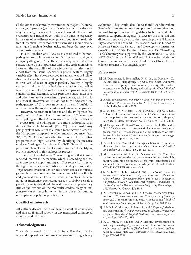
Mechanical transmission is a nonspecific process, whichcan take place when a biting insect initiates a blood mealon an infected host, starts to feed on infected blood, isinterrupted (by defensive movements of the host, e.g.), fliesoff from the infected host, and lands on another animal tobegin its blood meal again. When the insect first attempts tofeed on blood, its mouthparts can contain a small amount ofblood via capillary strength, estimated at 1–12 nl in tabanidsand 0.03 nl in Stomoxys [3]. The residual blood may bepartially inoculated into another animal during the earlystage of the next attempt to bite, when the insect inoculatesa small amount of saliva (necessary for its anticoagulantproperties) prior to sucking the blood of the second host[3–6]. A mathematical model has recently been developed;for cattle carrying a mean burden of 20–30 tabanids perhead, the model indicated that the probability of transmis-sion becomes significant when parasitaemia is above 106trypanosomes/mL [4]. Thus, in camels, which may exhibitvery high parasitaemia (>108 T. evansi/mL), tabanids andStomoxys may be responsible for the transmission of T.evansi; possibly, Haematobia (Figure 1(e)) and hippobosques(Figure 1(f)) might act as well.
In biting insects, trypanosomes do not generally survivefor very long. For example, their survival was estimated at30minwithT. vivax in tabanids and even shorter in Stomoxyssp. [7]. Experimental research shows that the transmissionis efficient when there is a short time lapse between twointerrupted blood meals, that is, less than 30 minutes [8, 9].Immediate mechanical transmission of this type can onlyoccur in a group of animals (e.g., intraherd transmission). Itleads to a high incidence of disease in a given herd. However,it may occur between herds of the same species (camels) orof different species (camels and goats, e.g.) at a water point.Transmission can also occur between wild and domesticherbivores, such as deer or capybaras when they graze with
horses, cattle, or buffalo. This occurs in extensive breedingconditions in Brazil [10], for example.
An alternative to the immediate transmission of try-panosomes occurs when blood from the insect’s gut or crop isregurgitated in the early stages of the blood feeding process.This could enable delayed transmission because parasites cansurvive in the stomach for 5–7 h in the case of T. vivax intabanids [7]. Trypanosomes could survive for even longerperiods in the crop of Stomoxys [11], where the absence ofdigestive secretion provides a more friendly environment.In experimental conditions of interrupted feeds, successfultransmissions were obtained after 4 hours with Tabanusnemocallosus, 8 h with T. rubidus (Figure 1(a)), 24 h withT. albimedius, and up to 72 h with T. striatus (Figure 1(b)).The probability of success decreases drastically from 1 : 10after 30min to 1 : 1,000 after 6 hours [2]. However, tabanidsare persistent feeders in natural conditions and, therefore,these results for delayed transmission would not apply. Oncetabanids have initiated a bloodmeal, theymake every attemptto complete it (even if it is interrupted), within a very shortperiod of time; that is, they do not wait 4–72 h. Once satisfied,tabanids do not look for a host before 5–7 days have elapsed.T. evansi cannot survive that long; therefore, the probabilityof delayed transmission by tabanids is very low [6].
On the contrary, in early experiments with Stomoxys, itwas shown that T. evansi could be transmitted 48 h after aninfective bloodmeal [12].This is unlikely to be due to residualblood on the mouthparts (survival was proven to be limitedto 30min in Stomoxys), but rather to the regurgitation ofinfected blood from the crop. However, it was demonstratedthat Stomoxys may naturally have two blood meals in thesame day or at 24-hour intervals [13]. This observationpotentially has a very high epidemiological impact because a“split blood meal” would allow transmission over long timeintervals. These intervals may range from a few hours to afew days.Thus, transmission may occur between herds in thesame place (stationary insects) or between herds attacked bythe samemobile insects. However, this experimental researchneeds to be confirmed. “Delayed mechanical transmission”might lead to the concept of “infective area,” such as a waterpoint, where an infected herd could infect Stomoxys at agiven time, which in turn would infect healthy animals (4–48hours later), in the absence of contact between infected anduninfected herds.
Mechanical transmission of T. evansi is thought to beessentially due to tabanids and Stomoxys. However, Hip-poboscids were previously suspected, especially in camelsand horses (Hippobosca equina and H. camelina) [2]. Otherinsects, such as Culicidae, Ceratopogonidaemay also have animportant role in transmission in particular local conditions.Experimental transmission of T. evansi has been successfulwith Aedes aegypti, Ae. Argenteus, and Anopheles fuliginosus.However, the epidemiological significance has not beendemonstrated (Kesler 1927 and Nieschulz 1928 quoted byGill, [2]). It is not possible to establish an exhaustive list ofthe potential mechanical vectors of T. evansi. However, themost important are the largest and most abundant bitinginsects.Themathematic model of trypanosome transmissionby tabanids developed by Desquesnes et al. [4] has shown

BioMed Research International 3
(a) Tabanus rubidus (18–25mm) (b) Tabanus striatus (13–18mm)
(c) Haematopota sp. (8–15mm) (d) Chrysops dispar (9–16mm)
(e) Stomoxys sp. & Haematobia sp. (5–9 & 2.5–4mm) (f) Hippobosca sp. (9–13mm)
Figure 1: Some of the potential vectors of Trypanosoma evansi in Thailand.
that the incidence of transmission is directly linked toparasitaemia and the number of biting insects around thehosts.The transmission of the infection is related to a numberof subparameters, including the size (and morphology) ofthe biting insect (volume of blood potentially transferredfrom one host to another) and the insect density. Thus, ahigh number of “small Stomoxys” can be as efficient as a lownumber of “large tabanids.”
Gill [2] mentioned that successful experimental trans-missions have been reported in no less than 29 Tabanussp., including Tabanus rubidus, T. ditaeniatus, T. immanis, T.rufiventris, T. malayensis, T. optatus, T. ceylonicus, T. partitus,T. striatus, and T. tenens. He also reported several successfultransmissions with Haematopota spp. (Figure 1(c)), H. cin-gulata, H. truncate, H. irrorata, H. pungens, Chrysops dispar(Figure 1(d)), C. flaviventris, C. fasciata, and even the lous
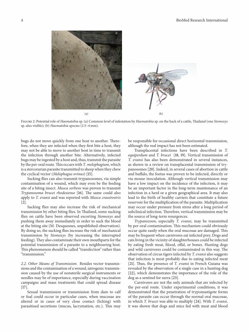
Haematopinus tuberculatus (Mitzmain, 1913, quoted by Gill,[2]). Lyperosia minuta was also suspected on the basis offield observations, although experimental transmissionswereunsuccessful [2]. Several demonstrations in experimentalconditions were also reported for Stomoxys (Figure 1(e)) byGill [2], with Stomoxys calcitrans and by other authors for S.niger, S. varipes, S. taeniatus, S. pallidus, and Haematoboscasqualida [8, 9].The role ofHaematobia sp., the smallest of theStomoxyine flies (2–4mm), has never been demonstrated,probably due to its small size, which makes it inconvenientfor laboratory experiments. However, it should be studiedbecause the density of Haematobia observed in the field isfrequently very high (Figures 1(e) and 2). In otherwords, theirrole may not be as negligible as their size!
Lastly, the potential role of reduviid bugs as mechanicalvectors was demonstrated experimentally [14]. However,
4 BioMed Research International
(a) (b)
Figure 2: Potential role ofHaematobia sp. (a) Common level of infestation byHaematobia sp. on the back of a cattle,Thailand (one Stomoxyssp. also visible); (b) Haematobia species (2.5–4mm).
bugs do not move quickly from one host to another. There-fore, when they are infected when they first bite a host, theymay not be able to move to another host in time to transmitthe infection through another bite. Alternatively, infectedbugsmay be ingested by a host and, thus, transmit the parasiteby the per-oral route.This occurswithT.melophagium, whichis a stercorarian parasite transmitted to sheepwhen they chewthe cyclical vector (Melophagus ovinus) [15].
Sucking flies can also transmit trypanosomes, via simplecontamination of a wound, which may even be the feedingsite of a biting insect. Musca sorbens was proven to transmitTrypanosoma brucei rhodesiense [16]. The same could alsoapply to T. evansi and was reported with Musca crassirostris[2].
Sucking flies may also increase the risk of mechanicaltransmission by other biting flies. In Thailand, some suckingflies on cattle have been observed escorting Stomoxys andpushing them away immediately in order to suck the bloodat the biting site (M. Desquesnes, unpublished observation).By doing so, the sucking flies increase the risk of mechanicaltransmission by Stomoxys (by increasing the interruptedfeeding).They also contaminate their ownmouthparts for thepotential transmission of a parasite to a neighbouring host.This phenomenon should be called “transfection” rather than“transmission.”
2.2. Other Means of Transmission. Besides vector transmis-sions and the contamination of a wound, iatrogenic transmis-sion caused by the use of nonsterile surgical instruments orneedles may be of importance, especially during vaccinationcampaigns and mass treatments that could spread disease[17].
Sexual transmission or transmission from dam to calfor foal could occur in particular cases, when mucosae arealtered or in cases of very close contact (licking) withparasitised secretions (mucus, lacrymation, etc.). This may
be responsible for occasional direct horizontal transmission,although the real impact has not been estimated.
Transplacental infections have been described in T.equiperdum and T. brucei [18, 19]. Vertical transmission ofT. evansi has also been demonstrated in several instances,as shown in a review on transplacental transmission of try-panosomes [20]. Indeed, in several cases of abortion in cattleand buffalo, the foetus was proven to be infected, directly orvia mouse inoculation. Although vertical transmission mayhave a low impact on the incidence of the infection, it maybe an important factor in the long-term maintenance of aninfection in a herd or a given geographical area. It may alsolead to the birth of healthy carriers that constitute a futurereservoir for the multiplication of the parasite. Multiplicationmay occur under pressure from stress after a long period ofsubclinical infection.Therefore, vertical transmission may bethe source of long-term resurgences.
Trypanozoon, especially T. evansi, may be transmittedby per-oral contamination. This mechanism could obviouslyoccur quite easily when the oral mucosae are damaged. Thismay be frequent when carnivores eat infected prey. Dogs andcats living in the vicinity of slaughterhouses could be infectedby eating fresh meat, blood, offal, or bones. Hunting dogsand wild carnivores could be contaminated in this way. Theobservation of circus tigers infected by T. evansi also suggeststhat infection is most probably due to eating infected meat[21]. Thus, the presence of T. evansi in French Guiana wasrevealed by the observation of a single case in a hunting dog[22], which demonstrates the importance of the role of thedog as a sentinel for surra [23].
Carnivores are not the only animals that are infected bythe per-oral route. Under experimental conditions, it wasdemonstrated that the penetration of trypomastigote formsof the parasite can occur through the normal oral mucosae,in which T. brucei was able to multiply [24]. With T. evansi,it was shown that dogs and mice fed with meat and blood

BioMed Research International 5
were infected [25], as well as rats fed on blood [26, 27].However, the parasite is unable to survive in the stomachof carnivores and rodents because of the pH conditions.Therefore, in these hosts, the penetration of the parasiteinevitably occurs through the oral mucosae. Conversely, theparasite can survive and pass through the oral mucosa, theoesophagus, and the stomach mucosa of vampire bats [28].
Other modes of transmission could be investigated, suchas leeches, ticks (as carriers), and other blood feeders.
2.3. Biological Vector: Vampire Bats. Transmission by thevampire bat is a new biological system that has been estab-lished in Latin America, thanks to the conquistadores whointroduced both T. evansi and its hosts on the subcontinent[15]. Vampire bats are infected by the oral route when theyleak blood from an infected prey (most often horses orcattle). As a host of T. evansi, bats may develop clinicalsymptoms and die during the initial phase of the disease(1 month). However, in the case of bats that survive, theparasite multiplies in the blood and is then found in thesaliva of chronically infected bats or in bats that do not showany clinical symptoms. Later, infected bats can contaminatetheir congeners by biting, thus acting as true reservoir hosts.They can also contaminate livestock, acting as permanentvectors, capable of contaminating their host for a long periodof time. Lastly, in the case of bats, the trypanosome may betransmitted from biter to bitten or vice versa [23]. Since thevampire bats can contaminate each other, a vampire colonycanmaintainT. evansi in the absence of themain host (horse),which makes them a true reservoir of the parasite. Whenfeeding on horses or cattle, vampire bats are true vectors, inas much as they initiate infection that biting insects can thenspread to other susceptible animals [15, 28, 29]. The vampirebatDesmodus rotundus acts as a host, reservoir, and biologicalvector of the parasite.
The differentmodes of transmission presented abovemayhave variable importance depending on the host and epi-demiological cycle. For example, biting insect transmission isvery important in livestock or large animals more generally,vampire bat transmission is important in horses and cattle,though only in Latin America, and per-oral transmissionis predominant in carnivores, vampire bats, and probablyrodents. However, important data is missing in terms of ourcurrent knowledge, including data on the link between largeanimals andwild rodents, how rodents are contaminated, andthe potential back infection from rodents to livestock or fromcarnivores to herbivores.This will be further discussed underthe epidemiology section.
3. Control
Disease control is generally presented last, following thedescription of epidemiology. However, we decided to discussdisease control first because trypanocide use is now partof regular livestock management in all areas endemic fortrypanosomoses. In other words, disease control has becomepart of disease epidemiology.Thus, it can only be understoodif we take into consideration the continuous and cosmopoli-tan use of trypanocide drugs for livestock.
The control of a vectorial disease is classically divided intotwo sections: pathogen control and vector control. There arealso various alternative means of controlling transmission,which can be combined as “means to prevent the infection.”
In the case of surra, in the absence of a vaccine againsttrypanosomes (due to a large repertoire of variable surfaceantigens), disease control is principally based on the use oftrypanocides andpreventivemanagementmethods to protectanimals from infection.
3.1. Chemical Control of Parasites. As a blood parasite, T.evansi can be killed by injecting various trypanocidal drugs,providing that concentration of the chemical in the serum islethal for the parasite. However, treatment might fail in thecase of extravascular invasion or chemoresistance.
Trypanocides can be divided into two categories. The“curative drugs” are used for treatment and have a short-termeffect.They can kill the parasites, although they do not alwayseliminate 100% of them. The “curative/preventive drugs” areused for chemoprophylaxis. They not only kill parasites butalso prevent any new infection or new circulation of parasites,due to the remanence of a sustainable curative dose in theserum of animals under chemoprophylaxis.
3.1.1. Curative and Chemoprophylactic Drugs. Curative drugsaim to eliminate parasites from a sick animal. A drug could beregarded as “curative” when the dose used is able to eliminateall parasites. The most widely used curative trypanocideagainst surra is diminazene aceturate. However, other drugscan be used, such as isometamidium chloride (both curativeand preventive), cymelarsan (so far, only recommended forcurative treatment of camels), suramin, and quinapyramine(curative and/or preventive) [30].
Diminazene aceturate (DA) is an aromatic diamidineused to control babesia and trypanosome infection in rumi-nants. A curative dose of DA is administered via intra-muscular injection to obtain a high concentration of thechemical in the circulating blood. The withdrawal period forthe consumption of produce fromcattle injectedwithDA is 21days for meat and 3 days for milk [31]. However, the chemicaldose in the serum actually suggests a longer withdrawalperiod of 30 and 21 days for meat and milk, respectively[32]. The dose recommended for the treatment of infectionsdue to parasites belonging to the Trypanozoon subgenus is7mg/kg bodyweight (bw) of DA, via intramuscular injection.The reality in the field often reveals that a dose of 3.5mg/kgbw is used to control surra.This could be for various reasons,including ignorance of the right dose or concern to savemoney by reducing the cost of treatment. Use of the “wrong”dose is based on the recommended dose for the treatment ofinfections by two otherAfricanTrypanosoma species:T. vivaxand T. congolense. Diminazene aceturate is recommendedin ruminants. Its use in horses and dogs is limited due topoor efficacy and tolerance in these species. Diminazeneaceturate has been used for a long time. Consequently,trypanosomes have developed chemoresistance inmost partsof the world [23, 31]. Using 3.5mg/kg bw to control T. evansican be considered as underdosing, as is often the case in
6 BioMed Research International
Thailand and more generally in South East Asia. This dosecan be regarded as a “premunition treatment,” when the hostremains infected, (although clinically cured), contrary to thecurative dose that eliminates all parasites. Such low dosetreatment can lead to the selection of chemoresistant strains.In Thailand, for example, the inefficiency of DA in bovines,horses, pigs, and elephants has frequently been reported [33–36].
Isometamidium chloride (IMC) belongs to the phenan-thridine family, as well as homidium chloride or bromide.However, the latter are highly toxic because they are DNAintercalating agents. Their mutagenic action was demon-strated early on [37, 38]. Therefore, their use in the fieldis not recommended. IMC is not known as a carcinogenicagent. It can be used for curative (0.5mg/kg bw) and pre-ventive (1mg/kg bw) treatment of trypanosome infectionsin ruminants and horses, via intramuscular or subcutaneousinjection. Alternate use ofDAand IMCconstitutes a “sanativepair,” whichmeans that once resistance develops to one of thedrugs, the other drug should be used to control the infection[30]. The withdrawal period for the consumption of producein cattle injected with IMC is 23 days. However, it is obviousthat the chemical can circulate in the blood for up to 4-5months after injection [39]. Consequently, a safe withdrawalperiod should be much longer, from around 3 months (when0.5mg/kg is injected) to 6months (when 1mg/kg is injected).These withdrawal periods make IMC poorly adapted to beefor dairy cattle. Horses have a limited tolerance to IMC [40],although it remains an alternative to DA.
Melarsomine dihydrochloride (Cymelarsan) is the latesttrypanocide to be developed. It was first available for com-mercial use in 1992. It is used to control surra in camels viadeep intramuscular injection at a dose rate of 0.25mg/kg bw[23]. Evaluations conducted on other host species suggestusing rates of 0.25–0.5mg/kg bw in horses, 0.5mg/kg bw incattle, and 0.75mg/kg bw in buffaloes [41–43].However, tran-sient side effects (nervous signs) were observed in buffaloestreated with 0.75mg/kg bw (Dargantes et al., unpublisheddata). Dogs have a satisfactory tolerance to the drug. It isrecommended for the treatment of heartworm (Dirofilariaimmitis) (Immiticide), at a dose of up to 2.5mg/kg bw (viadeep intralumbar injection). It can be used at a rate of 1-2mg/kg bw against T. evansi infections. However, in the caseof nervous infections in horses and dogs, even high doses,respectively, 0.5mg/kg bw and 2mg/kg bw, failed to cure theanimals [44, 45].
Suramin is an ureic component which was used in horsesand camels by intravenous injection. It was effective againstT. evansi infection, although it is no longer used.
Quinapyramine belongs to the group of aminoquinaldinederivatives. Quinapyramine methyl-sulphate can be used totreat the infection by subcutaneous injection at a dose of5mg/kg bw. A more effective combination of quinapyraminesulphate and quinapyramine chloride (Triquin) can be usedas a curative/preventive drug against T. evansi in horses andcamels, administered by subcutaneous injection at a dose of8mg/kg bw. Local tolerance is sometimes low. However, thedrug is quite efficient and the chemoprophylactic effect canlast up to 4months [46]. In cattle, the use of quinapyramine is
not recommended because it may induce cross-resistance toboth DA and IMC [47]. Its use should be restricted to horsesand camels only.
3.1.2. The Use of Trypanocides in Various Host Species. Buf-falo, cattle, and small ruminants infected by T. evansi canbe treated with DA (preferred drug) at a dose of 7mg/kgbw by intramuscular injection. The withdrawal period formeat consumption should be >30 days. In the case of strongclinical signs, especially when parasitaemia is high, an initialinjection of 3.5mg/kg bw DA may be given to reduce theparasitaemia and a second injection of 7mg/kg bw can begiven 5 days later to ensure that all the parasites are killed.
If the treatment is ineffective, the use of IMC is rec-ommended at a dose of 0.5mg/kg (withdrawal period formeat should be >90 days). Alternatively, the efficacy ofmelarsomine hydrochloride was recently demonstrated (nonervous signs were observed), at a dose of 0.5mg/kg bwby deep intramuscular injection in cattle [45] and buffaloes(Dargantes et al., unpublished data).
Horses, dogs, and cats can be treated with DA or IMCdespite being quite sensitive to the drugs. It is essential toprovide an adequate water supply to avoid a toxic effect onthe kidneys, which can be fatal. Similarly, in the case ofvery high parasitaemia in cattle, half a dose of DA or IMC,followed by a normal dose 5 days later can be administered.Given that horses have a low tolerance to DA and IMC,the normal recommended dose can also be split into twosubboosts (DA 2 × 3.5mg/kg and IMC 2 × 0.25mg/kg).However, the intervals between the subboosts should notbe too long, otherwise the curative drug concentration inthe plasma will not be reached. In such cases, injectingtwo subboosts with a 3–5-hour interval is recommended.DA treatment is not efficient in the case of nervous infec-tion. Results of DA or IMC treatment may be satisfying,although chemoresistance is often observed, which limits theeffectiveness of treatment. As an alternative, the efficacy ofmelarsomine dihydrochloride was evaluated in horses anddogs. The treatment can clear the parasite from the blood.However, in cases of nervous infection, it is inefficient andmay cause death in the patients [44, 45]. Another alternativeis the treatment of horses with quinapyramine sulphateand chloride (curative and chemoprophylactic effect), whichprovides durable protection to the animals. Nonetheless,we do not know whether the infected animals that receivesuch treatment are sterilised from the infection or whetherthey can carry the parasite in extravascular foci, such asjoint fluids, cerebrospinal fluid, and aqueous humour of theeye, as has been demonstrated in camels [48]. However,if parasites do survive in an extravascular refuge and laterattempt to reinvade the blood, they would be killed onreaching the blood given the chemoprophylactic drug’s long-lasting action. In such conditions, keeping horses alive inenzootic areas might require regular treatment with thechemoprophylactic drug. Indeed, horse owners usually treattheir animals regularly, both in Latin America and South EastAsia [46]. In dogs, treatment with quinapyramine is poorlydocumented, although drugs are available on the market

BioMed Research International 7
(Interquin). Another alternative treatment for dogs could betried out, using 5–8 serial injections with DA at 3.5mg/kgbw at a 2-3 week interval. In the absence of a trypanocidecapable of establishing curative treatment for dogs, this typeof strategy aims to enhance specific and protective immunityagainst the parasite. This premunition status can be expectedwithin some months.
In camels, although a number of trypanocides have beenused (DA, IMC, suramin, quinapyramine, etc.), melarsominedihydrochloride is the ideal product (dose: 0.25mg/kg bw),which can be increased up to 0.5mg/kg bw if fully curative(sterilising) treatment is required for international trading. Inenzootic areas, a dose of 3.5mg/kg of DA can also be used.However, it can induce severe side effects and might not besufficient to clear all parasites from the camel.
In pigs, little information is available on the controlpractices used for African trypanosomes. Quinapyraminemay be used, as well as DA, though the latter appears to be oflimited efficacy [36]. IMC and melarsomine dihydrochloridecould also be used. However, experimental evaluations arenecessary to validate the treatment protocols.
In Asian elephants, several attempts have been madewith DA. Lower doses, such as 5mg/kg bw, resulted inrelapses [49, 50], while 8mg/kg bw seems to be efficient[51]. Evaluation of melarsomine dihydrochloride at a doseof 0.2mg/kg bw could also be evaluated (Frans Van Gool,personal communication).
3.1.3. Strategies for the Use of Trypanocides. It is importantto determine a strategy and the objectives of a treatment,whichever trypanocide is used. Indeed, in most of the highlyenzootic situations, when the infection is not lethal, suchas T. evansi in bovines, the treatment does not necessarilyaim to completely eliminate the parasite from the animal.In practical terms, a “mild treatment” (3.5mg/kg bw DA,e.g.) might be sufficient to kill the majority of parasites,ensure clinical improvement, and induce the release of alarge amount of parasite antigens to enhance the host’simmune response. Animals that are treated in this way,but remain infected, can cope with the infection becausethey develop an adapted immune response. This leads tothe status of subclinical infection or healthy carrying. Fromthe clinical point of view, farmers may think that suchtreatment is curative. Maintaining an efficient immune statusis especially important for gestating animals in an enzooticsituation. However, it is important to emphasise that lowdose treatments potentially enhance the development ofchemoresistance.
In bovines, if the objective is to kill all parasites (to cleara farm from infection or prior to export, or for introducingan animal into a noninfected farm, etc.), a higher dose ofDA or other chemicals should be used, such as 7mg/kg DAbw (in the absence of chemoresistance), or 1mg/kg bw IMC,or 0.5–0.75mg/kg bw of melarsomine dihydrochloride. Aspreviouslymentioned, in dogs, a strategy of serial lowdoses ofDA injections (3.5mg/kg) could be attempted when sourcesof infection are out of control.
When the infection threat is lethal, such as T. evansi inhorses and dogs, a different strategy is generally preferred,
namely, to kill all parasites, as far as possible. The objective inthis case is to achieve fully curative or “sterilizing” treatment,which requires the use of (i) curative drugs, such as DA7mg/kg bw (obviously with a high probability of treatmentfailure) ormelarsomine dihydrochloride 0.5mg/kg bw; or (ii)chemoprophylactic drugs, such as quinapyramine sulfate andchloride 8mg/kg. However, in the case of an invasion of thenervous system, none of these drugs have yet been proven tobe efficient.
In horses, irrespective of the source of infection (from aninside extravascular focus in a carrier or from mechanicalvectors that bring parasites from neighbouring infectedhosts), the only option is to treat with quinapyramine sulfateand chloride at 8mg/kg bw to protect the blood from parasiteinvasion. However, if the parasites reach the nervous system,the disease is always fatal.
3.2. Preventing Infection. In addition to parasite preventionand control, vector control, or more generally, preventinginfection is an important part of disease control, especiallyfor highly susceptible species, such as horses and dogs.
3.2.1. Vector Control. In the case of tsetse-transmitted try-panosomes in Africa, vector control is quite effective atreducing the trypanosome pressure. The cyclical vectors canbe specifically targeted using insecticide impregnated screensand insect sterilisation techniques can be used in a limitedlivestock breeding area [52].
Conversely, the control of mechanical vectors is not easybecause of the diversity of tabanid species in a given area,their high mobility and prolificacy. In addition, the larvalstages of tabanids are generally spread over a wide areaand different species colonise various landscapes [53]. Theecological control of one species might help the developmentof another! Consequently, the ecological control of tabanidsis not usually an option.
Tabanid control using insecticide sprays was provento be efficient in small closed deforested areas in FrenchGuyana [54]. However, even in this case, tabanid infestationreappeared 2-3 years after the end of the control campaign[23]. When tabanid control is carried out in an open area,it is not sustainable because tabanids move in from thesurrounding areas to fill the ecological gap created by the con-trol campaign. Implementation of tabanid control is rarelyattempted because it is costly, unsatisfactory, unsustainable,and does not provide 100%cover from infection.Nonetheless,the control methods are described briefly below.
Stomoxys species differ from tabanids in that they developwithin the livestock area or the farm and are closely relatedto the farming systems [53]. Stomoxys population controlcan be achieved through management methods (see descrip-tion below). However, sustainable control using mechanicalvectors is not possible because of their high mobility andprolificacy. Indeed, an adult female tabanid or Stomoxysmay lay 100–200 eggs, 4-5 times in her lifetime. Therefore,total egg production ranges from 400 to 1000 eggs [53]. Bycomparison, a female tsetse fly produces one larva at a time,approximately 10 times, thus producing 10 flies in her lifetime.

8 BioMed Research International
Figure 3: Nzi trap. A universal trap able to catch tsetse flies,tabanids, and Stomoxys, especially efficient for large size tabanids.
These are known as the “R” and “K” reproduction strategies,respectively [55].
Mechanical vectors use the “R” strategy. Their extremeprolificacy means that if only 2% of the eggs reach the adultstage, the tabanid population remains stable [53]. Hence, ifmore that 2% of eggs reach the adult stage, the populationwillincrease. In order to control tabanid populations successfully,egg development must be kept below 2%.
The control of surra’s vector populations can be attemptedusing traps and/or impregnated screens or using insecticideson livestock. The most efficient traps for mechanical vectorsare theNzi (Figure 3) and theVavoua trap (Figure 4) [56, 57].The Nzi trap can catch large tabanid species and Stomoxys,while the Vavoua trap catches small tabanid species, such asChrysops (deer flies) and Stomoxys [58]. However, so far, thesetraps have been used to study insects and monitor controlcampaigns rather than for actual insect control. Sprayinginsecticides, such as deltamethrin on cattle, is efficient forcontrolling mechanical vectors [54, 59, 60]. However, theeffect is relatively short-lived, which makes efficient controlcostly. The use of targets or impregnated screens, like thoseused in Africa against tsetse flies, was not evaluated for thecontrol ofmechanical vectors. It is an attractive alternative forthe targeted control of biting insects. One of the traditionalmethods for controlling biting insects is the use of smokereleased by slow fire (Figure 5). The smoke repels the insects.However, because it only covers a limited protected area, theanimals in this area reduce their food intake [23]. Mosquitonets can be used to protect animals, though this is rarebecause of the expense. However, individual use does occur,such as for horses (Figure 6). It is possible to adapt fly-proofcorals or stables for groups of animals, for example, cattle(Figure 7). Insecticide impregnation of mosquito nets is analternative integrated method of control, which can helpreduce biting insects/vector populations.
In Latin America, the vampire bat can act as vector, host,and reservoir of T. evansi. Consequently, vampire bat controlis an integral part of surra control. The “Japanese net” can beused to catch vampire bats. It can also be used as a screento protect livestock. In this case, it should be set up at nightto create a screen between the bat colony refuge (forest area)and the livestock farm. Alternatively, the Japanese net can
Figure 4: Vavoua trap. A trap designed for tsetse flies, especiallyefficient for Chrysops and Stomoxys.
Figure 5: Smoke released to protect horses from biting flies, SuratThani, Thailand.
be used to catch a few bat specimens, which are then usedto kill the colony. Captured animals are coated with dropsof anticoagulant that contains an excipient, such as lanolin,before they are released. Once the animal has returned to thecolony, it spreads the chemical to the whole colony by lickingand contact. An anticoagulant, such as chlorophacinone, killsthe bats within a few days [23, 61].
3.2.2. OtherMethods to Prevent Infection. In situations whereit is difficult to control the biting insect populations, it may beeasier to control transmission, though not with 100% efficacy.Tabanids are naturally persistent feeders [62] and they do notleave one animal to bite another if the latter is more than50 metres away. Therefore, 200m is considered to be a safedistance for mechanical transmission by biting insects [62–64].
However, separating bovines from equines is highlyrecommended to avoid the transmission of T. evansi from abuffalo or cattle reservoir to highly sensitive horses. To avoidany risk of transmission (even that of occasional contact withanimals that have escaped), it is advisable to breed cattle andhorses in completely different areas that are at least severalkilometres apart.

BioMed Research International 9
Figure 6: Mosquito net system on a stable to protect a horse againstbiting flies in an area of high infestation, Ratacha Buri, Thailand.
Figure 7: Fly proof system with mosquito net for cattle stable(Nakhon Sawan, Thailand).
The case of carnivores is quite unusual. Carnivores maybe infected when they eat the bones, flesh, or blood of aninfected animal that has only just died. Rodents, which areomnivorous, may become infected like carnivores. T. evansican be transmitted via oral infection as demonstrated in a trialin which per-oral blood was given to rats and mice [26, 27].To avoid such infections, the dead animals’ carcasses shouldbe eliminated as soon as possible and dogs, especially straydogs, should be contained around slaughterhouses, as well ason livestock farms in general.
In addition to per-oral contamination, dogs may alsocontract the infection from biting flies, especially the dogfly, Stomoxys, when they live in the vicinity of reservoiranimals, such as cattle and horses. As mentioned above, hostspecies must be well separated to avoid the interspecific hostcirculation of parasites.
3.2.3. Preventing Introduction into a Noninfected Area. Aswas recently observed in Spain and France [65], healthy orinapparent carriers may be responsible for introducing theparasite from infected to noninfected areas. A number ofmeasures, that are currently being studied, could be appliedin order to avoid this type of introduction. The detection ofcarriers is based on laboratory detection according to theguide of methods recommended by the World Organisa-tion for Animal Health (WOAH) in its terrestrial manual,Chapter 2.1.17. It is available online at the following address:(http://www.oie.int/fileadmin/Home/fr/Health standards/tahm/2.01.17 TRYPANO SURRA.pdf)
Diagnosis techniques for surra are based on four types ofexamination (referred to below as “surra tests”): microscopicexamination, DNA detection by PCR, CATT/T. evansi andELISA T. evansi (detailed protocols are available at the abovelink).
For the international trading of animals, the followingguidelines could help avoid the introduction of infectedanimals into noninfected areas.
(i) Two quarantines should be applied for the interna-tional trade of equines and/or camelids (which couldbe extended to any mammal) from an infected coun-try to a noninfected country: a 4-week quarantine atthe exporting farm and a 4-week quarantine at theimporting farm.
(ii) To qualify for trading, an animal should originatefrom a noninfected farm in a nonsuspect area, andbe negative to surra tests twice at a 3-4 week intervalduring each of the quarantines.
(iii) A farm is considered to be in a nonsuspect area if therehave been no reports of surra in the previous 3 yearswithin a 30 km radius of the farm.
(iv ) A noninfected farm is a farm located in a nonsuspectarea, which only permits the introduction of animalsthat are negative to the surra tests and that originatefrom noninfected farms located in a nonsuspect area.To obtain the status of noninfected farm, all mammalspecies on the farm must be negative to surra teststwice at a 3-month interval. To maintain the statusof noninfected farm, all mammal species on the farmmust be negative to surra tests when tested every 10–12 months.
If thesemeasures were adopted, theywould considerably helpto control the circulation of animals infected with T. evansi.
4. Epidemiology
Surra is a disease that can show the following: (i) varioussymptoms in a given host (from subclinical evolution toabortion or death, with or without vascular, nervous, orgenital signs); (ii) various symptoms fromone host to another(mostly lethal in horses, acute or chronic in camels, variablein bovines and buffaloes, acute in dogs, and generally mildbut sometimes acute in pigs, sheep, and goats, etc.); as wellas (iii) various aspects in different places (surra in buffaloesand cattle is virtually absent in Latin American, although itis a major constraint in South East Asia). The epidemiologyof a disease depends on the characteristics of a pathogen,its hosts, reservoir, and vectors and their environment andinterrelations. Consequently, in the peculiar case of surra,which is a multispecies disease, it can exhibit highly variablecharacteristics because of its highly complex epidemiology.
The study of the epidemiology of surra requires var-ious specific diagnostic tools. Detailed procedures areavailable from the World Animal Health Organisation(WAHO/OIE) website, terrestrial manual, under Chapter2.1.17 Trypanosoma evansi infection (surra), as indicated
10 BioMed Research International
above. For this reason, we only present a summary ofthe techniques and their characteristics, as required for acomprehensive description of surra’s epidemiology.
4.1. Diagnostic Tools. As described above, clinical signs areonly indicative of surra.The definitive diagnosis involves lab-oratory analysis, either by using parasitological or moleculartools to demonstrate the presence of the infection or by usingserological tools to prove immune contact.
Parasitological examinations are usually conducted usingblood, although other biological materials can be used, suchas cerebrospinal fluid (in the case on nervous signs), jointfluid, or lymph node fluid. Microscopic observation (×400–500) of fresh blood is easy to carry out. However, it is oflimited sensitivity because it detects parasites when para-sitaemia is above 105 trypanosomes/mLof blood. Enrichmentmethods are widely used, namely, Hematocrit CentrifugeTechnique (HCT) [66] or dark ground Buffy Coat Method(BCM) [67]. They increase the sensitivity of the test downto 100–200 trypanosomes/mL. If high sensitivity is required,inoculating laboratory rodents can reveal infection. It low-ers the minimum level of parasitaemia detected to 20–50 parasites/mL.
In addition, molecular evidence of T. evansi DNA canbe tested using PCR with a number of primers specific forthe subgenus Trypanozoon, or to species levels [68]. Despitebeing relatively expensive and technical, PCR is generallyused to improve the sensitivity of the detection. Comparativestudies have led to the recommendation of TBR primers [69]as the most sensitive primers for detecting T. evansi [70], andthe Phenol-Chloroform method [71] as the most sensitiveDNApreparationmethod [72]. A combination of thesemeth-ods provided a sensitivity of around 5–10 trypanosomes/mLof blood (or other fluid).
In addition to the use of parasitological or moleculartools for detecting T. evansi infection, serological tests thatprove the immune contact between the host and the parasiteare quite useful. They can be applied to investigations atherd or population level (prevalence or incidence studies),follow-up (seasonal or interannual variations), or controlmethod assessment (trypanocide treatment or vector con-trol). The most common tools are the Card AgglutinationTest for T. evansi (CATT/T. evansi) [73] and the ELISA T.evansi [74, 75]. CATT can detect immunoglobulin M and,therefore, early infections, whereas ELISA is generally usedto detect immunoglobulin G, that is, established infections.Consequently, these tests are complementary and work welltogether. ELISA T. evansi is quite robust, regardless of thehost species. It provides the same range of sensitivity andspecificity (90–95%) in the various host species investigated,for example, camels, cattle, buffalo, and horses.The sensitivityof CATT T. evansi varies from one host to another. CATTseems highly sensitive in camels and horses, although it has avery low sensitivity in cattle (12%), even under experimentalconditions [41].
4.2. Africa and the Middle East. In Africa and the MiddleEast, T. evansi is responsible for an acute or chronic disease
principally found in camels, horses, and dogs in the north ofthe tsetse belt [76]. In camels kept close to the tsetse belt, somecases of T. brucei brucei have been recorded. T. congolenseinfections are fatal to camels. Therefore, camels should notbe allowed to enter the tsetse belt unless they are permanentlyprotected with the use of chemicals. Consequently, T. evansiis not found in the tsetse belt. Thus, the epidemiology ofsurra in Africa is mainly governed by camel infections. Thelatter are seasonal because the vectors’ activity is seasonal andthe disease is expressed seasonally, at times when animalsare exposed to stress from overwork, food shortages, and/orinsufficient or poor quality water [77]. For example, in Mau-ritania, by using CATT and IFAT blood smears, it was shownthatT. evansi infectionwaswidespread in the country, with anoverall prevalence of 1.3% by parasitological detection. Thislevel reached 18.4% to 31% with serological tests [78]. Otherhost species, such as goats, may be infected occasionally andcould act as a reservoir. However, their impact was neverdemonstrated [79]. In cattle, especially transhumant herds,that spend part of the year within the tsetse belt and the restof the year in the northern region, it is difficult to distinguishbetween infections due to T. brucei and T. evansi. The latter isprobably rare because, even under experimental conditions,the infection of African cattle by T. evansi proved to bedifficult as a consequence of their low susceptibility [80].The transmission of T. evansi can only occur if the “donorhost” exhibits high parasitaemia. This is because T. evansi ismechanically transmitted by biting insects (due to the verysmall amount of blood transferred from one host to another).In Africa, only camels and horses may be a source for thistype of transmission.Hosts that have a low susceptibility, suchas cattle and goats, are likely to constitute dead ends, evenif they may occasionally be infected when close to infectedcamels or horses. Finally, given that camels and horse cannotenter the tsetse belt without being at risk fromNagana, whichis fatal to both hosts, and because other hosts that could beinfected by T. evansi do not exhibit sufficient parasitaemiato play an important role in surra’s epidemiology, there isa reciprocal exclusion of Nagana in the southern territory(among tsetse flies, livestock, and wild animals) and surra,which is restricted to the northern region (amongmechanicalvectors and camels).
In the Middle East and towards Asia, the geographicaldistribution of T. evansi is closely related to that of camelsand dromedaries [15]. However, no difference was observedin terms of the pathogenic effects of the parasite in this hostspecies, which like horses are highly sensitive to the infection.
Overall, surra affects mainly camels with acute andchronic infections that cause death. Infection is contractedduring the rainy season when there is a peak level of bitinginsects. Camels constitute the main reservoir of T. evansi inthis region.
4.3. Latin America. In Latin America, the disease is calledMal de Caderas (Brazil),Murrina (Central America), or Der-rengadera (Venezuela) (Wells 1989). T. evansi is principallypathogenic in horses and induces outbreaks with very highmorbidity and mortality. It also affects buffaloes (Bubalus

BioMed Research International 11
bubalis). In Venezuela, although infection in buffaloes byT. evansi showed significant signs such as spleen, liver, andglandular enlargement, together with lymphoproliferation,the economic impact of infections has not been assessed[81]. Trypanosoma evansi regularly affects dogs (especiallyhunting dogs) and even cats. In both cases, the disease isusually fatal. In Latin American cattle, sheep, goats, and pigs,T. evansi is generally considered as a low pathogenic agent. Itis regularly found in awide range of wild reservoirs, includingcapybaras (Hydrochoerus hydrochaeris), which is the mostwell known, together with white tail deer (Odocoileus vir-ginianus chiriquensis), brocket deer (Mazama satorii), coati(Nasua nasua), vampire bats (Desmodus rotundus), wild pigs(Tayassu tajacu), Guinea pig (Cavia porcellus), wild dog(Canis azarae), and ocelot (Felis pardalis). Llamas are alsoreceptive to the disease and infected animals have beenfound, although little is known about its impact [23]. Thereis no obvious link between wild and domestic fauna. Insome places, the prevalence of infection may be very highin capybara and coati, while it remains low in horses [82].In French Guyana, the parasite has never been found inlivestock, including horses, but it was first described in 1995in a hunting dog, which was probably infected by wild faunawhenhunting in the forest [23]. Surra remains amajor diseasein Latin America, especially because horses (sensitive host)are used for herding cattle (reservoir) in extensive conditionsin Venezuela and Brazil, for example.
Overall in Latin America, surra is predominantly adisease that affects horses. However, a large range of wildand domestic mammals can act as a reservoir. In most cases,farmers use chemoprophylactic drugs regularly to protecthorses against T. evansi (isometamidium or quinapyramine).This treatment ensures that they stay alive and efficient forwork. As a result, two groups of livestock are kept in closecontact: a low susceptible reservoir made up of bovinesand a highly susceptible host made up of horses underchemoprophylaxis.
4.4. Asia. In Asia, the geographical distribution ofT. evansi isspreading steadily. It is present in large areas in India, China,and Russia [83, 84]. It is sometimes difficult to distinguish itfrom T. equiperdum [85]. It is present in Camelus bactrianusand horses in Mongolia, with low prevalence. It is morefrequent in Uzbekistan and Kazakhstan. In South East Asia,it affects principally horses, dogs, and buffaloes (Bubalusbubalis), as well as cattle, pigs, and deer. It has been describedin tigers in India [21], as well as in Thai elephants [86].
In the water buffalo, T. evansi causes production losses,abortion, and early calf mortality. It also has immuno-suppressing effects, which decrease the efficacy of some vac-cines (especially of the vaccine for hemorrhagic septicaemia).In bovines, its pathogenicity in Asia is superior to that ofAfrican and American strains. We do not know whetherthe difference is due to the presence of more sensitive dairybreeds in Asia, or if the local populations ofT. evansi aremorepathogenic to cattle, or both.
In India, surra is present all over the country in varioushosts, such as cattle, buffaloes, camels, donkeys, dogs, andhorses [87]. A recent survey carried out on horses showed
a maximum seroprevalence (20%) for T. evansi infection inUttar Pradesh. There was an overall seroprevalence of 11% innorth and north-western regions of India, which confirmedthat surra is endemic in equids in these areas [88].
In Thailand, a study on seroprevalence carried out indairy cattle demonstrated the presence of the parasite inmost parts of the country. The mean seroprevalence was 8%,ranging from 0 to 100% at farm level and 25% of dairy cattleare exposed to the infection [89]. Similar studies conductedon buffaloes and beef cattle showed seroprevalence of 10–12% (Desquesnes, unpublished data). Molecular evidence ofT. evansi was also obtained in various wild rodents [90, 91].However, their role in the epidemiology of the disease isnot known. In horses, several outbreaks are recorded everyyear and are frequently fatal. Indeed, serological studies showvery low evidence of positive animals; in other words, thereare few survivors after the outbreaks [44, 92]. Elephants areaffected by surra. Cases are reported rarely but regularly.They may be fatal or develop into a chronic or subclinicalevolution, depending on the case [50]. Surra outbreaks occurseasonally and are generally linked to the activity of bitingflies. In Northeast Thailand, seasonal occurrence is observedat the beginning of the rainy season (June-July) and in winter(October-November) [93]. Bovines (cattle and buffaloes)exhibit moderate signs and impact. However, they constitutea permanent threat to themselves and horses, which may dieor survive under permanent chemoprophylaxis.
In the Philippines, over the past decade, the numberand severity of surra outbreaks have increased dramatically.The highest mortality is in horses, carabao (Asian waterbuffalo), and cattle. As a result, the Philippine governmentnow regards surra as the second most important livestockdisease [94]. Indeed, surra has emerged as themost importantcause of livestock mortality in the Philippines, prompting thegovernment to implement a national control strategy.
In Indonesia, the disease appears in sporadic outbreaks,mainly in horses, buffaloes, cattle, and dogs, although it is alsopresent in sheep, goats, pigs, and wild animals [94–96]. Theparasite was found throughout most of the archipelago. Itsregular occurrence suggests the existence of enzootic stability,including an efficient reservoir [95, 96]. However, an updateof information is required.
In Vietnam, Laos, and Cambodia, the disease occurredespecially in horses, buffaloes, and cattle, although it wasgiven little attention. Serological surveys demonstrated thepresence of infection in all the areas investigated. Limitedmeans are available for carrying out studies on surra sincepriority is given to other diseases in these countries. Hence,the situation is not well documented.
Similarly in Malaysia, although the disease has beenknown for years, a national survey has not yet been organisedto evaluate its impact. Surra is regularly detected in horses,deer, pigs, buffaloes, cattle and, rarely, in dogs. It was alsoreported in Sumatran rhinoceroses [97]. In Malaysia, aseroprevalence survey carried out in 2012, for dourine inhorses usingCFTdid not reveal its presence [98]. Diagnosis isroutinely carried out using amouse inoculation test and buffycoat examination. Thin blood smears are also conducted inregional laboratories. Prophylactic treatment is administered
12 BioMed Research International
to livestock in high-risk areas where cattle and buffaloes livein close proximity to pigs or horses [99, 100]. In domesti-cated deer, the infection is observed regularly. Fulminatingparasitaemia is detected when the animals become weakand recumbent. Nervous symptoms are not clearly evident;however, fatality ismost often observed during the outbreaks.
Trypanosoma evansi is not present in Australia, but it mayspread eastward from Indonesia to Papua New Guinea andthen Australia [101].
Overall in Asia, surra is mainly a disease of horses andbuffaloes. It benefits from a large reservoir made up ofbuffaloes, cattle, deer, and possibly wild animals, such asdeer and rodents. On cattle farms, little attention is givento the disease, even though it may cause serious economiclosses, via abortion, weight loss, and immunosuppressiveeffects. An evaluation of the economic impact is neededto determine whether it would be profitable to eliminatethe infection. Horse breeders generally avoid close contactbetween buffaloes and horses to avoid the risk of infection,which is generally fatal and uncontrollable because of thelimited efficacy of trypanocides [34, 36]. When horses arebred in the same area as cattle or buffaloes, farmers regularlyuse chemoprophylactic drugs to protect horses against T.evansi (isometamidium or quinapyramine). Nonetheless, T.evansi remains a permanent threat to livestock throughoutSouth-East Asia, with a decreasing gradient of impact forhorses, buffaloes, dogs, cattle, deer, pigs, sheep, and goats.
5. Impact
There is limited information on the impact of surra amonglivestock in endemic countries, particularly (i) its impacton host population dynamics and demographics, (ii) theeconomic losses due to the disease, and (iii) social impacton animal owners. It is common knowledge that surra is aneconomically important disease, which causes highmortality,low milk and meat production, poor carcass quality, reducedreproductive performance, decreased draught power andmanure production, and immunosuppression in livestock[94, 102–105]. Yet, only few studies have quantified theeconomic value of the losses (including expenditure on diag-nosis, treatment, and replacement of lost animals) for limitedanimal species and for limited locations. Little information isavailable on the financial benefits of treating/controlling surrain infected animal populations.
The impact of surra on host population dynamics andreproduction has been extensively investigated for buffaloesin the southern Philippines [106]. Surra has a significant neg-ative impact on buffalo populations, causing high mortalityand reproductive losses. In particular, in surra endemic areas,buffalo herds have fewer calves, 50% lower calving rate andhigher removal rates (including adult mortality and early calfdeaths), than buffaloes in areas where surra is not detected.Higher mortality has been recorded amongst young buffalocows aged 2–8 years old in surra-endemic areas compared tosurra-free villages (9.1% versus 0.1% mortality, resp.). Giventhe decrease in the buffalo population in surra-endemicareas, replacement buffaloes are regularly imported becausethey provide essential draught power for farm operations
[106, 107]. This impact may also be true for other animalspecies that are susceptible to T. evansi. However, furtherinvestigations are required to validate the impact of surra onother hosts.
The low calving performance among buffaloes in Min-danao (in the Philippines) is closely linked to abortion andinfertility [107]. In Thailand, abortions and reproductivefailure due to surra have also been demonstrated in buffaloes[108], cattle [104, 109, 110], camels [111], and horses [34, 112].The death of buffalo cows during their most productivephase reduces their life expectancy (by almost half) andhas a major impact on farmers. Females at this age arehighly valued for draught power and as breeding animalsfor replacement or sale (to provide additional income) orhome consumption. Surra has been proven to causemortalityin buffalo after experimental [113] or natural infection [83,102, 114, 115].Whilst mortalities in draught buffalo caused bysurra could be partly associated with stress due to overwork,other factors such asmalnutrition, concurrent infections, andadverse climatic conditions may contribute to the animals’reduced resistance and higher susceptibility to the disease[102, 115]. Surra is also lethal in other livestock species, suchas horses [116–118], camels [111, 119], guanaco [120], cattle[86, 121], goats [122–124], sheep [125], and even pigs [126].Indeed, in the Philippines, serious outbreaks of surra withhighmortality rates have occurred in horses, buffaloes, cattle,small ruminants, and pigs [94, 102, 106, 127].
The financial losses due to surra are high, but treat-ment is cost effective. Recent estimates using a bioeconomicinfectious disease model suggest that a typical village in thePhilippines with livestock (80 buffaloes, 40 cattle, 200 pigs,150 goats/sheep, and 15 horses), affected with moderate tosevere surra, can lose as much as US $158,000 every year.However, it was demonstrated that the same village couldearn the same amount of money if treatment was used[128]. The model was developed using a vast amount of datafrom a 4-year field survey in Mindanao, in the Philippines,where surra is highly endemic. By comparison, the previousestimate of losses due to surra in the Philippines was only US$0.1 million yearly nationwide [102]. The previous estimatewas only based on the limited mortality data submitted tothe government [102], whilst the current data were basedon losses due to mortality, low reproduction, diagnosis,treatment costs, and replacement costs [128]. However, thepresent monetary estimates of losses due to surra may stillbe an underestimation because some factors were not takeninto account: losses from weight loss, carcass quality, milkproduction, draught output, and reduction in selling price.In Mindanao, the market price for T. evansi-infected animalsis very low (30–50% lower). The high financial losses causedby T. evansi infection in livestock in endemic areas have agreat social impact on poor farmers and their familieswho aredependent on their livestock for farm activities and income.The need to import replacement stock from other sources isalso an additional financial burden for marginal low-incomefarmers.
Financial losses due to surra can be avoided by adoptingan effective control approach that includes an effective controlstrategy. In surra-endemic areas in Pantanal, Brazil, where the

BioMed Research International 13
cattle industry is significant and horses are used for herdinglivestock, year-long monitoring and treatment of horseswith diminazene aceturate have been shown to be the mosteconomical treatment option, with a total net benefit of morethan US $2 million per year [116]. Nevertheless, this strategyassumed that the drug is 100% effective against T. evansi,which is unlikely to be the case, particularly in areas wheredrug resistance exists. Similarly, in the Philippines, targetedtreatment of all animals infected with surra throughoutthe year using a highly effective drug (e.g., melarsominedihydrochloride) is the most beneficial treatment strategy.Biannual mass treatment of all livestock species in a villageis also financially viable but may result in drug resistanceamongst T. evansi isolates [128].
As a conclusion on the epidemiology, impact, and controlof surra, a regular and sustained effective surveillance systemmust be carried out to monitor and assess the efficacyof treatment strategy, and support the control efforts. Thesuccess of any surveillance and control activities for surrais depending, amongst others, on (i) financial and resourcesallocation, (ii) support from the stakeholders (including localgovernment officials), (iii) commitment of the surveillanceand technical staff, and (iv) effective reporting system andclose cooperation with the farmers. Animal owners shouldbe properly educated on surra (e.g., impact, biology, andclinical signs), be empowered in monitoring their animalsfor any evidence of the disease, and be aware on whom andhow to report to concerned authorities for confirmation andtreatment. Regular monitoring and immediate treatment ofanimals with surra with an effective trypanocide were shownto be economically beneficial [128]. Random sampling oflivestock using a combination of appropriate diagnostic tests(serological and parasitological or molecular) must also beregularly carried out in endemic locations to assess efficacyof the control program and detect potential asymptomaticcarriers. Subclinical surra may occur in healthy animals (e.g.,buffaloes, cattle) [106, 129]); they are a real infection threat (aspotential sources of the parasite) to other animals, includinghighly susceptible ones such as horses, camels, and dogs[130, 131]. Therefore, identification and subsequent treatmentof subclinically infected livestock are significant to any effortsto control surra amongst livestock.
6. Zoonotic Aspects
Trypanosoma evansi is morphologically indistinguishablefrom the bloodstream form of Trypanosoma brucei spp., thecausative agents of human sleeping sickness (African HumanTrypanosomosis, HAT), that is, T. b. rhodesiense and T. b.gambiense and the pathogen of animal Nagana, T. b. brucei.However, the host range of T. evansi is restricted to nonhu-man animals because of its susceptibility to cytolysis by thetrypanolytic factor in normal human serum (NHS). The try-panolytic factor was first found as a consistent component ofhigh density lipoprotein. A later subfractional study showedthat this component was apolipoprotein L-1 (ApoL-1) [132,133]. The main components involved in NHS-mediated try-panolysis are the primate-specific apolipoprotein L-I (apoL1)and haptoglobin-related protein (Hpr), which are associated
with aminor subfraction ofHDLs and an IgM/apolipoproteinA-I (apoA1) complex, respectively, termed trypanosome lyticfactor (TLF) 1 and TLF2. The TLF1-Hpr-haemoglobin (Hb)complex binds to the trypanosome haptoglobin (Hp)-Hbreceptor, which triggers efficient uptake of TLF1 and subse-quent trypanosome lysis [134]. The trypanolytic activity ofApoL-1 is caused by the formation of an ionic pore in an acidpH environment [135]. This requires the translocation of themolecular membrane into a lysosomemembrane, possibly bythe haptoglobin-hemoglobin (Hp-Hb) receptor [136]. Apo L-1 are human apolipoproteins considered as the trypanolyticfactor present in NHS. They provide innate protection tohumans from infection by African trypanosomes, such as T.evansi, T. b. brucei, and others, with the exception of T. bruceirhodesiense and T. b. gambiense, which developed resistancemechanisms [137].Thus, T. evansi has long been considered anonhuman infective species similar to T. b. brucei. However,in 2005, a human case of trypanosomosis caused by T. evansiwas reported in a farmer from the Chandrapur district in theMaharashtra State, India [138–140].
In this case, the man had fluctuating trypanosome para-sitaemia associated with febrile episodes for several months.In the absence of central nervous system invasion, the patienthas been treated successfully with suramin. Contaminationby contact of a wound with infected animal blood wassuspected [140].
The infection was puzzling because the trypanosomesisolated from the patient were found to be typical T. evansibased on the analysis usingmolecular biology [141]. However,later it was demonstrated that the infection was due to theframeshift mutations in both Apo L-1 alleles in the patient[142]. This led to an unexpected termination of proteintranslation by internal stop codons [142], which resulted in atotal absence of Apo L-1.Without Apo L-1, the patient lost hisprotection against T. evansi and the infection thus developedhuman surra [140]. An investigation is urgently required onthe distribution of mutated Apo L-1 alleles in the populationsand the exposure of the population to T. evansi in prevalentareas in order to determine the potential of T. evansi to infecthumans.
A serologic screening was carried out in the surroundingarea near the first human case. Serum or blood from 1,806people from the patient’s village of origin was tested withthe CATT/T. evansi. The results showed that 4.5 to 22.7%were positive samples (with serum or blood, resp.). Notrypanosome was detected in the blood of 60 people thatwere highly positive. These results suggest that the humanpopulation is frequently exposed to T. evansi [143]. Thespecificity of the CATT has not been investigated in humansin Asia. Thus, further research is required.
Given the wide distribution of this parasite in developingcountries, a large part of the population is at risk from infec-tion, either by direct contact (percutaneous infection), orper-oral or, more likely, via bites from blood-sucking insectsthat have previously fed on infected animals. Thus, althoughthere are no reports on the prevalence of mutated Apo L-1alleles in the populations, people are still at risk, particularlyimmunosuppressed individuals living in the regions where T.evansi is endemic.

14 BioMed Research International
In fact, there were some suspected cases of humantrypanosomosis caused by T. evansi. The earliest case wasreported by Gill [2], in a scientist infected while pipettinginfected blood. The symptoms were insomnia, tachycardia,enlargement of liver, spleen, and lymph nodes, and loss ofrecent memory. In this case, nervous invasion was likely andwas successfully treated with atoxyl (p-aminophenylarsenicacid). More recently, a case was reported on ProMED-mailin 1999 [144]. The case was not officially published, buttrypanosomes isolated from the patient were confirmed asT. evansi using PCR (W. Gibson, University of Bristol, UK,personal communication). A further four cases of suspectedhuman trypanosomosis caused by T. evansi infection werereported in India with one mortality [145, 146]. However,no confirmatory reports have been obtained until now.Currently, a human infection by T. evansi was also reportedfrom Egypt although no details were provided regarding thestatus of the gene of Apo L-1 in the patient [147].
The resistance to ApoL-1 of pathogens of African sleepingsickness was demonstrated by at least two different strategieswith the neutralization by SRA in the case of T. b. rhodesiense[148] or the limited sublethal uptake of ApoL-1 in the case ofT. b. gambiense [149]. Actually, it was recently demonstratedthat the loss of the Hp-Hb receptor reduced the susceptibilityof trypanosomes to TLF-1, and to a lower extent to TLF-2, suggesting that both toxins can be taken up via theHp-Hb receptor, but those alternative pathways exist [150].Although SRA is absent in T. evansi, a pseudo gene calledSRABC has been confirmed [151]. Given the fact that T.evansi is directly transmitted by blood sucking insects withno development stages (life cycle) in the vector (like thosefound in T. brucei), the horizontal gene transfer of SRAobserved in T. b. rhodesiense is highly unlikely. However,tolerance to NHS among the T. evansi stocks was reported[152]. Unfortunately, it was noted that the tolerance to thesestocks could be enhanced by continued exposure to NHS[152]. Similar results were found in T. b. brucei, geneticallyvery close to T. evansi, after 9 months of in vivo selectionwithNHS [153]. It was suggested that the reduction of ApoL-1uptake might be associated with the low level of haptoglobin-hemoglobin receptor expression [154]. Whether T. evansicould develop any of the above strategies or others to resistApoL-1 lysis remains uncertain.The lack of innate protectionof ApoL-1 in humans or the development of new capacities inparasites to counter innate immune responses could lead tothe evolution of a major new trypanosome pathogen.
Lastly, although T. evansi is still not considered to be azoonosis, it is wise to remain cautious. The same applies toother trypanosomes, such as Trypanosoma lewisi, which asyet is considered to be atypical in humans [155].
In addition to these cases, other reports of cases wherehumans are infected by Trypanosoma lewisi and T. evansi[156–158] have led to the creation of a new network to coordi-nate information and research on atypical human infectionscaused by animal trypanosomes (NAHIAT). (The NAHIAT,Network on Atypical Human Infection by Animal Try-panosomes, was created in May 2011. It is coordinated by theInstitute of Research for Development (IRD) and the Centerfor International Collaboration on Agricultural Research for
Development (CIRAD)with the support of FAO,OIE,WHO,and a number of international research institutes and univer-sities. Contacts: Dr. PhilippeTruc<[email protected]> andDr. Marc Desquesnes <[email protected]>.) In thisnew context, further cases of human infections by T. evansihave already been reported [159].
7. Conclusions and Perspectives
The exact origin of T. evansi has not been fully clarified.In fact, in reality there may be “several” T. evansi [160].Nonetheless, most of the parasite’s dominant properties arewell known, with the exception of its particular abilityto induce immunosuppressive effects. This aspect requiresadditional investigation, which in turn could provide theopportunity to further our understanding of infectious par-asitic immunosuppression.
This brief review of the fundamental knowledge of T.evansi has provided the opportunity to emphasise the factthat this parasite has an unlimited geographical distribution.Its distribution is directly related to its almost unlimitedrange of hosts. The same applies to its unlimited rangeof potential reservoirs and its unlimited, nonspecific, andubiquitous range of potential vectors. Lastly, the loss of someDNAmaterial has made T. evansi a better parasite, inasmuchas it is less specific in terms of vector and geographicaldistribution! By losing its dependency on tsetse flies, whichare ecologically restricted to a specific area in Africa,T. evansihas travelled unlimited distances and found vicariant hostsand the necessary reservoirs and vectors for its successfulexpansion. It is important to emphasise that the geographicalexpansion of T. evansi is not limited, and recent outbreaksof surra in Spain and France are of serious concern for thehealth authorities. It is important to note that in Europeand Australia, this restless parasite should be monitoredclosely! Similarly, the undocumented situation in Turkey,Bulgaria, and neighbouring countries, such as Greece, shouldbe givenmore attention. Surramight well become establishedunseen in less susceptible domestic and wild fauna in Europe,before an outbreak is reported with fatal cases among themore susceptible domestic hosts. Sanitary measures shouldbe improved because, as the recent outbreaks in Spain andFrance have shown, healthy or inapparent carriers ofT. evansican be exported from infected to noninfected areas of theworld. The necessary measures are proposed in this paper.
The main vectors have been identified as Tabanids andStomoxys. However, theoretically the vectors are unlimitedand a number of questions have yet to be answered. Whatis the relative role of Haematobia sp., which is sometimesvery abundant in some hosts, such as camels and buffaloes?An even more important question is the following: what isthe potential of Stomoxys sp. for delayed transmission? Itcould play a role in interherd transmission, including the linkbetween domestic and wild animals. If this was the case, itwould determine the framework for controlling the diseasein situations where domestic animals are found in the sameareas as other domestic or wild hosts that potentially actas a reservoir for the parasite. Determining the capacity ofHaematobia and Stomoxys to transmit T. evansi, as well as
BioMed Research International 15
all the other mechanically transmitted pathogens (bacteria,viruses, and parasites), at intervals of a few hours or days is amajor challenge for research.The results would influence riskevaluation and means of controlling the parasite, especiallyin the case of new disease emergence in countries previouslyfree of infection. Other modes of transmission should also beinvestigated, such as leeches, ticks, and bugs that may evenact as passive carriers.
It is still unclear why T. evansi is considered to be non-pathogenic to cattle in Africa and Latin America and yet isa major pathogen in Asia. The answer may be found in thegenetic make-up of the parasites and/or the cattle themselves.However, the variability of the effects of surra infection ina host from the “same” species is a general feature. Highlyvariable effects have been recorded in cattle, as well as buffalo,sheep and even horses and dogs. Infected animals may diein over 90% of cases or appear perfectly healthy in highlyenzootic conditions. In the field, these variations may well berelated to a complex that includes host and parasite genetics,epidemiological situation, vector pressure, control measures,and other sanitary or zootechnical parameters, which couldbe seasonal. However, we still do not fully understand thepathogenicity of T. evansi in Asian cattle and buffalo. Itremains one of the greatestmysteries of this amazing parasite.
However, research in Indonesia and the Philippines hasconfirmed that South East Asian isolates of T. evansi aremore pathogenic than African isolates and that isolates ofT. evansi from the Philippines are more pathogenic thanthose collected in Indonesia [161, 162]. These results maypartly explain why surra is a much more severe disease inthe Philippines compared to other endemic countries [102,106, 107, 128]. Our ultimate objective is to help focus controlprogrammes on high-risk areas by identifying the presenceof these “pathogenic” strains using PCR. Research on theproteomic characterisation ofT. evansi is aimed at identifyingproteins involved in this pathogenic process.
The basic knowledge on T. evansi suggests that there isrenewed interest in the parasite, which is spreading and hasan economically important impact. This review has stressedthe highly variable characteristics exhibited by a taxon calledTrypanosoma evansi under various circumstances, in variousgeographical locations, and in interactions with specificallyand genetically varied hosts, reservoirs, and vectors.The largerange of interactive phenotypic aspects probably reveals agenetic diversity that should be evaluated via complementarystudies and reviews on the molecular epidemiology of Try-panosoma evansi in order to help further our understandingof the parasite’s polymorphic features.
Conflict of Interests
All authors declare that they have no conflict of interestsand have no financial activity for any mentioned commercialidentity inside the paper.
Acknowledgments
The authors would like to thank Frans Van-Gool for hisrenewed support for our investigations into drug efficacy
evaluation. They would also like to thank ChandrawathaniPanchadcharam for her input and personal communications.Wewish to express our sincere gratitude to theThailand Inter-national Cooperation Agency (TICA) for the financial anddiplomatic support given to the research project “MammalTrypanosomes inThailand.”This research was also funded byKasetsart University Research and Development Institution(Kor-Sor-Dor. 65.51), Kasetsart University. Dr. Zhao-RongLun’s laboratory was supported by the Grants (nos. 31071955,31272305) from the National Natural Science Foundation ofChina. The authors are very grateful to Isis Olivier for theefficient revising of our English paper.
References
[1] M. Desquesnes, P. Holzmuller, D.-H. Lai, A. Dargantes, Z.-R. Lun, and S. Jittaplapong, “Trypanosoma evansi and Surra:a review and perspectives on origin, history, distribution,taxonomy, morphology, hosts, and pathogenic effects,” BioMedResearch International, vol. 2013, Article ID 194176, 22 pages,2013.
[2] B. Gill, Trypanosomes and Trypanosomiases of Indian Livestock,Edited by ICAR, Indian Council of Agricultural Research, NewDelhi, India, 1st edition, 1977.
[3] L. D. Foil, W. V. Adams, J. M. McManus, and C. J. Issel,“Bloodmeal residues on mouthparts of Tabanus fuscicostatusand the potential for mechanical transmission of pathogens,”Journal of Medical Entomology, vol. 24, no. 6, pp. 613–616, 1987.
[4] M. Desquesnes, F. Biteau-Coroller, J. Bouyer, M. L. Dia, and L.Foil, “Development of a mathematical model for mechanicaltransmission of trypanosomes and other pathogens of cattletransmitted by tabanids,” International Journal for Parasitology,vol. 39, no. 3, pp. 333–346, 2009.
[5] W. L. Krinsky, “Animal disease agents transmitted by horseflies and deer flies (Diptera: Tabanidae),” Journal of MedicalEntomology, vol. 13, no. 3, pp. 225–275, 1976.
[6] M. Desquesnes, M. Dia, G. Acapovi, and W. Yoni, Lesvecteursmecaniques des trypanosomoses animales, generalites,morphologie, biologie, impacts et controle. Identification desespeces les plus abondantes en Afrique de l’Ouest. EditionCIRAD & CIRDES, 68 pages, 2005.
[7] S. A. Ferenc, H. L. Raymond, and R. Lancelot, “Essai detransmission mecanique de Trypanosoma vivax (Ziemann)(Kinetoplastida, Trypanosomatidae) par le taon neotropicalCryptotylus unicolor (Wiedermann) (Diptera, Tabanidae),” inProceedings of the 17th International Congress of Entomology, p.295, Vancouver, Canada, July 1988.
[8] A. L. Sumba, S. Mihok, and F. A. Oyieke, “Mechanical trans-mission of Trypanosoma evansi and T. congolense by Stomoxysniger and S. taeniatus in a laboratory mouse model,” Medicaland Veterinary Entomology, vol. 12, no. 4, pp. 417–422, 1998.
[9] S. Mihok, O. Maramba, E. Munyoki, and J. Kagoiya, “Mechan-ical transmission of Trypanosoma spp. by African Stomoxyinae(Diptera: Muscidae),” Tropical Medicine and Parasitology, vol.46, no. 2, pp. 103–105, 1995.
[10] R. C. Franke, M. Greiner, and D. Mehlitz, “Investigations onnaturally occurring Trypanosoma evansi infections in horses,cattle, dogs and capybaras (Hydrochaeris hydrochaeris) in Pan-tanal de Pocone (MatoGrosso, Brazil),”Acta Tropica, vol. 58, no.2, pp. 159–169, 1994.

16 BioMed Research International
[11] A. Coronado, J. F. Butler, J. Becnel, and J. Hogsette, “Artificialfeeding in the stable fly Stomoxys calcitrans and their relation-ship with the blood meal destination,” in Proceedings of the1st International Symposium and 2nd National Symposium onHemoparasites andTheir Vectors, pp. 50–51, 2004.
[12] G. Bouet and E. Roubaud, “Experiences de transmission destrypanosomiases animales d’Afrique occidentale francaise parles stomoxes,” Bulletin de la Societe de Pathologie Exotique, vol.5, pp. 544–550, 1912.
[13] S. E. Kunz and J.Monty, “Biology and ecology of Stomoxys nigraMacquart and Stomoxys calcitrans (L.) (Diptera, Muscidae) inMauritius,” Bulletin of Entomological Research, vol. 66, no. 4,pp. 745–755, 1976.
[14] P. Manz, Studies on the transmission of Trypanosoma evansi(Steel, 1885) via local tabanids (Diptera, Tabanidae) and SouthAmerican reduviids (Hempiptera, Reduviidae) [Ph.D. thesis],Ludwig-Maximilians-Universitat, Munich, Germany, 1985.
[15] C. A. Hoare, The Trypanosomes of Mammals. A ZoologicalMonograph, Blackwell Scientific Publications, Oxford, UK,1972.
[16] W. A. Lamborn, “The experimental transmission to man ofTreponema pertenue by the fly Musca sorbens W.D.,” Journal ofTropical Medicine & Hygiene, vol. 39, no. 20, pp. 235–239, 1936.
[17] A. M. R. Davila and R. A. M. S. Silva, “Animal trypanosomiasisin South America: current status, partnership, and informationtechnology,” Annals of the New York Academy of Sciences, vol.916, pp. 199–212, 2000.
[18] P. R. Gardiner and M. M. Mahmoud, “Salivarian trypanosomescausing disease in livestock outside sub-saharan Africa,” inParasitc Protozoa, J. R. Baker, Ed., vol. 3, pp. 1–68, AcademicPress, New York, NY, USA, 1990.
[19] G. Sina, G. Testa, N. Triolo, P. Trova, and B. Cramet, “Somenew cases of congenital human African trypanosomiasis (T.gambiense),”Medecine Tropicale, vol. 39, no. 1, pp. 57–63, 1979.
[20] D. Ogwu and S. Nuru, “Transplacental transmission of try-panosomes in animals and man. A review,” Veterinary Bulletin,vol. 51, no. 6, pp. 381–384, 1981.
[21] T. Bhaskararao, P. Balarama Raju, J. Hararama Das, and M.Hafeez, “Some observations on an outbreak of surra in circustigers,” Indian Veterinary Journal, vol. 72, pp. 1210–1221, 1995.
[22] M. Desquesnes, Les trypanosomoses du betail en AmeriqueLatine, etude speciale dans le Plateau des Guyanes [Ph.D. thesis],Lille II University, Lille, France, 1997.
[23] M.Desquesnes, “Livestock trypanosomoses and their vectors inLatinAmerica,” CIRAD-EMVTpublication, OIE, Paris, France,2004, http://www.oie.int/doc/ged/D9818.PDF.
[24] S. K. Moloo, G. J. Losos, and S. B. Kutuza, “Transmission ofTrypanosoma brucei to cats and dogs by feeding on infectedgoats,” Annals of Tropical Medicine and Parasitology, vol. 67, no.3, p. 287, 1973.
[25] A. K. Raina, R. Kumar, V. S. Rajora Sridhar, and R. P. Singh,“Oral transmission ofTrypanosoma evansi infection in dogs andmice,” Veterinary Parasitology, vol. 18, no. 1, pp. 67–69, 1985.
[26] A. S. da Silva, L. V. Ceolin, C. B. Oliveira, S. G. Monteiro, andR. L. Doyle, “Oral infection by Trypanosoma evansi in rats andmice,” Ciencia Rural, vol. 37, no. 3, pp. 897–900, 2007.
[27] T. Vergne, K. Kamyinkird, M. Desquesnes, and S. Jittapalapong,“Transmission of Trypanosoma evansi to rats and mice byingestion of contaminated blood,” Acta Protozoologica, vol. 50,pp. 133–136, 2011.
[28] C. A. Hoare, “Vampire bats as vectors and hosts of equine andbovine trypanosomes,”Acta Tropica, vol. 22, no. 3, pp. 204–209,1965.
[29] M. Desquesnes, Livestock Trypanosomoses and Their Vectors inLatin America, Edited by OIE, 2004.
[30] M. L. Dia and M. Desquesnes, Les trypanosomoses: utilisationrationnelle des trypanocides. Fiche technique, Sante animale,CIRDES, BP454 Bobo-Dioulasso, Burkina Faso, 3, p. 1–8, 2004.
[31] A. S. Peregrine and M. Mamman, “Pharmacology of dimi-nazene: a review,” Acta Tropica, vol. 54, no. 3-4, pp. 185–203,1993.
[32] R. E. Mdachi, G. A. Murilla, J. N. Omukuba, and V. Cag-nolati, “Disposition of diminazene aceturate (Berenil) intrypanosome-infected pregnant and lactating cows,” VeterinaryParasitology, vol. 58, no. 3, pp. 215–225, 1995.
[33] W. Kongkaew, R.Worasing,M.Desquesnes et al., “Investigationon an outbreak of equine trypanosomosis (surra) in a farm inSurat Thani Province—Southern Thailand.,” in Proceedings ofthe 1st Regional Conference of the Society for Tropical VeterinaryMedicine (STVM): A Change in Global Environment, Biodiver-sity, Diseases and Health, p. 32, Phuket, Thailand, June 2012.
[34] D. Tuntasuvan, W. Jarabrum, N. Viseshakul et al., “Chemother-apy of surra in horses and mules with diminazene aceturate,”Veterinary Parasitology, vol. 110, no. 3-4, pp. 227–233, 2003.
[35] D. Tuntasuvan and A. Luckins, “Status of Surra in livestock inThailand,” Journal of Protozoological Research, vol. 8, pp. 162–170, 1998.
[36] D. Tuntasuvan, K. Trongwanichnam, A. Sukruen, S. Borisut-suvan, K. Mohkaew, and T. Chompoochan, “Efficacy of dimi-nazene aceturate on the treatment of trypanosomosis in pigs,”Journal of the Thai Veterinary Medicine Association, vol. 54, no.1-2, pp. 49–55, 2003.
[37] J. T. Macgregor and I. J. Johnson, “In vitro metabolic acti-vation of ethidium bromide and other phenanthridiniumcompounds: mutagenic activity in Salmonella typhimurium,”Mutation Research, vol. 48, no. 1, pp. 103–108, 1977.
[38] J. McCann, E. Choi, E. Yamasaki, and B. N. Ames, “Detectionof carcinogens as mutagens in the Salmonella/microsome test:assay of 300 chemicals,” Proceedings of the National Academy ofSciences of the United States of America, vol. 72, no. 12, pp. 5135–5139, 1975.
[39] M. C. Eisler, J. Maruta, J. Nqindi et al., “Isometamidiumconcentrations in the sera of cattle maintained under a chemo-prophylactic regime in a tsetse-infested area of Zimbabwe,”TropicalMedicine and International Health, vol. 1, no. 4, pp. 535–541, 1996.
[40] M. Toro, E. Leon, R. Lopez et al., “Effect of isometamid-ium on infections by Trypanosoma vivax and T. evansi inexperimentally-infected animals,” Veterinary Parasitology, vol.13, no. 1, pp. 35–43, 1983.
[41] M. Desquesnes, K. Kamyingkird, T. Vergne, N. Sarataphan, R.Pranee, and S. Jittapalapong, “An evaluation of melarsominehydrochloride efficacy for parasitological cure in experimentalinfection of dairy cattle with Trypanosoma evansi in Thailand,”Parasitology, vol. 138, no. 9, pp. 1134–1142, 2011.
[42] Z.-R. Lun, Z.-P. Min, D. Huang, J.-X. Liang, X.-F. Yang,and Y.-T. Huang, “Cymelarsan in the treatment of buffaloesnaturally infected with Trypanosoma evansi in South China,”Acta Tropica, vol. 49, no. 3, pp. 233–236, 1991.
[43] R. C. Payne, I. P. Sukanto, S. Partoutomo, T. W. Jones, A.G. Luckins, and R. Boid, “Efficacy of cymelarsan in friesian

BioMed Research International 17
holstein calves infected with Trypanosoma evansi,” TropicalAnimal Health and Production, vol. 26, no. 4, pp. 219–226, 1994.
[44] W. Cherdchutham, M. Desquesnes, S. Yangtara, and S. Jitta-palapong, “Clinical observations and efficacy of diminazenediaceturate and melarsamine hydrochloride for the treatmentof surra in horses inThailand,” in Proceedings of the 1st RegionalConference of the Society for Tropical Veterinary Medicine(STVM): AChange inGlobal Environment, Biodiversity, Diseasesand Health, p. 25, Phuket, Thailand, June 2012.
[45] M. Desquesnes, K. Kamyingkird, T. Vergne et al., “Evaluationof melarsomine hydrochloride for the treatment of surra inThailand in cattle and dog,” in Proceedings of the 1st RegionalConference of the Society for Tropical Veterinary Medicine(STVM): AChange inGlobal Environment, Biodiversity, Diseasesand Health, Phuket, Thailand, June 2012.
[46] P. Finelle, “Chimiotherapie et chimioprevention de latrypanosomiase animale. Acquisition recentes et situationactuelle,” Les Cahiers de Medecine Veterinaire, vol. 42, pp.215–226, 1973.
[47] A. Peregrine, S. Kemei, and G. Ndoutamia, “Cross-resistancephenotypes associated with induction of resistance to isomet-amidium chloride and quinapyramine sulfate in Trypanosomacongolense,” in Proceedings of the 23rd meeting of the Inter-national Scientific Council for Trypanosomiasis Research andControl (ISCTRC ’95), abstract 508, p. 50, Banjul, The Gambia,September 1995.
[48] P. Tager-Kagan, J. Itard, and M. Clair, “Essai de l’efficacite duCymelarsan ND sur Trypanosoma evansi chez le dromadaire,”Revue D’elevage et de Medecine Veterinaire Des Pays Tropicaux,vol. 42, no. 1, pp. 55–61, 1989.
[49] W. Hin-On, P. Rodtian, A. Nuangmek, V. Uthaiwan, and M.Muangyai, “Surra in timber elephant from Northern Thailand,diagnosis and treatment; a case study,” in Proceedings of the 30thAnnual Conference of the Thai Veterinary Medical Association,pp. 373–385, Bangkok, Thailand, November 2004.
[50] O. Arjkumpa, M. Cheewasereechon, P. Suksaithaichana et al.,“Surra in timber elephant in Thungsong district, Nakhon-srithammarat province, Thailand: case investigation,” in Pro-ceedings of the 1st Regional Conference of the Society for TropicalVeterinary Medicine (STVM): A Change in Global Environment,Biodiversity, Diseases and Health, p. 26, Phuket, Thailand, June2012.
[51] P. Rodtian, W. Hin-on, and M. Muangyai, “A success dose ofeight mg per kg of diminazene aceturate in a timber elephantsurra treatment: case study,” in Proceedings of the 1st RegionalConference of the Society for Tropical Veterinary Medicine(STVM): AChange inGlobal Environment, Biodiversity, Diseasesand Health, p. 24, Phuket, Thailand, June 2012.
[52] D. Cuisance, H. Politzar, P.Merot, and I. Tamboura, “Les lachersde males irradies dans la campagne de lutte integree contre lesglossines dans la zone pastorale de Sideradougou Burkina Faso,”Revue D’elevage et de Medecine Veterinaire Des Pays Tropicaux,vol. 37, pp. 449–468, 1984.
[53] L. D. Foil and J. A. Hogsette, “Biology and control of tabanids,stable flies and horn flies,” Revue Scientifique et Technique, vol.13, no. 4, pp. 1125–1158, 1994.
[54] H. L. Raymond and J. Favre, Essai de lute contre les taonsde Gyane Francaise, effet d’un traitement de detiquage desbovins par aspersion au Butox 50ND sur l’abondance des taons.Rapport INRA/Roussel-UCLAF, 1991.
[55] E. Pianka, “On r- andK-selection,”TheAmericanNaturalist, vol.104, no. 940, pp. 592–597, 1970.
[56] S.Mihok, “The development of amultipurpose trap (the nzi) fortsetse and other biting flies,” Bulletin of Entomological Research,vol. 92, no. 5, pp. 385–403, 2002.
[57] C. Laveissiere and P. Grebaut, “The trapping of tsetse flies(Diptera: Glossinidae). Improvement of a model: the Vavouatrap,” Tropical Medicine and Parasitology, vol. 41, no. 2, pp. 185–192, 1990.
[58] M. L. Dia,M.Desquesnes, P. Elsen, R. Lancelot, andG. Acapovi,“Evaluation of new trap for tabanids and stomoxines,” Bulletinde la Societe Royale Belge d’Entomologie, vol. 140, pp. 64–73,2004.
[59] L. D. Foil, D. J. Leprince, and R. L. Byford, “Survival anddispersal of horse flies (Diptera: Tabanidae) feeding on cattlesprayed with a sublethal dose of fenvalerate,” Journal of MedicalEntomology, vol. 28, no. 5, pp. 663–667, 1991.
[60] D. J. Leprince, L. D. Foil, and R. L. Byford, “Evaluation ofpyrethroid ear tag and spray treatment of cattle against horseflies (Diptera: Tabanidae),” Journal of Entomological Science, vol.26, no. 2, pp. 271–280, 1991.
[61] S. B. Lihnart, A. R. Crespo, and G. C. Mitchell, “Control ofvampire bats by topical application of an anticoagulant chloro-phacinone,” Boletın de la Oficina Sanitaria Panamericana, vol.6, no. 2, pp. 31–38, 1972.
[62] L. Foil, D. Stage, W. V. Adams Jr., and C. J. Issel, “Observationsof tabanid feeding on mares and foals,” American Journal ofVeterinary Research, vol. 46, no. 5, pp. 1111–1113, 1985.
[63] A. T. M. Barros and L. D. Foil, “The influence of distance onmovement of tabanids (Diptera: Tabanidae) between horses,”Veterinary Parasitology, vol. 144, no. 3-4, pp. 380–384, 2007.
[64] L. Foil, “A mark-recapture method for measuring effects ofspatial separation of horses on tabanid (Diptera) movementbetween hosts,” Journal of Medical Entomology, vol. 20, no. 3,pp. 301–305, 1983.
[65] C. Gutierrez, M. Desquesnes, L. Touratier, and P. Buscher,“Trypanosoma evansi: recent outbreaks in Europe,” VeterinaryParasitology, vol. 174, no. 1-2, pp. 26–29, 2010.
[66] P. T. K. Woo, “The haematocrit centrifuge for the detection oftrypanosomes in blood,” Canadian Journal of Zoology, vol. 47,no. 5, pp. 921–923, 1969.
[67] M. Murray, P. K. Murray, and W. I. M. McIntyre, “An improvedparasitological technique for the diagnosis of African try-panosomiasis,” Transactions of the Royal Society of TropicalMedicine and Hygiene, vol. 71, no. 4, pp. 325–326, 1977.
[68] M.Desquesnes andA.M.R.Davila, “Applications of PCR-basedtools for detection and identification of animal trypanosomes:a review and perspectives,” Veterinary Parasitology, vol. 109, no.3-4, pp. 213–231, 2002.
[69] D. K. Masiga, A. J. Smyth, P. Hayes, T. J. Bromidge, and W. C.Gibson, “Sensitive detection of trypanosomes in tsetse flies byDNA amplification,” International Journal for Parasitology, vol.22, no. 7, pp. 909–918, 1992.
[70] M. Pruvot, K. Kamyingkird,M.Desquesnes, N. Sarataphan, andS. Jittapalapong, “A comparison of six primer sets for detectionof Trypanosoma evansi by polymerase chain reaction in rodentsandThai livestock,”Veterinary Parasitology, vol. 171, no. 3-4, pp.185–193, 2010.
[71] J. Sambrook and D. Russell, Molecular Cloning: A LaboratoryManual, Cold Spring Harbor Laboratory Press. Cold SpringHarbor, New York, NY, USA, 3rd edition, 2001.
[72] M. Pruvot, K. Kamyingkird,M.Desquesnes, N. Sarataphan, andS. Jittapalapong, “The effect of the DNA preparationmethod on

18 BioMed Research International
the sensitivity of PCR for the detection of Trypanosoma evansiin rodents and implications for epidemiological surveillanceefforts,” Veterinary Parasitology, vol. 191, no. 3-4, pp. 203–208,2013.
[73] E. Bajyana Songa and R. Hamers, “A card agglutination test(CATT) for veterinary use based on an early VAT RoTat 1/2 ofTrypanosoma evansi,” Annales de la Societe Belge de MedecineTropicale, vol. 68, no. 3, pp. 233–240, 1988.
[74] M. Desquesnes, G. Bossard, S. Thevenon et al., “Develop-ment and application of an antibody-ELISA to follow up aTrypanosoma evansi outbreak in a dromedary camel herd inFrance,” Veterinary Parasitology, vol. 162, no. 3-4, pp. 214–220,2009.
[75] M. Desquesnes, K. Kamyingkird, C. Kengradomkij, M. Pruvot,N. Sarataphan, and S. Jittapalapong, “Standardisation of anELISA forTrypanosoma evansi and its application to dairy cattleinThailand,” in Proceedings of the 15th Congress of the Federationof Asia Veterinary Association (FAVA), OIE Joint Symposium onEmerging Diseases, pp. 269–270, Bangkok, Thailand, October2008.
[76] M. L. Dia, Epidemiologie de la trypanosomose cameline aTrypanosoma evansi en Mauritanie [Ph.D. thesis], Universite deMontpellier I, Montpellier, France, 1997.
[77] M. L. Dia, C. Diop, M. Aminetou, P. Jacquiet, and A. Thiam,“Some factors affecting the prevalence of Trypanosoma evansiin camels in Mauritania,” Veterinary Parasitology, vol. 72, no. 2,pp. 111–120, 1997.
[78] M. L. Dia, P. Jacquiet, J. Y. Chollet et al., “Ten years ofresearch activities on tropical parasitology of dromedaries,cattle, and small ruminants in Mauritania,” Annals of the NewYork Academy of Sciences, vol. 916, pp. 404–409, 2000.
[79] P. Jacquiet, D. Cheikh, A. Thiam, and M. L. Dia, “Trypanoso-miasis caused by Trypanosoma evansi (Steel 1885), Balbiani1888 in small ruminants in Mauritania: results of experimentalinoculation and field surveys,” Revue d’elevage et de MedecineVeterinaire des Pays Tropicaux, vol. 46, no. 4, pp. 574–578, 1993.
[80] M. L. Dia and M. Desquesnes, “Infections experimentales debovins par Trypanosoma evansi: pathogenicite et efficacite dutraitement au Cymelarsan,” Revue Africaine de Sante et deProductions Animales, vol. 5, no. 1-2, pp. 37–41, 2007.
[81] H. Garcıa, M. E. Garcıa, G. Perez et al., “Trypanosomiasisin Venezuelan water buffaloes: association of packed-cell vol-umes with seroprevalence and current trypanosome infection,”Annals of Tropical Medicine and Parasitology, vol. 100, no. 4, pp.297–305, 2006.
[82] H. M. Herrera, A. M. R. Davila, A. Norek et al., “EnzootiologyofTrypanosoma evansi in Pantanal, Brazil,”Veterinary Parasitol-ogy, vol. 125, no. 3-4, pp. 263–275, 2004.
[83] Z. R. Lun, Y. Fang, C. J. Wang, and R. Brun, “Trypanosomiasisof domestic animals in China,” Parasitology Today, vol. 9, no. 2,pp. 41–45, 1993.
[84] N. Singh, K. M. Pathak, and R. Kumar, “A comparative eval-uation of parasitological, serological and DNA amplificationmethods for diagnosis of natural Trypanosoma evansi infectionin camels,” Veterinary Parasitology, vol. 126, no. 4, pp. 365–373,2004.
[85] V. T. Zablotskij, C. Georgiu, T. de Waal, P. H. Clausen, F.Claes, and L. Touratier, “The current challenges of dourine:difficulties in differentiating Trypanosoma equiperdum withinthe subgenus Trypanozoon,” Revue Scientifique et Technique del’OIE, vol. 22, no. 3, pp. 1087–1096, 2003.
[86] D. Tuntasuvan, N. Sarataphan, and H. Nishikawa, “Cerebraltrypanosomiasis in native cattle,” Veterinary Parasitology, vol.73, no. 3-4, pp. 357–363, 1997.
[87] R. Ravindran, J. R. Rao, A. K.Mishra et al., “Trypanosoma evansiin camels, donkeys and dogs in India: comparison of PCR andlight microscopy for detection,” Veterinarski Arhiv, vol. 78, no.1, pp. 89–94, 2008.
[88] R. Kumar, S. Kumar, S. Khurana, and S. C. Yadav, “Developmentof an antibody-ELISA for seroprevalence of Trypanosomaevansi in equids of North and North-Western regions of India,”Veterinary Parasitology, vol. 196, no. 3-4, pp. 251–257, 2013.
[89] M. Desquesnes, K. Kamyingkird, M. Pruvot et al., “Antibody-ELISA for Trypanosoma evansi: application in a serologicalsurvey of dairy cattle, Thailand, and validation of a locallyproduced antigen,” Preventive Veterinary Medicine, vol. 90, no.3-4, pp. 233–241, 2009.
[90] S. Jittapalapong, T. Inpankaew, N. Sarataphan et al., “Moleculardetection of divergent trypanosomes among rodents of Thai-land,” Infection, Genetics and Evolution, vol. 8, no. 4, pp. 445–449, 2008.
[91] C. Milocco, K. Kamyingkird, M. Desquesnes et al., “Moleculardemonstration of Trypanosoma evansi and Trypanosoma lewisiDNA in wild rodents from Cambodia, Lao PDR andThailand,”Transboundary and Emerging Diseases, vol. 60, no. 1, pp. 17–26,2013.
[92] W. Kongkaewa, R. Worasinga, M. Desquesnes et al., “Investiga-tion on an outbreak of equine trypanosomosis (surra) in a farmin SuratThani Province—SouthernThailand,” in Proceedings ofthe 1st Regional Conference of the Society for Tropical VeterinaryMedicine (STVM): A Change in Global Environment, Biodiver-sity, Diseases and Health, p. 32, Phuket, Thailand, June 2012.
[93] A. Kashemsant, M. Pholpark, S. Pholpark, S. Srihakim, andK. Leild, “Epidemiological pattern of Trypanosoma evansi inNortheastThailand and control measures,”Veterinary MedicineKasetsart University, vol. 40, no. 3-4, pp. 84–92, 1989.
[94] S. A. Reid, “Trypanosoma evansi control and containment inAustralasia,” Trends in Parasitology, vol. 18, no. 5, pp. 219–224,2002.
[95] R. C. Payne, I. P. Sukanto, and T.W. Jones, “Trypanosoma evansiinfection as a constraint to livestock productivity in Indonesia,”in Premier Seminaire International sur les TrypanosomosesAnimales Non Transmises par les Glossines, p. 164, Annecy,France, Octobre 1992.
[96] R. C. Payne, I. P. Sukanto, D. Djauhari et al., “Trypanosomaevensi infection in cattle, buffaloes and horses in Indonesia,”Veterinary Parasitology, vol. 38, no. 2-3, pp. 109–119, 1991.
[97] A. Mohamad, S. Vellayan, R. Radcliffe et al., “Trypanosomiasis(surra) in the captive Sumatran Rhinoceros (Dicerorhinussumatrensis sumatrensisi) in Peninsular Malaysia,” in Pro-ceedings of the International Conference of the Association ofInstitutions for Tropical Veterinary Medicine (AITVM ’04), vol.11, pp. 187–189, Petaling Jaya, Malaysia, August 2004.
[98] S. Chin, B. Premaalatha, A. Norazura, P. Chandrawathani, andM. Ramlan, “Serogiagnosis of dourine (Trypanosoma equiper-dum) from imported horses in Malaysia,” Malaysian Journal ofVeterinary Research. In press.
[99] T. S. Cheah, R. A. Sani, P. Chandrawathani, S. Bahri, and I.Dahlan, “Epidemiology of Trypanosoma evansi infection incrossbred dairy cattle in Malaysia,” Tropical Animal Health andProduction, vol. 31, no. 1, pp. 25–31, 1999.

BioMed Research International 19
[100] V. Arunasalam, P. Chandrawathani, and S. Sivanandan, “Anoutbreak of Trypansoma evansi infection in pigs,” JournalVeterinary Malaysia, vol. 7, pp. 71–73, 1995.
[101] S. A. Reid and D. B. Copeman, “Surveys in Papua New Guineato detect the presence of Trypanosoma evansii infection,”Australian Veterinary Journal, vol. 78, no. 12, pp. 843–845, 2000.
[102] M. F. Manuel, “Sporadic outbreaks of Surra in the Philippines,”Journal of Protozoology Research, vol. 8, pp. 131–138, 1998.
[103] G. J. Losos, “Diseases caused by Trypanosoma evansi, a review,”Veterinary Research Communications, vol. 4, no. 3, pp. 165–181,1980.
[104] S. Pholpark, M. Pholpark, C. Polsar, A. Charoenchai, Y. Paeng-passa, and Y. Kashiwazaki, “Influence of Trypanosoma evansiinfection on milk yield of dairy cattle in Northeast Thailand,”Preventive Veterinary Medicine, vol. 42, no. 1, pp. 39–44, 1999.
[105] R. C. Payne, D. Djauhari, S. Partoutomo, T. W. Jones, andR. A. Pearson, “Trypanosoma evansi infection in worked andunworked buffaloes (Bubalus bubalis) in Indonesia,” VeterinaryParasitology, vol. 40, no. 3-4, pp. 197–206, 1991.
[106] A. P. Dargantes, R. T. Mercado, R. J. Dobson, and S. A. Reid,“Estimating the impact of Trypanosoma evansi infection (surra)on buffalo population dynamics in Southern Philippines usingdata from cross-sectional surveys,” International Journal forParasitology, vol. 39, no. 10, pp. 1109–1114, 2009.
[107] A. P. Dargantes, Epidemiology, control and potential insectvectors of Trypanosoma evansi (surra) in village livestock inSouthern Philippines [Ph.D. dissertation], Murdoch University,Perth, Australia, 2010.
[108] K. F. Lohr, S. Pholpark, P. Siriwan, N. Leesirikul, L. Srikitjakarn,and C. Staak, “Trypanosoma evansi infection in buffaloes inNorth-eastThailand. II. Abortions,” Tropical Animal Health andProduction, vol. 18, no. 2, pp. 103–108, 1986.
[109] R. C. Payne, I. P. Sukanto, K. Bazeley, and T. W. Jones, “Theeffect of Trypanosoma evansi infection on the oestrous cycle ofFriesian Holstein heifers,” Veterinary Parasitology, vol. 51, no. 1-2, pp. 1–11, 1993.
[110] S. Jittapalapong, N. Pinyopanuwat, T. Inpankaew et al., “Preva-lence ofTrypanosoma evansi infection causing abortion in dairycows in central Thailand,” Kasetsart Journal, vol. 43, no. 1, pp.53–57, 2009.
[111] C. Gutierrez, J. A. Corbera, M. C. Juste, F. Doreste, and I.Morales, “An outbreak of abortions and high neonatal mortalityassociated with Trypanosoma evansi infection in dromedarycamels in the Canary Islands,” Veterinary Parasitology, vol. 130,no. 1-2, pp. 163–168, 2005.
[112] R. A. M. S. Silva, N. A. E. Arosemena, H. M. Herrera, C. A.Sahib, and M. S. J. Ferreira, “Outbreak of trypanosomosis dueto Trypanosoma evansi in horses of Pantanal Mato-grossense,Brazil,”Veterinary Parasitology, vol. 60, no. 1-2, pp. 167–171, 1995.
[113] R. Damayanti, R. J. Graydon, and P.W. Ladds, “The pathology ofexperimental Trypanosoma evansi infection in the Indonesianbuffalo (Bubalus bubalis),” Journal of Comparative Pathology,vol. 110, no. 3, pp. 237–252, 1994.
[114] R. C. Payne, D. Waltner-Toews, D. Djauhari, and T. W. Jones,“Trypanosoma evansi infection in swamp buffalo imported intoCentral Java,” Preventive Veterinary Medicine, vol. 11, no. 2, pp.105–114, 1991.
[115] K. F. Lohr, S. Pohlpark, L. Srikitjakarn, P. Thaboran, G. Better-mann, and C. Staak, “Trypanosoma evansi infection in buffaloesin North-EastThailand. I. Field investigations,” Tropical AnimalHealth and Production, vol. 17, no. 2, pp. 121–125, 1985.
[116] A. F. Seidl, A. S. Moraes, and R. A. M. S. Silva, “Trypanosomaevansi control and horse mortality in the Brazilian Pantanal,”Memorias do Instituto Oswaldo Cruz, vol. 96, no. 5, pp. 599–602,2001.
[117] C. M. Monzon, C. B. Hoyos, and G. A. Jara, “Outbreaks ofequine trypanosomiasis caused by Trypanosoma evansi in For-mosa Province, Argentina,” Revue Scientifique et Technique, vol.14, no. 3, pp. 747–752, 1995.
[118] F. Horchner, A. Schonefeld, and B. Wust, “Experimentalinfection of horses with Trypanosoma evansi. I. Parasitologicaland clinical results,” Annales de la Societe Belge de MedecineTropicale, vol. 63, no. 2, pp. 127–135, 1983.
[119] M. M. Mahmoud and A. R. Gray, “Trypanosomiasis due toTrypanosoma evansi (Steel, 1885) Balbiani, 1888. A review ofrecent research,”Tropical Animal Health and Production, vol. 12,no. 1, pp. 35–47, 1980.
[120] J. Kinne, U. Wernery, and R. Zachariah, “Surra in a guanaco(Lama guanicoe),” Journal of Camel Practice and Research, vol.8, no. 2, pp. 93–98, 2001.
[121] R. C. Payne, I. P. Sukanto, R. Graydon, H. Saroso, and S. H.Jusuf, “An outbreak of trypanosomiasis caused by Trypanosomaevansi on the island of Madura, Indonesia,” Tropical Medicineand Parasitology, vol. 41, no. 4, pp. 445–446, 1990.
[122] A. P. Dargantes, R. S. F. Campbell, D. B. Copeman, and S. A.Reid, “Experimental Trypanosoma evansi infection in the goat.II. Pathology,” Journal of Comparative Pathology, vol. 133, no. 4,pp. 267–276, 2005.
[123] D. K. Sharma, P. P. S. Chauhan, V. K. Saxena, and R. D. Agrawal,“Haematological changes in experimental trypanosomiasis inBarbari goats,” Small Ruminant Research, vol. 38, no. 2, pp. 145–149, 2000.
[124] J. J. N. Ngeranwa, P. K. Gathumbi, E. R. Mutiga, and G. J.O. Agumbah, “Pathogenesis of Trypanosoma (brucei) evansi insmall East African goats,”Research in Veterinary Science, vol. 54,no. 3, pp. 283–289, 1993.
[125] P. A. Audu, K. A. N. Esievo, G. Mohammed, and O. J.Ajanusi, “Studies of infectivity and pathogenicity of an isolate ofTrypanosoma evansi in Yankasa sheep,” Veterinary Parasitology,vol. 86, no. 3, pp. 185–190, 1999.
[126] V. Arunasalam, P. Chandrawathani, and S. Sivanandan, “Anoutbreak of Trypanosoma evansi infection in pigs,” JurnalVeterinar Malaysia, vol. 7, no. 2, pp. 71–73, 1995.
[127] J. Batolos and F. T. Somoray, “Outbreak of surra among pigsat the Artificial Breeding Center in Dagami, Leyte,” PhilippineAnimal Health Center News, vol. 2, pp. 5–9, 1989.
[128] R. J. Dobson, A. P. Dargantes, R. T. Mercado, and S. A.Reid, “Models for Trypanosoma evansi (surra), its controland economic impact on small-hold livestock owners in thePhilippines,” International Journal for Parasitology, vol. 39, no.10, pp. 1115–1123, 2009.
[129] R. C. Payne, D. Djauhari, S. Partoutomo, T. W. Jones, andR. A. Pearson, “Trypanosoma evansi infection in worked andunworked buffaloes (Bubalus bubalis) in Indonesia,” VeterinaryParasitology, vol. 40, no. 3-4, pp. 197–206, 1991.
[130] R. A. M. S. Silva, H. M. Herrera, and A. T. M. Barros,“Trypanosomiasis due to Trypanosoma evansi in the Pantanal,Brazil,” Trypnews, vol. 3, no. 1, pp. 1–2, 1995.
[131] F. N. C. Enwezor andA. K. B. Sackey, “Camel trypanosomosis—a review,” Veterinarski Arhiv, vol. 75, no. 5, pp. 439–452, 2005.
[132] E. Pays, B. Vanhollebeke, L. Vanhamme, F. Paturiaux-Hanocq,D. P. Nolan, and D. Perez-Morga, “The trypanolytic factor of

20 BioMed Research International
human serum,” Nature Reviews Microbiology, vol. 4, no. 6, pp.477–486, 2006.
[133] L. Vanhamme, F. Paturiaux-Hanocq, P. Poelvoorde et al., “Apol-ipoprotein L-I is the trypanosome lytic factor of human serum,”Nature, vol. 422, no. 6927, pp. 83–87, 2003.
[134] B. Vanhollebeke and E. Pays, “The trypanolytic factor of humanserum: many ways to enter the parasite, a single way to kill,”Molecular Microbiology, vol. 76, no. 4, pp. 806–814, 2010.
[135] D. Perez-Morga, B. Vanhollebeke, F. Paturiaux-Hanocq et al.,“Apolipoprotein L-I promotes trypanosome lysis by formingpores in lysosomal membranes,” Science, vol. 309, no. 5733, pp.469–472, 2005.
[136] B. Vanhollebeke, G. de Muylder, M. J. Nielsen et al., “A hap-toglobin-hemoglobin receptor conveys innate immunity toTrypanosoma brucei in humans,” Science, vol. 320, no. 5876, pp.677–681, 2008.
[137] E. Pays and B. Vanhollebeke, “Mutual self-defence: the try-panolytic factor story,”Microbes and Infection, vol. 10, no. 9, pp.985–989, 2008.
[138] P. P. Joshi, V. R. Shegokar, R. M. Powar et al., “Humantrypanosomiasis caused by Trypanosoma evansi in India: thefirst case report,” American Journal of Tropical Medicine andHygiene, vol. 73, no. 3, pp. 491–495, 2005.
[139] World Health Organisation, “A new form of human trypanoso-miasis in India. Description of the first human case in the worldcaused by Trypanosoma evansi,” The Weekly EpidemiologicalRecord, vol. 80, no. 7, pp. 62–63, 2005.
[140] R. M. Powar, V. R. Shegokar, P. P. Joshi et al., “A rare case ofhuman trypanosomiasis caused byTrypanosoma evansi,” IndianJournal of Medical Microbiology, vol. 24, no. 1, pp. 72–74, 2006.
[141] P. Truc, W. Gibson, and S. Herder, “Genetic characterization ofTrypanosoma evansi isolated from a patient in India,” Infection,Genetics and Evolution, vol. 7, no. 2, pp. 305–307, 2007.
[142] B. Vanhollebeke, P. Truc, P. Poelvoorde et al., “Human Try-panosoma evansi infection linked to a lack of apolipoprotein L-I,” The New England Journal of Medicine, vol. 355, no. 26, pp.2752–2756, 2006.
[143] V. R. Shegokar, R. M. Powar, P. P. Joshi et al., “Short report:human trypanosomiasis caused by Trypanosoma evansi in avillage in India: preliminary serologic survey of the local popu-lation,” American Journal of Tropical Medicine and Hygiene, vol.75, no. 5, pp. 869–870, 2006.
[144] D. Rajasingham, Trypanosomiasis?—Sri Lanka. ProMed19990707.1130, 1999.
[145] S. Das, Trypanosomiasis—India (West Bengal). ProMed20050120.0197, 2005, http://www.promedmail.org/direct.php?id=20050120.0197.
[146] S. Mehta, Trypanosomiasis-India (Maharashtra). ProMed20071215.4039, 2007, http://www.promedmail.org/direct.php?id=20071215.4039.
[147] F. M. Haridy, M. T. El-Metwally, H. H. M. Khalil, and T.A. Morsy, “Trypanosoma evansi in dromedary camel: with acase report of zoonosis in greater Cairo, Egypt,” Journal of theEgyptian Society of Parasitology, vol. 41, no. 1, pp. 65–76, 2011.
[148] H. V. Xong, L. Vanhamme, M. Chamekh et al., “A VSG expres-sion site-associated gene confers resistance to human serum inTrypanosoma rhodesiense,”Cell, vol. 95, no. 6, pp. 839–846, 1998.
[149] R. Kieft, P. Capewell, C. M. R. Turner, N. J. Veitch, A. MacLeod,and S. Hajduk, “Mechanism of Trypanosoma brucei gambiense(group 1) resistance to human trypanosome lytic factor,” Pro-ceedings of the National Academy of Sciences of the United Statesof America, vol. 107, no. 37, pp. 16137–16141, 2010.
[150] W. Bullard, R. Kieft, P. Capewell, N. J. Veitch, A. Macleod, andS. L. Hajduk, “Haptoglobin-hemoglobin receptor independentkilling of African trypanosomes by human serum and try-panosome lytic factors,” Virulence, vol. 3, no. 1, pp. 72–76, 2012.
[151] D.-H. Lai, Q.-P. Wang, Z. Li et al., “Evolution of the serumresistance-associated SRA gene in African trypanosomes,” Chi-nese Science Bulletin, vol. 54, no. 7, pp. 1275–1278, 2009.
[152] D.-H. Lai, Q.-P.Wang, Z. Li, A. G. Luckins, S. A. Reid, and Z.-R.Lun, “Investigations into human serum sensitivity expressed bystocks of Trypanosoma brucei evansi,” International Journal forParasitology, vol. 40, no. 6, pp. 705–710, 2010.
[153] S. D. Faulkner, M. W. Oli, R. Kieft et al., “In vitro generation ofhuman high-density-lipoprotein-resistant Trypanosoma bruceibrucei,” Eukaryotic Cell, vol. 5, no. 8, pp. 1276–1286, 2006.
[154] R. Kieft, N. A. Stephens, P. Capewell, A. MacLeod, and S. L.Hajduk, “Role of expression site switching in the developmentof resistance to human Trypanosome Lytic Factor-1 in Try-panosoma brucei brucei,” Molecular and Biochemical Parasitol-ogy, vol. 183, no. 1, pp. 8–14, 2012.
[155] Z. R. Lun, S. A. Reid, D. H. Lai, and F. J. Li, “Atypical human try-panosomiasis: a neglected disease or just an unlucky accident?”Trends in Parasitology, vol. 25, no. 3, pp. 107–108, 2009.
[156] N. Sarataphan, M. Vongpakorn, B. Nuansrichay et al., “Diagno-sis of a Trypanosoma lewisi-like (Herpetosoma) infection in asick infant fromThailand,” Journal of Medical Microbiology, vol.56, no. 8, pp. 1118–1121, 2007.
[157] S. Howie, M. Guy, L. Fleming et al., “A Gambian infant withfever and an unexpected blood film,” PLoS Medicine, vol. 3, no.9, p. e355, 2006.
[158] L. Touratier and S. Das, Confirmation et suspicion de try-panosomose humaine a Trypanosoma evansi dans des zones aforte prevalence de Surra. Premiere rencontre algero-francaisede parasitologie. Alger, Algeria, pp. 11, November 2006.
[159] F. M. Haridy, M. T. El-Metwally, H. H. M. Khalil, and T.A. Morsy, “Trypanosoma evansi in dromedary camel: with acase report of zoonosis in greater Cairo, Egypt,” Journal of theEgyptian Society of Parasitology, vol. 41, no. 1, pp. 65–76, 2011.
[160] D. H. Lai, H. Hashimi, Z. R. Lun, F. J. Ayala, and J. Lukes, “Adap-tations of Trypanosoma brucei to gradual loss of kinetoplastDNA: Trypanosoma equiperdum and Trypanosoma evansi arepetitemutants ofT. brucei,”Proceedings of the National Academyof Sciences of the United States of America, vol. 105, no. 6, pp.1999–2004, 2008.
[161] S. A. Reid, A. P. Dargantes, A. Husein, and C. C. Constantine,“Geographic variation in the pathogenicity of Trypanosomaevansi,” in Proceedings of the Annual Scientific Meeting of theAustralian Society for Parasitology, Hobart, Australia, 2002.
[162] H. Mekata, S. Konnai, C. N. Mingala et al., “Isolation, cloning,and pathologic analysis of Trypanosoma evansi field isolates,”Parasitology Research, vol. 112, no. 4, pp. 1513–1521, 2013.

Submit your manuscripts athttp://www.hindawi.com
Hindawi Publishing Corporationhttp://www.hindawi.com Volume 2014
Anatomy Research International
PeptidesInternational Journal of
Hindawi Publishing Corporationhttp://www.hindawi.com Volume 2014
Hindawi Publishing Corporation http://www.hindawi.com
International Journal of
Volume 2014
Zoology
Hindawi Publishing Corporationhttp://www.hindawi.com Volume 2014
Molecular Biology International
GenomicsInternational Journal of
Hindawi Publishing Corporationhttp://www.hindawi.com Volume 2014
The Scientific World JournalHindawi Publishing Corporation http://www.hindawi.com Volume 2014
Hindawi Publishing Corporationhttp://www.hindawi.com Volume 2014
BioinformaticsAdvances in
Marine BiologyJournal of
Hindawi Publishing Corporationhttp://www.hindawi.com Volume 2014
Hindawi Publishing Corporationhttp://www.hindawi.com Volume 2014
Signal TransductionJournal of
Hindawi Publishing Corporationhttp://www.hindawi.com Volume 2014
BioMed Research International
Evolutionary BiologyInternational Journal of
Hindawi Publishing Corporationhttp://www.hindawi.com Volume 2014
Hindawi Publishing Corporationhttp://www.hindawi.com Volume 2014
Biochemistry Research International
ArchaeaHindawi Publishing Corporationhttp://www.hindawi.com Volume 2014
Hindawi Publishing Corporationhttp://www.hindawi.com Volume 2014
Genetics Research International
Hindawi Publishing Corporationhttp://www.hindawi.com Volume 2014
Advances in
Virolog y
Hindawi Publishing Corporationhttp://www.hindawi.com
Nucleic AcidsJournal of
Volume 2014
Stem CellsInternational
Hindawi Publishing Corporationhttp://www.hindawi.com Volume 2014
Hindawi Publishing Corporationhttp://www.hindawi.com Volume 2014
Enzyme Research
Hindawi Publishing Corporationhttp://www.hindawi.com Volume 2014
International Journal of
Microbiology