trisomy eandt-e fistula* - bmj
TRANSCRIPT
Journal of Medical Genetics (1970). 7, 70.
Trisomy E and T-E Fistula*ANNEMARIE SOMMER and JAY L. GROSFELD
From the Department of Pediatric Medicine and the Division of Pediatric Surgery, Ohio State University,College of Medicine, and Children's Hospital, Columbus, Ohio, U.S.A.
One of the more common chromosomal abnor-malities which has been well delineated and docu-mented is trisomy of an E group chromosome.Since the first report of this syndrome by Edwardset al. (1960) and also by Patau et al. (1960), data onthis anomaly have accumulated rapidly, and mostcases are diagnosed on clinical features-only to beconfirmed by chromosome analysis. The pitfallsin diagnosis have been recently described by Taylor(1968) who analysed 27 cases of E and 27 of D tri-somy in detail. A high degree of overlap betweenthe two conditions is evident, and in this reportonly one feature (webbed neck) finally emerged asone stigmata not found in D trisomy as well. Oneof the anomalies that has only rarely been reportedin the E trisomy is tracheo-oesophageal (T-E)fistula.We wish to report two cases ofEdwards syndrome
that were seen at Columbus Children's Hospital inMarch 1969. These infants were referred to oursurgical service because of multiple congenitalanomalies, including oesophageal atresia and T-Efistulas. In both cases, the diagnosis of trisomy 18was confirmed by chromosome analysis.
Case ReportsCase 1. This female child was born to a 33-year-old
gravida 5 para 5 mother at 37 weeks' gestation, at whichtime the infant was delivered by caesarian section be-cause of placenta praevia. The pregnancy was compli-cated by polyhydramnios. The infant was given aone-minute Apgar score of 4 and needed respiratoryassistance. The birthweight was 1474 g. At less than24 hours of age, the infant was transferred to ColumbusChildren's Hospital because of multiple congenitalanomalies, respiratory distress, and suspected oesopha-geal atresia and T-E fistula. On physical examinationshe was found to be in moderate distress, with a pulserate of 180/min. and respiration of 60/min. Crown-heel length was 46 cm. and head circumference 31 cm.The baby had 'peculiar' facies and a head which appeared
Received 22 September 1969.* Supported in part by NIH Grant No. 1 F03 GM 39046-OIAI.
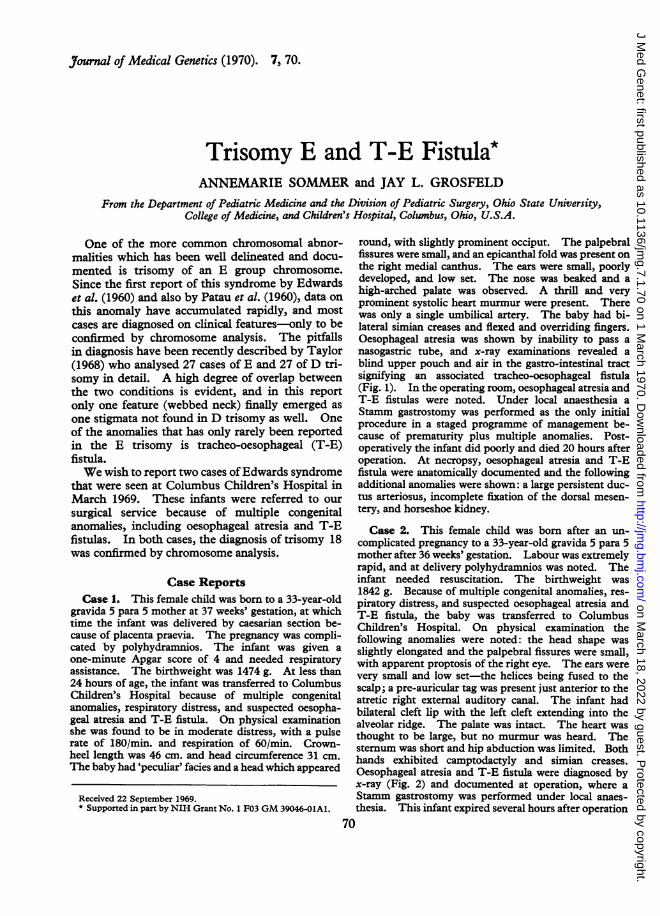
round, with slightly prominent occiput. The palpebralfissures were small, and an epicanthal fold was present onthe right medial canthus. The ears were small, poorlydeveloped, and low set. The nose was beaked and ahigh-arched palate was observed. A thrill and veryprominent systolic heart murmur were present. Therewas only a single umbilical artery. The baby had bi-lateral simian creases and flexed and overriding fingers.Oesophageal atresia was shown by inability to pass anasogastric tube, and x-ray examinations revealed ablind upper pouch and air in the gastro-intestinal tractsignifying an associated tracheo-oesophageal fistula(Fig. 1). In the operating room, oesophageal atresia andT-E fistulas were noted. Under local anaesthesia aStamm gastrostomy was performed as the only initialprocedure in a staged programme of management be-cause of prematurity plus multiple anomalies. Post-operatively the infant did poorly and died 20 hours afteroperation. At necropsy, oesophageal atresia and T-Efistula were anatomically documented and the followingadditional anomalies were shown: a large persistent duc-tus arteriosus, incomplete fixation of the dorsal mesen-tery, and horseshoe kidney.
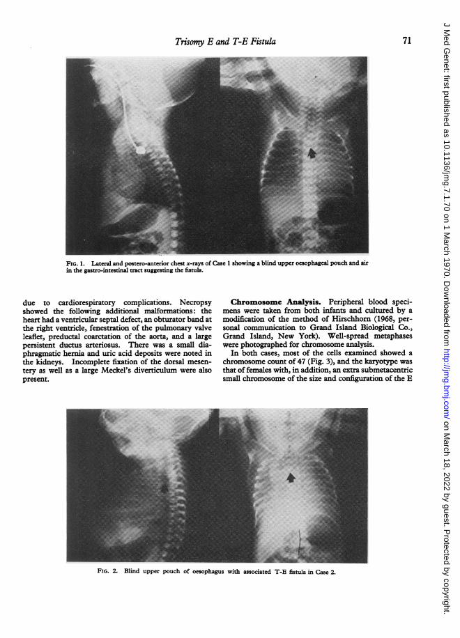
Case 2. This female child was born after an un-complicated pregnancy to a 33-year-old gravida 5 para 5mother after 36 weeks' gestation. Labour was extremelyrapid, and at delivery polyhydramnios was noted. Theinfant needed resuscitation. The birthweight was1842 g. Because of multiple congenital anomalies, res-piratory distress, and suspected oesophageal atresia andT-E fistula, the baby was transferred to ColumbusChildren's Hospital. On physical examination thefollowing anomalies were noted: the head shape wasslightly elongated and the palpebral fissures were small,with apparent proptosis of the right eye. The ears werevery small and low set-the helices being fused to thescalp; a pre-auricular tag was present just anterior to theatretic right external auditory canal. The infant hadbilateral cleft lip with the left cleft extending into thealveolar ridge. The palate was intact. The heart wasthought to be large, but no murmur was heard. Thesternum was short and hip abduction was limited. Bothhands exhibited camptodactyly and simian creases.Oesophageal atresia and T-E fistula were diagnosed byx-ray (Fig. 2) and documented at operation, where aStamm gastrostomy was performed under local anaes-thesia. This infant expired several hours after operation
70
on March 18, 2022 by guest. P
rotected by copyright.http://jm
g.bmj.com
/J M
ed Genet: first published as 10.1136/jm
g.7.1.70 on 1 March 1970. D
ownloaded from
Trisomy E and T-E Fistula
FIG. 1. Lateral and postero-anterior chest x-rays of Case 1 showing a blind upper oesophageal pouch and airin the gastro-intestinal tract suggesting the fistula.
due to cardiorespiratory complications. Necropsyshowed the following additional malformations: theheart had a ventricular septal defect, an obturator band atthe right ventricle, fenestration of the pulmonary valveleaflet, preductal coarctation of the aorta, and a largepersistent ductus arteriosus. There was a small dia-phragmatic hernia and uric acid deposits were noted inthe kidneys. Incomplete fixation of the dorsal mesen-tery as well as a large Meckel's diverticulum were alsopresent.
Chromosome Analysis. Peripheral blood speci-mens were taken from both infants and cultured by amodification of the method of Hirschhorn (1968, per-sonal communication to Grand Island Biological Co.,Grand Island, New York). Well-spread metaphaseswere photographed for chromosome analysis.
In both cases, most of the cells examined showed achromosome count of 47 (Fig. 3), and the karyotype wasthat of females with, in addition, an extra submetacentricsmall chromosome of the size and configuration of the E
FIG. 2. Blind upper pouch of oesophagus with associated T-E fistula in Case 2.
71
on March 18, 2022 by guest. P
rotected by copyright.http://jm
g.bmj.com
/J M
ed Genet: first published as 10.1136/jm
g.7.1.70 on 1 March 1970. D
ownloaded from

72 Sommer and Grosfeld
1-3 ~ -1. nbslg
.I
19 - 20
FIG. 3. Karyotype of patients (Case 1 (lower) and Case 2 (upper)) both showing trisomy E.
on March 18, 2022 by guest. P
rotected by copyright.http://jm
g.bmj.com
/J M
ed Genet: first published as 10.1136/jm
g.7.1.70 on 1 March 1970. D
ownloaded from

Trisomy E and T-E Fistula
group chromosomes. A few cells with a chromosomecomponent of less than 47 were found. Analysis ofthese losses showed them to be completely random.A distribution of the chromosome count is presented
in the Table.
TABLECHROMOSOME COUNT DISTRIBUTION
43 44 45 46 47 48 49 >49 Total
Case 1 1 1 1 27 30Case 2 1 29 30
Discussion
The incidence of trisomy E has variously beenestimated as 1 in 4000 (Lubs and Ruddle, 1969)live births to 1 in 6766 (Taylor, 1968). The postu-lated mechanism responsible for the occurrence oftrisomy is meiotic non-disjunction.The most frequent features (Butler et al., 1965)
of this syndrome are hypertonia, antero-posteriorelongation of the skull, webbed neck, a high-pitchedcry (probably due to an abnormally shaped palate),and micrognathia. The ears are low set, malform-ed, and rotated, and the palpebral fissures are usuallysmall. Camptodactyly of the fingers is seen and thethumbs are often distally implanted or rudimentaryand retroflexible. The feet have a calcaneo-valgus deformity or a prominent calcaneus('rockerbottom feet'), the halluces are often short,broad, and dorsiflexed, and partial syndactyly of thetoes is common. The toe-nails are often hypoplas-tic. The sternum is short and the chest may benarrow or shield-shaped. Abnormal hip andshoulder abduction is common. Other frequentfindings include single umbilical artery, redundantdowny hirsutism of the forehead and back, con-genital heart disease, renal anomalies, inguinalhernias, and general mental and motor retardationwith failure to thrive.Necropsy findings show an even greater degree of
overlap of the D and E trisomies (Taylor, 1968), butMeckel's diverticulum, pyloric stenosis, and even-
tration of the diaphragm are more frequent inEdwards' syndrome. The brain in trisomy E may
be superficially normal or it may have abnormallyfew convolutions or dilated cerebral ventricles. InPatau's syndrome, the olfactory nerves are usuallymissing and the optic nerves may be absent.Our patients presented with several of the features
mentioned for trisomy E, i.e. slightly elongatedskull, small palpebral fissures, low-set malformedears, congenital heart disease, camptodactyly, andsingle umbilical artery. Necropsy also did not
reveal any features not previously reported inEdwards' syndrome.However, in addition, both infants had docu-
mented tracheo-oesophageal fistulas. These find-ings are not commonly included in the manifestationof trisomy 18.
This syndrome, though not compatible withlong-term survival, as a rule is thought to be some-what less severe in its manifestations as comparedto trisomies of even larger autosomes, such as a Dgroup chromosome. Most of the attention has beenfocused on the cranio-facial and limb anomalies inEdwards' syndrome (Butler et al., 1965), but theclinical spectrum is growing rapidly. The amountof genetic information carried on the extra number18 autosome, while presumably having specificeffect, has not been fully defined. The autosomaltrisomies known to date have some stigmata incommon, leaving only a narrow range of clinicallydistinguishing features. All defects seen do essen-tially correspond to disturbance occurring at theembryological anlage during the fourth through tothe seventh weeks of gestation in man (Rohde,Hodgman, and Cleland, 1964). The action ofenvironment on the genetic material has not beenestablished for the trisomies of the larger autosomes,though some causal relation between the occurrenceof hepatitis and Down's syndrome has been sug-gested (Stoller and Collmann, 1965).A review of all cases of trisomy E seen in our
hospital, including the present two cases, did notuncover any unusual disturbances during preg-nancy.
SummaryTwo infants with trisomy E, oesophageal atresia,
and T-E fistulae are reported. T-E fistula hasonly rarely been reported in this syndrome, butthese cases might warrant inclusion of this malfor-mation in the clinical spectrum for suspecting thischromosomal disorder.
We are grateful to Dr. H. William Clatworthy, Jr. forallowing us to study his patients and for his advice in thepreparation of this report.
REFERENCESButler, L. J., Snodgrass, G. J. A. I., France, N. E., Sinclair, L., and
Russell, A. (1965). E (16-18) trisomy syndrome: analysis of 13cases. Archives of Disease in Childhood, 40, 600-611.
Edwards, J. H., Harnden, D. G., Cameron, A. H., Crosse, V. M.,and Wolff, 0. H. (1960). A new trisomic syndrome. Lancet, 1,787-790.
Lubs, H. A., and Ruddle, F. H. (1969). Chromosomal abnormalitiesin 400 consecutive newborns. Mammalian Chromosome Newsletter,10, No. 1.
73
on March 18, 2022 by guest. P
rotected by copyright.http://jm
g.bmj.com
/J M
ed Genet: first published as 10.1136/jm
g.7.1.70 on 1 March 1970. D
ownloaded from

74 Sommer and Grosfeld
Patau, K., Smith, D. W., Therman, E., Inhorn, S. L., and Wagner, Stoller, A., and Collmann, R. D. (1965). Incidence of infectiveH. P. (1960). Multiple congenital anomaly caused by an extra hepatitis followed by Down's syndrome nine months later. Lancet,autosome. Lancet, 1, 790-793. 2,1221-1223.
Rohde, R. A., Hodgman, J. E., and Cleland, R. S. (1964). Multiple Taylor, A. I. (1968). Autosomal trisomy syndrome: a detailedcongenital anomalies in the E1-trisomy (group 16-18) syndrome. study of 27 cases of Edward's syndrome and 27 cases of Patau'sPediatrics, 33, 258-270. syndrome (review article). JournalofMedical Genetics, 5,227-252.
on March 18, 2022 by guest. P
rotected by copyright.http://jm
g.bmj.com
/J M
ed Genet: first published as 10.1136/jm
g.7.1.70 on 1 March 1970. D
ownloaded from