trigeminal ganglion cell processes are spatially ordered prior to
TRANSCRIPT

The Journal of Neuroscience, October 1992, 12(10): 3946-3955
Trigeminal Ganglion Cell Processes Are Spatially Ordered Prior to the Differentiation of the Vibrissa Pad
Reha S. Erzurumlu and Sonal Jhaveri
Department of Brain and Cognitive Sciences, Massachusetts Institute of Technology, Cambridge, Massachusetts 02139
The rodent trigeminal system is characterized by the punc- tate organization of its afferents and neurons that replicate the distribution of mystacial vibrissae and sinus hairs on the snout. We have examined the development of topographic equivalence between the sensory periphery on the snout and the brainstem trigeminal nuclei in rats. Lipophilic tracers Dil (1 ,l’-dioctodecyl-3,3,3’,3’-tetramethylindocarbocyanine perchlorate) and DiA [4-(4-dihexadecylaminostyryl)-Kmeth- ylpyridinium iodide] were used to label trigeminal ganglion cells and their processes differentially from discrete regions of the presumptive vibrissa field in fixed embryos. Our re- sults show that trigeminal ganglion cell processes are spa- tially ordered as they reach their peripheral and central tar- gets on embryonic day 12 (E12). Peripheral processes of dorsomedially situated ganglion cells course dorsally toward the presumptive vibrissa field, and those of ventrolaterally situated ganglion cells project ventrally. On E 13, the central processes of dorsomedially situated ganglion cells enter the brainstem medially whereas those of ventrolaterally situated ganglion cells enter laterally. This spatial order of trigeminal ganglion cell processes precedes the emergence of vibrissa rows in the periphery and the differentiation of brainstem trigeminal nuclei. Thus, the subsequent transfer of the vi- brissa-related pattern to the brainstem trigeminal nuclei oc- curs along a preexisting, spatially aligned bridge formed by the trigeminal ganglion cells.
Primary sensory afferents that connect the periphery to the CNS navigate through a variety of substrates and across considerable distances before contacting target cells, yet these fibers succeed in conveying the spatial organization of the sensory periphery to central relay nuclei with remarkable precision. Understanding how this comes about has been a major endeavor in develop- mental neurobiology for many years.
Periphery-related neuronal patterns are a distinct feature of the rodent trigeminal system. In both rats and mice, the whiskers on the snout are arranged in five rostrocaudally aligned rows and the number of vibrissa follicles in each row is constant (Yamakado and Yohro, 1970; Van Exan and Hardy, 1980). During perinatal life, afferents and cellular elements of the cen- tral trigeminal pathway leading to the neocortex form facsimiles of the peripheral pattern (Woolsey and Van der Loos, 1970;
Received Nov. 29, 1991; revised Apr. 29, 1992; accepted May 7, 1992. We thank Drs. L. I. Mortin, S. L. Pallas, and G. E. Schneider for their comments
on an earlier version of the manuscript. Supported by NIH Grant NS27678. Correspondence should be addressed to Dr. R. S. Erzurumlu, E25-634, M.I.T.,
Cambridge, MA 02 139. Copyright 0 1992 Society for Neuroscience 0270-6474/92/123946-10$05.00/O
Van der Loos, 1976; Belford and Killackey, 1979; Killackey and Belford, 1979; Erzurumlu et al., 1980; Ivy and Killackey, 1982; Ma and Woolsey, 1984; Bates and Killackey, 1985). These cen- tral patterns are drastically altered if the trigeminal innervation of the vibrissa pad is disturbed during a sensitive period in development (Van der Loos and Woolsey, 1973; Belford and Killackey, 1980; Jeanmonod et al., 198 1; Killackey and Shinder, 198 1; Durham and Woolsey, 1984). In contrast, an array of vibrissal follicles develops, independent of trigeminal inner- vation, on uninnervated embryonic mouse snout grown in cul- ture (And& and Van der Loos, 1982, 1983).
The construction of vibrissa-related central patterns is thought to occur under the directive guidance of periphery (Van der Loos and DGrfl, 1978; Van der Loos and Welker, 1985; Woolsey, 1987). However, little is known about developmental strategies used by trigeminal ganglion cells to match topographically equivalent regions of the periphery and brainstem. Nor is it known how the punctate organization of the whiskers is trans- ferred to afferent arbors and their target cells along the entire trigeminal neuraxis leading to the neocortex. In this study, we present evidence of an orderly patterning in the early outgrowth of trigeminal ganglion cell processes; this intrinsic spatial or- ganization may provide a foundation upon which periphery- related patterns are elaborated.
Materials and Methods
Timed-pregnant Sprague-Dawley rats were obtained from Taconic Farms. Day of sperm positivity is designated as embyronic day 0 (EO). Embryos were delivered by cesarian section, after overdosing the dams with sodium pentobarbital. At least six embryos were used for each time point (El l-El 8). The heads were fixed in 8% paraformaldehyde (S. Senft, personal communication) in 0.1 M phosphate buffer. Small crystals of fluorescent tracers Dil (1, I’-dioctodecyl-3,3,3’,3’-tetrameth- ylindocarbocyanine perchlorate) and DiA [4-(4-dihexadecylaminosty- ryl)-N-methylpyridinium iodide] (Molecular Probes) (Godement et al., 1987) were inserted into emerging vibrissa rows in varying combinations (Fig. 1).
During morphogenesis of the vibrissa pad, the dorsal two rows of whiskers (rows A and B) arise from the lateral nasal process whereas the ventral three (rows C, D, and E) derive from the maxillary process; the two prominences are separated by the nasolacximal groove (Ya- makado and Yohro, 1979; Van Exan and Hardy, 1980). Before E14, prior to the appearance of vibrissa rows, crystals of Dil and DiA were placed either one in the lateral nasal process above the nasolacrimal groove and the other in the maxillary prominence, or one dorsally and the other ventrally, both in the maxillary process (Fig. 1). In older embryos, alternating crystals of Dil and DiA were placed in rows A, C, and E (caudally in rows A and E and rostrally in row C); in some cases, one crystal was placed rostrally and the other caudally in row C. Labeled heads were stored in fixative at 37°C for 2-8 weeks, embedded in 2.5% agar, and sectioned (100-200 pm thick) on a vibratome. Sections were mounted from buffer; counterstained with 0.025% bisbenzimide; viewed with rhodamine, fluorescein, and ultraviolet filters; and photographed.

The Journal of Neuroscience, October 1992, 12(10) 3947
Figure I. Photomicrographs of E 12 (A) and E 13 (B) rat heads. On E 12 no vibrissal ridges are visible on the maxillary process, whereas on E 13 shallow depressions within the maxillary process indicate the beginning of ridge formation. No vibrissal follicles are present at either age. Arrowheads point to the nasolacrimal groove that separates the lateral nasal process from the maxillary process. Solid and open circles indicate the position of Dil and DiA implants, respectively. The levels indicated by lines,4 and B on the El2 head correspond to coronal sections shown in Figure 2. Lines A, B, and C on the El 3 head indicate the levels of coronal sections shown in Figure 3.
Results Trigeminal ganglion cells and their peripheral processes Although neurogenesis in the rat trigeminal ganglion begins after E9.5 (Rhoades et al., 1991), placement of carbocyanine dyes
superficially in the differentiating snout on El 1 did not label any ganglion cells. Only in those cases where the dye crystal was implanted deep within the mesenchyme, apposed to the emerg- ing ganglion, were a few bipolar cell profiles encountered (data not shown). This observation suggests that processes of ganglion
Figure 2. Differential labeling in the infraorbital nerve and trigeminal gan- alion following implantation of DiA dorsally and I%1 ventrally in the max- illary process of an El2 rat. A-C are photomicrographs of the same coronal section at the level of the infmorbital foramen (plane A in Fig. lA), and D- F are from one at the level of the tri- geminal ganglion (plane B in Fig. 1A). A and D are photographed with a UV filter to show the cell nuclei stained with bisbenzimide; B and E are photo- graphed with a rhodamine isothiocya- nate (RITC) filter to show Dil labeling, and C and F with a fluorescein isothio- cyanate (FITC) filter to reveal the DiA labeling. DiA-labeled cells and pro- cesses are situated dorsal to Dil-labeled ones. Broken line in D outlines the tri- geminal ganglion; single arrowheads point to DiA, and double arrowheads, to Dil labeling. D, dorsal; L, lateral. Scale bar, 200 pm.
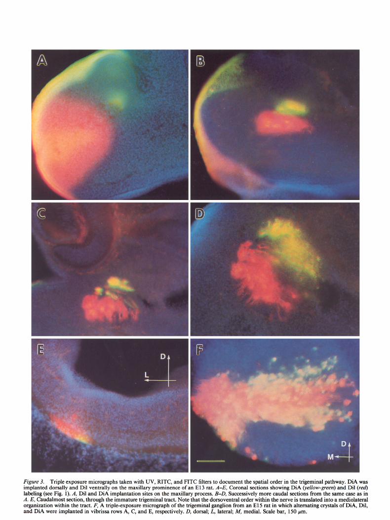
Figure 3. Triple exposure micrographs taken with UV, RITC, and FITC filters to document the spatial order in the trigeminal pathway. DiA was implanted dorsally and Dil ventrally on the maxillary prominence of an El3 rat. A-E, Coronal sections showing DiA (y&w-green) and Dil (red) labeling (see Fig. 1). A, Dil and DiA implantation sites on the maxillary process. B-D, Successively more caudal sections from the same case as in A. E, Caudalmost section, through the immature trigeminal tract. Note that the dorsoventral order within the nerve is translated into a mediolateral organization within the tract. F, A triple-exposure micrograph of the trigeminal ganglion from an El 5 rat in which alternating crystals of DiA, Dil, and DiA were implanted in vibrissa rows A, C, and E, respectively. D, dorsal; L, lateral; M, medial. Scale bar, 150 pm.

The Journal of Neuroscience. October 1992, fZ(10) 3949
Figure 4. Topographic order in trigeminal ganglion of El 5 rats. A and B show the same horizontal section from an animal in which the face was labeled with a dorsoventral sequence of Dil, DiA, and Dil in whisker rows A, C, and E, respectively. A, A single band of labeling in the middle of the ganglion is seen with the FITC filter (DiA labeling). B, Two bands of labeling are seen with the RITC filter (Dil labeling). C and D, Coronal section through another El 5 trigeminal ganglion following placement of DiA (C, FITC filter) caudally and Dil (0, RITC filter) rostrally in row C. In D, many of the cells labeled with DiA also show through the RITC filter. D, dorsal; L, lateral; C, caudal.
cells first begin to grow out on El 1 (cf. Erzurumlu and Killackey, 1983).
On E 12, neurogenesis in the ganglion is occurring at peak levels but morphological differentiation of the vibrissa pad is not yet evident (English et al., 1980; Forbes and Welt, 1981; Altman and Bayer, 1982; Erzurumlu and Killackey, 1983; Rhoades et al., 199 1) (see Figs. 1,8). At this time, tracers placed dorsally and ventrally within the maxillary process revealed numerous labeled trigeminal ganglion cells and their axons. These labeled profiles were topographically aligned with respect to the dorsoventral axis of the face: when Dil was placed in the ventral part of the presumptive vibrissa field and DiA in the dorsal part, discrete fascicles of axons and specific groups of ganglion cells were labeled (Fig. 2).
On E 13, shallow depressions along the maxillary process de- marcate the presumptive location of vibrissa rows (Fig. 1). How-
ever, no evidence of vibrissa follicle development is visible. Ganglion cell axons directed toward the rudimentary vibrissa field form a roughly circular bundle of fascicles within the in- fraorbital foramen. These fascicles fan out as they approach the maxillary and lateral nasal processes. A distinct topography was evident along the entire trigeminal pathway (Fig. 3). Placements of Dil and DiA crystals along the dorsoventral axis of the pre- sumptive vibrissa field led to labeling of discrete populations of axon fascicles, ganglion cell bodies, and central processes. Cells located in the dorsomedial portion of the ganglion ex- tended peripheral processes that traveled in fascicles toward dorsal targets; cells situated ventrolaterally traveled likewise to ventral target fields. Thus, the spatial order of the developing infraorbital nerve fascicles was aligned with the dorsoventral axis of the presumptive vibrissa pad and consequently of future vibrissa rows. The topographic order of the ganglion cells with

3950 Erzurumlu and Jhaveri l Spatial Order in the Trigeminal Nerve
Figure 5. Spatial order in the infraorbital nerve (A) and the trigeminal ganglion (B) of an El 8 rat. On the left side, Dil was placed in rows A and E, whereas on the right side Dil was placed in row C. Both micrographs are photographed with an RITC filter. D, dorsal; L, lateral.
respect to their target rields was best seen in coronal or sagittal sections through the head: discrete injections of Dil and DiA in the periphery led to bands of labeled cells that were oriented dorsolaterally to ventromedially (Fig. 3).
Vibrissa rows (A-E) can be identified on the snout by El4 and become quite distinct on El 5. At these ages, crystals of DiA, Dil, and DiA were placed in alternating rows (A, C, and E, respectively) on one side of the face, whereas Dil, DiA, and Dil crystals were applied likewise on the other side of the face. With such a procedure, the vast majority of cells as well as their processes were labeled differentially from corresponding loci in their peripheral fields (Figs. 4, 5; see also Fig. 7).
We were also able to place two different dye crystals along the rostrocaudal axis of a given vibrissal row in El4 and older embryos. The resultant labeling pattern showed that the rostro- caudal axis of the vibrissa pad is represented mediolaterally in the ganglion (Fig. 4). Between El4 and El 8, the topography with respect to both the dorsoventral and rostrocaudal axes of the vibrissa pad becomes increasingly distinct in the trigeminal gan- glion and in its projections (see Figs. 5, 7).
Central processes and arbors of trigeminal ganglion cells
The entry of trigeminal ganglion cell processes into the CNS is best seen in sagittal sections (Fig. SF). On El 2 and El 3, tracer implants in the periphery revealed axons with large lamellate
growth cones approaching the rhombencephalon (Fig. 6A). As they encountered the surface of the brainstem on E13, these processes formed T-shaped swellings (about 125-150 Km in length), presaging their bifurcation into the ascending and de- scending branches ofthe central trigeminal tract (Fig. 6). Growth cones with filopodia tipped the leading edges of the T-shaped swellings on both sides. Furthermore, at the leptomeningeal- CNS interface, the bifurcating fibers were not all of the same caliber: a subpopulation of labeled fibers was consistently thin- ner than others (Fig. 6E). These differences may be a reflection of the dual origin (placodal vs. neural crest) of the ganglion cells (Hamburger, 1961; LeDourain et al., 1986; Nichols, 1986), or they may reflect a differentiation into the large versus small fibers seen in the mature trigeminal tract.
Centrally directed processes of ganglion cells were also spa- tially ordered from the onset of trigeminal tract formation. As early as El 3, axons of dorsally situated ganglion cells entered the brainstem medially, whereas those of ventrally situated gan- glion cells entered laterally (Fig. 3). Thus, the dorsoventral axis of the peripheral field is rotated 90” and translated into a medial- to-lateral dimension within the brainstem. With further mat- uration of the hindbrain, this axis shifts an additional 90”, re- sulting in an almost upside-down representation of the face in the trigeminal tract (compare Figs. 3 and 7).
On El4 and later, axons contributing to the trigeminal tract

The Journal of Neuroscience, October 1992, f2(10) 3951
Figure 6. Profiles of growth cones that tip the central processes of trigeminal ganglion cells. All the micrographs are from E 13 rat heads sectioned in the sagittal plane. The growth cones are large with lamellipodia and filopodia as they approach the CNS (A). Upon entering the developing brainstem, the leading edge of the central process bifurcates to form large, T-shaped swellings that presage the ascending and descending processes of the central trigeminal fibers (B-D). E, Some T-shaped processes are very thin. F, Low-power view of the bifurcating central processes at their entry point into the CNS. Arrowheads point to the bifurcation points of single fibers. gV, trigeminal ganglion; 0, dorsal; C, caudal. Scale bar: 50 pm for A-E; 200 pm for F.
began emitting collaterals at right angles to the pial surface, to stem trigeminal nuclei, reflecting a 180” inversion with respect invade the newly forming brainstem sensory trigeminal nuclei to the dorsoventral axis of the vibrissa pad. In addition, col- (Fig. 7). Placement of alternating crystals of Dil and DiA in lateral branches were consistently longer within the subnucleus vibrissa rows A, C, and E also revealed that radial collaterals interpolaris of the brainstem trigeminal complex than those seen and their arbors, emitted from parent axons in the trigeminal in the principal sensory nucleus (Fig. 7) despite the fact that tract, were segregated along the dorsoventral axis of the brain- invasion of both nuclei and arbor formation within all brainstem

3952 Erzurumlu and Jhaveri l Spatial Order in the Trigeminal Nerve
Figure 7. Topographic order in the trigeminal tract of El4 rats. Crystals of Dil, DiA, and Dil were placed in rows A, C, and E, respectively, on the left side of the face, and DiA, Dil, and DiA were implanted in rows A, C, and E, respectively, on the right side of the face. A and B, The same coronal section through the brainstem, photographed with an RITC (A) and an FITC (B) filter, revealed alternating bands of label in the trigeminal tract. Note that by this age, the dorsoventral axis of the face representation is shifted nearly 180” in the trigeminal tract. Higher-magnification views show that axons of the trigeminal tract have begun emitting radially oriented collaterals into the newly differentiated brainstem trigeminal nuclei. C, Labeling pattern in the subnucleus interpolaris of the brainstem trigeminal complex, seen with an RITC filter, in an El 6 rat with Dil implanted in rows A and E. Note that the length and size of the arbors within the principal sensory nucleus (0) are considerably smaller than those in the subnucleus interpolaris (E). D, dorsal; L, lateral. Scale bar: 500 pm for A and B, 200 pm for C, 50 pm for D and E.
trigeminal targets began around the same time. Thus, although the central primary afferent arbors have not yet segregated into the disjunctive patches characteristic of the more mature ani- mal, they are focalized, at least with respect to the dorsoventral axis of the face.
Discussion Connectivity between the vibrissa pad and the brainstem tri- geminal complex is established relatively early in development (Fig. 8). Previous studies have established that in the rat, tri-

The Journal of Neuroscience, October 1992, fZ(10) 3953
GV axons enter BSTC
Vibrissa pad differentiation Vibrissa-related
ra ------ patterns emerge inCNS
GV neurocenesis e
-/ 0 9 10 11 13 A 112 1 14 15 16 17 18
t
I Embryonic days
CONCEPTION
geminal ganglion cells are generated between E9.5 and E14.5, with a peak of neurogenesis on El2 (Forbes and Welt, 1981; Altman and Bayer, 1982; Rhoades et al., 199 1). The peripheral processes of these cells invade the superficial epidermis of the maxillary process after El 3 (English et al., 1980; Erzurumlu and Killackey, 1983). Brainstem trigeminal nuclei follow a similar though shorter neurogenetic timetable, between El2 and El5 (Altman and Bayer, 198Oa-c). Our study shows that newly dif- ferentiated trigeminal ganglion cells extend peripheral and cen- tral processes in an orderly fashion before the differentiation of their respective targets. Such order in initial outgrowth of pri- mary sensory neurons has also been reported for the embryonic chick trigeminal and chick and rat dorsal root ganglia (Noden, 1980; Honig, 1982; Scott, 1982; Wessels et al., 1988).
Possible role of chemospeciJc interactions in the establishment of topography During development, sensory axons may navigate along path- ways labeled by ephemeral molecular cues (see Dodd and Jesse& 1988, for a review) or along gradients of target-derived diffusible “chemoattractants” (see Tessier-Lavigne and Placzek, 199 1, for a review). One such example of chemotropic interaction be- tween afferents and their targets is derived from tissue culture experiments involving the embryonic mouse trigeminal system. E 1 O-E 11 mouse trigeminal ganglion cells, when cocultured with maxillary and hyoid processes, extend neurites preferentially toward the maxillary process (Lumsden and Davies, 1983, 1986). Based on the assumption that at the time of explantation of the target tissues the maxillary process has not yet been explored by the pioneering trigeminal ganglion cell processes, Lumsden and Davies suggested that the epithelium of the maxillary pro- cess produces a specific “attractant” that guides the trigeminal net&es. Recently, Stainier and Gilbert (1990, 199 l), using spe- cific antibodies to newly differentiated trigeminal ganglion cells, showed that the pioneer axons of the mouse trigeminal ganglion invade their peripheral fields by El 0 (Stainier and Gilbert, 1990). These observations leave open the possibility that, by the time the so-called “virgin” peripheral explants were taken for co- culturing in Lumsden and Davies’ experiments, the pioneer trigeminal axons have already explored and perhaps even al- tered the state of differentiation of the target tissue.
Our results do not challenge the notion of chemotropic factors attracting trigeminal fibers to peripheral targets. However, if
19 20 21 22
t BIRTH
Figure 8. Summary diagram of se- lected morphogenetic events that occur during the time that trigeminal ganglion cell processes bridge the periphery and the brainstem (BSTC). The rectangle indicates the period during which the trigeminal ganglion (G v cell processes invade their peripheral and central tar- gets. The data presented in this diagram have been compiled from Belford and Killackev (1979). Altman and Baver (198Oa-& English et al. (1980) Forbes and Welt (198 l), Erzurumlu and Kil- lackey (1983) Rhoades et al. (1991), and the present study. For purposes of comparison, ages of the embryos in all these studies are adjusted so that the day of sperm positivity is EO.
chemotaxis were the only factor influencing the growth of gan- glion cell processes, they would extend diffusely toward the source of the attractant. We show that there is a considerable degree of order between trigeminal fibers as they emerge from the gan- glion. The mechanisms whereby uus occurs remain to be de- termined. The order may be established by axoaxonal interac- tions mediated via cell surface molecules (Trisler, 1982, 1990; Trisler and Collins, 1987) or by differential gradients of target- associated molecules (Thanos et al., 1984; O’Leary et al., 1990; Baier and Bonhoeffer, 1992). It is highly likely that the chemical milieu within which the leading wavefront of axons and growth cones navigate toward their targets plays a major role in axonal pathway formation (Landmesser. et al., 1988). The elaborate bifurcation of a single growth cone into a T-shaped swelling, which occurs only at the leptomeningeal-CNS interface of the brainstem, is also strongly suggestive of surface-associated po- sitional cues.
Spatial order in the trigeminal ganglion cell processes and the formation of somatotopy in the brainstem Previously, topographic order in the trigeminal ganglion pro- jections has been noted in studies utilizing silver stains or ret- rograde labeling of ganglion cells in perinatal rats (Erzurumlu and Killackey, 1982, 1983; Rhoades et al., 1990). However, these studies do not provide direct clues as to whether or not the initial outgrowth of the trigeminal nerve is ordered. In an experiment addressing these issues, Davies and Lumsden (1986) counted the number of axonal profiles in different fascicles of serially sectioned segments of the infraorbital nerve in embry- onic mice. They found that the number of axonal profiles in a given fascicle varied considerably over a distance of 200 pm. They concluded that the early outgrowth of the trigeminal gan- glion projection was diffuse and that vibrissa-related topography emerged from selective elimination of inappropriately con- nected fibers (Davies and Lumsden, 1984, 1986). However, the local axonal disorder reported by Davies and Lumsden (1986) does not imply a lack of global order in the trigeminal ganglion projections. Conversely, our observation of spatial order in the peripheral and central projections of the trigeminal ganglion cells (labeled from their peripheral target fields) need not pre- clude free exchange of fibers between neighboring fasciculi along short segments of the nerve. Finally, the overall spatial order at the outset of trigeminal path formation and at the peak of

3954 Erzurumlu and Jhaveri * Spatial Order in the Trigeminal Nerve
trigeminal ganglion neurogenesis argues against the hypothesis that the somatotopic organization of trigeminal ganglion pro- jections emerges from an early exuberance.
The observation of spatial order along axonal pathways is also related to the establishment of axial orientation of periph- ery-related maps along the rest of the trigeminal neuraxis. While the punctate arrangement of vibrissae on the snout is replicated by the segregation of afferents and neurons in the brainstem trigeminal complex, the ventrobasal thalamus, and the primary somatosensory cortex (Woolsey and Van der Loos, 1970; Van der Loos, 1976; Belford and Killackey, 1979; Erzurumlu et al., 1980; Ivy and Killackey, 1982; Ma and Woolsey, 1984; Bates and Killackey, 1985; Woolsey, 1987) the face map is oriented differently in each of these vibrissa representation areas (cf. Erzurumlu and Killackey, 1980; Bernard0 and Woolsey, 1987). We have described a 180” inversion of the vibrissal map at the level of the brainstem. This shift is brought about by a gradual rotation in the positions of the central processes of ganglion cells due to the changing conformation of the brainstem. It is likely that a combination of axial ordering within fiber tracts and conformational changes in the developing brain is, to a consid- erable degree, also responsible for the absolute orientation of the map in the thalamus and cortex. At least a crude topography in the connections between dorsal thalamus and neocortex is seen prior to the appearance of barrels (Crandall and Caviness, 1984; Dawson and Killackey, 1985; Molnar and Blakemore, 1990) indicating that formation of such ordered connections in the CNS occurs independent of the sensory periphery. In fact, in anophthalmic mice, the axial orientation of the geniculo- cortical map develops normally in the absence of retinal input (Godement et al., 1979; Kaiserman-Abramoff et al., 1980; for map orientation in the Siamese cat, see also Guillery and Kaas, 1973; Shatz and Le Vay, 1979). Furthermore, while neonatal disruption of the vibrissa nerves alters the pattern of central representations, the orientation of the face maps is not affected (Belford and Killackey, 1979; Killackey and Belford, 1979). Even in the case of mice with supernumerary whiskers and corresponding extra barrels, the axial orientation of the face maps is unaltered (Welker and Van der Loos, 1986). Collec- tively, these observations argue for the existence of separate strategies for determining axial orientation of a peripheral map and periphery-related pattern formation (cf. Killackey, 1985; Welker and Van der Loos, 1986).
We propose that pattern formation in the rodent trigeminal system can be divided into two distinct phases. In the first phase, the axial framework of the trigeminal neuraxis is laid down in an orderly fashion without guidance from the periphery. This organization is “hard-wired” by the spatial arrangement of de- veloping sensory afferents. In the second phase, peripheral pat- terns guide the modular patterning of neuronal elements within the constraints of this axial framework. The available data on the consequences of peripheral alterations in the formation of vibrissa-related patterns in the CNS strongly suggest that it is the second phase of vibrissa-related pattern construction that is extrinsically malleable and under directive guidance of the sen- sory periphery.
References Altman J, Bayer SA (1980a) Development of the brain stem in the
rat. I. Thymidine-radiographic study of the time of origin of neurons of the lower medulla. J Comp Neurol 194: l-35.
Altman J, Bayer SA (1980b) Development of the brain stem in the
rat. II. Thymidine-radiographic study of the time of origin of neurons of the upper medulla, excluding the vestibular and auditory nuclei. J Comp Neurol 194:37-56.
Altman J, Bayer SA (1980~) Development of the brain stem in the rat. IV. Thymidine-radiographic study of the time of origin of neurons in the pontine region. J Comp Neurol 194:905-929.
Altman J, Bayer SA (1982) Development of the cranial nerve ganglia and related nuclei in the rat. Adv Anat Embryo1 Cell Biol 74: l-89.
And& FL, Van Der Loos H (1982) Whisker patterns form in cultured non-innervated muzzle skin from mouse embryos. Neurosci Lett 30: 3741.
Andrts FL, Van Der Loos H (1983) Cultured embryonic non-inner- vated mouse muzzle is capable of generating a whisker pattern. Int J Dev Neurosci 1:319-338.
Baier H, Bonhoeffer F (1992) Axon guidance by gradients of a target- derived component. Science 255472475.
Bates CA, Killackey HP (1985) The organization of the neonatal rat’s brainstem trigeminal complex and its role in the formation of central trigeminal patterns. J Comp Neurol 240:265-287.
Belford GR, Killackey HP (1979) Vibrissae representation in subcor- tical trigeminal centers of the neonatal rat. J Comp Neurol 183:305- 322.
Belford GR, Killackey HP (1980) The sensitive period in the devel- opment of the trigeminal system of the neonatal rat. J Comp Neurol 193:335-350.
Bernard0 KL, Woolsey TA (1987) Axonal trajectories between mouse somatosensory thalamus and cortex. J Comp Neurol258:542-564.
Crandall JE, Caviness VS Jr (1984) Thalamocortical connections in newborn mice. J Comp Neurol228:542-556.
Davies AM, Lumsden AGS (1984) Relation of target encounter and neuronal death to nerve growth factor responsiveness in the devel- oping mouse trigeminal ganglion. J Comp Neuro1223: 124-l 37.
Davies AM. Lumsden AGS (1986) Fasciculation in the early mouse trigeminal nerve is not ordered in relation to the emerging pattern of whisker follicles. J Comp Neurol 253: 13-24.
Dawson DR, Killackey HP (1985) Distinguishing topography and somatotopy in the thalamocortical projections of the developing rat. Dev Brain Res 17:309-3 13.
Dodd J, Jesse11 TM (1988) Axon guidance and the patterning of neu- ronal vroiections in vertebrates. Science 232~692-699.
Durham D: Woolsey TA (1984) Effects of neonatal whisker lesions on mouse central trigeminal pathways. J Comp Neurol233:424-447.
Enelish KB. Burgess PR. Kavka-Van Norman D (1980) Develovment lf rat Merkel iells. J Comp Neurol 194:475-496. ’ -
Erzurumlu RS, Killackey HP (1980) Diencephalic projections of the subnucleus interpolaris of the brainstem trigeminal complex in the rat. Neuroscience 5:1891-1902.
Erzurumlu RS, Killackey HP (1982) Order in the developing rat tri- geminal nerve. Brain Res 3:305-3 10.
Erzurumlu RS, Killackey HP (1983) Development of order in the rat trigeminal system. J Comp Neurol 213:365-380.
Erzurumlu RS, Bates CA, Killackey HP (1980) Differential organi- zation of thalamic projection cells in the brainstem trigeminal com- plex of the rat. Brain Res 198:427-433.
Forbes DJ, Welt C (198 1) Neurogenesis in the trigeminal ganglion of the albino rat: a quantitative autoradiographic study. J Comp Neurol 199:133-147.
Godement P, Saillour P, Imbtrt M (1979) Thalamic afferents to the visual cortex in congenitally anophthalmic mice. Neurosci Lett 13: 271-278.
Godement P, Vanselow J, Thanos S, Bonhoeffer F (1987) A study of developing visual systems with a new method for staining neurons and their vrocesses in fixed tissue. Development 10 1:697-7 13.
Guillery RW, Kaas JH (1973) The transfer of abnormal visual field representations from the dorsal lateral geniculate nucleus to the visual cortex in Siamese cats. Brain Res 59:61-95.
Hamburger V (196 1) Experimental analysis of the dual origin of the trigeminal ganglion in the chick embryo. J Exp Zoo1 148:91-123.
Honig MG (1982) The development of sensory projection patterns in embryonic chick hind limb. J Physiol (Lond) 330: 175-302.
Ivy GO, Killackey HP (1982) Ephemeral cellular segmentation in the thalamus of the neonatal rat. Dev Brain Res 2: 1-17.
Jeanmonod D, Rice FL, Van der Loos H (198 1) Mouse somatosensory cortex: alterations in the barrel field following receptor injury at dif- ferent postnatal ages. Neuroscience 6: 1503-l 535.

The Journal of Neuroscience, October 1992, 72(10) 3955
Kaiserman-Abramoff IR, Graybiel AM, Nauta WJH (1980) The tha- lamic projection to cortical area 17 in a congenitally anophthalmic mouse strain. Neuroscience 5:41-52.
Killackey HP (1985) Intrinsic order in the developing rat trigeminal system. In: Development, organization, and processing in somato- sensory pathways (Rowe M, Willis WD, eds), pp 43-52. New York: Liss.
Killackey HP, Belford GR (1979) The formation of afferent patterns in the somatosensory cortex of the neonatal rat. J Comp Nemo1 183: 285-304.
Killackey HP, Shinder A (198 1) Central correlates of peripheral pat- tern alterations in the trigeminal system of the rat. II. The effect of nerve section. Dev Brain Res 1:121-126.
Landmesser L, Dahm L, Schultz K, Rutishauser U (1988) Distinct roles for adhesion molecules during innervation of embryonic chick muscle. Dev Biol 130:645-670.
LeDouarin NM, Fontaine-P&us J, Couly G (1986) Cephalic ecto- dermal placodes and neurogenesis. Trends Neurosci 9: 175-l 80.
Lumsden AGS, Davies AM (1983) Earliest sensory nerve fibers are guided to peripheral targets by attractants other than nerve growth factor. Nature 306:786-788.
Lumsden AGS, Davies AM (1986) Chemotropic effect of specific tar- get epithelium in the developing mammalian nervous system. Nature 323:538-539.
Ma PKM, Woolsey TA (1984) Cytoarchitectonic correlates of the vibrissae in the medullary trigeminal complex of the mouse. Brain Res 306:374-379.
Molnar Z, Blakemore C (1990) Relationship of corticofugal and cor- ticopetal projections in the prenatal establishment of projections from thalamic nuclei to specific cortical areas of the rat. Proc Physiol Sot (Lond) 104P.
Nichols DH (1986) Mesenchyme formation from the trigeminal plac- odes of the mouse embryo. Am J Anat 176: 19-3 1.
Noden DM ( 1980) Somatotopic organization of the embryonic chick trigeminal ganglion. J Camp-Neural 190:429444. -
O’Leary DDM, Bicknese AR, De Carlos JA, Heffner CD, Koester SE, Kutka LJ, Terashima T (1990) Target selection by cortical axons: alternative mechanisms to establish axonal connections in the de- veloping brain. Cold Spring Harbor Symp Quant Biol 55:453468.
Rhoades RW, Chiaia NL, Macdonald GJ (1990) Topographic orga- nization of the peripheral projections of the trigeminal ganglion in the fetal rat. Somatosens Mot Res 7:67-84.
Rhoades RW, Enfiejian HL, Chiaia NL, Macdonald GJ, Miller MW, McCann P, Goddard CM (199 1) Birthdates of trigeminal ganglion cells contributing axons to the infraorbital nerve and-specific &b&Sal follicles in the rat. J Comn Neurol 307:163-175.
Scott SA (1982) The development of segmental pattern of skin sensory innervation in embryonic chick hindlimb. J Physiol (Lond) 333:203- 220.
Shatz CJ, Le Vay S (1979) Siamese cat: altered connections of visual cortex. Science 204:328-330.
Stainier DYR, Gilbert W (1990) Pioneer neurons in the mouse tri- eeminal sensorv svstem. Proc Nat1 Acad Sci USA 87:923-927.
Stiinier DYR, Gilbert W (199 1) Neuronal differentiation and matu- ration in the mouse trigeminal sensory system, in vivo and in vitro. J Comp Neurol 3 11:300-3 12.
Tessier-Lavigne M, Placzek M (199 1) Target attraction: are developing axons guided by chemotropsim? Trends Neurosci 14:303-3 10.
Thanos S, Bonhoeffer F, Rutishauser U (1984) Fiber-fiber interaction and tectal cues influence the development of the chicken retinotectal projection. Proc Nat1 Acad Sci USA 8 1: 1906-l 9 10.
Trisler D (1982) Are molecular markers of cell position involved in the formation of neural circuits? Trends Neurosci 5: l-5.
Trisler D (1990) Cell recognition and pattern formation in the devel- oping nervous system. J Exp Biol 153: 1 l-28.
Trisler D, Collins F (1987) Corresponding spatial gradients of TOP molecules in the developing retina and optic tectum. Science 237: 1208-1209.
Van der Loos H (1976) Barreloids in mouse somatosensory thalamus. Neurosci Lett 2: l-6.
Van der Loos H, DBrfl J (1978) Does the skin tell the somatosensory cortex how to construct a map of the periphery? Neurosci Lett 7:23- 30.
Van der Loos H, Welker E (1985) Development and plasticity of somatosensory brain maps. In: Development, organization and pro- cessing in somatosensory pathways (Rowe M, Willis WD, eds), pp 53-67. New York: Liss.
Van der Loos H, Woolsey TA (1973) Somatosensory cortex: structural alterations following early injury to sense organs. Science 179:395- 397.
Van Exan RJ, Hardy MH (1980) A spatial relationship between in- nervation and the early differentiation of vibrissa follicles in the em- bryonic mouse. J Anat 13 1:643-656.
Welker E, Van der Loos H (1986) Quantitative correlation between barrel-field size and the sensory innervation of the whiskerpad: a comparative study in six strains of mice bred for different patterns of mvstacial vibrissae. J Neurosci 6:3355-3373.
Wessels WJT, Feierabend HKP, Marani E (1988) Development of the sensory innervation in the rat hindlimb. Sot Neurosci Abstr 14:422.
Woolsev TA (1987) Structural. functional and biochemical nlasticitv in the rodent brain. In: Developmental neurobiology of mammals (Chagas C, Linden R, eds), pp 347-380. Vatican City: Pontific Acad- emy of Sciences.
Woolsey TA, Van der Loos H (1970) The structural organization of layer IV in the somatosensory region (SI) of mouse cerebral cortex. Brain Res 17:205-242.
Yamakado M, Yohro T (1979) Subdivision of mouse vibrissae on an embryological basis, with descriptions of variations in the number and arrangement of sinus hairs and cortical barrels in BALB/c (nu/ f; nude, nu/nu) and hairless (hr/hr) strains. Am J Anat 155:153- 174.