triclosanbindingsmet . - csic
TRANSCRIPT

The Binding of Triclosan to SmeT, the Repressor of theMultidrug Efflux Pump SmeDEF, Induces AntibioticResistance in Stenotrophomonas maltophiliaAlvaro Hernandez1., Federico M. Ruiz2., Antonio Romero2, Jose L. Martınez1*
1 Centro Nacional del Biotecnologıa, CSIC, Cantoblanco, Madrid, Spain, 2 Centro de Investigaciones Biologicas, CSIC, Madrid, Spain
Abstract
The wide utilization of biocides poses a concern on the impact of these compounds on natural bacterial populations.Furthermore, it has been demonstrated that biocides can select, at least in laboratory experiments, antibiotic resistantbacteria. This situation has raised concerns, not just on scientists and clinicians, but also on regulatory agencies, which aredemanding studies on the impact that the utilization of biocides may have on the development on resistance andconsequently on the treatment of infectious diseases and on human health. In the present article, we explored thepossibility that the widely used biocide triclosan might induce antibiotic resistance using as a model the opportunisticpathogen Stenotrophomonas maltophilia. Biochemical, functional and structural studies were performed, focusing onSmeDEF, the most relevant antibiotic- and triclosan-removing multidrug efflux pump of S. maltophilia. Expression of smeDEFis regulated by the repressor SmeT. Triclosan released SmeT from its operator and induces the expression of smeDEF, thusreducing the susceptibility of S. maltophilia to antibiotics in the presence of the biocide. The structure of SmeT bound totriclosan is described. Two molecules of triclosan were found to bind to one subunit of the SmeT homodimer. The bindingof the biocide stabilizes the N terminal domain of both subunits in a conformation unable to bind DNA. To our knowledgethis is the first crystal structure obtained for a transcriptional regulator bound to triclosan. This work provides the molecularbasis for understanding the mechanisms allowing the induction of phenotypic resistance to antibiotics by triclosan.
Citation: Hernandez A, Ruiz FM, Romero A, Martınez JL (2011) The Binding of Triclosan to SmeT, the Repressor of the Multidrug Efflux Pump SmeDEF, InducesAntibiotic Resistance in Stenotrophomonas maltophilia. PLoS Pathog 7(6): e1002103. doi:10.1371/journal.ppat.1002103
Editor: Partho Ghosh, University of California San Diego, United States of America
Received November 10, 2010; Accepted April 19, 2011; Published June 30, 2011
Copyright: � 2011 Hernandez et al. This is an open-access article distributed under the terms of the Creative Commons Attribution License, which permitsunrestricted use, distribution, and reproduction in any medium, provided the original author and source are credited.
Funding: This work was supported by the Spanish Ministry of Science and Innovation (grant numbers BIO2008-00090 to JLM and BFU2008-02595 and CSD2009-00088 to AR), by the European Union [grant numbers KBBE-227258 (BIOHYPO) and C-HEALTH/0601 [PAR] to JLM) and the Regional Government of Madrid (grantnumber PBIO-0214-2006 to AR). The funders had no role in study design, data collection and analysis, decision to publish, or preparation of the manuscript.
Competing Interests: The authors have declared that no competing interests exist.
* E-mail: [email protected]
. These authors contributed equally to this work.
Introduction
The widespread use of biocides in toothpastes, soaps, household
cleaning agents, surface disinfectants and as additives in different
materials (from textiles to the concrete used in germ-free buildings)
etc., all with the aim of preventing microbial colonization [1–5],
could have an undesired impact on natural bacterial populations
[1,6–8]. Biocides have been associated with the in vitro selection of
bacterial mutants showing reduced susceptibility to antibiotics
(cross-resistance) without the need for any antibiotic-selective
pressure [9–12], although whether this occurs in the wild is less
clear. Triclosan is one of the most widely used biocides [13]. Using
different models it has been shown that resistance to triclosan can
be conferred by the expression of multidrug (MDR) efflux pumps
capable of expelling antibiotics [9,11,14,15]. Mutants overex-
pressing MDR efflux pumps are easily obtained under antibiotic
selective pressure [16–18]. It has also been shown that triclosan
can select for mutants that constitutively overproduce such pumps
and which are thus less susceptible to antibiotics [9,11,14,15]. The
constitutive overexpression of MDR efflux pumps is very often due
to mutations in the local transcriptional regulators that control
pumps expression or, in a few cases, to mutations in their operator
DNA sequences [19–21].
The expression of chromosomally-encoded MDR efflux pumps
is tightly controlled by specific transcriptional regulators (usually
repressors). Under normal growing conditions in the laboratory,
MDR efflux pumps are expressed at a very low level (if they are
expressed at all) [9,11,14,15]. However, their expression can be
activated by the binding of effectors to their repressors and the
consequent inhibition of the binding of such repressors to their
operators [22–27]. Although most work on bacterial efflux pumps
has focused on their impact on antibiotic resistance, antibiotics
are not always the natural inducers of their expression [20]. In
fact, in spite of the broad range of substrates that efflux pumps
can expel, only a narrow group of ligands can act as effectors
capable of triggering the transcription of the operons encoding
these pumps.
The present work explores whether the biocide triclosan can
activate the expression of MDR efflux pumps. Previous work has
shown that triclosan selects mutants that overproduce the
Stenotrophomonas maltophilia MDR efflux pump SmeDEF [11]. This
efflux pump belongs to the resistance-nodulation-cell division
family and is a tripartite efflux pump formed by an inner
membrane protein, which is the transporter itself (SmeE), an outer
membrane protein (SmeF) and a membrane fusion protein
(SmeD). S. maltophilia is often isolated from the rhizosphere and
PLoS Pathogens | www.plospathogens.org 1 June 2011 | Volume 7 | Issue 6 | e1002103

from water sources [28,29]. Besides this environmental origin, this
bacterial species is an opportunistic pathogen, which presents low
susceptibility to several antibiotics [30,31], and is involved in
different types of infections with a considerable mortality rate [32].
Infections by S. maltophilia include bacteremia [33], endocarditis
[34], infections in patients with cancer [35] and respiratory tract
infections, including those suffered by cystic fibrosis patients
[36–38] among others. The genome of this bacterial pathogen
harbors a large number of antibiotic resistance determinants
[39,40], including antibiotic inactivating enzymes [41–43], a qnr
determinant [44–46] and different MDR efflux pumps, like
SmeABC, SmeDEF, SmeJKL and SmeYZ, being SmeDEF the
most important MDR efflux pump known to confer antibiotic
resistance in S. maltophilia [47–52]. The expression of SmeDEF is
regulated by SmeT, a transcriptional repressor encoded upstream
of smeDEF in the complementary DNA strand [53,54]. SmeT
belongs to the TetR family of transcriptional repressors. The
members of this family show a characteristic helix-turn-helix
DNA-binding motif at their N-terminal end and a C-terminal
region involved in both dimerization and effector binding [55].
The structural analysis of SmeT has revealed this repressor to have
close similarities to TetR, QacR and TtgR [53] and to a lesser
extent with CprB [56], EthR [57], CmeR [58], AcrR [59], ActR
[60], IcaR [61], members of the TetR family of repressors.
However, unlike them, SmeT has extensions at its termini that
might modulate its interaction with DNA as well as the nature and
size of the effector-binding pocket (when empty, this pocket is the
smallest of all those of the TetR family members). SmeT binds to a
28 bp-long pseudopalindromic region in the promoter regions of
smeDEF and smeT with a Km (app.), calculated from the data
presented in [53] in the range of 1 mM, which is similar to that
found for the TetR regulator TtgR [62]. The binding of SmeT to
its operator region simultaneously represses smeDEF and smeT
transcription by the steric interference of RNA polymerase
binding to DNA [54]. Constitutive overexpression of smeDEF
occurs in mutants selected by triclosan or antibiotics, and these
show changes in SmeT that preclude the binding of the repressor
to its operator [48,51]. The possibility of the binding of effectors to
SmeT inducing smeDEF expression has been suggested [53], but
never demonstrated.
SmeDEF has a wide range of substrates that includes antibiotics,
solvents, biocides and dyes [47,49]. However, no information is
available on the inducers of this efflux pump. To ascertain whether
the biocide triclosan, which is a substrate of SmeDEF, might also
activate its expression, a number of functional and structural
analyses were performed. The data collected support the idea that
triclosan can induce the expression of smeDEF and consequently
reduce the susceptibility of S. maltophilia to antibiotics. This
induction is due to the binding of triclosan to the pump repressor
SmeT, which impedes its binding to its operator region, and
triggers the expression of the most important MDR system in S.
maltophilia, SmeDEF. The X-ray crystal structure of the SmeT-
triclosan complex indicates that the biocide stabilizes the protein
structure in a conformation unable to bind DNA. To our
knowledge, this crystal structure is the first structural evidence of
the ability of triclosan to act as an effector via its binding to a
transcriptional regulator. Given that this regulator (SmeT)
mediates the susceptibility of S. maltophilia to antibiotics by
repressing smeDEF expression, the present results provide infor-
mation that aids our understanding of the molecular basis of
biocide-induced antibiotic resistance.
Results
Triclosan modifies the fluorescence spectra of SmeTTo measure the binding kinetics of triclosan to SmeT, we
determined the changes of fluorescence of SmeT in the presence of
triclosan. This method has been previously used for analyzing the
interactions of triclosan with the enoyl-acyl carrier protein
reductase [63] and is a good alternative to isothermal titration
calorimetry for molecules with low solubility in water as triclosan.
The triclosan addition to SmeT samples resulted in a concentra-
tion-dependent quenching of the intrinsic protein fluorescence.
The fluorescence variations, relative to the untreated samples,
were best fitted by a single hyperbola, assuming an stoichiometry
of two molecules of triclosan per SmeT dimer and yielding an
Author Summary
The wide utilization of biocides for different purposes,including toothpastes, soaps, house-hold compoundssurfaces’ disinfectants and even their use as additives ofdifferent materials (from textiles to concrete used in germ-free buildings) to avoid their colonization by microorgan-isms, poses a concern on the impact of these compoundson natural bacterial populations. Furthermore, it has beendemonstrated that such biocides can select, at least inlaboratory experiments, bacteria resistant to antibiotics.This situation has raised concerns on the impact that theutilization of biocides may have on the development onresistance and consequently on the treatment of infectiousdiseases. In the present article we study whether biocidescan induce phenotypic resistance to antibiotics, a processthat would be barely detectable unless purposely searchedout. In the article, we present functional, biochemical andstructural data showing that the widely used biocidetriclosan induces antibiotic resistance, mediated by thebinding of the biocide to SmeT, the transcriptionalregulator of the expression of the Stenotrophomonasmaltophilia multidrug efflux pump SmeDEF, which canextrude an ample range of antibiotics. Our study providesan unambiguous link between the presence of this biocideand the increased efflux of antibiotics by the opportunisticpathogen S. maltophilia.
Figure 1. Effect of triclosan concentration on the fluorescenceof SmeT. SmeT was treated with increasing concentrations of triclosanat room temperature. The change in fluorescence, relative to theuntreated sample, was plotted against triclosan concentration. Thehyperbola shows the best fit of the data. The chemical formula oftriclosan (5-chloro-2-(2,4-dichlorophenoxy) phenol) is shown into thebox.doi:10.1371/journal.ppat.1002103.g001
Biocide Triclosan Induces Antibiotic Resistance
PLoS Pathogens | www.plospathogens.org 2 June 2011 | Volume 7 | Issue 6 | e1002103

apparent Kd value of 0.6360.15 mM (Figure 1). Values for the Kd
in the low micro molar range have been described for the binding
of drugs to different transcriptional regulators of the TetR family
[24,64]. These results confirmed that in the solution state triclosan
interacts with SmeT at concentrations similar to those described
for known effectors of other members of the TetR family of
transcriptional regulators.
Triclosan releases SmeT from its operator DNAIt has been shown that the binding of other members of the
TetR family to their cognate DNA operators is modulated in
response to effectors such as antibiotics, detergents or plant
exudates [55]. To determine whether triclosan is able to induce a
conformational change in SmeT and thus modify its DNA binding
properties, EMSA was performed with SmeT and a 30-bp DNA
fragment containing its operator either in the presence or absence
of the biocide. In vivo, SmeT is usually bound to its operator thus
repressing transcription of smeDEF. However, the entrance of an
effector into its binding pocket might release the effector-SmeT
complex from the DNA. To mimic this situation, triclosan was
added to preformed SmeT-DNA complexes. As shown in Figure 2,
the addition of triclosan to the DNA-SmeT complex resulted in
the loss of the retarded band, indicating the separation of the
components. The addition to the preformed complex of
ciprofloxacin, which is a substrate of SmeDEF [49], did not
release SmeT from its operator (not shown). These results suggest
that the structural changes suffered by SmeT upon triclosan
binding render it unable to bind to its cognate operator DNA.
Triclosan induces the transcription of smeDEFSince the addition of triclosan precludes the in vitro binding of
SmeT to its cognate operator, smeDEF expression ought to be
induced by this biocide. To ascertain whether our in vitro data
match the physiological in vivo response, the levels of the mRNA
from the smeD gene in the presence and absence of the biocide
were measured by real time RT-PCR (Figure 3). The expression of
smeD was also measured in S. maltophilia D457R. This strain is a
natural mutant, derived from the wild-type D457, which has been
selected in the presence of antibiotics [50]. The multidrug resistant
strain D457R harbors an inactive allele of SmeT as the
consequence of a Leu166Gln change [51,54]. Because of this,
the strain D457R constitutively expresses high levels of smeDEF
[49] and it is thus a good control for measuring the level of
expression of smeDEF under non-repressing conditions. As shown
in Figure 3, triclosan increased 8.7-fold the expression levels of
smeD compared to the levels observed for cells growing without the
biocide. In comparison, S. maltophilia D457R, in which smeDEF
transcription is fully de-repressed, showed a 13.7-fold increase for
smeD mRNA. These results indicate that triclosan de-represses the
transcription of smeDEF in agreement with the data obtained with
the EMSA assays described above.
The genome of S. maltophilia harbors genes encoding several
putative MDR efflux pumps [40]. Among them, SmeABC,
SmeJKL and SmeYZ are known to be involved in antibiotic
resistance in S. maltophilia [40,65,66]. To determine whether the
effect of triclosan was specific for smeDEF or whether other MDR
pumps are induced by the biocide, the expression of smeC, smeJ
and smeY was examined. As shown in Figure 3, none of these
MDR pumps were induced by triclosan, indicating that the effect
of this biocide is specific for smeDEF.
Triclosan reduces the susceptibility of S. maltophilia toquinolones
Since triclosan induced the expression of smeDEF, it was
predicted that the susceptibility of S. maltophilia to antibiotics would
be lower in the presence of the biocide. To test this, Etest assays
were performed with the biocide and ciprofloxacin. Ciprofloxacin
was chosen because the constant over-production of SmeDEF in
the strain S. maltophilia D457R, which harbors a defective SmeT
repressor, results in an 8-fold increase in the MIC value for this
quinolone [48], and because ciprofloxacin does not induce smeDEF
expression (AH, unpublished results).
For these assays, a square of dried Whatman paper previously
soaked with triclosan was placed just below the point of the Etest
strip corresponding to the minimal inhibitory concentration
(MIC) of ciprofloxacin, and the MICs in the presence or in the
absence of the biocide were determined. A 2.5-fold increase in
ciprofloxacin MIC was observed in the presence of triclosan
(from 0.75 mg/ml to 2 mg/ml), indicating that the biocide
Figure 2. Triclosan breaks the SmeT-DNA complex. The c-[32P]labeled 30 bp operator DNA (2 nM, 10000 cpm) of SmeT was incubatedwith 0.2 mM SmeT for 20 min at room temperature (lane 1).Subsequently, increasing concentrations (0.1 mM and 0.2 mM) oftriclosan were added and the mixture further incubated at roomtemperature for 15 min more. Retarded complexes were separated in a6% non-denaturing polyacrylamide gel. The positions of free DNA andthe retarded SmeT-DNA complex are indicated with arrows.doi:10.1371/journal.ppat.1002103.g002
Figure 3. Triclosan increases the mRNA levels of smeD. Theamount of smeC, smeD, smeJ and smeY mRNAs in the presence oftriclosan (purple columns) was measured by real time RT-PCR and thefold changes estimated with respect to the value determined for thewild type strain grown in the absence of the biocide (blue columns). Asshown, the expression of smeD was the only one induced by triclosan.D457R (yellow columns) is a mutant strain in which smeDEF is fully de-repressed due to a mutation that inactivates SmeT. This mutation didnot affect the expression of the other tested MDR efflux pumps.doi:10.1371/journal.ppat.1002103.g003
Biocide Triclosan Induces Antibiotic Resistance
PLoS Pathogens | www.plospathogens.org 3 June 2011 | Volume 7 | Issue 6 | e1002103

induced resistance to antibiotics, although the increase in MIC
was lower than that observed in the S. maltophilia D457R mutant,
which constitutively expresses smeDEF at high level [48]. To
further confirm that triclosan transiently reduces the susceptibility
of S. maltophilia to quinolones, growth curves were plotted for S.
maltophilia cultures with or without triclosan in the presence or
absence of sub-MIC concentrations of these antibiotics. As shown
in Figure 4, at the tested concentrations the presence of the
biocide alone slightly slowed the growth of S. maltophilia.
However, when bacterial growth was inhibited by adding the
antibiotics, the presence of the biocide favored bacterial growth,
antagonizing the inhibitory effect of the quinolones. This
indicates that the biocide exerts a dual effect on bacterial viability
and thus on the susceptibility of S. maltophilia to antibiotics
(Figure 4). Triclosan inhibits bacterial growth but simultaneously
induces the expression of drug-detoxification elements. This
mixed effect might be the cause of the moderate increase in MIC
values observed in the presence of triclosan, in spite of highly
increased smeD expression.
Binding of triclosan to SmeT: overview of the structure ofthe complex
To gain more insight into the structural basis of the induced
expression of SmeDEF by triclosan, SmeT was co-crystallized with
this biocide and the structure solved by X-ray diffraction. The
structure of the SmeT-triclosan complex involves one homodimer
in the asymmetric unit, as seen for other TetR family members
[55]. Each SmeT polypeptide chain is composed of 9 a-helices
(a1–a9) divided into two structurally distinct domains (Figure 5).
The smaller N-terminal domain, composed of the first 3 helices
(a1: residues 14–27, a2: residues 24–41, a3: residues 45–49) and
the beginning of the fourth, mediates DNA binding through the
N-terminal helices a2 and a3, which are almost perpendicular to
each other and constitute the DNA-binding helix-turn-helix motif.
The larger C-terminal domain, which is mainly involved in ligand
binding and dimerization, is composed of helices 4 to 9 (a4:
residues 54–76, a5: residues 85–102, a6: residues 104–114, a7:
residues 127–149, a8: residues 159–179 and a9: residues 186–
201). The dimerization surface, which is mostly formed by a8 and
Figure 4. Effect of triclosan and quinolones alone or in combination on the growth of S. maltophilia. Bacterial growth in presence of3 mg/ml triclosan (solid brown lines) was slightly impaired compared with that in absence of the biocide (solid blue lines). However, in the presence ofan antibiotic concentration that precluded bacterial growth this effect was reverted, and bacteria growing with triclosan and antibiotic (dotted brownlines) had an ameliorated growth compared to those growing in presence of the antibiotics but without the biocide (dotted blue lines).doi:10.1371/journal.ppat.1002103.g004
Biocide Triclosan Induces Antibiotic Resistance
PLoS Pathogens | www.plospathogens.org 4 June 2011 | Volume 7 | Issue 6 | e1002103

a9 helices, has a hydrophobic character despite the highly
negatively charged solvent-exposed surface of the C-terminal
domain. This interface involves 48 residues of chain A and 46
residues of chain B, an area of 1718 A2 and 1796 A2 respectively.
The hydrophobic interactions are complemented by a network of
at least eight hydrogen bonds and two salt bridges between Arg134
(chain A) and Glu180 (chain B) and between Arg164 and Asp189
of both monomers. The monomers are almost identical, with a
root mean square deviation (rmsd) of 0.708 A for all atoms
(0.680 A for Ca atoms). The biggest difference between monomers
is found at the N terminal domain, with a rmsd value of 0.712 A
compared to 0.611 A for the C-terminus.
The SmeT-triclosan structure revealed several structural
differences with respect to the apo SmeT structure [53] that shed
light into the role of the biocide at the binding pocket and its
subsequent stabilization of the protein folding specially at
disordered regions that could not be modeled in the apo SmeT
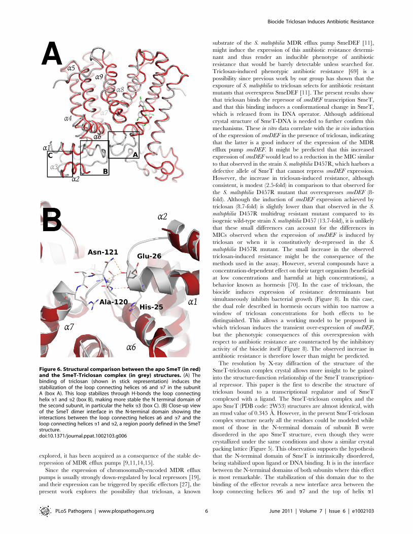
structure (Figure 6a). Residues 10–12, 21–34 and 45–55 that
appeared disordered in apo SmeT become well ordered upon
triclosan binding. Therefore, a new interface area between the
loop connecting helices a6 and a7 (residues Ser116 – Arg123) and
the top of helix a1 (residues His25 – Gly28) and the beginning of
the loop connecting a1 and a2 (residues Val29 – Thr33) could be
seen as a result of this ordering (Figure 6b). In this area, poorly
defined in the SmeT structure [53], hydrogen-bonds formed
between Ala120 and Asn121 of one monomer and His25 and
Glu26 of the other, respectively, help to stabilize this region.
Triclosan binding siteA closer inspection of the electron density maps in the C-
terminal domain showed a clear density in one of the subunits (A)
(Figure 7a). This density was readily interpretable and allows the
unambiguous placement of two molecules of the biocide (in
agreement with the stoichiometry determined by fluorescence
measurements) and the subsequent refinement. This is the first
structural evidence showing a transcriptional regulator in complex
with triclosan. However, no equivalent dense area was seen in
subunit B. The volume of the triangle-shaped ligand binding
cavity is 1030 A3, which represents a notable increase with respect
to the volume calculated for the apo SmeT structure (630 A3), but
similar to the calculated values for other TetR proteins.
One of the triclosan (TCL1) molecules binds to the bottom of
the ligand binding site, in close contact with helices a6, a7 and a8.
The phenolic hydroxyl group forms a strong hydrogen bond with
the Nd atom of His167 (placed at 2.2 A), anchoring this molecule
almost parallel to helix a8. In the apo SmeT structure this residue
is placed at an alternative conformation into the empty binding
site, with the Cd and Ne atoms pointing to the interior of the
cavity. The position and orientation of the triclosan molecule is
further stabilized through its 2,4-dichlorophenoxy ring via two
edge-to-face aromatic ring interactions, on one side with Phe133
(3.9 A) and on the other with the 2,4-dichlorophenoxy ring of the
second triclosan molecule (TCL2) (4.1 A). In this scenario the
chlorine atoms engage stabilizing interactions with hydrophobic
residues at the binding site. Thus, the 5-Cl atom makes contact
with the side chains of Val170 (3.5 A), Leu166 (4.1 A), Met140
(3.3 A) and Met93 (3.5 A). The 2-Cl atom faces Gly132 (3.2 A),
the Ce atom of His67 (3.3 A), and the phenol ring of TCL2
(4.4 A). The 4-Cl atom is surrounded by the side chains of Met110
(3.4 A), Leu114 (3.6 A) and Met113 (3.6 A). This atom, together
with the phenolic ring of TCL2, displaces these last two residues to
expand the active site cavity (see above) and maybe to
accommodate a second triclosan molecule (Figure 7b). The
hydroxyl group of the phenolic ring of TCL2 forms an H-bond
with the Nd atom of His67 (2.3 A). The 2,4-dichlorophenoxy ring
of TCL2 stacks against the phenol ring of Phe70 (3.4 A). This
residue was modeled in double conformation in the apo SmeT
structure but only the so-called open conformation is seen in the
structure of the SmeT-triclosan complex (Figure 7c). The density
for this residue in subunit B is weaker than in subunit A and a
single conformation of the side chain was modeled with an
occupancy of 50%. None of the triclosan molecules interacts with
residues from subunit B.
Discussion
In recent years, the possibility that widely-used biocides might
co-select for antibiotic resistance has been suggested to pose a
potential risk to the successful treatment of infectious diseases
[1,6–8]. Although presently there is no clear evidence of the
selection of antibiotic-resistant mutants by biocides in the wild,
risk-assessment studies are required since in vitro experiments have
shown that exposure of bacterial populations to certain biocides,
such as triclosan, indeed leads to selection for mutants with
reduced susceptibility to antibiotics [11,14,67,68]. On most
occasions on which the molecular basis of this resistance has been
Figure 5. Overview of SmeT-triclosan structure. Stereo cartoon representation of the SmeT-Triclosan complex structure, with triclosan in stickrepresentation. Helices are shown in different colors; for clarity they are labeled only in monomer B.doi:10.1371/journal.ppat.1002103.g005
Biocide Triclosan Induces Antibiotic Resistance
PLoS Pathogens | www.plospathogens.org 5 June 2011 | Volume 7 | Issue 6 | e1002103
explored, it has been acquired as a consequence of the stable de-
repression of MDR efflux pumps [9,11,14,15].
Since the expression of chromosomally-encoded MDR efflux
pumps is usually strongly down-regulated by local repressors [19],
and their expression can be triggered by specific effectors [27], the
present work explores the possibility that triclosan, a known
substrate of the S. maltophilia MDR efflux pump SmeDEF [11],
might induce the expression of this antibiotic resistance determi-
nant and thus render an inducible phenotype of antibiotic
resistance that would be barely detectable unless searched for.
Triclosan-induced phenotypic antibiotic resistance [69] is a
possibility since previous work by our group has shown that the
exposure of S. maltophilia to triclosan selects for antibiotic resistant
mutants that overexpress SmeDEF [11]. The present results show
that triclosan binds the repressor of smeDEF transcription SmeT,
and that this binding induces a conformational change in SmeT,
which is released from its DNA operator. Although additional
crystal structure of SmeT-DNA is needed to further confirm this
mechanisms. These in vitro data correlate with the in vivo induction
of the expression of smeDEF in the presence of triclosan, indicating
that the latter is a good inducer of the expression of the MDR
efflux pump smeDEF. It might be predicted that this increased
expression of smeDEF would lead to a reduction in the MIC similar
to that observed in the strain S. maltophilia D457R, which harbors a
defective allele of SmeT that cannot repress smeDEF expression.
However, the increase in triclosan-induced resistance, although
consistent, is modest (2.5-fold) in comparison to that observed for
the S. maltophilia D457R mutant that overexpresses smeDEF (8-
fold). Although the induction of smeDEF expression achieved by
triclosan (8.7-fold) is slightly lower than that observed in the S.
maltophilia D457R multidrug resistant mutant compared to its
isogenic wild-type strain S. maltophilia D457 (13.7-fold), it is unlikely
that these small differences can account for the differences in
MICs observed when the expression of smeDEF is induced by
triclosan or when it is constitutively de-repressed in the S.
maltophilia D457R mutant. The small increase in the observed
triclosan-induced resistance might be the consequence of the
methods used in the assay. However, several compounds have a
concentration-dependent effect on their target organism (beneficial
at low concentrations and harmful at high concentrations), a
behavior known as hormesis [70]. In the case of triclosan, the
biocide induces expression of resistance determinants but
simultaneously inhibits bacterial growth (Figure 8). In this case,
the dual role described in hormesis occurs within too narrow a
window of triclosan concentrations for both effects to be
distinguished. This allows a working model to be proposed in
which triclosan induces the transient over-expression of smeDEF,
but the phenotypic consequences of this overexpression with
respect to antibiotic resistance are counteracted by the inhibitory
activity of the biocide itself (Figure 8). The observed increase in
antibiotic resistance is therefore lower than might be predicted.
The resolution by X-ray diffraction of the structure of the
SmeT-triclosan complex crystal allows more insight to be gained
into the structure-function relationship of the SmeT transcription-
al repressor. This paper is the first to describe the structure of
triclosan bound to a transcriptional regulator and of SmeT
complexed with a ligand. The SmeT-triclosan complex and the
apo SmeT (PDB code: 2W53) structures are almost identical, with
an rmsd value of 0.345 A. However, in the present SmeT-triclosan
complex structure nearly all the residues could be modeled while
most of those in the N-terminal domain of subunit B were
disordered in the apo SmeT structure, even though they were
crystallized under the same conditions and show a similar crystal
packing lattice (Figure 5). This observation supports the hypothesis
that the N-terminal domain of SmeT is intrinsically disordered,
being stabilized upon ligand or DNA binding. It is in the interface
between the N-terminal domains of both subunits where this effect
is most remarkable. The stabilization of this domain due to the
binding of the effector reveals a new interface area between the
loop connecting helices a6 and a7 and the top of helix a1
Figure 6. Structural comparison between the apo SmeT (in red)and the SmeT-Triclosan complex (in grey) structures. (A) Thebinding of triclosan (shown in stick representation) induces thestabilization of the loop connecting helices a6 and a7 in the subunitA (box A). This loop stabilizes through H-bonds the loop connectinghelix a1 and a2 (box B), making more stable the N terminal domain ofthe second subunit, in particular the helix a3 (box C). (B) Close-up viewof the SmeT dimer interface in the N-terminal domain showing theinteractions between the loop connecting helices a6 and a7 and theloop connecting helices a1 and a2, a region poorly defined in the SmeTstructure.doi:10.1371/journal.ppat.1002103.g006
Biocide Triclosan Induces Antibiotic Resistance
PLoS Pathogens | www.plospathogens.org 6 June 2011 | Volume 7 | Issue 6 | e1002103

(Figure 6b). An analogous contribution to the dimer interface
through the DNA binding domain has been described in the TtgR
complex [23].
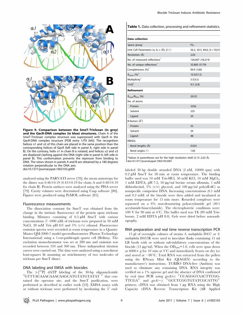
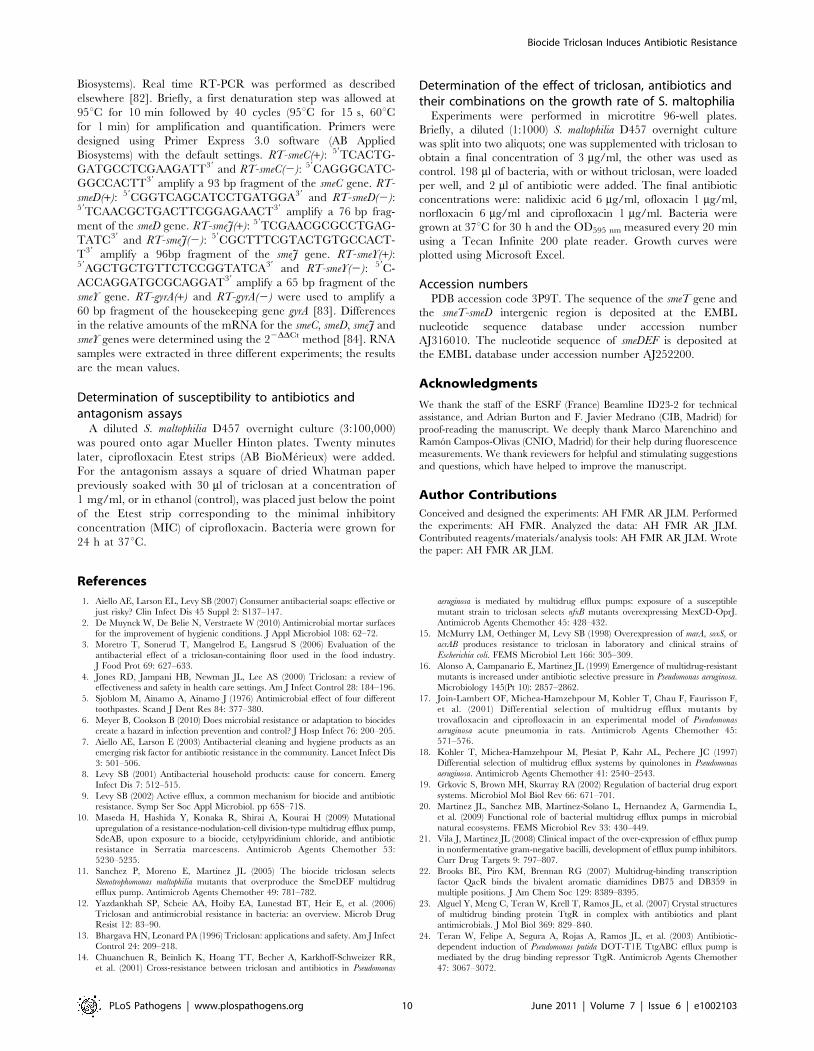
Due to the stabilization of the SmeT structure, the distance
between the a-3 recognition helices of both subunits could be
measured in the SmeT-triclosan complex, with 44.6 A between
the C-alpha atoms of Tyr49 (the N terminal of subunit B in the
apo SmeT structure had to be modeled in order to estimate this
length). This value is 10 A longer than the distance between the B-
form DNA major grooves, where these helices should bind
(Figure 9). This conformation prevents the repressor from binding
to DNA, as observed for the QacR dimer [25]. Further, the
conformational change associated with the binding of triclosan
causes the release of SmeT from its DNA operator, as seen in the
EMSA assay.
The present work shows that two molecules of triclosan enter
the hydrophobic binding pocket of SmeT. The binding of two
molecules to one subunit of a dimer has been reported for other
member of the TetR family, e.g. the binding of two molecules of
phloretin to TtgR [23] or the binding of two different drugs to
QacR [64]. One molecule of triclosan binds at the bottom of the
binding pocket, parallel to helix a6, in a geometry similar to the
phloretin molecule when binding to the high affinity site of TtgR.
The second molecule, binds close to the dimer interface, and also
interacts with this helix through the phenolic ring (Figure 6b). The
first molecule of triclosan stacks against the phenol ring of Phe70, a
residue identified as the gatekeeper of the ligand binding pocket.
Supporting this hypothesis, the so-called open conformation is the
only one seen in the structure of the SmeT-triclosan complex. In
contrast, Phe70 has a double conformation in the apo SmeT
structure.
The ligand binding site of SmeT is mainly formed by
hydrophobic residues - there are just three polar residues: His67,
His167 and Ser96. This lack of charged residues may be related to
the polyspecific substrate recognition of SmeT. Additionally the
binding site can increase its volume almost 2-fold by displacing the
side chains of residues with double conformations in the apo SmeT
structure namely Phe70, Met113 and Leu114. The different side
chains that can be exposed, or covered, may also facilitate
interactions with a wide range of compounds. Remarkably, some
residues displaced by ligand binding (Met110 and Met113) belong
to helix a6, which interacts through hydrogen bonds with residues
from helix a1. These interactions are described as decisive in
QacR and TetR deactivation due to ligand binding [19]. Through
these interaction networks between helices, the QacR and TetR
changes the relative orientation of the N-terminal domains to
generate a ligand bound structure unable to bind DNA. The
present data suggest that SmeT may modify its shape after
triclosan binding, provoking the release of DNA by a similar
mechanism (Figure 9).
The structure of the complex SmeT-triclosan, together with the
experimental data supporting its role in smeDEF pump derepres-
sion, is the first structural evidence that triclosan can act as an
Figure 7. Structural details of the triclosan binding site. (A) TheFo-Fc omit electron density map contoured at 2.4 s around thetriclosan molecules is shown in red. (B) Key interactions (H-bonds inorange) between the triclosan molecules and the binding pocketresidues of SmeT (distances less than 4 A). The most important residuesin the formation of the binding pocket are shown and labeled. (C) Thebinding of two molecules of triclosan displaces residues His-167, Leu-114 and Met-113. These last two residues belong to helix a6, whichinteracts trough H bonds with helix a1, part of the DNA binding domainof SmeT.doi:10.1371/journal.ppat.1002103.g007
Biocide Triclosan Induces Antibiotic Resistance
PLoS Pathogens | www.plospathogens.org 7 June 2011 | Volume 7 | Issue 6 | e1002103

effector of an MDR system regulator, and provides an unambig-
uous link between the presence of this biocide and the increased
efflux of antibiotics by the opportunistic pathogen S. maltophilia.
Materials and Methods
Protein purification and crystallizationSmeT protein was expressed and purified as previously
described [53]. Briefly, the smeT gene was cloned into the
pTYB1 vector (IMPACT-CN system, New England Biolabs) and
the protein expressed in the E. coli strain ER2566 (IMPACT-CN
system, NEB). After induction with 0.5 mM IPTG, the culture
was grown overnight at 15uC. Cells were disrupted by sonication
and centrifuged. The supernatant with the SmeT-Intein-CBD
fusion protein was loaded into a Poly-Prep chromatography
column (Bio-Rad) containing chitin beads (NEB). After overnight
incubation in cleavage buffer (20 mM Tris-HCl, 0.5 M NaCl,
100 mM dithiothreitol (DTT), 1 mM EDTA, pH 8.0) at 4uC,
SmeT was eluted and the remaining DTT present in the sample
removed by dialysis against Tris-buffered saline. Finally, the
sample was loaded into a Sephacryl S100 gel filtration column
(GE Healthcare) in a buffer containing 300 mM NaCl and
20 mM Tris pH 8.0.
Prior to concentration, the sample was incubated with triclosan
(5-chloro-2-(2,4-dichlorophenoxy) phenol), previously dissolved in
DMSO, for 4 h at 4uC in a 1:10 molar ratio. The SmeT-triclosan
complex was concentrated to 5 mg/ml and crystallized using the
sitting-drop vapor diffusion technique in a solution containing
175 mM Li2SO4, 100 mM Tris pH 8.5 and 27% v/v PEG MME
2K. Crystals grew to approximately 5006200620 mm within 3
days at 22uC. These crystals were transferred into a cryoprotectant
solution (175 mM Li2SO4, 100 mM Tris pH 8.5 and 30% v/v
PEG MME 2K) and flash-cooled at 100uK.
Crystallographic data collection and processingCrystallographic data were collected at the Beamline ID23-2 of
the European Synchrotron Radiation Facility (ESRF) and processed
using Mosflm [71] and SCALA software [72]. The crystal
parameters were equivalent to those of the apo SmeT structure
[53]. The crystals belonged to the space group P21 and had unit cell
dimensions of a = 56.5, b = 59.5, c = 84.8 A, and b= 102.7u. The
Mathews coefficient for a dimer in the asymmetric unit was
2.82 A3/Da, which corresponds to a solvent content of 56.4%.
Details of data collection, crystal parameters and data-processing
statistics are given in Table 1.
Model refinement and validationDifference Fourier techniques were used to solve the structure
since unit cell dimensions are almost identical to those of the
SmeT structure (PDB code 2w53 [53]). Molecular Replacement
using MOLREP [73] has confirmed that the crystal packing of the
SmeT-TCL complex is identical to that of the SmeT protein. The
translation-libration-screw (TLS) groups (3 per chain) were defined
using the TLSMD server [74]. Iterative cycles of manual building
and TLS-restrained refinement cycles were performed using
COOT [75] and REFMAC software [76] to final Rfree and
Rcryst values of 25 and 20% respectively (Table 1). The final
model comprised residues 6 to 218 of chain A, 10 to 218 of chain
B, 225 water molecules, 2 sulphate anions and 2 triclosan
molecules. The overall electron density map was of high quality
but of poor definition for residues 30–31 and 120–121 in chain B,
perhaps due to the high flexibility of these regions. Analysis of the
geometry for the final model was performed using Molprobity
software [77]; 98.3% of the residues fell within the favored regions
of the Ramachandran plot and none within the disallowed regions.
The anisotropy of the atomic displacement parameters was
Figure 8. Effect of triclosan on the susceptibility to antibiotics of S. maltophilia. Panel A: In the basal state, SmeT (orange circles) is bound toits cognate operator DNA repressing the transcription of smeDEF. The fact that damage is caused by ciprofloxacin (yellow stars) inside the bacteria isrepresented by crinkly arrows. Panel B: The entrance of triclosan (black spots) causes bacterial damage and, simultaneously, the binding of thebiocide to SmeT releases the protein from its operator allowing high-level transcription of smeDEF. Once the pump is expressed, its substrates -triclosan and ciprofloxacin - are pumped out of the bacteria. However the toxic effect of triclosan prevents the cells reaching the level of resistanceobserved in a mutant defective in SmeT (see text for more details).doi:10.1371/journal.ppat.1002103.g008
Biocide Triclosan Induces Antibiotic Resistance
PLoS Pathogens | www.plospathogens.org 8 June 2011 | Volume 7 | Issue 6 | e1002103
analyzed using the PARVATI server [78]; the mean anisotropy for
the dimer was 0.4660.19 (0.4360.19 for chain A and 0.4860.19
for chain B). Protein surfaces were analyzed using the PISA server
[79]. Cavity volumes were determined using Casp software [80].
Figures were produced using PyMOL software [81].
Fluorescence measurementsThe dissociation constant for SmeT was obtained from the
change in the intrinsic fluorescence of the protein upon triclosan
binding. Mixtures consisting of 0.5 mM SmeT with various
concentrations (7–1000 nM) of triclosan were prepared in 50 mM
NaCl, 20 mM Tris pH 8.0 and 5% (v/v) ethanol. Fluorescence
emission spectra were recorded at room temperature in a Quanta-
Master QM-2000-7 model spectrofluorometer (Photon Technology
International) using a 1-cm-pathlength quartz cell (Hellma). The
excitation monochromator was set at 280 nm and emission was
recorded between 310 and 360 nm. Three independent titration
curves were carried out. The data were analyzed using a non-linear
least-squares fit assuming an stoichiometry of two molecules of
triclosan per SmeT dimer.
DNA labeling and EMSA with biocidesThe c-[32P] dATP labeling of the 30-bp oligonucleotide
59GTTTACAAACAAACAAGCATGTATGTATAT 39 that con-
tained the operator site, and the SmeT purification, were
performed as described in earlier work [53]. EMSA assays with
or without triclosan were performed by incubating the 59 end-
labeled 30 bp double stranded DNA (2 nM, 10000 cpm) with
0.2 mM SmeT for 20 min at room temperature. The binding
buffer used was 10 mM Tris-HCl, 50 mM KCl, 10 mM MgCl2,
1 mM EDTA, pH 7.2, 50 mg/ml bovine serum albumin, 1 mM
dithiothreitol, 5% (v/v) glycerol, and 100 mg/ml poly(dI-dC) as
nonspecific competitor DNA. Increasing concentrations (0.1 mM
and 0.2 mM) of the biocide were then added and incubated at
room temperature for 15 min more. Retarded complexes were
separated on a 6% non-denaturing polyacrylamide gel (40:1
acrylamide:bisacrylamide). The electrophoretic conditions were:
100 V for 90 min at 4uC. The buffer used was TE (89 mM Tris-
borate, 2 mM EDTA pH 8.0). Gels were dried before autoradi-
ography.
RNA preparation and real time reverse transcription PCR15 ml of overnight cultures of strains S. maltophilia D457 or S.
maltophilia D457R were used to inoculate flasks containing 15 ml
LB broth with or without sub-inhibitory concentrations of the
biocide (15 mg/ml). When the OD600<1.0, cells were spun down
at 60006g for 10 min at 4uC and immediately frozen on dry ice
and stored at 280uC. Total RNA was extracted from the pellets
using the RNeasy Mini Kit (QIAGEN) according to the
manufacturer’s instructions. TURBO DNA-free (Ambion) was
used to eliminate any remaining DNA. RNA integrity was
verified on a 1% agarose gel and the absence of DNA confirmed
by real time PCR using gyrA(+): 59CCAGGGTAACTTCGG-
TTCGA39 and gyrA(2): 59GCCTCGGTGTATCGCATTG39
primers. cDNA was obtained from 1 mg RNA using the High
Capacity cDNA Reverse Transcription Kit (AB Applied
Figure 9. Comparison between the SmeT-Triclosan (in grey)and the QacR-DNA complex (in blue) structures. Chain A of theSmeT-Triclosan complex structure was superposed with QacR in theQacR-DNA complex structure (PDB entry 1JT0 [64]). The recognitionhelices a1 and a2 of this chain are placed in the same position than thecorresponding helices of QacR (left side in panel A, right side in panelB). On the contrary, helix a1 in chain B is rotated, and helices a2 and a3are displaced clashing against the DNA (right side in panel A, left side inpanel B). This conformation prevents the repressor from binding toDNA. The views shown in panels A and B are obtained by a 180 degreesrotation perpendicular to the DNA axis.doi:10.1371/journal.ppat.1002103.g009
Table 1. Data collection, processing and refinement statistics.
Data collection
Space group P21
Unit Cell Parameters (a, b, c (A); b (u) 56.2, 59.3, 84.6; b= 102.9
Resolution (A) 2,02
No. of measured reflections* 126,007 (18,214)
No. of unique reflections* 35,800 (5179)
Completeness (%)* 99.9 (100)
Rmeas (%)* 10.3(57.2)
Multiplicity* 3.5(3.5)
I/s(I)* 9.5 (2.8)
Refinement
Rcryst/Rfree (%) 20/25
No. of atoms
Protein 3356
Solvent 225
Ligand 34
B-factors (A2)
Protein 35
Solvent 34
Ligand 48
RMSDs
Bond lengths (A) 0.023
Bond angles (u) 1.85
*Values in parentheses are for the high resolution shell (2.13–2.02 A).doi:10.1371/journal.ppat.1002103.t001
Biocide Triclosan Induces Antibiotic Resistance
PLoS Pathogens | www.plospathogens.org 9 June 2011 | Volume 7 | Issue 6 | e1002103
Biosystems). Real time RT-PCR was performed as described
elsewhere [82]. Briefly, a first denaturation step was allowed at
95uC for 10 min followed by 40 cycles (95uC for 15 s, 60uCfor 1 min) for amplification and quantification. Primers were
designed using Primer Express 3.0 software (AB Applied
Biosystems) with the default settings. RT-smeC(+): 59TCACTG-
GATGCCTCGAAGATT39 and RT-smeC(2): 59CAGGGCATC-
GGCCACTT39 amplify a 93 bp fragment of the smeC gene. RT-
smeD(+): 59CGGTCAGCATCCTGATGGA39 and RT-smeD(2):59TCAACGCTGACTTCGGAGAACT39 amplify a 76 bp frag-
ment of the smeD gene. RT-smeJ(+): 59TCGAACGCGCCTGAG-
TATC39 and RT-smeJ(2): 59CGCTTTCGTACTGTGCCACT-
T39 amplify a 96bp fragment of the smeJ gene. RT-smeY(+):59AGCTGCTGTTCTCCGGTATCA39 and RT-smeY(2): 59C-
ACCAGGATGCGCAGGAT39 amplify a 65 bp fragment of the
smeY gene. RT-gyrA(+) and RT-gyrA(2) were used to amplify a
60 bp fragment of the housekeeping gene gyrA [83]. Differences
in the relative amounts of the mRNA for the smeC, smeD, smeJ and
smeY genes were determined using the 22DDCt method [84]. RNA
samples were extracted in three different experiments; the results
are the mean values.
Determination of susceptibility to antibiotics andantagonism assays
A diluted S. maltophilia D457 overnight culture (3:100,000)
was poured onto agar Mueller Hinton plates. Twenty minutes
later, ciprofloxacin Etest strips (AB BioMerieux) were added.
For the antagonism assays a square of dried Whatman paper
previously soaked with 30 ml of triclosan at a concentration of
1 mg/ml, or in ethanol (control), was placed just below the point
of the Etest strip corresponding to the minimal inhibitory
concentration (MIC) of ciprofloxacin. Bacteria were grown for
24 h at 37uC.
Determination of the effect of triclosan, antibiotics andtheir combinations on the growth rate of S. maltophilia
Experiments were performed in microtitre 96-well plates.
Briefly, a diluted (1:1000) S. maltophilia D457 overnight culture
was split into two aliquots; one was supplemented with triclosan to
obtain a final concentration of 3 mg/ml, the other was used as
control. 198 ml of bacteria, with or without triclosan, were loaded
per well, and 2 ml of antibiotic were added. The final antibiotic
concentrations were: nalidixic acid 6 mg/ml, ofloxacin 1 mg/ml,
norfloxacin 6 mg/ml and ciprofloxacin 1 mg/ml. Bacteria were
grown at 37uC for 30 h and the OD595 nm measured every 20 min
using a Tecan Infinite 200 plate reader. Growth curves were
plotted using Microsoft Excel.
Accession numbersPDB accession code 3P9T. The sequence of the smeT gene and
the smeT-smeD intergenic region is deposited at the EMBL
nucleotide sequence database under accession number
AJ316010. The nucleotide sequence of smeDEF is deposited at
the EMBL database under accession number AJ252200.
Acknowledgments
We thank the staff of the ESRF (France) Beamline ID23-2 for technical
assistance, and Adrian Burton and F. Javier Medrano (CIB, Madrid) for
proof-reading the manuscript. We deeply thank Marco Marenchino and
Ramon Campos-Olivas (CNIO, Madrid) for their help during fluorescence
measurements. We thank reviewers for helpful and stimulating suggestions
and questions, which have helped to improve the manuscript.
Author Contributions
Conceived and designed the experiments: AH FMR AR JLM. Performed
the experiments: AH FMR. Analyzed the data: AH FMR AR JLM.
Contributed reagents/materials/analysis tools: AH FMR AR JLM. Wrote
the paper: AH FMR AR JLM.
References
1. Aiello AE, Larson EL, Levy SB (2007) Consumer antibacterial soaps: effective or
just risky? Clin Infect Dis 45 Suppl 2: S137–147.
2. De Muynck W, De Belie N, Verstraete W (2010) Antimicrobial mortar surfacesfor the improvement of hygienic conditions. J Appl Microbiol 108: 62–72.
3. Moretro T, Sonerud T, Mangelrod E, Langsrud S (2006) Evaluation of the
antibacterial effect of a triclosan-containing floor used in the food industry.
J Food Prot 69: 627–633.
4. Jones RD, Jampani HB, Newman JL, Lee AS (2000) Triclosan: a review ofeffectiveness and safety in health care settings. Am J Infect Control 28: 184–196.
5. Sjoblom M, Ainamo A, Ainamo J (1976) Antimicrobial effect of four different
toothpastes. Scand J Dent Res 84: 377–380.
6. Meyer B, Cookson B (2010) Does microbial resistance or adaptation to biocides
create a hazard in infection prevention and control? J Hosp Infect 76: 200–205.
7. Aiello AE, Larson E (2003) Antibacterial cleaning and hygiene products as anemerging risk factor for antibiotic resistance in the community. Lancet Infect Dis
3: 501–506.
8. Levy SB (2001) Antibacterial household products: cause for concern. Emerg
Infect Dis 7: 512–515.
9. Levy SB (2002) Active efflux, a common mechanism for biocide and antibioticresistance. Symp Ser Soc Appl Microbiol. pp 65S–71S.
10. Maseda H, Hashida Y, Konaka R, Shirai A, Kourai H (2009) Mutational
upregulation of a resistance-nodulation-cell division-type multidrug efflux pump,SdeAB, upon exposure to a biocide, cetylpyridinium chloride, and antibiotic
resistance in Serratia marcescens. Antimicrob Agents Chemother 53:
5230–5235.
11. Sanchez P, Moreno E, Martinez JL (2005) The biocide triclosan selectsStenotrophomonas maltophilia mutants that overproduce the SmeDEF multidrug
efflux pump. Antimicrob Agents Chemother 49: 781–782.
12. Yazdankhah SP, Scheie AA, Hoiby EA, Lunestad BT, Heir E, et al. (2006)
Triclosan and antimicrobial resistance in bacteria: an overview. Microb DrugResist 12: 83–90.
13. Bhargava HN, Leonard PA (1996) Triclosan: applications and safety. Am J Infect
Control 24: 209–218.
14. Chuanchuen R, Beinlich K, Hoang TT, Becher A, Karkhoff-Schweizer RR,et al. (2001) Cross-resistance between triclosan and antibiotics in Pseudomonas
aeruginosa is mediated by multidrug efflux pumps: exposure of a susceptible
mutant strain to triclosan selects nfxB mutants overexpressing MexCD-OprJ.
Antimicrob Agents Chemother 45: 428–432.
15. McMurry LM, Oethinger M, Levy SB (1998) Overexpression of marA, soxS, or
acrAB produces resistance to triclosan in laboratory and clinical strains of
Escherichia coli. FEMS Microbiol Lett 166: 305–309.
16. Alonso A, Campanario E, Martinez JL (1999) Emergence of multidrug-resistant
mutants is increased under antibiotic selective pressure in Pseudomonas aeruginosa.
Microbiology 145(Pt 10): 2857–2862.
17. Join-Lambert OF, Michea-Hamzehpour M, Kohler T, Chau F, Faurisson F,
et al. (2001) Differential selection of multidrug efflux mutants by
trovafloxacin and ciprofloxacin in an experimental model of Pseudomonas
aeruginosa acute pneumonia in rats. Antimicrob Agents Chemother 45:
571–576.
18. Kohler T, Michea-Hamzehpour M, Plesiat P, Kahr AL, Pechere JC (1997)
Differential selection of multidrug efflux systems by quinolones in Pseudomonas
aeruginosa. Antimicrob Agents Chemother 41: 2540–2543.
19. Grkovic S, Brown MH, Skurray RA (2002) Regulation of bacterial drug export
systems. Microbiol Mol Biol Rev 66: 671–701.
20. Martinez JL, Sanchez MB, Martinez-Solano L, Hernandez A, Garmendia L,
et al. (2009) Functional role of bacterial multidrug efflux pumps in microbial
natural ecosystems. FEMS Microbiol Rev 33: 430–449.
21. Vila J, Martinez JL (2008) Clinical impact of the over-expression of efflux pump
in nonfermentative gram-negative bacilli, development of efflux pump inhibitors.
Curr Drug Targets 9: 797–807.
22. Brooks BE, Piro KM, Brennan RG (2007) Multidrug-binding transcription
factor QacR binds the bivalent aromatic diamidines DB75 and DB359 in
multiple positions. J Am Chem Soc 129: 8389–8395.
23. Alguel Y, Meng C, Teran W, Krell T, Ramos JL, et al. (2007) Crystal structures
of multidrug binding protein TtgR in complex with antibiotics and plant
antimicrobials. J Mol Biol 369: 829–840.
24. Teran W, Felipe A, Segura A, Rojas A, Ramos JL, et al. (2003) Antibiotic-
dependent induction of Pseudomonas putida DOT-T1E TtgABC efflux pump is
mediated by the drug binding repressor TtgR. Antimicrob Agents Chemother
47: 3067–3072.
Biocide Triclosan Induces Antibiotic Resistance
PLoS Pathogens | www.plospathogens.org 10 June 2011 | Volume 7 | Issue 6 | e1002103

25. Schumacher MA, Miller MC, Grkovic S, Brown MH, Skurray RA, et al. (2001)Structural mechanisms of QacR induction and multidrug recognition. Science
294: 2158–2163.
26. Heldwein EE, Brennan RG (2001) Crystal structure of the transcriptionactivator BmrR bound to DNA and a drug. Nature 409: 378–382.
27. Wade H (2010) MD recognition by MDR gene regulators. Curr Opin StructBiol 20: 489–496.
28. Berg G, Eberl L, Hartmann A (2005) The rhizosphere as a reservoir for
opportunistic human pathogenic bacteria. Environ Microbiol 7: 1673–1685.
29. Ribbeck-Busch K, Roder A, Hasse D, de Boer W, Martinez JL, et al. (2005) A
molecular biological protocol to distinguish potentially human pathogenicStenotrophomonas maltophilia from plant-associated Stenotrophomonas rhizophila.
Environ Microbiol 7: 1853–1858.
30. Sanchez MB, Hernandez A, Martinez JL (2009) Stenotrophomonas maltophilia drugresistance. Future Microbiol 4: 655–660.
31. Looney WJ, Narita M, Muhlemann K (2009) Stenotrophomonas maltophilia: an
emerging opportunist human pathogen. Lancet Infect Dis 9: 312–323.
32. Falagas ME, Kastoris AC, Vouloumanou EK, Rafailidis PI, Kapaskelis AM,
et al. (2009) Attributable mortality of Stenotrophomonas maltophilia infections: asystematic review of the literature. Future Microbiol 4: 1103–1109.
33. Wang WS, Liu CP, Lee CM, Huang FY (2004) Stenotrophomonas maltophilia
bacteremia in adults: four years’ experience in a medical center in northernTaiwan. J Microbiol Immunol Infect 37: 359–365.
34. Crum NF, Utz GC, Wallace MR (2002) Stenotrophomonas maltophilia endocarditis.Scand J Infect Dis 34: 925–927.
35. Aisenberg G, Rolston KV, Dickey BF, Kontoyiannis DP, Raad II, et al. (2007)
Stenotrophomonas maltophilia pneumonia in cancer patients without traditional riskfactors for infection, 1997–2004. Eur J Clin Microbiol Infect Dis 26: 13–20.
36. Turrientes MC, Baquero MR, Sanchez MB, Valdezate S, Escudero E, et al.
(2010) Polymorphic mutation frequencies of clinical and environmentalStenotrophomonas maltophilia populations. Appl Environ Microbiol 76: 1746–1758.
37. Valdezate S, Vindel A, Maiz L, Baquero F, Escobar H, et al. (2001) Persistenceand variability of Stenotrophomonas maltophilia in cystic fibrosis patients, Madrid,
1991–1998. Emerg Infect Dis 7: 113–122.
38. Spicuzza L, Sciuto C, Vitaliti G, Di Dio G, Leonardi S, et al. (2008) Emergingpathogens in cystic fibrosis: ten years of follow-up in a cohort of patients.
Eur J Clin Microbiol Infect Dis 28: 191–195.
39. Ryan RP, Monchy S, Cardinale M, Taghavi S, Crossman L, et al. (2009) The
versatility and adaptation of bacteria from the genus Stenotrophomonas. Nat Rev
Micro 7: 514–525.
40. Crossman LC, Gould VC, Dow JM, Vernikos GS, Okazaki A, et al. (2008) The
complete genome, comparative and functional analysis of Stenotrophomonas
maltophilia reveals an organism heavily shielded by drug resistance determinants.
Genome Biol 9: R74.
41. Avison MB, Higgins CS, Ford PJ, von Heldreich CJ, Walsh TR, et al. (2002)Differential regulation of L1 and L2 beta-lactamase expression in Stenotrophomonas
maltophilia. J Antimicrob Chemother 49: 387–389.
42. Okazaki A, Avison MB (2007) Aph(39)-IIc, an aminoglycoside resistance
determinant from Stenotrophomonas maltophilia. Antimicrob Agents Chemother
51: 359–360.
43. Okazaki A, Avison MB (2008) Induction of L1 and L2 beta-lactamase
production in Stenotrophomonas maltophilia is dependent on an AmpR-typeregulator. Antimicrob Agents Chemother 52: 1525–1528.
44. Sanchez MB, Hernandez A, Rodriguez-Martinez JM, Martinez-Martinez L,
Martinez JL (2008) Predictive analysis of transmissible quinolone resistanceindicates Stenotrophomonas maltophilia as a potential source of a novel family of Qnr
determinants. BMC Microbiology 8: 148.
45. Sanchez MB, Martinez JL (2010) SmQnr contributes to intrinsic resistance toquinolones in Stenotrophomonas maltophilia. Antimicrob Agents Chemother 54:
580–581.
46. Shimizu K, Kikuchi K, Sasaki T, Takahashi N, Ohtsuka M, et al. (2008) Smqnr,
a new chromosome-carried quinolone resistance gene in Stenotrophomonas
maltophilia. Antimicrob Agents Chemother 52: 3823–3825.
47. Zhang L, Li XZ, Poole K (2001) SmeDEF multidrug efflux pump contributes to
intrinsic multidrug resistance in Stenotrophomonas maltophilia. Antimicrob AgentsChemother 45: 3497–3503.
48. Alonso A, Martinez JL (2001) Expression of multidrug efflux pump SmeDEF by
clinical isolates of Stenotrophomonas maltophilia. Antimicrob Agents Chemother 45:1879–1881.
49. Alonso A, Martinez JL (2000) Cloning and characterization of SmeDEF, a novelmultidrug efflux pump from Stenotrophomonas maltophilia. Antimicrob Agents
Chemother 44: 3079–3086.
50. Alonso A, Martinez JL (1997) Multiple antibiotic resistance in Stenotrophomonas
maltophilia. Antimicrob Agents Chemother 41: 1140–1142.
51. Sanchez P, Alonso A, Martinez JL (2004) Regulatory regions of smeDEF inStenotrophomonas maltophilia strains expressing different amounts of the multidrug
efflux pump SmeDEF. Antimicrob Agents Chemother 48: 2274–2276.
52. Alonso A, Morales G, Escalante R, Campanario E, Sastre L, et al. (2004)Overexpression of the multidrug efflux pump SmeDEF impairs Stenotrophomonas
maltophilia physiology. Journal of Antimicrobial Chemotherapy 53: 432–434.
53. Hernandez A, Mate MJ, Sanchez-Diaz PC, Romero A, Rojo F, et al. (2009)Structural and Functional Analysis of SmeT, the Repressor of the Stenotropho-
monas maltophilia Multidrug Efflux Pump SmeDEF. J Biol Chem 284:14428–14438.
54. Sanchez P, Alonso A, Martinez JL (2002) Cloning and characterization of
SmeT, a repressor of the Stenotrophomonas maltophilia multidrug efflux pump
SmeDEF. Antimicrob Agents Chemother 46: 3386–3393.
55. Ramos JL, Martinez-Bueno M, Molina-Henares AJ, Teran W, Watanabe K,
et al. (2005) The TetR family of transcriptional repressors. Microbiol Mol Biol
Rev 69: 326–356.
56. Natsume R, Ohnishi Y, Senda T, Horinouchi S (2004) Crystal structure of a
gamma-butyrolactone autoregulator receptor protein in Streptomyces coelicolor
A3(2). J Mol Biol 336: 409–419.
57. Dover LG, Corsino PE, Daniels IR, Cocklin SL, Tatituri V, et al. (2004) Crystal
structure of the TetR/CamR family repressor Mycobacterium tuberculosis EthR
implicated in ethionamide resistance. J Mol Biol 340: 1095–1105.
58. Gu R, Su CC, Shi F, Li M, McDermott G, et al. (2007) Crystal structure of the
transcriptional regulator CmeR from Campylobacter jejuni. J Mol Biol 372:
583–593.
59. Li M, Gu R, Su CC, Routh MD, Harris KC, et al. (2007) Crystal structure of the
transcriptional regulator AcrR from Escherichia coli. J Mol Biol 374: 591–603.
60. Willems AR, Tahlan K, Taguchi T, Zhang K, Lee ZZ, et al. (2008) Crystal
structures of the Streptomyces coelicolor TetR-like protein ActR alone and in
complex with actinorhodin or the actinorhodin biosynthetic precursor (S)-
DNPA. J Mol Biol 376: 1377–1387.
61. Jeng WY, Ko TP, Liu CI, Guo RT, Liu CL, et al. (2008) Crystal structure of
IcaR, a repressor of the TetR family implicated in biofilm formation in
Staphylococcus epidermidis. Nucleic Acids Res 36: 1567–1577.
62. Krell T, Teran W, Mayorga OL, Rivas G, Jimenez M, et al. (2007)
Optimization of the palindromic order of the TtgR operator enhances binding
cooperativity. J Mol Biol 369: 1188–1199.
63. Kapoor M, Reddy CC, Krishnasastry MV, Surolia N, Surolia A (2004) Slow-
tight-binding inhibition of enoyl-acyl carrier protein reductase from Plasmodium
falciparum by triclosan. Biochem J 381: 719–724.
64. Schumacher MA, Miller MC, Grkovic S, Brown MH, Skurray RA, et al. (2002)
Structural basis for cooperative DNA binding by two dimers of the multidrug-
binding protein QacR. EMBO J 21: 1210–1218.
65. Li XZ, Zhang L, Poole K (2002) SmeC, an outer membrane multidrug efflux
protein of Stenotrophomonas maltophilia. Antimicrob Agents Chemother 46:
333–343.
66. Chang LL, Chen HF, Chang CY, Lee TM, Wu WJ (2004) Contribution of
integrons, and SmeABC and SmeDEF efflux pumps to multidrug resistance in
clinical isolates of Stenotrophomonas maltophilia. J Antimicrob Chemother 53:
518–521.
67. Pumbwe L, Chang A, Smith RL, Wexler HM (2006) Clinical significance of
overexpression of multiple RND-family efflux pumps in Bacteroides fragilis isolates.
J Antimicrob Chemother 58: 543–548.
68. Cottell A, Denyer SP, Hanlon GW, Ochs D, Maillard JY (2009) Triclosan-
tolerant bacteria: changes in susceptibility to antibiotics. J Hosp Infect 72: 71–76.
69. Bailey AM, Constantinidou C, Ivens A, Garvey MI, Webber MA, et al. (2009)
Exposure of Escherichia coli and Salmonella enterica serovar Typhimurium to
triclosan induces a species-specific response, including drug detoxification.
J Antimicrob Chemother 64: 973–985.
70. Calabrese EJ (2005) Paradigm lost, paradigm found: the re-emergence of
hormesis as a fundamental dose response model in the toxicological sciences.
Environ Pollut 138: 379–411.
71. Leslie AG (2006) The integration of macromolecular diffraction data. Acta
Crystallogr D Biol Crystallogr 62: 48–57.
72. CCP4 (1994) The CCP4 suite: programs for protein crystallography. Acta
Crystallogr D Biol Crystallogr 50: 760–763.
73. Vagin A, Teplyakov A (1997) MOLREP: an Automated Program for Molecular
Replacement. J Appl Cryst 30: 1022–1025.
74. Painter J, Merritt EA (2006) TLSMD web server for the generation of multi-
group TLS models. J Appl Cryst 39: 109–111.
75. Emsley P, Lohkamp B, Scott WG, Cowtan K (2010) Features and Development
of Coot. Acta Crystallographica Section D - Biological Crystallography 66:
22–25.
76. Murshudov GN, Vagin AA, Dodson EJ (1997) Refinement of macromolecular
structures by the maximum-likelihood method. Acta Crystallogr D Biol Crystal-
logr 53: 240–255.
77. Chen VB, Arendall WB, 3rd, Headd JJ, Keedy DA, Immormino RM, et al.
(2010) MolProbity: all-atom structure validation for macromolecular crystallog-
raphy. Acta Crystallogr D Biol Crystallogr 66: 12–21.
78. Merritt EA (1999) Expanding the Model: Anisotropic Displacement Parameters
in Protein Structure Refinement. Acta Crystallographica D 55: 1109–1117.
79. Krissinel E, Henrick K (2007) Inference of macromolecular assemblies from
crystalline state. J Mol Biol 372: 774–797.
80. Dundas J, Ouyang Z, Tseng J, Binkowski A, Turpaz Y, et al. (2006) CASTp:
computed atlas of surface topography of proteins with structural and
topographical mapping of functionally annotated residues. Nucleic Acids Res
34: W116–118.
81. DeLano WL (2002) The PyMOL Molecular Graphics System. DeLano
Scientifics, Palo Alto, CA, USA.
82. Morales G, Ugidos A, Rojo F (2006) Inactivation of the Pseudomonas putida
cytochrome o ubiquinol oxidase leads to a significant change in the
transcriptome and to increased expression of the CIO and cbb3-1 terminal
oxidases. Environ Microbiol 8: 1764–1774.
Biocide Triclosan Induces Antibiotic Resistance
PLoS Pathogens | www.plospathogens.org 11 June 2011 | Volume 7 | Issue 6 | e1002103

83. Gould VC, Avison MB (2006) SmeDEF-mediated antimicrobial drug resistance
in Stenotrophomonas maltophilia clinical isolates having defined phylogeneticrelationships. J Antimicrob Chemother 57: 1070–1076.
84. Livak KJ, Schmittgen TD (2001) Analysis of relative gene expression data using
real-time quantitative PCR and the 2(-Delta Delta C(T)) Method. Methods 25:402–408.
Biocide Triclosan Induces Antibiotic Resistance
PLoS Pathogens | www.plospathogens.org 12 June 2011 | Volume 7 | Issue 6 | e1002103