trends in colloid and interface science xvi || salt-induced aggregation in cationic liposome...
TRANSCRIPT

Introduction
Liposomes are spherical structures composed by a closedlipid bilayer that encompasses an aqueous core disjoinedfrom an external continuous medium [1, 2]. Liposomes inaqueous suspension are an interesting system not only asa model colloidal system in fundamental researchconcerning self-assembling molecules but also as a drugdelivery vehicle, where the enclosed water core can beused to solubilise active substances and the biocompat-ible bilayer can be used as carrier to join the site ofinfection or disease [3, 4, 5].
In particular, liposomes, built up from cationic lipids,have been widely used for cell transfection in vitro andare being investigated in gene therapy and geneticengineering for the delivery of genes into mammaliancells.
The success of such a method depends on the stabilityof the liposomes and, in recent years, a number ofamphiphile systems has been developed, each of themshows a different efficiency, depending on variousparameters such as the composition of the incubation
medium (pH, ionic strength), the nature of the lipidcomponent, the incubation time before transfection, andso on.
Despite their widespread use, the critical factorsdetermining the transfection activity of these systemsare not clear yet and, although the structure of theresulting aggregates should play an important role, upuntil now, little effort has been put into understandingthe more fundamental aspects concerning the struc-ture and the relationship between the transfectionefficiency and the morphological characteristics of theaggregates.
In order to elucidate the mechanism governing theformation and the stability of cationic liposome aggre-gates of various sizes, induced by electrostatic interac-tions, we have investigated the conformational behaviourof liposomes in the presence of simple uni- and divalentsalt electrolyte solutions, at various charge ratios. Wehave monitored the formation of aggregates and theirchange in size over time by means of the dynamic lightscattering technique, because of its unique feature tostudy the structure and the dynamics of colloidal-size
Progr Colloid Polym Sci (2004) 123: 78–82DOI 10.1007/b11646� Springer-Verlag 2004
A. Di Biasio
F. Bordi
C. Cametti
Salt-induced aggregation in cationicliposome suspensions
A. Di BiasioDipartimento di Matematica e Fisica,Universita’ di Camerino, Camerino, Italy
F. Bordi Æ C. Cametti (&)Dipartimento di Fisica,Universita’ di Roma ‘‘La Sapienza’’,Piazzale A. Moro 5,00185 Rome, Italy
A. Di Biasio Æ F. Bordi Æ C. CamettiIstituto Nazionale per la Fisica dellaMateria (INFM), Unita’ di Roma 1,Rome, Italy
Abstract The simple salt-inducedaggregation of small unilamellardioleoyltrimethylammoniumpropane[DOTAP] vesicles is investigated bymeasuring the change in the effectiveradius with time, using dynamic lightscattering techniques. At small saltconcentration (lower than0.5–0.6 mol/L), an aggregationmechanism results in the formationof stable liposome structures ofmoderate size, before that the usualirreversible coagulation prevails, athigher salt concentration. The
steady-state size reached by theseaggregates, after their initial growth,is governed by binding counterionsto liposome surfaces, resulting in ascreening effect and in a reduction ofelectrostatic repulsive forces. Theseliposomal structures with hydrody-namic radius three or four timeslarger than that of the initial lipo-somes are stable as colloidal disper-sions. These structures may bepotentially useful to promote effi-cient DNA transfection of animalcells in tissue cultures.
aggregates in solution. We studied the early-stageformation of liposome aggregates, 100–1000 nm indiameter, using a cationic lipid (dioleoyltrimethylammo-niumpropane, DOTAP) in the presence of NaCl, CaCl2,MgCl2 electrolyte solutions at different concentrationsand have examined the time evolution of the hydrody-namic radius of the resulting aggregates from the initialvalue up to a steady-state, before the irreversiblecoagulation. The salt concentration investigated (in therange from 0.2 to 0.6 mol/L) is well below the criticalvalue yielding the irreversible coagulation, so we observein the initial process of aggregation, small clusters ofparticles that act on an individual basis.
Experimental
Dioleoyltrimethylammoniumpropane [DOTAP] was purchasedfrom Avanti Polar Lipids (Alabaster, Al) and used without furtherpurification. The amount of lipid component (10 mg/mL) wasdissolved in chloroform-methanol (1:1, vol/vol) in a suitable vial.The solvent was then evaporated to give a dry lipid film and thenresuspended in pure water (electrical conductivity less than10)6 ohm/cm). The formation of liposomes was induced byultrasonication and the resulting mixture was extracted through apolycarbonate membrane filter (pore size 100 nm) using anextruder (Lipex Biomembranes) until a homogeneous liposomalsuspension of unilamellar vesicles was obtained.
The hydrodynamic radius of the diffusing particle aggregates inthe aqueous suspension (at an initial concentration of about 1012
particle/mL) was measured by means of the dynamic lightscattering method [6, 7, 8]. The intensity-intensity correlationfunction was obtained from a standard laboratory-built spectrom-eter equipped with an He-Ne laser operating at 10 mW and632.8 nm wavelength. Measurements were collected at a scatteringangle of 90�. The normalised field autocorrelation function g(1)(t),obtained from the intensity autocorrelation function through theSiegert relationship [8], has been expanded according to the methodof cumulants [9] as
gð1ÞðtÞ ¼ exp � < C > t þ 1=2l2t2 � � � � :� �
ð1Þ
where <G> is the average decay rate and l2 characterises thewidth of the size distribution.
The hydrodynamic radius R was obtained from the Stokes-Einstein equation
R ¼ KBTq2
6pg < C >ð2Þ
with q the scattering vector, KBT the thermal energy and g thesolvent viscosity. The polydispersity index can be obtained fromthe ratio of the second to the first cumulant
P ¼ffiffiffiffiffil2p
< C >ð3Þ
Characterisation of size and size distribution of liposomesuspensions carried out by means of the dynamic light scatteringtechnique has been discussed in detail elsewhere [10, 11]. Vesicleaggregation was induced by mixing equal volumes of liposomesuspension and a simple salt solution (NaCl, CaCl2, MgCl2) to afinal concentration varying from 0.2 to 0.6 mol/L and monitoringthe change in radius upon time. Each measurement took typicallybetween 30 and 60 s and the sample time was adjusted between
measurements in order to obtain the optimal autocorrelationfunction. All measurements were performed at the temperature of25.0 ± 0.1 �C.
Results and discussion
Thanks to the fine balance of the different forces existingbetween two approaching lipid bilayers, most often theliposomal structure is stable over an extended period oftime. Among these forces, a crucial role is played by theattractive van der Waals forces and the repulsiveelectrostatic forces, the mechanism of their mutualinteractions being described within the DLVO theory[12, 13]. Further contributions may derive from attrac-tive hydrophobic interaction, steric repulsion, whenflexible polymers are adsorbed onto the lipid bilayer,and repulsive effects of the head group hydration [14].Alteration of one component of this intricate balancemakes the liposomes unstable and they tend to aggregatein a process known as flocculation or to coagulate in aninfinite cluster leading to the partial or total sedimenta-tion of the liposomal structures.
Before salt addition, all the liposome suspensionsinvestigated were stable over time, consisting in vesiclesabout 140 nm in radius dispersed in the aqueous phasewith a typical polydispersity between 0.1 and 0.2.
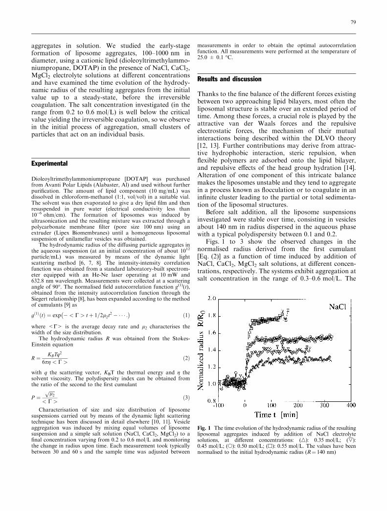
Figs. 1 to 3 show the observed changes in thenormalised radius derived from the first cumulant[Eq. (2)] as a function of time induced by addition ofNaCl, CaCl2, MgCl2 salt solutions, at different concen-trations, respectively. The systems exhibit aggregation atsalt concentration in the range of 0.3–0.6 mol/L. The
Fig. 1 The time evolution of the hydrodynamic radius of the resultingliposomal aggregates induced by addition of NaCl electrolytesolutions, at different concentrations: (n): 0.35 mol/L; (,):0.45 mol/L; (s): 0.50 mol/L; (h): 0.55 mol/L. The values have beennormalised to the initial hydrodynamic radius (R¼ 140 nm)
79

kinetic aggregate growth seems to be restricted to thisinterval, the aggregation behaviour being observedneither below nor above this interval of salt concentra-tion. At concentrations higher than this value, a rapidcoagulation occurs, resulting, at the end of the process, inan approximately complete phase separation.
In the systems investigated, in the presence of smallamount of added salt, the aggregation process starts withthe formation of structures of increasing size, but after aninitial stage, the rate of change of the radius continually
decreases, until an approximately steady-state is reachedand the aggregation ends. It is noteworthy that, after thisaggregation, the system undergoes a new stationarycondition with particles (or aggregates of particles) ofradius three or four times larger than their value at thebeginning of the process. Fig. 4 shows the ability ofmonovalent and divalent salts to promote liposomeaggregation in the condition of the experiment. As can beseen, divalent salts are able to induce larger aggregatesthan monovalent salts do, at the same molar concentra-tions. However, when the steady-state hydrodynamicradius is plotted as a function of the ionic strength, thesedifferences disappear and mono- and divalent saltsinduce the same aggregation effect. This observationconfirms that the important parameter in determiningthe salt-induced flocculation is the counterion concen-tration present in the aqueous phase, i.e., in this case, theconcentration of chloride ions that bind the cationicDOTAP. The reduction of the electrostatic repulsionbetween vesicles through binding counterions to the lipid
Fig. 2 The time evolution of the hydrodynamic radius of the resultingliposomal aggregates induced by addition of CaCl2 electrolytesolutions, at different concentrations: (s): 0.25 mol/L; (h):0.30 mol/L; (n): 0.35 mol/L; (,): 0.40 mol/L. The values have beennormalised to the initial hydrodynamic radius (R¼ 140 nm)
Fig. 3 The time evolution of the hydrodynamic radius of the resultingliposomal aggregates induced by addition of MgCl2 electrolytesolutions, at different concentrations: (s): 0.25 mol/L; (h): 0.30mol/L; (n): 0.40 mol/L; (,): 0.50 mol/L; (e): 0.60 mol/L. The valueshave been normalised to the initial hydrodynamic radius (R¼ 140 nm)
Fig. 4 The hydrodynamic radius, normalised to its initial value, of thesteady-state aggregates reached after the addition of different simplesalt electrolyte solutions: (s): NaCl; (h): CaCl2; (e): MgCl2. Data areplotted as a function of the molar concentration (A) and as a functionof the ionic strength of the electrolyte solution (B). The dotted linesare the second order polynomial best fit
80
surface induces aggregation and/or fusion, giving rise tostable structures in the same size range. This behaviourcan offer new possibilities in biomedical applicationsbecause the tendency of such liposomes to associate withone another, maintaining separate structures, can becontrolled by varying the ionic concentration of theappropriate counterion in the external medium.
In the usual salt-induced aggregation of colloidalsuspensions, the kinetics of aggregation is generallydescribed by dynamical scaling using the fractal mor-phology of the clusters [15, 16, 17]. Within this context,the average hydrodynamic radius of the aggregates in thediffusion limited cluster aggregation (DLCA) regimeobeys a power-law behaviourR(t)»R0t
z with z¼ 1/Df, theinverse of the fractal dimension of the clusters. Theexponent z characterises the aggregation mechanism.
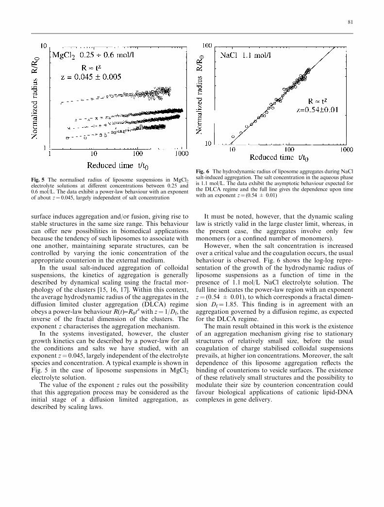
In the systems investigated, however, the clustergrowth kinetics can be described by a power-law for allthe conditions and salts we have studied, with anexponent z¼ 0.045, largely independent of the electrolytespecies and concentration. A typical example is shown inFig. 5 in the case of liposome suspensions in MgCl2electrolyte solution.
The value of the exponent z rules out the possibilitythat this aggregation process may be considered as theinitial stage of a diffusion limited aggregation, asdescribed by scaling laws.
It must be noted, however, that the dynamic scalinglaw is strictly valid in the large cluster limit, whereas, inthe present case, the aggregates involve only fewmonomers (or a confined number of monomers).
However, when the salt concentration is increasedover a critical value and the coagulation occurs, the usualbehaviour is observed. Fig. 6 shows the log-log repre-sentation of the growth of the hydrodynamic radius ofliposome suspensions as a function of time in thepresence of 1.1 mol/L NaCl electrolyte solution. Thefull line indicates the power-law region with an exponentz¼ (0.54 ± 0.01), to which corresponds a fractal dimen-sion Df¼ 1.85. This finding is in agreement with anaggregation governed by a diffusion regime, as expectedfor the DLCA regime.
The main result obtained in this work is the existenceof an aggregation mechanism giving rise to stationarystructures of relatively small size, before the usualcoagulation of charge stabilised colloidal suspensionsprevails, at higher ion concentrations. Moreover, the saltdependence of this liposome aggregation reflects thebinding of counterions to vesicle surfaces. The existenceof these relatively small structures and the possibility tomodulate their size by counterion concentration couldfavour biological applications of cationic lipid-DNAcomplexes in gene delivery.
Fig. 5 The normalised radius of liposome suspensions in MgCl2electrolyte solutions at different concentrations between 0.25 and0.6 mol/L. The data exhibit a power-law behaviour with an exponentof about z¼ 0.045, largely independent of salt concentration
Fig. 6 The hydrodynamic radius of liposome aggregates during NaClsalt-induced aggregation. The salt concentration in the aqueous phaseis 1.1 mol/L. The data exhibit the asymptotic behaviour expected forthe DLCA regime and the full line gives the dependence upon timewith an exponent z¼ (0.54 ± 0.01)
81

References
1. Lasic DD (1993) Liposomes: fromphysics to applications. Elsevier,Amsterdam
2. Hunter DG, Frisken BJ (1998) BiophysJ 74:2990–3002
3. Kreuter J (ed) (1994) Colloidal drugdelivery systems. Marcel Dekker, NewYork
4. Zuidam NJ, Barenholz Y (1998) Bio-chim Biophys Acta 1386:115–128
5. Pedroso de Lima MC, Simoes S, Fan-eca H, Duzgunes N (2001) Adv DrugDelivery Rew 47:277–294
6. Cumming HZ, Pusy PN (1977) In:Cummings ZH, Pike ER (eds), Photoncorrelation spectroscopy and veloci-metry. Plenum Press, New York
7. Chu B (1974) Laser light scattering.Plenum Press, New York
8. Pecora R (1985) Dynamic light scat-tering. Plenum Press, New York
9. Koppel DE (1972) J Chem Phys57:8414–8420
10. Hallet FR, Craig T, Marsh J, Nickel B(1989) Can J Spectrosc 34:63–70
11. Jin AJ, Huster D, Gawrisch K, NossalR (1999) Eur Biophys J 28:187–199
12. Verwey EJW, Overbeek JT (1948)Theory and stability of liophobic col-loids. Elsevier, Amsterdam
13. Ninham BW (1981) Pure Appl Chem53:2153–2147
14. Silvander M (1999) Structure and sta-bility of liposomes: interactions withmicelle-forming surfactants. UppsalaDissertation N. 455, Faculty of Scienceand Technology, Acta UniversitatisUpsaliensis, Upsala
15. Lin MY, Lindsay HM, Weitz AA,Klein R, Ball RC, Meakin P (1990)J Phys Cond. Matter 2:3093–3113
16. Cametti C, Codastefano P, Tartaglia P(1989) J Colloid Interface Sci 131:409–422
17. Schaefer DW, Martin JE, Wiltzius P,Cannel PS (1984) Phys Rev Lett52:2371–2374
82