trends and sources report on zoonotic agents in belgium most commonly reported zoonotic infections...
TRANSCRIPT

report on zoonotic agents in belgium
working group on foodborne infections and intoxications
trends and sources

• Federal Agency for the Safety of the Food Chain (FAVV-AFSCA)
• Scientific Institute of Public Health (WIV-ISP)
• Veterinary and Agrochemical Research Centre (CODA-CERVA)
3
Zoonoses are diseases or infections that are transmissible from animals to humans. The infection
of humans can be acquired directly from animals or indirectly through the ingestion of con-
taminated foodstuffs. It’s important that humans in contact with animals are aware of possible
transmission of a zoonotic disease and that consumers are informed about potential zoonotic
pathogens which can cause foodborne illness.
Surveillance of zoonoses remains an enormous task as well as an opportunity for all competent
authorities. Measures and systems of disease surveillance, diagnosis and control must be imple-
mented on a national level and have to be based on a suitable regulatory framework and an ap-
propriate level of funding. Active collaboration between all actors of the food chain, stakeholders,
industry, scientists, experts of the national reference laboratories and other laboratories, special-
ists of the competent authorities, technical committees have to bring together their expertise,
experiences, methods and findings. Only a collaborative approach and effective partnership at all
levels will achieve success to control zoonoses and to improve food safety.
The most commonly reported zoonotic infections in humans are those caused by bacterial
zoonotic agents that can be shed by asymptomatic farm animals. Since 2005 Campylobacteriosis
is the most frequently reported zoonotic disease in humans. Broiler and other poultry meat are an
important source of foodborne Campylobacter infections. Salmonellosis is the second most fre-
quently reported zoonotic disease in humans. The most important cause of foodborne outbreaks
(FBO’s) are Noroviruses followed by Campylobacter in 2008 and Salmonella in 2009 as the second
most important cause of foodborne outbreaks. The major sources of Salmonella in foodborne
outbreaks are table eggs, poultry meat and pig meat. Salmonella and Campylobacter reduction
remain an important task.
Bacterial diseasesBrucellosis
CampylobacteriosisEscherichia coli (VTEC)
LeptospirosisListeriosis
MRSAQ-fever
SalmonellosisTuberculosis
YersiniosisViral diseases
Avian influenzaHantaviruses
RabiesWest Nile virus
Parasitic diseasesCysticercosis
EchinococcosisSarcosporidiosis
ToxoplasmosisTrichinellosis
Prion diseasesFoodborne outbreaks
Executive summary
4
Table of contentsExecutive summary 3
Table of contents 4
Preface 7
Introduction 8
Belgian reference laboratories for zoonotic agents 11
Acronyms, abbreviations and special terms 15
General information 16
Susceptible human population 16
Susceptible animal populations 17
Animals slaughtered 2005 – 2009 20
bacterial diseases 22
Brucellosis 23
Zoonotic brucellosis 23
Brucellosis in cattle 24
Brucellosis in sheep and goats 26
Brucellosis in pigs 27
Brucellosis in humans 28
Campylobacteriosis 29
Campylobacter in food 30
Antimicrobial resistance in strains isolated
from meat and meat products 32
Campylobacter in humans 35
Escherichia coli (VTEC) infections 39
Verotoxin producing Escherichia coli 39
Verotoxin producing Escherichia coli in cattle 40
Escherichia coli O157 in food 40
Verotoxinogenic Escherichia coli in humans 41
Leptospirosis 43
Leptospirosis 43
Leptospirosis in animals 44
Leptospirosis in humans 44
Listeriosis 47
Listeriosis 47
Listeria monocytogenes in food 48
Listeria monocytogenes in humans 50
MRSA 53
MRSA 53
MRSA in pigs 54
MRSA in humans 54
Conclusions 55
Q-fever 57
Coxiella burnetii 57
Q-fever in animals 58
Q-fever in humans 60
Salmonellosis 63
Salmonella 63
Salmonella in animal feed 64
Salmonella in poultry 65
Salmonella in pigs 70
Salmonella in food (meat and meat products) 72
Salmonella in humans 77
Antimicrobial resistance 80
Tuberculosis 89
Zoonotic tuberculosis (Mycobacterium bovis) 89
Mycobacterium bovis in cattle 90
Mycobacterium bovis in humans 91
Human tuberculosis (Mycobacterium tuberculosis) 92

5
Yersiniosis 93
Yersinia enterocolitica 93
Yersinia enterocolitica in food 94
Yersiniosis in humans 94
viral diseases 98
Avian influenza 99
General 99
Monitoring in birds 100
Influenza in humans: monitoring 102
Hantaviruses 105
Hanta disease 105
Hantaviruses in animals 106
Rabies 111
Rabies 111
Rabies in animals 112
West Nile virus 115
West Nile virus 115
West Nile virus in animals 116
West Nile virus in humans 118
parasitic diseases 120
Cryptosporidiosis 121
Cryptosporidiosis 121
Cryptosporidiosis in animals 122
Cryptosporidiosis in humans 122
Cysticercosis 127
Cysticercosis 127
Cysticercosis in cattle 128
Echinococcosis 131
Echinococcosis 131
Echinococcus in food animals 132
Echinococcus in wildlife (foxes) 132
Echinococcus in humans 134
Sarcosporidiosis 135
Sarcosporidiosis 135
Sarcosporidiosis in animals 135
Toxoplasmosis 137
Toxoplasma gondii 137
Toxoplasmosis in animals 138
Toxoplasmosis in humans 138
Trichinellosis 141
Trichinella 141
Trichinella in food animals 142
Trichinella in wildlife 142
prion diseases 144
TSE 145
Transmissible spongiform encephalopathies 145
TSE in animals 146
TSE in humans 150
foodborne outbreaks 152
Foodborne outbreaks 153
Foodborne outbreaks in humans 154
Major etiological agents 154
Prevention of foodborne outbreaks 161

trends and sources

7
Preface
All European member states have the obligation to yearly submit an official report on the
monitoring of zoonoses and zoonotic agents to the European Food Safety Authority (EFSA)
based on article 9 of Directive 2003/99/EC of the European Parliament and the Council. In
that report all the relevant official monitoring programmes on animals in primary produc-
tion as well as on feed and food are presented. The report specifies all available data from
monitoring and research activities, as well as laboratory findings from the previous year and
includes results from antimicrobial susceptibility testing and FBOs.
Similarly, based on article 1 of Council Decision 2119/98/EC, data on zoonotic infections in
humans are officially reported each year to the European Centre for Disease Prevention and
Control (ECDC).
Based on these two official reports, the Federal Agency for the Safety of the Food Chain
(FASFC), together with the federal scientific institutions Veterinary and Agrochemical Re-
search Centre (VAR) and Scientific Institute of Public Health (IPH) agreed to publish a booklet
which contains this same information combined with data of previous years to indicate some
trends of diseases or sources of infection. This Trends and Sources report will focus especially
on the data of 2008 and on the data of 2009 as this report is a bi-annual edition. The aim of
this booklet is to inform professional readers as well as persons who have a general inter-
est in animal and human infections and in the safety of our food and at last but not least to
inform the consumer.
We hope that the reader will enjoy this seventh edition of the Belgian Trends and Sources
report on zoonotic agents.
Luc Vanholme
FAVV – AFSCA - FASFC
Hein Imberechts
CODA – CERVA - VAR
Geneviève Ducoffre
WIV – ISP - IPH
Katelijne Dierick
WIV – ISP - IPH
Preface
Introduction
Belgian reference laboratories for zoonotic agents
Acronyms, abbreviations and special terms
General information

8
Introduction
This report compiles especially the data of 2008 and 2009 and
compares these data to some data of previous years on zoonoses
and zoonotic agents, and is derived from the official documents
reported to EFSA and ECDC. For this reason, it is a unique docu-
ment in which laboratory results from the primary production,
from food, from feed and from clinical public health sources are
combined. In addition to the compulsory reporting on zoonoses
and zoonotic agents as listed in the European Directive 2003/99/
EC, this document contains data on other foodborne agents that
may be of interest to the reader, e.g. on avian influenza, transmis-
sible spongiform encephalopathies (TSE, e.g. mad cow disease) or
Norovirus infections. For the second time, the parasitic infection
cryptosporidiosis and the emerging disease West Nile Fever are
described.
Together with the general descriptive information on the diseases
or the infections themselves, their evolution over time, some rec-
ommendations on prevention of the infection are provided. This
booklet should meet the expectations of those concerned with
the possible (micro)biological contamination of our food.
The FASFC organises diverse monitoring and eradication pro-
grammes in, among others, the primary production and in the
transformation and distribution sectors. From their description
follows that much effort is being paid to control the contami-
nation of foodstuffs with pathogens. Some infectious diseases
have successfully been reduced or even eliminated (for instance
brucellosis, mad cow disease) and for others (for instance campy-
lobacteriosis) further programmes should be developed. In addi-
tion to the continuous effort from the authorities, the consumer
plays also an important role. Indeed, respect for the cold chain
and simple hygiene measures in the kitchen may be very efficient
in preventing foodborne contaminations and illness.
Most of the data in this report are from the following sources:
The Federal Agency for the Safety of the Food Chain (FASFC – •
FAVV - AFSCA);
The Scientific Institute of Public Health (IPH – WIV - ISP);•
The Veterinary and Agrochemical Research Centre (VAR – •
CODA - CERVA).
This report was coordinated by L. Vanholme (FASFC), H. Im-
berechts (VAR), K. Dierick and G. Ducoffre (IPH). Obviously, the
professional help of many other experts was needed to assemble
all available analytical data and information. The authors wish
to thank all those who collaborated actively in any way to this
Trends and Sources report, and especially (in alphabetical order):
S. Bertrand, National Reference Laboratory for Salmonella •
and Shigella, Bacterial Diseases Division , Scientific Institute of
Public Health;
K. Beulens, Ph. Heinen, J. Hooyberghs, C. Keppens, K. Ver-•
meersch, J. Wits, and J.-P. Maudoux, Control Policy Directorate,
Federal Agency for the Safety of the Food Chain;

9
N. Botteldoorn, National Reference Laboratory for foodborne •
outbreaks and antimicrobial resistance, Food Pathogens Divi-
sion, Scientific Institute of Public Health;
P. Butaye, Department of Bacteriology and Immunology, Veteri-•
nary and Agrochemical Research Centre;
Y. Carlier, National Reference Laboratory for Echinococcus, •
Laboratory of Parasitology, Faculty of Medicine, Université Libre
de Bruxelles;
L. Claes and P. Dorny, National Trichinella Reference Centre, •
Veterinary Department, Institute of Tropical Medicine Antwerp;
P. Cras, TSE humans, Department Neurology and Neuropathol-•
ogy, Faculty of Medicine, University of Antwerp;
S. Decraeye, National Reference Laboratory for Toxoplasmose, •
Food Pathogens Division, Scientific Institute of Public Health;
M. Delmée, UCL St-Luc and J. Verhaegen, UZ Leuven, National •
Reference Laboratory for Yersinia enterocolitica;
S. Denayer, National Reference Laboratory for foodborne out-•
breaks and antimicrobial resistance, Food Pathogens Division,
Scientific Institute of Public Health;
O. Denis, National Reference Laboratory for Staphylococcus •
aureus, Hôpital Erasme;
L. De Zutter, Department of Veterinary Public Health and Food •
Safety, Faculty of Veterinary Medicine, University of Ghent;
A. Dobly, Department of Bacteriology and Immunology, Veteri-•
nary and Agrochemical Research Centre;
J.-J. Dubois, Scientific secretariat of the Superior Health Council;•
M. Fauville-Dufaux, National Reference Laboratory for Tubercu-•
losis and Mycobacterium, Bacterial Diseases Division, Scientific
Institute of Public Health;
D. Fretin and M. Govaerts, National Reference Laboratory for •
Brucellosis, Laboratory of Bacterial diseases and Immunology,
Veterinary and Agrochemical Research Centre;
E. Goossens, Department of Bacteriology and Immunology, •
Veterinary and Agrochemical Research Centre;
M. Govaerts, Department of Bacteriology and Immunology, •
Veterinary and Agrochemical Research Centre;
P. Heyman, Research Laboratory for Vector-borne Diseases, •
Belgian Ministry of Defence, Queen Astrid Military Hospital,
Brussels;
Ph. Houdart, Crisis prevention and crisis management division, •
Federal Agency for the Safety of the Food Chain;

10
M. Lambert, A. Sevenants, K. Mennens, A. Scipioni and Ph. •
Dodion, Control Directorate, Federal Agency for the Safety of
the Food Chain;
C. Letellier, M. Dispas, Department of Virology, Veterinary and •
Agrochemical Research Centre;
A. Linden, Department of Bacteriology and Pathology of Bacte-•
rial diseases, Faculty of Veterinary Medicine, University of Liège;
J.-Y. Michelet, National Reference Laboratory for Biotoxins, Food •
Division, Scientific Institute of Public Health;
M. Naranjo, National Reference Laboratory for foodborne out-•
breaks and antimicrobial resistance, Food Pathogens Division,
Scientific Institute of Public Health;
D. Pierard, National Reference Laboratory for Enterohemor-•
rhagic Escherichia coli, Public Health, Microbiology Section, UZ
Brussels;
B. Pochet, Laboratories Directorate, Federal Agency for the •
Safety of the Food Chain;
S. Quoilin, Public Health and Surveillance Division, Scientific •
Institute of Public Health;
S. Roels, A.Dobly, Department of Pathology and Prionology, •
Veterinary and Agrochemical Research Centre;
E. Thiry, Department of Virology and Pathology of viral animal •
diseases, Faculty of Veterinary Medicine, University of Liège;
T. van den Berg, B. Lambrecht, O. Poncin, S. Marché and S. Van •
Borm, Department of Small Stock Pathology, Veterinary and
Agrochemical Research Centre;
M. Van Esbroeck, National Reference Centre for Coxiella bur-•
netii, Cryptosporidium, Leptospira and West Nile virus, Institute
of Tropical Medicine, Antwerp;
S. Van Gucht, National Reference Laboratory for Rabies, Viral •
Diseases Division, Scientific Institute of Public Health;
X. Van Huffel, Control Policy Directorate, Secretariat of the •
Scientific Committee, Federal Agency for the Safety of the Food
Chain;
O. Vandenberg, National Reference Laboratory for Campylo-•
bacter, Department of Microbiology, University Medical Center
St. Peter;
D. Vangeluwe and O. Poncin, Royal Institute of Natural Science;•
O. Vangeluwe, Museum of Natural Sciences;•
E. Vanopdenbosch and S. Roels, Department of Biocontrol, •
Veterinary and Agrochemical Research Centre;
M. Wanlin, Fondation contre les Affections Respiratoires et pour •
l’Education à la Santé, FARES – VRGT;
C. Wildemauwe, National Phage Typing Centre, Bacterial Diseas-•
es Division , Scientific Institute of Public Health;
F. Wuillaume, Public Heatlh and Surveillance Division, Scientific •
Institute of Public Health;
M. Yde, National Reference Laboratory for Listeria, Bacterial •
Diseases Division, Scientific Institute of Public Health.

11
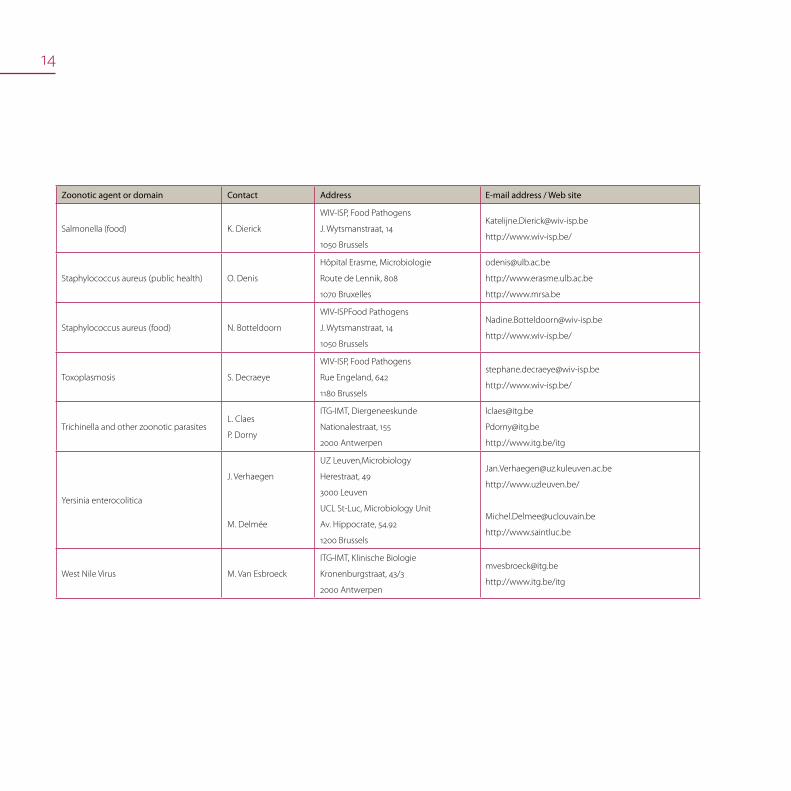
Belgian reference laboratories for zoonotic agents
Zoonotic agent or domain Contact Address E-mail address / Web site
Avian Influenza T. van den Berg
CODA-CERVA
Groeselenberg 99
1180 Brussels
http://www.var.fgov.be/
Biotoxins J.-Y. Michelet
WIV-ISPJ.
Wytsmanstraat 14
1050 Brussels
http://www.wiv-isp.be/
Brucella (public and animal health) D. Fretin
CODA-CERVA
Groeselenberg 99
1180 Brussels
http://www.var.fgov.be/
BSE / TSE S. Roels
CODA-CERVA
Groeselenberg 99
1180 Brussels
http://www.var.fgov.be/
Campylobacter (public health) O. Vandenberg
CHU St-Pierre
Microbiology Rue Haute, 322
1000 Brussels
http://www.stpierre-bru.be/
Campylobacter (food) N. Botteldoorn
WIV-ISP, Food Pathogens
J. Wytsmanstraat, 14
1050 Brussels
http://www.wiv-isp.be/
Clostridium botulinum Y. Fikri
WIV-ISP, Food Pathogens
Rue Engeland, 642
1180 Brussels
http://www.wiv-isp.be/
Cryptosporidium M. Van Esbroeck
ITG-IMT, Klinische Biologie
Kronenburgstraat, 43/3
2000 Antwerpen
http://www.itg.be/itg
Echinococcus multilocularis Y. Carlier
Fac. de Médecine U.L.B., Parasitologie
Route de Lennik, 808
1070 Brussels
http://www.ulb.ac.be/

12
Zoonotic agent or domain Contact Address E-mail address / Web site
Escherichia coli VTEC and EHEC
(public health)D. Pierard
UZ Brussel, Microbiology
Laarbeeklaan, 101
1090 Brussels
http://www.uzbrussel.be/
Escherichia coli VTEC and EHEC
(animal health)H. Imberechts
CODA-CERVA
Groeselenberg, 99
1180 Brussels
http://www.var.fgov.be/
Escherichia coli VTEC and EHEC (food) S. Denayer
WIV-ISP, Food Pathogens
J. Wytsmanstraat, 14
1050 Brussels
http://www.wiv-isp.be/
Foodborne outbreaks
and Food Microbiology
K. Dierick
N. Botteldoorn
WIV-ISP, Food Pathogens
J. Wytsmanstraat, 14
1050 Brussels
http://www.wiv-isp.be/
Hantavirus P. Heyman
Queen Astrid Military Hospital
Bruynstraat, 2
1120 Brussels
http://www.smd.be/rlvbd
Human influenza I. Thomas
WIV-ISP, Viral diseases
J. Wytsmanstraat, 14
1050 Brussels
http://www.wiv-isp.be/flu
Leptospira M. Van Esbroeck
ITG-IMT, Klinische Biologie
Kronenburgstraat, 43/3
2000 Antwerpen
http://www.itg.be/itg
Listeria monocytogenes (public health) M. Yde
WIV-ISP, Bacterial diseases
J. Wytsmanstraat, 14
1050 Brussels
http://www.wiv-isp.be/
Listeria monocytogenes (food) N. Botteldoorn
WIV-ISP, Food Pathogens
J. Wytsmanstraat, 14
1050 Brussels
http://www.wiv-isp.be/

13
Zoonotic agent or domain Contact Address E-mail address / Web site
Mycobacterium
M. Fauville-Dufaux
(public health)
F. Portaels
(public health)
M. Govaerts
(animal health)
WIV-ISP, Bacterial diseases
Rue Engeland, 642
1180 Brussels
ITG-IMT, Mycobacteriology
Nationalestraat, 155
2000 Antwerpen
CODA-CERVA
Groeselenberg, 99
1180 Brussels
http://www.wiv-isp.be/
http://www.itg.be/itg
http://www.var.fgov.be/
Phage typing centre (Salmonella, Staphy-
lococcus)C. Wildemauwe
WIV-ISP, Bacterial diseases
Rue Engeland, 642
1180 Brussels
http://www.wiv-isp.be/
Q-Fever (Coxiella burnetii)
M. Van Esbroeck
D. Fretin
ITG-IMT, Klinische Biologie
Kronenburgstraat, 43/3
2000 Antwerpen
CODA-CERVA
Groeselenberg, 99
1180 Brussels
http://www.itg.be/itg
http://www.var.fgov.be/
Rabies S. Van Gucht
WIV-ISP, Viral diseases
Rue Engeland, 642
1180 Brussels
http://www.wiv-isp.be/
Salmonella (public health) S. Bertrand
WIV-ISP, Bacterial diseases
Rue J. Wytsman, 14
1050 Brussels
http://www.wiv-isp.be/
Salmonella (animal health) H. Imberechts
CODA-CERVA
Groeselenberg, 99
1180 Brussels
http://www.var.fgov.be/
14
Zoonotic agent or domain Contact Address E-mail address / Web site
Salmonella (food) K. Dierick
WIV-ISP, Food Pathogens
J. Wytsmanstraat, 14
1050 Brussels
http://www.wiv-isp.be/
Staphylococcus aureus (public health) O. Denis
Hôpital Erasme, Microbiologie
Route de Lennik, 808
1070 Bruxelles
http://www.erasme.ulb.ac.be
http://www.mrsa.be
Staphylococcus aureus (food) N. Botteldoorn
WIV-ISPFood Pathogens
J. Wytsmanstraat, 14
1050 Brussels
http://www.wiv-isp.be/
Toxoplasmosis S. Decraeye
WIV-ISP, Food Pathogens
Rue Engeland, 642
1180 Brussels
http://www.wiv-isp.be/
Trichinella and other zoonotic parasitesL. Claes
P. Dorny
ITG-IMT, Diergeneeskunde
Nationalestraat, 155
2000 Antwerpen
http://www.itg.be/itg
Yersinia enterocolitica
J. Verhaegen
M. Delmée
UZ Leuven,Microbiology
Herestraat, 49
3000 Leuven
UCL St-Luc, Microbiology Unit
Av. Hippocrate, 54.92
1200 Brussels
http://www.uzleuven.be/
http://www.saintluc.be
West Nile Virus M. Van Esbroeck
ITG-IMT, Klinische Biologie
Kronenburgstraat, 43/3
2000 Antwerpen
http://www.itg.be/itg

15
Acronyms, abbreviations and special terms
ADNS Animal Disease Notification System
ARSIAAssociation Régionale de Santé et d’Identification
Animales, Walloon Regional Animal Health Association
BAPCOC Belgian Antibiotic Policy Coordination Committee
CFT Complement Fixation Test
CLSI Clinical and Laboratory Standards Institute
CODA – CERVA - VAR
Veterinary and Agrochemical Research Centre
Centrum voor Onderzoek in Diergeneeskunde en
Agrochemie
Centre d’Etude et de Recherches Vétérinaires et
Agrochimiques
CRL Community Reference Laboratory
DGZ VlaanderenDierengezondheidzorg Vlaanderen,
Flanders Regional Animal Health Association
ECDC European Centre for Disease Prevention and Control
EFSA European Food Safety Authority
ELISA Enzyme Linked Immuno Sorbent Assay
FASFC - FAVV – AFSCA
Federal Agency for the Safety of the Food Chain
Federaal Agentschap voor de Veiligheid van de
Voedselketen
Agence Fédérale pour la Sécurité de la Chaîne
alimentaire
FBO Foodborne outbreak
FPSFederal Public Service (Ministry) – Public Health, Food
Chain Security and Environment
IFA Indirect Fluorescent Antibody assay
IFN Interferon
IPH - WIV - ISP
Scientific Institute of Public Health
Wetenschappelijk Instituut Volksgezondheid
Institut Scientifique de Santé Publique
ITM - ITG – IMT
Institute of Tropical Medicine
Instituut voor Tropische Geneeskunde
Institut de Médecine Tropicale
MIRU-VNTRMycobacterial Interspersed Repetitive Units –
Variable Number of Tandem Repeats
NRCSSNational Reference Centre for
Salmonella and Shigella, IPH
NRL National Reference Laboratory
OIE World Organisation for Animal Health
PCR Polymerase Chain Reaction
RBT Rose Bengal Test
RFLP Restriction Fragment Length Polymorphism
RT PCR Reverse Transcriptase Polymerase Chain Reaction
SanitelRegistration and Identification
Database of farm animals
SAT Slow Agglutination Test
SFM French Society for Microbiology
WHO World Health Organization

16
General information
Susceptible human populationThe evolution of the total human population in Belgium from 2004 to 2009 is shown in table 1
Evolution in the total human population 2005-2009 Table1.
Source: National statistical authority, http://statbel.fgov.be/
2005 2006 2007 2008 2009
Total 10.445.852 10.511.382 10.584.534 10.666.866 10.750.000
Male 5.111.325 5.143.821 5.181.408 5.224.309
Female 5.334.527 5.367.561 5.403.126 5.442.557
Brussels 1.006.749 1.018.804 1.031.215 1.048.491
Flanders 6.043.161 6.078.600 6.117.440 6.161.600
Wallonia 3.395.942 3.413.978 3.435.879 3.456.775
Foreigners 870.862 900.473 932.161 971.448
All figures on human population 2009 are not available at the
finalisation date of this report due to the implementation of a
new internal data exchange procedure and the use of a new IT
application for data exchange of the statistical authority of FPS
Economy, S.M.E.’s, Self-employed and Energy. Only a global total
figure for 2009 is available.
The total number of immigrants in Belgium is important. For-
eigners are found in four typical areas: former coal basins, cities,
border regions and some concentrations in the triangle between
Brussels, Antwerp and Ghent, or in the triangle between Brussels,
Mons and Namur. Immigration and tourism may play an impor-
tant role in the introduction and the transmission of zoonotic or
other infectious diseases.
17
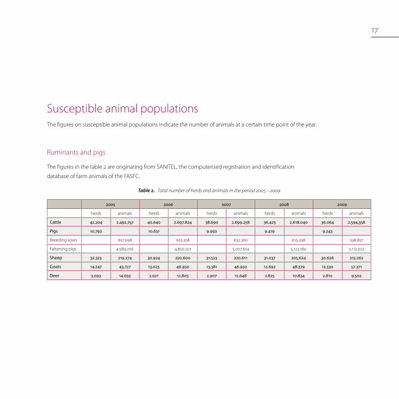
Susceptible animal populationsThe figures on susceptible animal populations indicate the number of animals at a certain time point of the year.
Ruminants and pigs
The figures in the table 2 are originating from SANITEL, the computerised registration and identification
database of farm animals of the FASFC.
Total number of herds and animals in the period 2005 – 2009Table2.
2005 2006 2007 2008 2009
herds animals herds animals herds animals herds animals herds animals
Cattle 42.204 2.492.757 40.640 2.697.824 38.690 2.699.258 36.423 2.618.040 36.064 2.594.358
Pigs 10.792 10.631 9.950 9.419 9.243
Breeding sows 657.998 653.358 632.360 615.298 598.857
Fattening pigs 4.989.016 4.850.501 5.007.614 5.123.189 5.113.202
Sheep 32.323 219.274 30.924 220.600 31.523 220.611 31.037 205.624 30.626 215.262
Goats 14.247 43.727 13.025 46.950 13.381 46.950 12.692 48.379 12.530 57.371
Deer 3.093 14.655 2.021 12.805 2.907 12.648 2.825 10.834 2.810 9.502

18
Evolution of the total number of pig herds, period 2005–2009Figure1. Evolution of the total number of breeding pigs, period 2005–2009Figure2.
Evolution of the total number of fattening pigs, period 2005–2009Figure3.
Evolution of the total number of cattle herds, period 2005–2009Figure4. Evolution of the total number of bovines, period 2005–2009Figure5.
Next figures represent the evolution of total number of respectively prcine and bovine herds and animals over last years.
19
Poultry
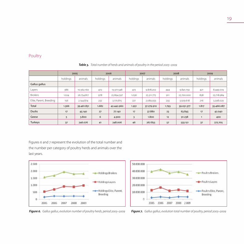
Total number of herds and animals of poultry in the period 2005–2009Table3.
2005 2006 2007 2008 2009
holdings animals holdings animals holdings animals holdings animals holdings animals
Gallus gallus
Layers 386 10.562.160 472 13.377.548 472 9.878.202 444 9.841.759 421 8.449.074
Broilers 1.024 26.754.817 978 25.894.597 1.036 25.311.775 911 25.700.000 838 23.718.984
Elite, Parent, Breeding 156 2.144.874 232 3.170.815 221 2.089.933 233 3.509.618 216 3.298.029
Total 1.566 39.461.851 1.682 42.442.960 1.951 37.279.910 1.793 39.051.377 1.817 35.466.087
Ducks 17 45.140 27 77.140 17 37.880 23 63.845 17 42.040
Geese 5 3.800 6 4.900 3 1.800 12 91.238 1 400
Turkeys 37 246.076 41 248.006 46 267.855 57 533.151 37 272.705
Figures 6 and 7 represent the evolution of the total number and
the number per category of poultry herds and animals over the
last years.
Gallus gallus, evolution number of poultry herds, period 2005–2009Figure6. Gallus gallus, evolution total number of poultry, period 2005–2009Figure7.

20
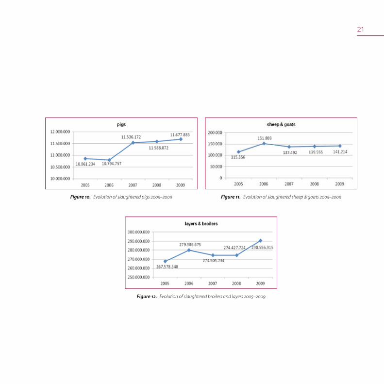
Animals slaughtered 2005 – 2009Number of slaughtered animals in the period 2005–2009Table4.
2005 2006 2007 2008 2009
Cattle 523.795 496.181 495.492 522.557 480.068
Calves 313.115 327.467 306.961 301.102 319.188
Pigs 10.861.234 10.794.757 11.536.172 11.588.072 11.677.883
Solipeds 11.542 10.728 10.064 9.173 8.910
Sheep 112.771 148.767 133.192 135.071
Goats 2.585 3.036 6363 6143
115.356 151.803 137.492 139.555 141.214
Broiler 237.670.666 247.721.072 0 242231046 262935369
Layer 29.907.674 32.265.603 0 32.196.678 27.621.546
267.578.340 279.986.675 274505734 274427724 290556915
Figures 8 to 12 represent the evolution of the total number of slaughtered bovines, porcines, solipeds, sheep & goats and broilers &
layers over the last years.
Evolution of slaughtered cattle 2005–2009Figure8. Evolution of slaughtered calves 2005–2009Figure9.
21
Evolution of slaughtered pigs 2005–2009Figure10. Evolution of slaughtered sheep & goats 2005–2009Figure11.
Evolution of slaughtered broilers and layers 2005–2009Figure12.
trends and sources
bacterial diseases

23
BrucellosisDavid Fretin, Luc Vanholme
Zoonotic brucellosisBrucellosis is an infectious disease caused by bacterial species of
the genus Brucella. Most species have a specific animal reservoir
that can cause human disease: B. abortus in cattle, B. melitensis in
sheep and goats, B. suis in pigs and B. canis in dogs. Transmission
occurs through contact with infected animals, contaminated
animal tissue or through ingestion of contaminated products.
In people brucellosis is characterised by flu-like symptoms such
as fever, headache, back pain and physical weakness. Nocturnal
sweating is frequently observed. Infections of the central nervous
system or lining of the heart may occur.
In the non-”officially brucellosis free” Mediterranean countries, •
the consumption of raw milk or raw cheese from sheep and
goats is thought to be the major source of contamination (B.
melitensis).
In Northern Europe, besides some occupational human cases •
of B. abortus infections, the majority of brucellosis cases are
imported and are mainly caused by B. melitensis.
Zoonotic brucellosis
Brucellosis in cattle
Brucellosis in sheep and goats
Brucellosis in pigs
Brucellosis in humans

24
Brucellosis in cattle
Belgium is officially free from bovine brucellosis since the 25th of
June 2003 (Commission Decision 2003/467/EC establishing the
official tuberculosis, brucellosis and enzootic-bovine-leucosis-free
status of certain Member states and regions of Member states as
regards bovine herds).
Vaccination has been prohibited in Belgium since 1992.
An intensified bovine brucellosis eradication programme started
in Belgium in 1988. In case of active brucellosis, i.e. excretion of
Brucella, the plan consisted in the culling of all animals of the
infected herd (total depopulation), the slaughtered animals were
compensated based on the replacement value.
The annual herd prevalence notified was in 1988 was 1.13% and
has fallen below 0.01% since 1998. On 27th March 2000, the last
case of bovine brucellosis was identified. No infected herd was
detected in Belgium since then.
Surveillance programme and methods used
Since Belgium obtained an official brucellosis free status, the
eradication programme has been replaced by a surveillance
programme.
Dairy cattle herds are checked at least 4 times a year via tank milk
control. Tank milk is examined by means of the milk ring test. If
tank milk is positive, all individual animals of the herd older than
2 years are tested serologically. Beef cattle older than 2 years are
serologically monitored once every three years.
The herds are selected on the basis of geographical localisation.
Furthermore, all female animals older than 1 year and breeding
bulls are serologically tested at purchase. Each abortion or prema-
ture birth in animals at risk is subject to compulsory notification
to the FASFC and testing for brucellosis is obligatory. Aborting
females should be kept in isolation until the results of the investi-
gation exclude Brucella infections.
Blood sera are analysed by micro-agglutination as screening test;
in case of a positive result, an indirect ELISA test is performed as
confirmatory test. Bacteriological examination is done in case of
serological and/or epidemiological suspicion. An animal is legally
suspected of brucellosis in case of a positive ELISA. If, according to
the epidemiology an animal or herd is found to be at risk, a bac-
teriological investigation always takes place. Hence, a brucellosis
animal is defined as an animal in which Brucella has been isolated
and a cattle herd is considered as infected if one of its animals is
positive for brucellosis by culture.
Table 5 indicates the evolution of the total individual serological
tests related to the monitoring programme of beef cattle and the
mandatory examination at purchase. The evolution of the total
number of bulk milk tests is in line with the continuous decrease
in number of dairy herds over the last years.
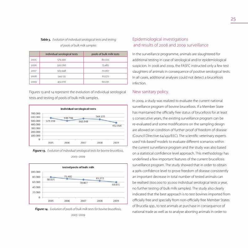
25
Evolution of individual serological tests for bovine brucellosis, Figure13.
2005–2009
Evolution of pools of bulk milk tests for bovine brucellosis, Figure14.
2005–2009
Evolution of individual serological tests and testing Table5.
of pools of bulk milk samples
individual serological tests pools of bulk milk tests
2005 579.390 80.025
2006 500.766 73.482
2007 563.948 70.067
2008 544.135 65.572
2009 452.016 60.031
Figures 13 and 14 represent the evolution of individual serological
tests and testing of pools of bulk milk samples.
Epidemiological investigations and results of 2008 and 2009 surveillance
In the surveillance programme, animals are slaughtered for
additional testing in case of serological and/or epidemiological
suspicion. In 2008 and 2009, the FASFC instructed only a few test
slaughters of animals in consequence of positive serological tests.
In all cases, additional analyses could not detect a brucellosis
infection.
New sanitary policy.
In 2009, a study was realized to evaluate the current national
surveillance program of bovine brucellosis. If a Member State
has maintained the officially free status of brucellosis for at least
5 consecutive years, the existing surveillance program can be
re-evaluated and some modifications on the sampling design
are allowed on condition of further proof of freedom of disease
(Council Directive 64/432/EEC). The scientific veterinary experts
used ‘risk-based’ models to evaluate different scenarios within
the current surveillance program and the study was also based
on a statistical confidence level approach. This methodology has
underlined a few important features of the current brucellosis
surveillance program. The study showed that in order to obtain
a 99% confidence level to prove freedom of disease consistently
an important decrease in total number of tested animals can
be realised (600.000 to 30.000 individual serological tests a year,
no further testing of bulk milk samples). The study also clearly
indicated that the best approach is to test bovines imported from
officially free and specially from non-officially free Member States
of Brucella spp., to test animals at purchase in consequence of
national trade as well as to analyse aborting animals in order to

26
Number of notified abortions, 2005–2009Figure15.
early detect (re-)infection. Regarding the passive surveillance
(abortions), the study indicated that there is a need to increase
the number of analysed abortions. In consequence of this study
and an opinion of the Scientific Committee of the FASFC, a
new surveillance program will be applied for winter screening
2009–2010 starting mid November 2009.
For more details on the study and the scientific advices see:
CCVD report “Evaluation of the Belgian surveillance program •
for bovine brucellosis and leucosis”. S. Welby, Y. Van der Stede, C.
Letellier, D. Fretin, J.-Y. Houtain, M. Lomba, S. Stoop, M. Van-
robaeys, L. Van Schoubroeck, J. Hooyberghs, L. Vanholme, L.
Lengelé, G. Lamsens, E. Pottie.
Advice 26-2009 of the Scientific Committee of the FASFC on •
the evaluation of an alleviated brucellosis and leucosis surveil-
lance program and on propositions for a new surveillance
program of other bovine diseases.
Addendum of Advice 26-2009 of the Scientific Committee of •
the FASFC
Advice 05-2010 of the Scientific Committee of the FASFC on the •
new surveillance policy of animal diseases – part cattle (other
matrices), small ruminants and pigs.
Advice 10-2010 of the Scientific Committee of the FASFC on the •
new surveillance policy of animal diseases – part poultry and
solipeds.
Regarding the passive surveillance by examination of abortions,
next figure 15 represents the total number of notified abortions in
the period 2005 – 2009.
Brucellosis in sheep and goatsBelgium is official free for sheep and goat brucellosis (B. melitensis)
since 29 March 2001 (Commission Decision 2001/292/EC amend-
ing Decision 93/52/EEC recording the compliance by certain
Member States or regions with the requirements relating to
brucellosis (Brucella melitensis) and according them the status of
a Member State or region officially free of the disease).
Surveillance programme
Serum samples taken in the framework of national monitoring for
Visna-Maedi and at export were examined for Brucella melitensis
specific antibodies by means of ELISA (about 5% of the sheep and
goats population should be tested by legal provisions). Positive
samples were subsequently tested with Rose Bengal test (RBT)
and Complement Fixation test (CFT). A sample is classified as
positive for brucellosis only if it is positive in all three tests.

27
Number of tests of Brucella melitensis, 2005 – 2009Figure16.
Brucellosis in pigs
Surveillance programme in pigs and epidemiological investigations
Serological screening for Brucella is done in breeding pigs that
are brought together (e.g. at a fair), at artificial insemination
centres or in animals intended for trade. The methods used are
Rose Bengal test (RBT), Slow Agglutination test (SAT) according
to Wright, CFT and ELISA. Bacteriological examination for Brucella
and Yersinia is done in case of positive serology.
Sometimes, false positive serological reactions are reported.
These are due to a Yersinia enterocolitica O:9 infection and are
confirmed by Yersinia spp. isolation in the absence of Brucella
spp. isolation.
The domestic pig population is free of brucellosis (last Brucella
isolation in pigs in Belgium was in 1969). In 2007, 2008 and 2009
all samples were negative.
Since 2001, yearly serum samples from sheep and goats were
tested at the NRL. In addition, serum samples from sheep for
export were analysed. In 2008 and 2009, respectively 3.375 and
2.321 samples were tested. Serological positive reacting animals
after serial and repeated testing were finally negative. The NRL
has confirmed infections of Yersinia enterocolitica O:9 in sheep.
Those infections are associated with false positive serology in the
tests ELISA, RBT and possibly CFT of brucellosis. The phenomenon
of FPSR (false positive serological reactors) as known for bovines is
also observed in sheep.
Next figure 16 represents the total number of tests realised by the
NRL for Brucella melitensis.

28
Brucellosis in humansThe last indigenous case of Brucella was reported in 1997. It is
helpful to note that B. suis biovar 2, the only known biovar circu-
lating in Belgium among wild boars, shows only limited patho-
genicity for humans, if pathogenic at all.
In 2007, the NRL confirmed two cases of Brucella melitensis biovar
2 and one case of Brucella melitensis biovar 3. The country of origin
of these three imported cases was not known.
In 2008, a suspected case was finally negative by culture.
In 2009, the NRL confirmed one imported case of Brucella
melitensis biovar 1 on a patient from Lebanon where brucellosis is
endemic.
29
CampylobacteriosisKatrien Beullens, Katelijne Dierick, Geneviève Ducoffre, Olivier Vandenberg, Luc Vanholme
Campylobacteriosis
Campylobacteriosis continued to be the most commonly
reported gastrointestinal bacterial pathogen in humans in
Belgium since 2005. Campylobacteriosis in humans is caused by
thermotolerant Campylobacter spp. Typically, the infective dose
of these bacteria is low. The species most commonly associated
with human infection are C. jejuni followed by C. coli and C. lari,
but other Campylobacter species are also known to cause human
infections.
The incubation period in humans averages from two to five
days. Patients may experience mild to severe illness, with general
clinical symptoms including watery, often bloody diarrhea, ab-
dominal pain, fever, headache and nausea. Usually, infections are
self-limiting and last only a few days. Infrequently, complications
as reactive arthritis and neurological disorders occur. C. jejuni has
become the most recognised cause of Guillain-Barré syndrome,
a polio-like form of paralysis that can result in respiratory and
severe neurological dysfunction and even death.
Campylobacteriosis
Campylobacter in food
Antimicrobial resistance in strains isolated from meat and meat products
Campylobacter in humans

30
Thermotolerant Campylobacter spp. are widespread in nature.
The principal reservoir is the alimentary tract of wild and do-
mesticated birds and mammals. Thermotolerant Campylobacter
spp. are prevalent in food animals such as poultry, cattle, pigs
and sheep; in pets, including cats and dogs; in wild birds and in
environmental water sources. Animals are mostly asymptomatic
carriers.
The bacteria can contaminate various foodstuffs, including meat,
raw milk and dairy products, and less frequently fish, fishery prod-
ucts and fresh vegetables. Contact with live poultry, consumption
of undercooked poultry meat, drinking water from untreated
water sources, and contact with pets have been identified as
important sources of infection.
The contamination of poultry carcasses and meat with Campy-
lobacter is monitored by the FASFC since 2000. The incidence of
positive poultry samples is high and remains stable. Poultry meat
has to be well cooked before consumption and cross-contamina-
tion should be avoided during preparation.
Campylobacter in food
Monitoring programme
In 2008 and 2009, a monitoring programme in Belgian slaughter-
houses, meat cutting plants, processing plants and retail trades
representative of the Belgian production of poultry carcasses and
meat, pork carcasses and minced meat of all species was realised
by the FASFC. In addition, samples from raw milk cheese and live
bivalve molluscs were also analysed.
Specially trained staff of the FASFC performed the sampling. Dif-
ferent sample sizes (25g, 0.01g and 600 cm²) were analysed and in
some cases an enumeration was performed. For broiler carcasses
at slaughter and cutting meat at processing plants, independent
samples were taken per matrix in order to estimate prevalence of
the contamination at 95% confidence level.
Results of the 2008-2009 monitoring
The results of the monitoring of the FASFC are shown in the next
table 6.

31
Zoonosis monitoring programme – Campylobacter in foodTable6.
Sample Sample sizePercentage of positive
samples 2008
Percentage of positive
samples 2009
Broiler
Carcasses at slaughter 25g (caeca) 53.8% (n=292) 31% (n=337)
Carcasses at slaughter 1g 33% (n=185) 32% (n=261)
Carcasses at retail 0.01g 19.1% (n=115) 25.4% (n=118)
Meat cuts (skinned or with skin) at processing plant 1g 7.3% (n=523) 8.6% (n=513)
Layer
Carcasses at slaughter 25g (caeca) 96% (n=76) -
Carcasses at slaughter 1g 27% (n=37) 37% (n=317)
Carcasses at retail 0.01g - 15% (n=60)
Poultry
Meat cuts (with skin) at retail 0.01g 7.0% (n=57) 21.7% (n=92)
Meat cuts (without skin) at retail 0.01g 0.0% (n=69) 0.0% (n=91)
Minced meat at retail Enumeration (M=100 cfu/g) 2.2% (n=183) 0.0% (n=71)
Meat preparation at processing plant 0.01g 1.6% (n=123) 0.0% (n=112)
Meat preparation at retail Enumeration (M=100 cfu/g) 0.0% (n=94) 0.0% (n=100)
Pork
Carcasses at slaughter 600 cm² 16.6% (n=500) 14% (n=656)
Minced meat (intended to be eaten raw) at retail (all species) Enumeration (M=10 cfu/g) 0.0% (n=129) 0.0% (n=184)
Minced meat (intended to be eaten cooked) at retail (all species) Enumeration (M=100 cfu/g) 0.0% (n=125) 0.0% (n=183)
Raw milk cheese at retail 25g 0.0% (n=46) 0.0% (n=49)
Raw milk cheese at farm 25g 0.0% (n=45) 0.0% (n=40)
Live bivalve molluscs at retail 25g 0.0% (n=61) 0.0% (n=94)
The contamination rate of pork carcasses raised until 2006 and remained stable later on. In 2007, the contamination rate decreased a
bit to increase again in 2008 and to decrease in 2009. From 2008 on, the Campylobacter spp. contamination has been enumerated. The
results show a low level of contamination.
Evolution of the Campylobacter prevalence of pork carcasses 2005-2009Table7.
Sampling level 2004 2005 2006 2007 2008 2009
Pork Carcasses 600 cm² 4.9% 7.2% 13.4% 12.2% 16.6% 14.0%

32
Antimicrobial resistance in strains isolated from meat and meat products
Surveillance programme and method used
In 2008 and 2009, respectively 484 and 461 Campylobacter
strains, were isolated in the zoonoses monitoring program.
Isolates originating from poultry (carcasses of broilers, filets, meat
preparations, turkey and carcasses of spent hens) and pork were
examined for antimicrobial susceptibility by the NRL. Compared
to 2007, the number of analysed isolates has more than doubled.
Seventy-five and 77 strains were respectively isolated from pork
meat or carcasses in 2008 and 2009, while 402 and 407 poultry-
derived isolates (broiler meat or carcasses, spent hens and turkey)
were analysed. C. coli was the most prevalent strain isolated from
pork carcasses (90%/71% in 2008/2009), while for poultry meat
C. jejuni was the most isolated Campylobacter strain (78%/72% in
2008/2009), followed by C. coli (21%/28% in 2008/2009).
Minimum Inhibitory Concentrations (MIC) were determined by
using E®-test on blood agar plates. The antimicrobials tested and
the breakpoints (following the CLSI standards) used are listed in
the following table 8.
Campylobacter in meat and meat products: list of antimicrobials Table8.
tested and breakpoints used
Antimicrobial Breakpoints (µg / ml)
C. jejuni C. coli
Ampicillin 16 16
Tetracycline 2 2
Nalidixic acid 16 32
Ciprofloxacin 1 1
Erythromycin 4 16
Gentamicin 1 2
The percentage of resistant strains of Campylobacter in meat is
reported in the next table 9.
Antimicrobial susceptibility testing of Campylobacter in food: Table9.
percentage of resistant strains
Poultry meat Pork
C. jejuni C. coli C. coli
2008
(n=313)
2009
(n=292)
2008
(n=86)
2009
(n=115)
2008
(n=70)
2009
(n=55)
Tetracycline 44% 45% 87% 64% 86% 73%
Ciprofloxacin 43% 41% 81% 71% 31% 36%
Nalidixic acid 46% 45% 86% 69% 31% 36%
Gentamicin 20% 25% 10% 23% 23% 26%
Erythromycin 8% 12% 14% 20% 21% 20%
Ampicillin 29% 46% 29% 30% 13% 18%

33
Antimicrobial resistance in Campylobacter from poultry meat
In total, 402 and 407 Campylobacter strains were isolated from
poultry in 2008 and 2009, respectively, and tested for antimicro-
bial susceptibility (313/86-C. jejuni/C. coli in 2008 and 292/115-C.
jejuni/C. coli in 2009). In 2008 and 2009, 20% and 17% of all
Campylobacter strains, respectively, were sensitive to all tested
antibiotics.
In 2008, tetracycline and nalidixic acid resistance were most
dominantly present (54%), followed closely by resistance to
ciprofloxacin (52%). When comparing different food matrices
investigated, it is clear that Campylobacter isolates from the more
processed meat matrices showed higher antibiotic resistance.
For example, isolates from minced poultry meat showed a high
ampicillin resistance (62.5%), and isolates from poultry meat
preparations had high resistance to tetracycline (92%), while both
matrices contained little or no isolates sensitive to all antibiotics
tested. On the other hand, 40% of all isolates from spent hens
were sensitive to all antibiotics tested. In 2009, resistance profiles
were similar: respectively 52%, 50% and 50% of the tested strains
were resistant to nalidixic acid, ciprofloxacin and tetracycline.
Overall, antibiotic resistance was more prevalent in C. coli than
in C. jejuni (see graph), with in 2008 and 2009 only 3 and 8 strains
sensitive to all antibiotics, respectively. The number of multire-
sistant strains, resistant to three or more antibiotics, significantly
decreased from 80% in 2008 to 61% in 2009. This trend was also
observed for resistance to ciprofloxacin (81%/71%-2008/2009),
nalidixic acid (86%/69%-2008/2009) and tetracycline (87%/64%-
2008/2009), but remains high. For C. jejuni, 25% and 21% of all
strains were sensitive to all antibiotics tested, while 38% and 41%
were resistant to three or more antibiotics in 2008 and 2009, re-
spectively. High prevalence of resistance was observed for nalidix-
ic acid (46%/45%-2008/2009), tetracycline (44%/45%-2008/2009)
and ciprofloxacin (43%/41%-2008/2009). Resistance of C. jejuni to
ampicillin increased from 36% in 2008 to 46% in 2009.
Compared to previous years, general resistance has increased (see
figures 17 and 18), mostly due to adaptation of the breakpoint val-
ues used to assess resistance. This is most obvious for gentamicin.
0
20
40
60
80
100
AMP TET NAL CIP ERY GEN
A. Evolution of the percentage resistant C. jejuni strains in poultry meat
2004 2005 2006 2007 2008 2009
Evolution of the percentage resistant Campylobacter jejuni strains Figure17.
in poultry
34
Evolution of the percentage resistant Campylobacter coli strains Figure18.
in poultry
0
20
40
60
80
100
AMP TET NAL CIP ERY GEN
B. Evolution of the percentage resistanct C. coli strains in poultry meat
2004 2005 2006 2007 2008 2009
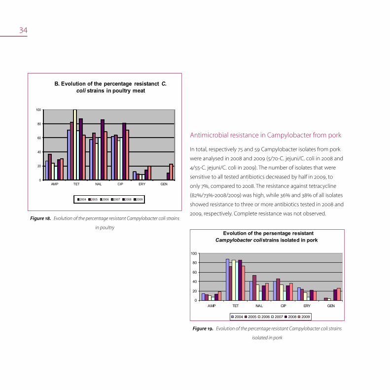
Antimicrobial resistance in Campylobacter from pork
In total, respectively 75 and 59 Campylobacter isolates from pork
were analysed in 2008 and 2009 (5/70-C. jejuni/C. coli in 2008 and
4/55-C. jejuni/C. coli in 2009). The number of isolates that were
sensitive to all tested antibiotics decreased by half in 2009, to
only 7%, compared to 2008. The resistance against tetracycline
(82%/73%-2008/2009) was high, while 36% and 38% of all isolates
showed resistance to three or more antibiotics tested in 2008 and
2009, respectively. Complete resistance was not observed.
Evolution of the percentage resistant Campylobacter coli strains Figure19.
isolated in pork
Evolution of the persentage resistantCampylobacter colistrains isolated in pork
0
20
40
60
80
100
AMP TET NAL CIP ERY GEN
2004 2005 2006 2007 2008 2009
35
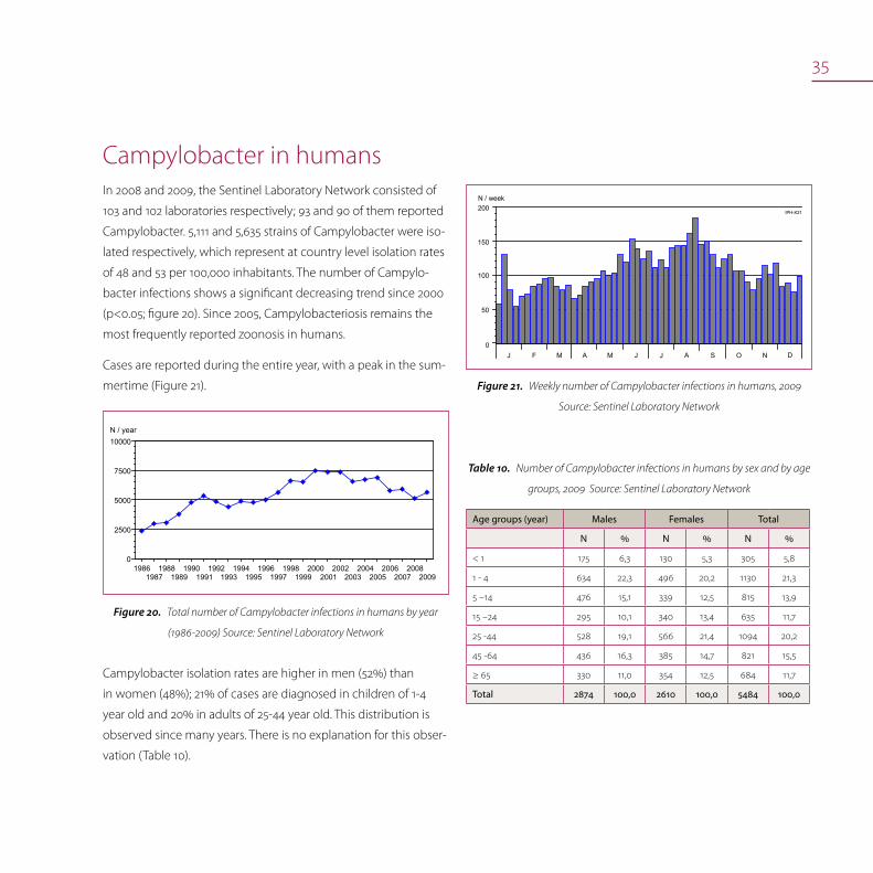
Campylobacter in humansIn 2008 and 2009, the Sentinel Laboratory Network consisted of
103 and 102 laboratories respectively; 93 and 90 of them reported
Campylobacter. 5,111 and 5,635 strains of Campylobacter were iso-
lated respectively, which represent at country level isolation rates
of 48 and 53 per 100,000 inhabitants. The number of Campylo-
bacter infections shows a significant decreasing trend since 2000
(p<0.05; figure 20). Since 2005, Campylobacteriosis remains the
most frequently reported zoonosis in humans.
Cases are reported during the entire year, with a peak in the sum-
mertime (Figure 21).
19861987
19881989
19901991
19921993
19941995
19961997
19981999
20002001
20022003
20042005
20062007
20082009
0
2500
5000
7500
10000N / year
Total number of Campylobacter infections in humans by year Figure20.
(1986-2009) Source: Sentinel Laboratory Network
N / week
J F M A M J AJ S O N D
IPH-K21
0
100
200
150
50
Weekly number of Campylobacter infections in humans, 2009 Figure21.
Source: Sentinel Laboratory Network
Campylobacter isolation rates are higher in men (52%) than
in women (48%); 21% of cases are diagnosed in children of 1-4
year old and 20% in adults of 25-44 year old. This distribution is
observed since many years. There is no explanation for this obser-
vation (Table 10).
Number of Campylobacter infections in humans by sex and by age Table10.
groups, 2009 Source: Sentinel Laboratory Network
Age groups (year) Males Females Total
N % N % N %
< 1 175 6,3 130 5,3 305 5,8
1 - 4 634 22,3 496 20,2 1130 21,3
5 –14 476 15,1 339 12,5 815 13,9
15 –24 295 10,1 340 13,4 635 11,7
25 -44 528 19,1 566 21,4 1094 20,2
45 -64 436 16,3 385 14,7 821 15,5
≥ 65 330 11,0 354 12,5 684 11,7
Total 2874 100,0 2610 100,0 5484 100,0

36
Since the beginning of the registration (1983), the incidence in
Flanders is higher than in Wallonia. This was confirmed in 2009
with an estimated incidence of 61/100,000 inhabitants in Flanders,
42/100,000 inhabitants in Wallonia and 34/100,000 inhabitants
in Brussels-Capital Region. The incidence is very high in a few
districts since many years and also in 2009:
134/100,000 inhabitants in Mouscron, 122/100,000 inhabitants
in Eeklo, 109/100,000 inhabitants in Mechelen and 109/100,000
inhabitants in Leuven (Figure xx). It would be useful to make a
study to further explore the reasons for the observed difference
in occurrence of Campylobacteriose between districts in Belgium.
Incidence of Campylobacter infections in humans by district (N/105 inhab., 2009) Figure22.
Source: Sentinel Laboratory Network
AL AR AT
AW B
BG BS CR DK DM DN EK GT HS HV HY
IP KR LG LV MC MH MN MR MS NC NM NV OD OS PV RS SG SN TG TH TL TN TR VR VT VV WR
: Aalst : Arlon : Ath : Antwerpen : Brussels : Brugge : Bastogne : Charleroi : Diksmuide : Dendermonde : Dinant : Eeklo : Gent : Hasselt : Halle-Vilvoorde : Huy : Ieper : Kortrijk : Liège : Leuven : Mouscron : Mechelen : Mons : Marche-en-Fam. : Maaseik : Neufchâteau : Namur : Nivelles : Oudenaarde : Oostende : Philippeville : Roeselare : Soignies : St.-Niklaas : Tongeren : Turnhout : Tielt : Thuin : Tournai : Veurne : Virton : Verviers : Waremme
> 75.00
> 0.00 - 25.00
Incidence/100,000 inhabitants[3.73 - 134.43]
> 25.00 - 50.00
> 50.00 - 75.0052.83
IPH-K21
VR
BG
DK TLGT
SN
ALOD
MH
HV
LV
AWTH
MS
HS
TG
NV
MRDN
CR
NC
AR
MC TR
KR
SG
IP
TN PV
VT
BS
VVLG
HY
WR
TG
MN NM
B
DM
EK
RS
AT
OS

37
Epidemiology Enteric Campylobacter
Data were obtained from the NRL for human Enteric Campy-
lobacter. Since there is not obligation for clinical laboratories
to send human isolates to the NRL to confirm the presence of
Campylobacter, a correct epidemiological situation of Campylo-
bacter in human populations cannot be made.
Data about human Campylobacter cases were only obtained
from two laboratories deserving the Brugmann, Queen Fabiola,
Bordet and St.-Peter University Hospitals located in Brussels.
Methods used
From January 2008 to December 2009, a total of 18,737 stool
specimens from 12,084 patients submitted to the NRL were
routinely examined for Campylobacter spp. using one selective
medium and a filtration method.
Results of surveillance
By the application of this comprehensive isolation procedure,
enteric Campylobacter was isolated in 631 patients, giving a
prevalence of Campylobacter infection of 5.2%. Similar reported
prevalence has been reported from other European countries.
Antimicrobial resistance of human isolates
Methods used
A total of 2447 human Campylobacter strains isolated in two
laboratories serving the Brugmann, Queen Fabiola, Bordet and
St-Peter University Hospitals located in Brussels, Belgium from
January 2001 to 2009 were examined for their resistance.
Three antibiotics of therapeutic interest were tested in disk diffu-
sion according to Kirby-Bauer, following SFM recommendations.
Results of 2009 surveillance
In 2009, resistance was mostly found to ampicillin (53.6%), and to
ciprofloxacin (39.7%). Most Campylobacter were susceptible to
erythromycin (87.1%). These results point out the need to monitor
antibiotic resistance in Campylobacter from human and food
(Table 11).
Resistance of Campylobacter in Belgium fecal isolates, trend from Table11.
2001 till 2009, based on data from the NRL (Laboratory for Microbiology,
St.-Peter University Hospital, Brussels)
Antim
icrobial A
gent
2001
2002
2003
2004
2005
2006
2007
2008
2009
N = 280
N = 266
N = 212
N = 291
N = 260
N = 246
N = 263
N = 284
N = 345
% Resistant
% Resistant
% Resistant
% Resistant
% Resistant
% Resistant
% Resistant
% Resistant
% Resistant
Ampicillin 13,9 15,8 16,0 29,2 32,3 51,6 57,4 50,7 53,6
Erythromycin 3,2 0,0 9,4 6,5 6,9 8,1 5,2 3,5 2,9
Ciprofloxacin 18,9 22,6 24,5 28,1 33,1 50,4 49,8 40,8 39,7
(Sources: Trends of antimicrobial resistance of Campylobacter strains isolated in two
laboratories serving the Brugmann, Queen Fabiola, Bordet and St.-Peter University Hos-
pitals located in Brussels, Belgium from January 2001 to 2009. Antimicrobial susceptibility
testing was realized by disk diffusion methods following the SFM recommendations).

38
Invasive Campylobacter in humans
In 2009, the NRL for Campylobacter confirmed 28 invasive
Campylobacter isolates. Among these, C. fetus was recovered in
15 patients. The other strains were C. jejuni and C. coli in 11 and 2
cases respectively (Table 12).
Repartition by biotype of invasive Campylobacter in humans, 2009 Table12.
Source: NRL
N Campylobacter jejuni C. coli C. fetus
I II III IV I II subsp.fetus
28 10 1 1 1 15

39
Verotoxin producing Escherichia coli
Verotoxin producing Escherichia coli in cattle
Escherichia coli O157 in food
Verotoxinogenic Escherichia coli in humans
Escherichia coli (VTEC) infectionsKatrien Beullens, Hein Imberechts, Denis Pierard, Luc Vanholme
Verotoxin producing Escherichia coli
Verotoxigenic Escherichia coli (VTEC) is a group of E. coli character-
ised by the ability to produce ‘verocytotoxins’ or ‘shiga like toxins’.
Human pathogenic VTEC usually have additional virulence factors
that are important for the development of disease in man and are
called EHEC (enterohemorrhagic E.coli). EHEC infections in man
are usually associated with a minor number of O:H serogroups. Of
these, the O157:H7 or the O157:H- serogroup (EHEC O157) are the
ones most frequently reported to be associated with the human
disease. Other pathogenic serotypes of E. coli, e.g. O26, O91, O103,
O111 and O145 may also be involved.
Human EHEC infections are mostly sporadic. Human infection
may occur after consumption of contaminated food or water,
after contact with contaminated water, or by direct transmission
from person to person or through contact with infected animals.
The clinical symptoms range from mild to bloody diarrhoea
through haemorraghic colitis, which is often accompanied by ab-
dominal cramps, usually without fever. VTEC infections can result
in haemolytic uremic syndrome (HUS), characterised by acute
renal failure, anaemia and lowered platelet counts. HUS develops
in up to 10% of patients infected with E. coli O157 and is the lead-
ing cause of acute renal failure in young children.

40
Animals are a reservoir for VTEC, and VTEC have been isolated
from many different animal species. The gastrointestinal tract of
healthy ruminants seems to be the foremost important reservoir
for VTEC. Cattle are the principal reservoir of VTEC. The organ-
ism is excreted in the faeces. Food of bovine origin is frequently
reported as a source for human VTEC infections. Other important
food sources include faecally contaminated vegetables and drink-
ing water.
Prevention mainly relies on bio-security measures at farm-level
and hygienic measures at the level of the slaughterhouses. Since
August 2005, the sampling of cattle at farms that had sent E. coli
O157 positive animals to the abattoir is not compulsory any more.
Since then, epidemiological investigations and additional sam-
pling at the farm of origin is only done following EHEC infections
in humans.
In Belgium, approximately 50 to100 sporadic human cases are
registered per year.
Verotoxin producing Escherichia coli in cattleSurveillance programme, measures and methods used
No surveillance programme was in place in 2008 or 2009. In case
a cattle farm is suspected to be at the origin of an EHEC outbreak
in humans, a representative number of animals this farm is sam-
pled (faecal, feed, environmental samples). FASFC officials inform
the owner that EHEC circulate on his farm and encourage the
implementation of hygienic measures, i.e. cleaning and disinfec-
tion of milk reservoirs and milking equipment, and cleaning of
animals before transport to the slaughterhouse.
The ISO 16654:2001method was used for isolation of E. coli O157.
Briefly, the samples are enriched in mTSB with novobiocin and
treated by immunomagnetic separation. Subsequently, the
suspected colonies on CT-SMAC are latex agglutinated for the de-
tection of E. coli O157. Confirmation of serotype (O group) is done
by means of slow tube agglutination after heating of the bacterial
cultures. Virulence factors are determined by PCR for toxin genes
stx1 and stx2 and for eae (intimin). Enterohemolysis is done on
appropriate culture media.
Epidemiological investigations and results of 2008-2009 surveillance
In 2008 and 2009, no cattle farms were sampled to identify the
source of an E. coli O157 outbreak. Only one bovine VTEC strain
was sent to the NRL (animal health) for typing, with pathotype
stx1 eae.
Escherichia coli O157 in foodMonitoring programme
E. coli O157 was analysed in diverse beef and dairy products.
Notification is mandatory since March 2004 (Ministerial Decree on
mandatory notification in the food chain). For enterohemorrhagic
E. coli, absence in 25g in ready-to-eat food products put on the
market is mandatory.

41
Results of the 2008-2009 monitoring
The results of the monitoring by the FASFC are shown in the
following table 13.
Zoonosis monitoring programme - E. coli O157, 2008 -2009Table13.
Sample Prevalence 2008 Prevalence 2009
Beef
Carcasses 0.7% (n=1353 1.0% (n=995)
Fresh meat at cutting plant 0.0% (n=766) 0.0% (n=291)
Minced meat (steak tartare)
at processing - 0.0% (n=294)
Minced meat (steak tartare)
at retail 0.6% (n=159) 0.0% (n=147)
Meat preparations (steak
tartare with herbs and sauce)
at retail
0.0% (n=155) 1.0% (n=147)
Cheese
From raw cow’s milk, at farm 0.0% (n=35) 0.0% (n=59)
From raw cow’s milk, at retail 0.0% (n=95) 0.0% (n=106)
From raw milk, at processing 0.0% (n=21) 0.0% (n=40)
From raw sheep’s milk,
at farm 0.0% (n=18) 0.0% (n=15)
From raw sheep’s milk,
at retail 0.0% (n=44) 0.0% (n=63)
From raw goat’s milk, at farm 0.0% (n=18) 4.0% (n=25)
From raw goat’s milk, at retail 0.0% (n=50) 0.0% (n=64)
ButterAt farm 0.0% (n=121) 0.0% (n=132)
From raw milk, at retail 0.0% (n=48) 0.0% (n=24)
Cream At farm 0.0% (n=51) 0.0% (n=46)
Verotoxinogenic Escherichia coli in humansOnly few clinical laboratories examine human stools for the
presence of E. coli O157. Therefore, a correct incidence of VTEC in
human populations cannot be given.
In 2008, the NRL confirmed 103 verotoxigenic E coli isolated from
102 patients. Among these:
73 typical VTEC isolates, positive for two factors of additional •
virulence: the presence of the gene eae (intimin) gene and
enterohemolysin (EHEC virulence plasmid) gene.
30 atypical VTEC isolates, negative for intimin and/or •
enterohemolysin.
In 2009, the NRL confirmed 112 verotoxigenic E coli isolated from
110 patients. Among these:
86 typical VTEC isolates, positive for two factors of additional •
virulence: the presence of the gene eae (intimin) gene and
enterohemolysin (EHEC virulence plasmid) gene.
26 atypical VTEC isolates, negative for intimin and/or enterohe-•
molysin.
The number of isolates confirmed annually by the NRL is increas-
ing but probably still corresponds to a large rate of underdiagno-
sis (Table 14 on next page).

42
E. coli: evolution in number of isolates in humans, 1998-2009. Table14.
Source: NRL
1998
1999
2000
2001
2002
2003
2004
2005
2006
2007
2008
2009
Number of isolates 48 53 47 46 46 47 45 47 46 47 103 112
Number of typical isolates 38 46 33 36 37 40 36 36 36 40 73 86
Number of O157 isolates 25 33 26 29 26 21 29 27 21 25 55 61
In 2008, 11 strains (all from serotype O157:H7) were associated with
haemolytic uremic syndrome (HUS). Six patients were less than 5
years old (of which one was infected with two strains, see below),
three were older children (respectively 6, 7 and 11 years old) and
two were adults (55 and 59 years old). These last patients were
involved in a small outbreak of bloody diarrhea in a psychiatric
hospital, related to ground pork meat (that was later shown by
PCR to contain also ground beef ). These isolates were atypical
since they were urease positive; PFGE analysis showed that 9
other urease positive O157 isolated from the same province in the
same period as well as two other provinces were indistinguisha-
ble, but no common source could be identified. According to the
information available at the NRL, all other cases were not related.
In 2009, 15 strains from 14 patients – 9 of serotype O157:H7, 3
O157:H-, 1 O103, 1 O111 and 1 not typable - were associated with
haemolytic uremic syndrome (HUS), comprising 7 children less
than 6 years old, 6 older children (6 to 13 years old), one 48 years
old adult and one patient with unknown age. According to the
information available at the NRL, all these cases were unrelated.
In conclusion, the number of O157 isolates has more than dou-
bled in 2008 and increased slightly in 2009. No explanation was
found for this fact.

43
Leptospirosis
Leptospirosis in animals
Leptospirosis in humans
LeptospirosisJean-Jacques Dubois, Geneviève Ducoffre, Els Goossens, Marjan Van Esbroeck
Leptospirosis
Leptospirosis or Weill’s disease is a disease caused by Leptospira
interrogans sensu lato, which is divided into more than 26 sero-
groups and more than 230 serovars. This aerobic mobile spiro-
chete is able to survive for short times outside the host in a warm
and humid environment (stagnant water, muddy soils). It endures
mostly and for longer periods in host reservoirs, mainly rodents.
After infection, a short bacteraemia is followed by the invasion of
mainly kidneys and liver, in which leptospires can survive for years
and can be intermittently excreted. Accidental hosts, infected
through contact with contaminated water or soil (or by contact
with infected animals) can develop mild flu-like symptoms. Fatal
subacute kidney and or liver failure can also occur.

44
Laboratory tests
The standard serological test to detect leptospirosis is the mi-
croscopic agglutination test, which is sensitive and specific and
allows a first identification at serogroup level. This test requires
however the maintenance of a panel of reference strains, so only
a limited number of laboratories are able to perform this assay.
Other serological tests used are rapid agglutination tests and
ELISA’s, which perform well for a rapid detection, but are less
specific. Isolation of the antigen is very difficult and laborious.
Antigen detection is possible by immunofluorescence tech-
niques and molecular techniques, but do not allow a typing at
serogroup or serovar level.
Leptospirosis in animalsData from the NRL indicate that in 2008-2009 Leptospira infec-
tions are only seldom diagnosed. The microscopic agglutina-
tion test of 429 cattle and 606 pig sera demonstrated about 10
samples positive in each animal species. However, in 371 horse
sera tested, antibodies against serovars Australis, Pyrogenes and
Grippotyphosa were found in 10,0%, 4.6% and 4.0% of the cases,
respectively.
Leptospirosis in humansLeptospirosis occurs worldwide but is most common in tropi-
cal and subtropical areas with high rainfall. The disease is found
mainly wherever humans come into contact with the urine of
infected animals or a urine-polluted environment.
Animal-human transmission occurs through direct contact with
urine of a natural host via a wound, the mucous membrane of the
mouth, the nose or the eyes or indirectly by contaminated water
or food. The longer the exposure the higher the risk of infection.
Human to human transmission is possible but extremely rare. The
incubation period is usually 6 to 12 days with a range of 2 to 30
days.
The professions at highest risk of acquiring the disease are sew-
age workers, but also farmers, veterinarians, slaughterhouse staff,
garbage collectors, etc. Certain hobbies can also lead to con-
tamination: persons involved in water sports such as swimming,
kayaking, diving, surfing, fishing and (wind)surfing are at risk.
This disease is mainly observed at the end of summer and during
autumn.
In Belgium, the number of human cases remain so far limited.
Clinical manifestations
Leptospirosis may induce a wide variety of clinical manifestations.
Symptoms are divided into 4 main clinical categories:
moderate influenza-like complaints•
Weil syndrome with jaundice and renal failure•
meningitis, encephalitis•
difficult breathing, including coughs and breathlessness.•
Classically this disease has two phases with an abrupt beginning
marked by a high fever (≥ 40°C), shivers and muscle ache for ap-
proximately one week.

45
After a recovery period of one to three days without symptoms a
second phase follows with multiple problems of internal organs
(e.g. kidney, liver). Generally the disease has a favourable outcome
if treatment is started quickly after the onset of disease.
Diagnosis
Diagnosis is based on clinical symptoms, risk factors and labora-
tory analyses. Bacterial culture is difficult and takes a long time.
Serology is done for diagnosis. Five to ten days after onset of
symptoms, antibodies against the leptospires can be detected in
the blood. A negative result at the beginning of an infection does
not exclude a diagnosis of leptospirosis.
Requests for analysis or confirmation of a screening result can be
sent to the NRL. It is recommended to analyse paired samples
taken with an interval of one or two weeks.
Treatment
Leptospirosis is treated by antibiotics. The earlier the treatment,
the fewer symptoms and complications the patient will have.
Therefore, it is recommended not to wait for laboratory results
before starting treatment. With appropriate antibiotics a full
recovery can be expected about a month after the start of treat-
ment.
Prevention
There is no vaccine against leptospirosis available. Professionals
with risk of leptospirosis are recommended to wear watertight
glasses, gloves, boots and clothing.
The following measures could be used in the prevention and
control of leptospirosis:
to increase awareness of the disease among the population, •
risk groups and health care providers;
to avoid contact with animal urine, infected animals or an •
infected environment;
to wear protective clothing;•
to wash the hands after any contact with a contaminated •
animal or object;
to cover all injuries and wounds with waterproof dressings •
before contact with contaminated freshwater or humid envi-
ronment;
not to swim or do any water sports in contaminated water;•
to stop access of rodents into housing by obstructing possible •
entrances;
to remove all rubbish and to keep areas around human habita-•
tions clean;
not to leave food around, especially in recreational areas where •
rats may be present.
Results of the 2008 surveillance
In the Institute of Tropical Medicine (NRL), a total of 596 human
sera have been examined for the presence of antibodies to Lept-
ospira by the microscopic agglutination technique (MAT).
Five confirmed cases have been diagnosed. All patients were
men. The age of the patients ranged from 21 to 66 years with a
median age of 42 years. Four patients were exposed in Belgium
(3) or France (1), one patient contracted the disease during or

46
after a stay in a (sub)tropical region (Kenia). Three patients were
exposed to water and/or rats, one patient was exposed to a horse
with leptospirosis. This infection goes back to October 2007 but
the diagnosis was made one year later. The way of contracting
the disease is not known for one patient. All infections were diag-
nosed between June and November.
Results of the 2009 surveillance
In 2009, a total of 579 human sera have been examined.
Eight confirmed cases have been diagnosed. All patients were
men. The age of the patients ranged from 25 to 66 years with a
median age of 46 years. Five patients were exposed in Belgium (4)
or France (1), 3 patients contracted the disease during or after a
stay in a (sub)tropical region (Indonesia, Thailand, Congo). Seven
patients were exposed to water and/or rats. The exposition of the
patient who came back from Indonesia is not known. All but one
infections were diagnosed between August and December.
47
Listeriosis
Listeria monocytogenes in food
Listeria monocytogenes in humans
ListeriosisKatrien Beullens, Geneviève Ducoffre, Marc Yde
ListeriosisThe bacterial genus Listeria currently comprises six species, but
human cases of listeriosis are almost exclusively caused by the
species Listeria monocytogenes. Listeriae are ubiquitous organ-
isms that are widely distributed in the environment, especially in
plant matter and soil. The principal reservoirs of Listeria are soil,
forage and water. Other reservoirs include infected domestic and
wild animals. The main route of transmission to both humans and
animals is believed to be through consumption of contaminated
food or feed. However, infection can also be transmitted directly
from infected animals to humans as well as between humans.
Cooking kills Listeria, but the bacteria are known to multiply at
temperatures down to 4°C, which makes the occurrence in ready-
to-eat foods with a relatively long shelf life of particular concern.
In humans severe illness mainly occurs in the unborn child,
infants, the elderly and those with compromised immune sys-
tems. Symptoms vary, ranging from mild flu-like symptoms and
diarrhea to life threatening infections characterised by septicemia
and meningoencephalitis. In pregnant women the infection can
spread to the foetus resulting in foetal death with abortion or
neonatal infection. Illness is often severe and mortality is high.
Human infections are rare yet important given the high mortality
rate associated with them. These organisms are among the most
important causes of death from foodborne infections in industri-
alized countries.

48
In domestic animals, especially cattle, sheep and goats, clinical
symptoms of listeriosis are usually encephalitis, abortion, mastitis
or septicaemia. However, animals may also be asymptomatic
intestinal carriers and shed the organism in significant numbers,
contaminating the environment.
General food hygiene rules are essential for the prevention of hu-
man listeriosis. As some persons are at high risk (pregnant wom-
en, the eldery, immuno-compromised people), they are advised
not to eat certain categories of food with proven elevated risk of
Li. monocytogenes contamination, such as unpasteurized milk and
butter, soft cheeses and ice cream made from unpasteurized milk,
any soft cheese crust, smoked fish, pâté, cooked ham, ‘rillettes’,
salami, cooked meat in jelly, raw minced meat from beef, pork
and poultry, steak tartar, raw fish and shellfish (oysters, mussels,
shrimps), fish, meat and surimi salads, insufficiently rinsed raw
vegetables, and unpeeled fruit. People should be made aware of
the considerable risk of infection by consuming ready-to-eat food
products.
Listeria monocytogenes in food
Monitoring programme
The matrices for Listeria monocytogenes analyses were diverse
products of beef, pork, dairy products, fish and ready-to-eat
products. Notification is mandatory since March 2004 (Ministerial
Decree on mandatory notification in the food chain). For Listeria
monocytogenes in ready-to-eat products put on the market, a
maximum limit of 100 cfu/g is set.
Results of the 2008-2009 monitoring
Zoonosis monitoring programme - Listeria monocytogenes in food, Table15.
2008-2009
Sample
Qua
ntity
analysed
Percen
tage
of p
ositi
ve
sam
ple
s 20
08
Percen
tage
of p
ositi
ve
sam
ple
s 20
09
Beef
Minced meat at retail
intended to be eaten
raw (steak tartare)
Enumeration
(M=100 cfu/g)0.0% (n=158)
0.0% (n=
98)
Meat preparation at re-
tail intended to be eaten
raw (steak tartare with
herbs and sauce)
Enumeration
(M=100 cfu/g)0.0% (n=160) 0.0% (n=98)
Pork
Cooked ham at process-
ing plant 25g 7.4% (n=54) 2.7% (n=37)
Cooked ham at retail Enumeration
(M=100 cfu/g)0.0% (n=59) 0.0% (n=76)
Raw ham at processing
plant
25g or enumeration
(M=100 cfu/g) (*)4.4% (n=159) 3.1% (n=96)
Raw ham at retail Enumeration
(M=100 cfu/g) 0.0% (n=33) 0.0% (n=34)
Pâté at processing plant 25g 1.7% (n=58) 0.0% (n=38)
Pâté at retail Enumeration
(M=100 cfu/g)0.0% (n=57) 0.0% (n=84)
White pudding at
processing plant 25g 5.3% (n=38) 2.4% (n=41)
White pudding at retail Enumeration
(M=100 cfu/g)0.0% (n=77) 0.0% (n=76)
Sausages at processing
plant
25g or enumeration
(M=100 cfu/g) (*)7.0% (n=157)
10.3%
(n=185)
Sausages at retail Enumeration
(M=100 cfu/g) 0.0% (n=34) 0.0% (n=69)
Mea
t
(uns
peci
fied)
Minced meat intended
to be eaten raw, at retail
Enumeration
(M=100 cfu/g)0.0% (n=161)
0.0%
(n=200)Minced meat intended
to be eaten raw, at
processing plant
25g or enumeration
(M=100 cfu/g) (*)- 13.8% (n=72)
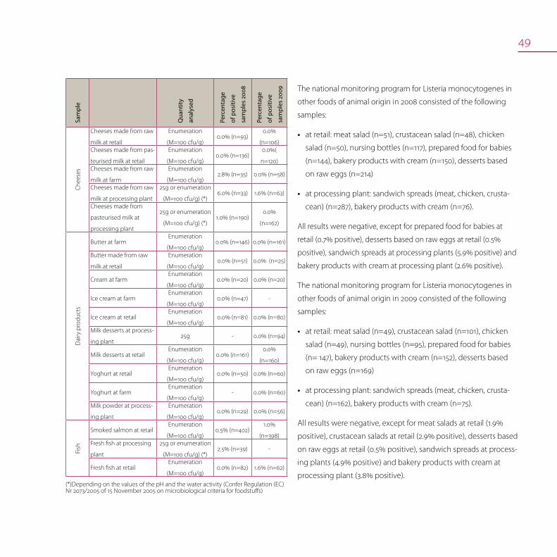
49
Sample
Qua
ntity
analysed
Percen
tage
of p
ositi
ve
sam
ple
s 20
08
Percen
tage
of p
ositi
ve
sam
ple
s 20
09
Che
eses
Cheeses made from raw
milk at retail
Enumeration
(M=100 cfu/g)0.0% (n=93)
0.0%
(n=106)Cheeses made from pas-
teurised milk at retail
Enumeration
(M=100 cfu/g)0.0% (n=136)
0.0%(
n=120)Cheeses made from raw
milk at farm
Enumeration
(M=100 cfu/g)2.8% (n=35) 0.0% (n=58)
Cheeses made from raw
milk at processing plant
25g or enumeration
(M=100 cfu/g) (*)6.0% (n=33) 1.6% (n=63)
Cheeses made from
pasteurised milk at
processing plant
25g or enumeration
(M=100 cfu/g) (*)1.0% (n=190)
0.0%
(n=162)
Dai
ry p
rodu
cts
Butter at farm Enumeration
(M=100 cfu/g)0.0% (n=146) 0.0% (n=161)
Butter made from raw
milk at retail
Enumeration
(M=100 cfu/g)0.0% (n=51) 0.0% (n=25)
Cream at farm Enumeration
(M=100 cfu/g)0.0% (n=20) 0.0% (n=20)
Ice cream at farm Enumeration
(M=100 cfu/g)0.0% (n=47) -
Ice cream at retail Enumeration
(M=100 cfu/g)0.0% (n=81) 0.0% (n=80)
Milk desserts at process-
ing plant25g - 0.0% (n=94)
Milk desserts at retail Enumeration
(M=100 cfu/g)0.0% (n=161)
0.0%
(n=160)
Yoghurt at retail Enumeration
(M=100 cfu/g)0.0% (n=50) 0.0% (n=60)
Yoghurt at farm Enumeration
(M=100 cfu/g)- 0.0% (n=60)
Milk powder at process-
ing plant
Enumeration
(M=100 cfu/g)0.0% (n=29) 0.0% (n=56)
Fish
Smoked salmon at retail Enumeration
(M=100 cfu/g)0.5% (n=402)
1.0%
(n=398)Fresh fish at processing
plant
25g or enumeration
(M=100 cfu/g) (*)2.5% (n=39) -
Fresh fish at retail Enumeration
(M=100 cfu/g)0.0% (n=82) 1.6% (n=62)
(*)Depending on the values of the pH and the water activity (Confer Regulation (EC) Nr 2073/2005 of 15 November 2005 on microbiological criteria for foodstuffs)
The national monitoring program for Listeria monocytogenes in
other foods of animal origin in 2008 consisted of the following
samples:
at retail: meat salad (n=51), crustacean salad (n=48), chicken •
salad (n=50), nursing bottles (n=117), prepared food for babies
(n=144), bakery products with cream (n=150), desserts based
on raw eggs (n=214)
at processing plant: sandwich spreads (meat, chicken, crusta-•
cean) (n=287), bakery products with cream (n=76).
All results were negative, except for prepared food for babies at
retail (0.7% positive), desserts based on raw eggs at retail (0.5%
positive), sandwich spreads at processing plants (5.9% positive) and
bakery products with cream at processing plant (2.6% positive).
The national monitoring program for Listeria monocytogenes in
other foods of animal origin in 2009 consisted of the following
samples:
at retail: meat salad (n=49), crustacean salad (n=101), chicken •
salad (n=49), nursing bottles (n=95), prepared food for babies
(n= 147), bakery products with cream (n=152), desserts based
on raw eggs (n=169)
at processing plant: sandwich spreads (meat, chicken, crusta-•
cean) (n=162), bakery products with cream (n=75).
All results were negative, except for meat salads at retail (1.9%
positive), crustacean salads at retail (2.9% positive), desserts based
on raw eggs at retail (0.5% positive), sandwich spreads at process-
ing plants (4.9% positive) and bakery products with cream at
processing plant (3.8% positive).
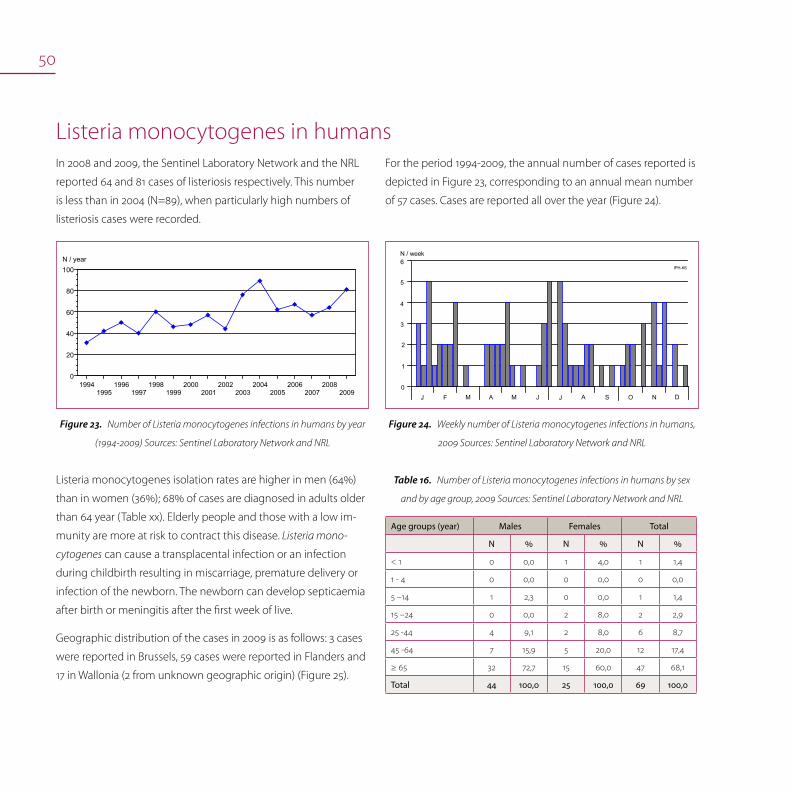
50
Listeria monocytogenes in humans
19941995
19961997
19981999
20002001
20022003
20042005
20062007
20082009
0
20
40
60
80
100N / year
Number of Listeria monocytogenes infections in humans by year Figure23.
(1994-2009) Sources: Sentinel Laboratory Network and NRL
N / week
J F M A M J AJ S O N D
IPH-K6
0
2
6
3
4
1
5
Weekly number of Listeria monocytogenes infections in humans, Figure24.
2009 Sources: Sentinel Laboratory Network and NRL
For the period 1994-2009, the annual number of cases reported is
depicted in Figure 23, corresponding to an annual mean number
of 57 cases. Cases are reported all over the year (Figure 24).
In 2008 and 2009, the Sentinel Laboratory Network and the NRL
reported 64 and 81 cases of listeriosis respectively. This number
is less than in 2004 (N=89), when particularly high numbers of
listeriosis cases were recorded.
Listeria monocytogenes isolation rates are higher in men (64%)
than in women (36%); 68% of cases are diagnosed in adults older
than 64 year (Table xx). Elderly people and those with a low im-
munity are more at risk to contract this disease. Listeria mono-
cytogenes can cause a transplacental infection or an infection
during childbirth resulting in miscarriage, premature delivery or
infection of the newborn. The newborn can develop septicaemia
after birth or meningitis after the first week of live.
Geographic distribution of the cases in 2009 is as follows: 3 cases
were reported in Brussels, 59 cases were reported in Flanders and
17 in Wallonia (2 from unknown geographic origin) (Figure 25).
Number of Listeria monocytogenes infections in humans by sex Table16.
and by age group, 2009 Sources: Sentinel Laboratory Network and NRL
Age groups (year) Males Females Total
N % N % N %
< 1 0 0,0 1 4,0 1 1,4
1 - 4 0 0,0 0 0,0 0 0,0
5 –14 1 2,3 0 0,0 1 1,4
15 –24 0 0,0 2 8,0 2 2,9
25 -44 4 9,1 2 8,0 6 8,7
45 -64 7 15,9 5 20,0 12 17,4
≥ 65 32 72,7 15 60,0 47 68,1
Total 44 100,0 25 100,0 69 100,0

51
AL AR AT
AW B
BG BS CR DK DM DN EK GT HS HV HY
IP KR LG LV MC MH MN MR MS NC NM NV OD OS PV RS SG SN TG TH TL TN TR VR VT VV WR
: Aalst : Arlon : Ath : Antwerpen : Brussels : Brugge : Bastogne : Charleroi : Diksmuide : Dendermonde : Dinant : Eeklo : Gent : Hasselt : Halle-Vilvoorde : Huy : Ieper : Kortrijk : Liège : Leuven : Mouscron : Mechelen : Mons : Marche-en-Fam. : Maaseik : Neufchâteau : Namur : Nivelles : Oudenaarde : Oostende : Philippeville : Roeselare : Soignies : St.-Niklaas : Tongeren : Turnhout : Tielt : Thuin : Tournai : Veurne : Virton : Verviers : Waremme
IPH-K6
0.00
0.76
Incidence/100,000 inhabitants[0.00 - 5.58]
> 0.00 - 0.75
> 0.75 - 1.50
> 1.50
VR
BG
DK TLGT
SN
ALOD
MH
HV
LV
AWTH
MS
HS
TG
NV
MRDN
CR
NC
AR
MC TR
KR
SG
IP
TN PV
VT
BS
VVLG
HY
WR
TG
MN NM
B
DM
EK
RS
AT
OS
Incidence of Listeria monocytogenes infections in humans by district (N/105 inhab., 2009) Figure25.
Sources: Sentinel Laboratory Network and NRL
In 2008, the NRL received 53 strains of Listeria monocytogenes of
clinical human origin, representing a small increase of 1.9% with
respect to 2007. Distribution of the serovars revealed the domi-
nating position of 1/2a with 41.5% of the isolates and 4b (33.9%),
followed by 1/2b (18.9%) and 1/2c (1.9%). Two strains (3.8%) were
defined as belonging to serogroup 4. Five strains were related to
pregnancy (2 strains isolated from the mother and 2 strains from
the newborn, 1 strain from the mother and the newborn). Eight
strains were isolated from patients with a meningo-encephalitis
form of listeriosis; 36 strains were isolated from blood and 1 strain
from pus, 1 strain from ascite fluid and 1 strain from bile.

52
In 2009, the NRL received 64 strains of Listeria monocytogenes
of clinical human origin, representing a large increase of 20.8%
with respect to 2008. Distribution of the serovars revealed the
dominating position of 1/2a with 62.5% of the isolates; the other
serovars were represented with 4b (26.5%), 1/2b (9.4%) and 1/2c
(1.6%). Two strains were related to pregnancy (1 strain isolated
from the mother and newborn and 1 strain from the newborn).
Eight strains were isolated from patients with a meningo-en-
cephalitis form of listeriosis; 50 strains were isolated from blood, 1
strain from pus and 2 strains from synovial fluid.
An episode of listeriosis was observed in the period October
2008 – January 2009; 8 patients were infected with Listeria
monocytogenes (serovar 1/2a, identical pulsotype). The source of
contamination could not be determined.
53
MRSAPatrick Butaye, Olivier Denis, Luc Vanholme
MRSAStaphylococcus aureus is an important pathogen both for humans
and for animals. S. aureus are bacteria commonly carried on the
skin or in the nose of healthy people. Last decades, resistance of
this pathogen to methicillin and all other beta-lactam antimicro-
bial agents has been of special concern, especially since these
so-called methicillin-resistant Staphylococcus aureus (MRSA)
also tend to accumulate resistance genes to most other (broad-
spectrum) antibiotics as well, making it difficult to treat infections
by these strains.
MRSA is a well recognised pathogen in humans, causing mainly
septicaemia, pneumonia and skin infections. Some clones of
these MRSA strains have been spread among hospitals and rest
homes (HA [Hospital acquired] MRSA) causing major outbreaks.
In addition, MRSA clones related to communities were identified
(CA [Community Acquired] MRSA). These latter strains were found
different from the nosocomial isolates and are generally less
resistant to antimicrobial agents other than beta-lactams.
MRSA
MRSA in animals
MRSA in humans

54
MRSA have also been identified in association with infection in
horses, cattle and dogs. Transfer of these bacteria from animals to
man through direct contact have been demonstrated. Recently,
a new type of MRSA (i.e. ST398) was found in pigs. The strain was
also isolated from the pig owner, his family, a patient and the
nurse in the hospital. Further studies confirmed that MRSA ST398
was present in various countries in pigs, but also in other species,
including dogs.
MRSA in pigsA survey was performed in 2007 to determine the prevalence of
MRSA colonization among the pig farms and in the individual
pigs.
In 2008 and 2009 no official monitoring programme was in place.
MRSA in humansThe NRL of Staphylococci – MRSA of the Université Libre de Brux-
elles provided specialized analysis:
Identification, characterisation and antimicrobial resistance pat-•
tern of atypical Staphylococci strains by means of
- Phenotypic methods such as colony morphology, biochemi-
cal tests, minimal inhibiting concentrations (MIC determina-
tion), glycopeptide susceptibility (population analysis)
- Genotypic methods: identification of different Staphylococci
species by sequence analysis of gene rpoB, detection by
PCRs for nuc gene (for S. aureus identification), mecA gene
(encoding oxacillin resistance), mupA gene (encoding mupi-
rocine resistance) and of resistance genes to macrolides-
lincosamides-streptogramines (MLS), tetracyclines and
aminoglycosides.
Detection of toxin genes: PCRs for exfoliative toxins A, B and •
D, Panton Valentine Leucocidin (PVL), Toxic Shock Syndrome
Toxin-1 (TSST-1) and staphylococcal enterotoxins including sea,
seb, sec, sed, see, seg, seh, sei, selj, selk, sell, selm, seln, selo,
selp, selq, selr.
Molecular typing, by Pulsed Field Gel Electrophoresis (PFGE) •
after SmaI macrorestriction, by spa sequence and typing of the
Staphylococcal Chromosome Cassette mec (SCCmec typing),
detection of the agr group, detection of the presence of the
arcA gene as marker of the pathogenic locus of the Arginine
Catabolic Mobile Element (ACME).
The NRL especially analysed clinical strains for additional suscep-
tibility testing or for searching virulence genes, and collections
of staphylococcal strains isolated during local outbreaks for
epidemiological studies.
Since 1992, the NRL organises epidemiological surveillance of
MRSA by means of biannual surveys in collaboration with the IPH
and the Belgian Infection Control Society (BICS). The objectives
are to follow the evolution of genotypes and of antimicrobial
resistance profiles of MRSA isolates from patients admitted to
Belgian acute-care hospitals. Detailed information on the results
of the last survey conducted in 2008 is available on the website
http://www.mrsa.be.

55
Since 1999, the NRL participated in the EARSS programme (Euro-
pean Antimicrobial Surveillance System) in collaboration with the
section of epidemiology of IPH. Information about the protocol
and the data of the EARSS programme is available at the address
http:// www.rivm.nl/earss/.
Conclusions
The number of PVL-positive MRSA cases reported in 2008 in-
creased compared to the previous years (43 cases in 2008 versus
25-30 recorded from 2005 to 2007). These PVL-positive strains
caused mainly skin and soft-tissue infections. By molecular typ-
ing, the ST80-SCCmec IV clone, which is widely disseminated in
Europe, was still predominant. However, MRSA strains belonging
to ST8-SCCmec IV USA-300 clone were more frequently isolated.
The emergence of USA300 MRSA is of increasing concern in view
of severe and fatal infections recently reported from Belgium.
Since 2003, MRSA clone ST398 strains of animal origin (livestock-
associated MRSA, LA-MRSA) are regularly isolated in humans in
contact with livestock animals, including veterinarians and swine
farmers. The swine population appeared to be highly colonized
by ST398 LA-MRSA clone. This clone was also recovered from a
number of other animal species including bovine, equine, poultry
and pets. These LA-MRSA strains were infrequently associated
with active disease in humans.
The proportion of MRSA among patients with S. aureus bacterae-
mia in acute-care hospitals decreased continuously after a peak
(33%) in 2004 to stabilize at 23% in 2008 (EARSS data). This obser-
vation corresponded to the results of the national surveillance
programme NSIH conducted by the IPH. Since 2005, a dramatic
decrease has halved the incidence of nosocomial MRSA acquisi-
tion to 1.7 cases per 1000 admissions in 2008 (data available at
http://www.nsih.be/).
The last microbiological survey conducted in 2008 showed that
the majority (86%) of MRSA strains belonged to 4 epidemic
clones: spa CC38 ST45-SCCmec IV (formerly PFGE type B2), spa
CC8 ST8-SCCmec IV (PFGE type A20), spa CC2 ST5-SCCmec II
(PFGE type G10) and spa CC2 ST5-SCCmec IV (PFGE type C3).
These MRSA clones are still widely disseminated in Belgian acute-
care hospitals causing local outbreaks as confirmed by the NLR
in 2008-2009. LA-MRSA and PVL-positive MRSA strains only rep-
resent a small proportion (<3%) of the burden of MRSA strains in
the population of patients admitted in acute-care hospitals. Their
frequency has remained stable since their first detection in 2003.

56

57
Q-feverDavid Fretin, Marjan Van Esbroeck, Luc Vanholme, Katie Vermeersch
Coxiella burnetii Q-fever is a zoonotic disease caused by Coxiella burnetii. Q-fever
(Q for query) is a systemic disease caused by an obligate intracel-
lular bacterium Coxiella burnetii that is highly resistant to heat,
drying and many common chemical and physical agents. This
resistance enables the bacteria to survive for a long period in the
environment. Coxiella burnetii occurs worldwide except in New
Zealand.
Natural reservoirs are more than 40 species of ticks and free-
living vertebrates, primarily rodents. Ticks or their excreta may
spread Coxiella burnetii to domestic animals, e.g. sheep, goats,
cattle and dogs. These animals may display a cycle that does not
involve ticks since Coxiellae can multiply in the trophoblast of
the placenta. The placentas and amniotic fluids of these animals
contain large numbers of bacteria which contaminate pastures
and soil. Once animal secreta or excreta have dried, infectious
dust is created.
Coxiella burnetii
Q-fever in animals
Q-fever in humans

58
Q-fever in animalsCattle, sheep, and goats are the main reservoirs but a wide variety
of other animals can be contaminated, including domesticated
pets. Coxiella burnetii usually does not cause clinical disease in
these animals, although an increased abortion rate and fertility
problems in cattle, sheep and goats are observed. The emer-
gence of these common symptoms over a longer period of time
leads finally to the suspicion of Q-fever.
Organisms are excreted in milk, urine and faeces by infected
animals. Animals shed the organisms especially during parturition
within the amniotic fluids and the placenta. Airborne transmis-
sion can occur in premises contaminated by placental material,
birth fluids or excreta from infected animals. Airborne inhalation
is the most important transmission route of infection.
In 2008 and 2009, only limited testing by the NRL was performed
on individual animal level of breeding bulls at the Artificial In-
semination Centers and for confirmation of laboratory diagnosis
in case of a clinical suspicion in case of an increased number of
abortions of ruminants.
In 2008, the NRL analysed 314 sera of bovine animals. In total 25
bovine sera were positive.
In 2009, the NRL analysed 119 sera of breeding bulls at the
Artificial Insemination centers, all sera were negative by RT-PCR.
Individual analysis of 117 abortions of cattle resulted in 8 positive
cases by RT-PCR. In case of RT-PCR tests for diagnosis, 69 PCR’s
were positive on a total of 208 analyses. In case of serology by
ELISA for diagnosis, 40 ELISA’s were positive on a total of 165
analyses. Screening of goats by PCR, 9 tests were positive on a
total of 56 and 15 sera were positive. 6 sera analysed of sheep
were negative.
The NRL tested 44 tank milk samples of dairy cattle by PCR, 4 sera
were positive. Tank milk samples of goats were tested by RT-PCR
and ELISA. Respectively 9 and 15 samples were positive on a total
of 60 and 56 tankmilk samples. Only one tankmilk sample of
sheep was tested by PCR and ELISA with a negative result of both
tests (table 17).
NRL alayses for Q-feverTable17.
NRL
testanimals
tested
animals
positive
cattleartificial insemination
centerPCR 119 0
cattleclinical investigation
abortionPCR 117 8
cattle diagnosis PCR 208 69
cattle diagnosis ELISA 165 40
testherds
tested
herds
positive
cattle monitoring tankmilk PCR 44 4
goats monitoring tankmilk PCR 60 9
goats monitoring tankmilk ELISA 56 15
sheep monitoring tankmilk PCR 1 0
sheep monitoring tankmilk ELISA 1 0
In 2009, also ARSIA and DGZ (Regional veterinary health laborato-
ries) did analyses for Q-fever (table 18).
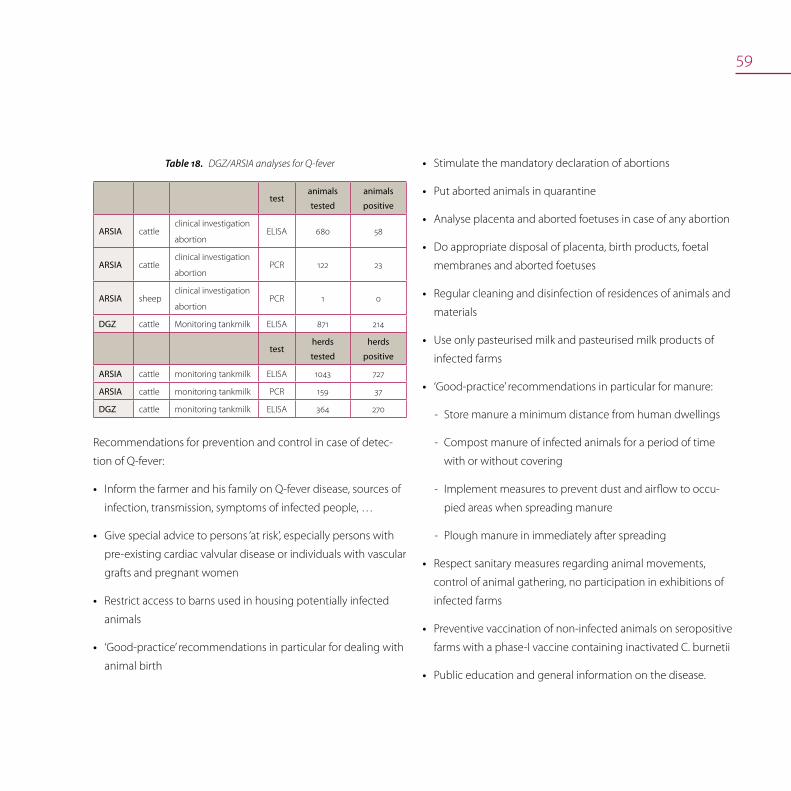
59
DGZ/ARSIA analyses for Q-feverTable18.
testanimals
tested
animals
positive
ARSIA cattleclinical investigation
abortionELISA 680 58
ARSIA cattleclinical investigation
abortionPCR 122 23
ARSIA sheepclinical investigation
abortionPCR 1 0
DGZ cattle Monitoring tankmilk ELISA 871 214
testherds
tested
herds
positive
ARSIA cattle monitoring tankmilk ELISA 1043 727
ARSIA cattle monitoring tankmilk PCR 159 37
DGZ cattle monitoring tankmilk ELISA 364 270
Recommendations for prevention and control in case of detec-
tion of Q-fever:
Inform the farmer and his family on Q-fever disease, sources of •
infection, transmission, symptoms of infected people, …
Give special advice to persons ‘at risk’, especially persons with •
pre-existing cardiac valvular disease or individuals with vascular
grafts and pregnant women
Restrict access to barns used in housing potentially infected •
animals
‘Good-practice’ recommendations in particular for dealing with •
animal birth
Stimulate the mandatory declaration of abortions•
Put aborted animals in quarantine•
Analyse placenta and aborted foetuses in case of any abortion•
Do appropriate disposal of placenta, birth products, foetal •
membranes and aborted foetuses
Regular cleaning and disinfection of residences of animals and •
materials
Use only pasteurised milk and pasteurised milk products of •
infected farms
‘Good-practice’ recommendations in particular for manure:•
- Store manure a minimum distance from human dwellings
- Compost manure of infected animals for a period of time
with or without covering
- Implement measures to prevent dust and airflow to occu-
pied areas when spreading manure
- Plough manure in immediately after spreading
Respect sanitary measures regarding animal movements, •
control of animal gathering, no participation in exhibitions of
infected farms
Preventive vaccination of non-infected animals on seropositive •
farms with a phase-I vaccine containing inactivated C. burnetii
Public education and general information on the disease.•
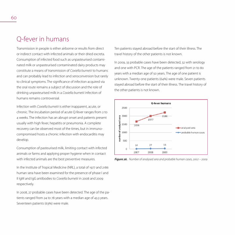
60
Q-fever in humansTransmission in people is either airborne or results from direct
or indirect contact with infected animals or their dried excreta.
Consumption of infected food such as unpasteurised contami-
nated milk or unpasteurised contaminated dairy products may
constitute a means of transmission of Coxiella burnetii to humans
and can probably lead to infection and seroconversion but rarely
to clinical symptoms. The significance of infection acquired via
the oral route remains a subject of discussion and the role of
drinking unpasteurised milk in a Coxiella burnetii infection of
humans remains controversial.
Infection with Coxiella burnetii is either inapparent, acute, or
chronic. The incubation period of acute Q-fever ranges from 2 to
4 weeks. The infection has an abrupt onset and patients present
usually with high fever, hepatitis or pneumonia. A complete
recovery can be observed most of the times, but in immuno-
compromised hosts a chronic infection with endocarditis may
develop.
Consumption of pasteurised milk, limiting contact with infected
animals or farms and applying proper hygiene when in contact
with infected animals are the best preventive measures.
In the Institute of Tropical Medicine (NRL), a total of 1977 and 2186
human sera have been examined for the presence of phase I and
II IgM and IgG antibodies to Coxiella burnetii in 2008 and 2009
respectively.
In 2008, 27 probable cases have been detected. The age of the pa-
tients ranged from 24 to 78 years with a median age of 43.3 years.
Seventeen patients (63%) were male.
Ten patients stayed abroad before the start of their illness. The
travel history of the other patients is not known.
In 2009, 33 probable cases have been detected, 32 with serology
and one with PCR. The age of the patients ranged from 21 to 80
years with a median age of 50 years. The age of one patient is
unknown. Twenty-one patients (64%) were male. Seven patients
stayed abroad before the start of their illness. The travel history of
the other patients is not known.
Number of analysed sera and probable human cases, 2007 – 2009Figure26.

61

62
63
SalmonellosisKatrien Beullens, Sophie Bertrand, Patrick Butaye, Katelijne Dierick, Hein Imberechts, Christophe Keppens, Luc Vanholme, Katie Vermeersch
SalmonellaSalmonella is an important zoonotic pathogen and a major cause
of registered bacterial foodborne infections, both in individuals
and in communities. The genus Salmonella is currently divided
into two species: S. enterica and S. bongori. S. enterica is further
divided into six sub-species and most Salmonella belong to the
subspecies S. enterica subsp. enterica. Members of this subspe-
cies have usually been named based on where the serovar or
serotype was first isolated. Mostly, the organisms are identified
by genus followed by serovar, e.g. S. Virchow. More than 2.500
serovars of zoonotic Salmonella exist and the prevalence of the
different serovars change over time.
Human salmonellosis is usually characterised by the acute onset
of fever, abdominal pain, nausea, and sometimes vomiting. Symp-
toms are often mild and most infections are self-limiting, lasting a
few days. However, in some patients, the infection may be more
serious and the associated dehydration can be life threatening.
In these cases, as well as when Salmonella causes septicaemia,
effective antimicrobials are essential for treatment. Salmonellosis
has also been associated with long-term and sometimes chronic
sequelae e.g. reactive arthritis.
Salmonellosis
Salmonella in animal feed
Salmonella in poultry
Salmonella in pigs
Salmonella in cattle
Salmonella in food
Salmonella in humans
Antimicrobial resistance

64
The common reservoir of Salmonella is the intestinal tract of a
wide range of domestic and wild animals which result in a variety
of foodstuffs covering both food of animal and plant origin as
sources of infections. Transmission often occurs when organ-
isms are introduced in food preparation areas and are allowed to
multiply in food, e.g. due to inadequate storage temperatures,
inadequate cooking or cross contamination of ready-to-eat food.
The organism may also be transmitted through direct contact
with infected animals or faecally contaminated environments and
humans.
Food prepared with contaminated raw eggs, egg products or
insufficiently heated poultry meat or pork is the source of the
human Salmonella infection. Therefore, surveillance programmes
that in time detect Salmonella contaminations early in the whole
food chain (feed, living animals, slaughterhouses, cutting plants,
retail sector, restaurants) together with sanitary measures to
reduce contamination are essential. In addition, good hygiene
practices during food preparation in the kitchen, adequate refrig-
eration and adequate heating also help to prevent Salmonella
infections.
In animals, sub-clinical infections are common. The organism may
rapidly spread between animals in a herd or flock without detec-
tion and animals may become intermittent or persistent carriers.
Infected cows may succumb to fever, diarrhea and abortion.
Within calf herds, Salmonella may cause outbreaks of diarrhea
with high mortality. Fever and diarrhea are less common in pigs
than in cattle. Sheep, goats and poultry usually show no signs of
infection.
Salmonella in animal feedIn June 2008 the Panel on Biological Hazards of EFSA identified
Salmonella spp. as the major hazard for microbial contamina-
tion of animal feed. For other microbiological hazards feed was
regarded a far less important source of contamination. This
opinion confirmed the strategy of the FASFC to focus its efforts
on microbiological contamination in feed on Salmonella. Special
attention is given to the sampling procedure using n=5 and tak-
ing into account the heterogenic nature of a possible Salmonella
contamination of feed. Feed materials of animal origin, oilseeds
and wheat bran are considered as ‘at-risk’ products.
Using a statistically substantiated risk evaluation the FASFC re-
evaluates and performs an official control program every year.
Compound feed and feed materials are sampled and analysed
for absence of Salmonella in 25g. Every detection of Salmonella
is treated as a non-conformity, but the actions taken are depend-
ing on the serotype detected and the type of feed. In 2008 and
2009, more stringent actions were taken if the contamination
concerned 5 critical serotypes in poultry feed and finishing feed.
Those critical serotypes were S. Typhimurium, S. Enteritidis, S.
Virchow, S. Hadar and S. Infantis. For 2010 a new strategy will be
implemented determining more critical serotypes and fine-
tuning the actions depending on the type of feed and the place
in the feed chain where the contamination is detected.
In 2008 a total of 709 samples of feed were analysed for the
detection of Salmonella. Out of those, 103 were feed materials of
animal origin. 7 were found positive (serotypes detected: S. Infan-
tis, S. Jerusalem, S. Livingstone, S. Ohio, S. Senftenberg, S. Yoruba, S.
6,7:-:-, S. 6,7:z10:- and S. Paratyphi B var. Java).
65
101 were other feed materials. 10 were found positive (sero-
types detected: S. Banana, S. Cubana, S. Kentucky, S. Lexington, S.
Mbandaka, S. Rissen, S. Senftenberg, S. Worthington, S. Yoruba, S.
3,10:-:1,5 and S. 3,19:-:-). 505 samples were from compound feeding-
stuffs. 17 were found positive (serotypes detected: S. Brandenburg,
S. Enteritidis, S. Havana, S. Jeruzalem, S. Lexington, S. Livingstone, S.
Minnesota, S. Orion, S. Senftenberg, S. Worthington, S. 6,7:-:- and S.
3,19:-:-).
In 2009 a total of 714 samples of feed were analysed for the detec-
tion of Salmonella. Out of those 65 were feed materials of animal
origin. 1 was found positive (2 serotypes detected: S. Bredeney
and S Montevideo). 112 were other feed materials. 3 samples were
found positive (serotypes detected: S. Emek, S. Infantis, S. Rissen
and S. Lexington). 537 samples were from compound feeding-
stuffs. 10 were found positive (serotypes detected: S. Anatum, S.
Cerro, S. Jeruzalem, S. Livingstone, S. Minnesota, S. Oranienburg, S.
Rissen, S. Schwarzengrund and S. Senftenberg).
Salmonella in poultry
Salmonella in breeders and hatcheries
Control programme in breeders
The regional animal health associations (i.e. “Association Ré-
gionale de Santé et d’Identification Animales” [ARSIA (http://
www.arsia.be/)] and Dierengezondheidszorg Vlaanderen [DGZ
Vlaanderen (http://www.dgz.be/)]) organise the official sampling
in the framework of the Belgian Salmonella control programme
in breeders.
All breeder flocks are routinely sampled for Salmonella at delivery
as day-old chicks (imported and domestic flocks). At the farm,
pieces (5 by 5 cm) of the inner linings of the delivery boxes of
the day-old chicks are taken by the owner, i.e. one sample for the
hen-chicks and one for the cock-chicks. Each sample consists of
20 pieces of inner linings. The two samples are analysed separate-
ly. In addition, 20 living hen-chicks and 20 living cock-chicks are
tested serologically. The samples have to be taken the day of the
delivery and have to reach the lab within 24h of sampling. Official
samples of breeding flocks are taken at the age of 16 weeks, 22
weeks, 46 and 56 weeks by technicians of DGZ or ARSIA. Routine
sampling is performed by the operator at the age of 4 weeks and
every two weeks during production. The sampling is conform
Regulation (EC) No 1168/2005 and consist of 5 pair of overshoes
pooled in two samples. All samples are immediately analysed in
the laboratories of DGZ or ARSIA according to ISO 6579:2002 Amd
1:2007.
The official programme also controls the hygiene level of hatcher-
ies 4 times a year. These are done during visits of the technician
at non-hatching days and include various sites of the hatchery,
including hatching drawers. Rodac samples are taken and both
total bacteria and moulds are counted. After appropriate incuba-
tion, an index or code is given to the number of colonies per sur-
face of approximately 22 cm² in order to facilitate comparisons. In
addition, a specific Salmonella control is done 4 times a year, on
pooled samples from dead-in-shell chicks and on fluff and meco-
nium. These samples are sent to the laboratory by the owner.
66
Case definition, notification, sanitary measures
and vaccination
A poultry breeding flock is considered Salmonella positive when
S. Enteritidis, S. Typhimurium, S. Virchow, S. Hadar or S. Infantis is
isolated from at least one sample. Confirmatory samples (5 faeces
samples and 2 dust samples) during rearing or production may
be requested by the farmer and are taken by or under the super-
vision of the competent authority. The results of these analyses
are binding. Provisional measures are taken during the course of
analysis.
The isolation of zoonotic Salmonella is notifiable to the FASFC by
the approved laboratories since January 2004.
Several measures are taken in case of positive breeding flocks:
prohibition of incubation of hatching eggs, removal and destruc-
tion of incubated hatching eggs and canalisation of not yet
incubated hatching eggs for pasteurisation. In addition, positive
flocks are logistically slaughtered within the month, after removal
of the positive flock, the house is thoroughly cleaned and disin-
fected and a Salmonella control is performed of the house using
2 samples each consisting of 25 swabs. Cleaning and disinfection
is repeated until the Salmonella control is negative.
Vaccination against S. Enteritidis is mandatory; vaccination against
S. Typhimurium is strongly recommended for parent flocks. Both
attenuated and inactivated vaccines are available.
Epidemiological investigations and results of 2008-2009
surveillance
In 2008, 224 rearing flocks and 550 flocks during production were
tested. Six rearing flocks were positive for Salmonella (S. Typhimu-
rium, S. Infantis, S. Mbandaka, S. Montevideo, S. Senftenberg and S.
O4) and 45 flocks in production of which 3 for S. Enteritidis, 2 for
S. Typhimurium, 9 for S. Senftenberg, 8 for S. Agona, 3 each for S.
Mbandaka, S. Montevideo and S. Cochise, 2 each for S. Anatum, S.
Lexington and S. Rissen and 1 each for S. Cubana, S. Livingstone, S.
Muenchen, S. Orion, S. Ruiri, S. Tennessee, S. O4 and S. O3,19:-:-.
In 2009, 302 rearing flocks and 526 flocks during production
were tested. Three rearing flocks were positive for Salmonella
(1 S. Hadar, 1 S. Cubana and 1 S. O3,19:-) and 16 flocks in produc-
tion of which 6 for S. Senftenberg, 3 for S. Mbandaka, 2 each for S.
Lexington and S. Minnesota and 1 each for S. Corvallis, S. Livingstone
and S. O9,46:-:-.
Salmonella in layers
Surveillance programme in commercial laying hen flocks
All laying hen flocks on farms with at least 200 laying hens must
follow the national Salmonella control programme in layers.
Flocks are sampled by the owner at the age of one day (at arrival)
16 weeks, 24, 39 and 54 weeks and in the last three weeks of
production.
The day-old-chicks are sampled in the same way as the breeders.
For all other sampling, two samples are taken each consisting of,
depending on the housing system, either one pair of overshoes or
in case of cages, 150g naturally pooled faeces or mixed fresh faeces.
67
The two samples are pooled to one sample at the laboratory. One
flock of all farms with at least 200 laying hens is also sampled by
the FASFC. This official sampling consists of two pair of overshoes
pooled to one sample at the laboratory and 1 dust sample.
Case definition, notification, sanitary measures
and vaccination
A laying hen flock is declared positive if S. Enteritidis or S. Typhimu-
rium is isolated. Confirmatory samples (5 faeces samples and 2
dust samples) during rearing or production may be requested by
the farmer. The results of these analyses are binding. Provisional
measures are taken during the course of analysis.
The isolation of zoonotic Salmonella is notifiable to the FASFC
since January 2004.
In case of positive findings in layers, eggs are placed on the
market as B-eggs for heat treatment. The poultry house must be
cleaned and disinfected after slaughter of the positive flock and a
Salmonella control of the house is performed in the same way as
for breeding flocks.
Vaccination against S. Enteritidis is mandatory for layers; vaccina-
tion against S. Typhimurium is strongly encouraged. Both attenu-
ated and inactivated vaccines are available.
Epidemiological investigations and results of 2008-2009
surveillance
In 2008 of the 293 batches of day-old chicks, none were found
positive for Salmonella. During rearing of the 293 flocks, 5 were
positive for Salmonella (1 for S. Enteritidis, 3 for S. Jerusalem and 1
for S. Lexington). During production, of the 649 flocks, 76 flocks
were positive for Salmonella (23 for S. Enteritidis, 7 for S. Mbandaka,
6 for S. Livingstone, 4 each for S. Infantis, S. Rissen and S. Senften-
berg, 3 each for the Salmonella group O4 and the Salmonella
group O6, 2 each for S. Hillingdon, S. Lexington, S. Virchow, and
the Salmonella group O9, 1 each for S. Banana, S. Blegdam, S.
Braenderup, S. Derby, S. Dublin, S. Gateshead, S. Grumpensis, S. Jeru-
salem, S. Orion, S. Typhimurium and S. Paratyphi B var. Java.
In 2009 of the 283 batches of day-old-chicks, none were found
positive for Salmonella. During rearing, 283 flocks were sampled
of which two were positive for Salmonella (1 S. Typhimurium en 1
S. O4: l). During production, 763 flocks were sampled of which 54
were positive for Salmonella (26 for S. Enteritidis, 6 for S. Infantis, 3
each for S. Typhimurium and S. Rissen, 2 each for S. Agona, S. Ana-
tum, S. Livingstone, S. Mbandaka and S. Virchow, 1 each for S. O4:d:-,
S. O6,7:-:-, S. Derby, S. Hadar, S. Idikan, S. Jerusalem, S. Minnesota and
S. Yoruba). There were 2 flocks positive for an untypable Salmo-
nella spp. and 4 flocks were positive for 2 different serotypes.
Salmonella in broilers
Surveillance programme in commercial broiler flocks
Day-old-chicks are sampled on farms with the highest sanitary
qualification, following the same procedure as for laying hens. All
broiler flocks with a capacity of 200 or more broilers are sampled
by the holder in the last three weeks before slaughter. The sample
consists of two pair of overshoes. Ten percent of the holdings are
also sampled by the FASFC.
Case definition, notification, sanitary measures
68
and vaccination
A flock is positive if zoonotic Salmonella is found. The isolation of
zoonotic Salmonella is notifiable to the FASFC since January 2004.
The positive Salmonella result is reported to the slaughterhouse
by means of the food chain information system.
As of 2009, it is forbidden to use antimicrobials as treatment for
Salmonella. Before restocking the house, the house is cleaned,
disinfected and the presence of Salmonella is checked by means
of two samples each consisting of 25 swabs taken throughout the
house. A hygienogram is performed. If Salmonella is found or if
the hygienogram indicates that too many germs are present, the
Salmonella control and/or the hygienogram have to be repeated
after the next round. In addition to the previous measures, the
house has to be disinfected by an external company and thinning
must be performed as last holding of the day if 2 consecutive
flocks in 1 house are positive for the same serotype. If 3 consecu-
tive flocks are positive for the same Salmonella serotype, an
epidemiological investigation of the farm and optimizing the
biosecurity and hygiene measures on the farm are added to the
list of measures.
Epidemiological investigations and results of 2008-2009
surveillance
In 2008, 5.074 flocks were sampled as day-old chicks of which 4
were positive for Salmonella. 7.775 flocks were sampled within
three weeks before slaughter. 234 were positive for Salmonella.
Serotyping was not mandatory in 2008.
In 2009, 5.226 flocks were sampled as day-old-chicks of which 7
were positive for Salmonella (1 for S. Enteritidis, 1 for S. Typhimu-
rium, 1 for S. Paratyphi B var. Java and 4 unspecified). 8.049 flocks
were sampled in the last three weeks before slaughter. 247 flocks
were positive for Salmonella. The most frequently found sero-
types were: S. Typhimurium (26 flocks), S. Paratyphi B (22 flocks), S.
Paratyphi B var. Java (18 flocks), S. Enteritidis (14 flocks), S. Agona (18
flocks), S. Livingstone (17 flocks), S. Minnesota (12 flocks), S. Virchow
(12 flocks), S. Rissen (11 flocks) and S. Hadar (9 flocks).
Salmonella serotypes of the 2008–2009 surveillance in layers and broilers
Laboratory findings of the NRL show that in 2008 and 2009 a
total 951 and 1142 Salmonella strains from poultry origin were
analysed, respectively, which is considerably more than the 745
strains tested in 2007. As for the strains known to originate from
the layer flocks, the majority (39.1% of 92 in 2008 and 59.0% of
122 in 2009) belonged to serotype Enteritidis. Other serotypes
in 2008 were Livingstone (9.8%) and Typhimurium (7.6%), and in
2009 Infantis (8.2%), Typhimurium and Livingstone (both 4.1%). The
majority of broiler isolates tested in 2008 (n=372) were S. Enteritidis
(54.3%), and also serotypes Typhimurium (8.1%), Paratyphi B (4.8%),
Virchow (4.0%) and Infantis (3.5%) were found. In 2009, the most
common broiler isolates (n=192) were S. Paratyphi B (32.3%), but
also Hadar (9.9%), Agona (7.8%), Minnesota (5.7%) and Virchow
(5.7%) were frequently identified.
The number of poultry isolates yearly sent to the NRL varies
between about 700 and almost 1.500. In 2009, almost one third of
poultry strains belonged to serotype Enteritidis, which is less than
in 2008 and 2007.

69
The proportion of Typhimurium strains is since 2001 less than 10%
and reached 6.7% in 2009. The proportion of Salmonella Paratyphi
B strains, after their peak in 2006, fell down in recent years, but
reached again 20% in 2009. Serotypes Hadar, Infantis and Virchow
represented about 2 to 3% of poultry isolates in 2009.
Evolution of the percentages of the principal Salmonella serotypes Figure27.
isolated from poultry between 1998 and 2009
The bars represent the total number of poultry isolates per year, and
refer to the right axis; the lines represent the percentage of each sero-
type per year and refer to the left axis
Salmonella in turkeys, geese, ducks, pigeons and other poultry
Surveillance programme and sampling
The national control programme for Salmonella in turkeys, geese,
ducks and guinea fowl is performed according to the sanitary
qualification act. Sanitary Qualification A is mandatory for all com-
mercial breeding flocks. Flocks are at least sampled as day-old
chicks, at the age of 26 weeks when entering the production unit
if this is on a different farm than the rearing unit, and within the
last 3 weeks before slaughter. Meat production flocks are sampled
within three weeks of slaughter if the holding has a capacity of
more than 5.000 birds (Sanitary Qualification B). On a voluntary
basis, one-day-old birds may also be sampled.
Samples for day-old-birds are taken at the farm, and consist of
pieces (5 by 5 cm) of the inner linings of delivery boxes. Two sam-
ples, each composed of 20 pieces of inner linings, are taken for
each flock, one for the hen chicks and one for the cock chicks. The
two samples are analysed separately according to ISO 6579.
The owner of breeding flocks takes faeces samples from the
delivery boxes at time of delivery. A sample consists of 60 x 5
to 10g sub-samples taken from every flock with different origin
of rearing. The samples have to be examined by an accredited
laboratory within 48 hours. At 26 weeks, 60 blood samples are
taken of each breeding flock for the surveillance of Salmonella. If
one or more blood samples are positive, faecal samples are taken
to confirm the results.
Within 3 weeks before slaughter, the owner takes a pooled faecal
sample consisting of 60 x 1g sub-samples of each flock.
70
Alternatively, the sampling may consist of a pooled faecal sample
of 60 x 1g taken by hand, or recovered from two pair of overshoes
that were pooled for analysis.
Case definition, sanitary measures and vaccination policy
A flock is considered positive if zoonotic Salmonella serotypes
were isolated. Measures are taken only at time of slaughter: if the
flock is Salmonella positive, at time of slaughter, it is slaughtered
at the end of the day (logistic / sanitary slaughter).
There is no vaccination policy for breeding flocks, nor for meat
production flocks. Notification of zoonotic Salmonella to the
FASFC is compulsory since January 2004.
Results of the investigation in 2008-2009 surveillance
In 2008, 167 meat turkey flocks were tested of which 6 were posi-
tive for Salmonella. Serotyping was not performed.
For meat turkey flocks following results were found in 2009: 155
flocks were sampled at the end of production of which 6 were
positive for Salmonella spp. (1 each for S. Blockley, S. Indiana
and S. Kottbus, 1 isolate was not typable and 2 isolates were not
serotyped).
2 flocks of guinea fowl were tested, both negative for Salmonella.
Data from the NRL Salmonella indicate that in 2008 eleven of the
13 pigeon strains analysed were S. Typhimurium var. Copenhagen,
which is a common finding for these birds. In 2009, all 12 pigeon
strains analysed were identified as S. Typhimurium var. Copenha-
gen. Two serotyped turkey strains were S. Indiana and S. Kottbus,
and four pheasant isolates belonged to serotypes Gallinarum,
Indiana, Senftenberg and Typhimurium.
Salmonella in pigs
Serology
Surveillance programme in fattening pigs
Similar to former years, in 2008-2009 the blood samples from
fattening and growing pigs that were taken for the monitoring
of Aujeszky’s disease were also analysed for the surveillance of
Salmonella. On each farm, which is visited every 4 months, 10 to
12 blood samples were taken. The analysis for Salmonella-specific
antibodies was done in the veterinary laboratories ARSIA and
DGZ by means of a commercially available ELISA kit, following the
manufacturer’s instructions.
Case definition, sanitary measures and vaccination policy
A herd is considered as a ‘Salmonella risk herd’ if 3 consecutive mean
S/P ratio’s were above 0,6. The S/P ratio is considered to be related to
the presence of specific antibodies against Salmonella spp.
Following measures are taken on a Salmonella risk herd: comple-
tion of checklist on state of play on biosecurity, management
and hygiene, formulating and implementing a herd specific
Salmonella action plan, based on the results of the checklist and
bacteriological evaluation of the farm.
Pigs were not vaccinated in 2008-2009, since no vaccine was
authorised in Belgium.

71
Results in 2008-2009
In 2008, a total of 188.257 serological analyses were performed.
Of these, 39.875 samples (21,2%) had a S/P ratio above 0,6. 40,2%
of the 6.658 farms monitored had at least once a mean S/P ratio
above 0,6.
In 2009, a total of 171.767 serological analyses were performed,
28.270 (16,5%) of the samples had a S/P ratio above 0,6; 32,6% of
the 6.395 farms monitored had at least once a mean S/P ratio
above 0,6. 231 farms were considered as Salmonella risk farms for
the first time in 2009, 84 for a second time.
Bacteriology
One of the measures on ‘Salmonella risk herds’ is a bacteriological
investigation of housing environment of the pigs. Four samples
are taken by the responsible farm veterinarian. Samples were also
taken for research activities.
Laboratory findings from the NRL showed that, as compared
to 2007, the number of pig strains more than doubled in 2008
(n=481 in 2007 and n=1.017 in 2008) most likely due to the
mandatory bacteriological investigation on Salmonella risk herds.
Significantly less S. Typhimurium isolates were found (48.5%; 65.2%
in 2007), but considerably more S. Derby (15.6%; 7.2% in 2007). In
2009, the number of pig strains tested resembled that of 2007
(n=536, 1 017). Again more S. Typhimurium isolates were found
(63.8%) as compared to 2008), but an equal proportion of S. Derby
(13.4%; 15.6% in 2008). Nine percent of pig strains were only par-
tially typable, and belonged to group B Salmonella.
S. Typhimurium still is the most prevalent serotype among pig
isolates, representing more than 60% of pig Salmonella. Serotype
Derby is the second most important serotype, and represents
about 13% of the strains.
Evolution of the percentage of the principal Salmonella serotypes Figure28.
isolated from pigs between 1998 and 2009
The bars represent the total number of pig isolates per year, and refer
to the right axis; the lines represent the percentage of each serotype
per year and refer to the left axis.

72
Salmonella in food (meat and meat products)Monitoring programme
In 2008 and 2009, sampling for Salmonella monitoring was done
on the following matrices: carcasses, trimmings and raw ham
of pork, minced meat and meat preparations of beef, carcasses,
meat cuts and meat preparations of broilers, layer carcasses and
minced meat and meat preparations of diverse species.
Sampling of pork carcasses was done by means of swabs. The car-
cass samples of broilers and layers at slaughter consisted of neck
skin. Different contamination levels were analysed: 25g, 600 cm²,
1g and 0.1g. Sampling was done by specially trained staff.
Notification is mandatory since March 2004 (Ministerial Decree
on mandatory notification in the food chain). For Salmonella,
absence in 25g in ready-to-eat food is requested.
Salmonella in meat and meat products.
Epidemiological investigations and results of 2008 surveillance
The results of the monitoring – Salmonella in meat and meat productsTable19.
Species Sample size Prevalence Predominant serotypeOther serotypes
(in decreasing order)
Beef
Minced meat (steak tartare) at retail (n=127) 25g 1.6%
Meat preparation (steak tartare with herbs and
sauce) at retail (n=125)25g 0.1% Typhimurium
Pork
Carcasses at slaughter (n=281)
Meat cuts at processing plant (n=122)
600cm²
25g
14.6%
5.7%
Typhimurium
Typhimurium
Derby, Brandenburg, Livingstone, Rissen, Ohio,
Paratyphi B, Infantis
Trimmings at processing plant (n=99) 25g 6.1% Typhimurium
Raw ham at retail (n=31) 25g 0.0%
Raw ham at processing plant (n=57) 25g 1.8% Typhimurium
Broilers
Carcasses at slaughter (n=157) 1g 7% Paratyphi B Typhimurium, Virchow, Infantis
Carcasses at slaughter (n=193) 25g (caeca) 9.8% Paratyphi B
Carcasses at retail (n=90) 25g 11.1% Paratyphi B Typhimurium, Agona, Hadar
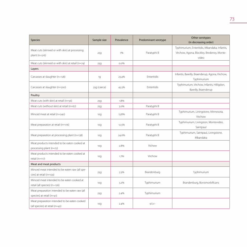
73
Species Sample size Prevalence Predominant serotypeOther serotypes
(in decreasing order)
Meat cuts (skinned or with skin) at processing
plant (n=516)25g 7% Paratyphi B
Typhimurium, Enteritidis, Mbandaka, Infantis,
Virchow, Agona, Blockley, Bredeney, Monte-
video
Meat cuts (skinned or with skin) at retail (n=74) 25g 0.0%
Layers
Carcasses at slaughter (n=128) 1g 23.4% EnteritidisInfantis, Bareilly, Braenderup, Agona, Virchow,
Typhimurium
Carcasses at slaughter (n=200) 25g (caeca) 45.5% EnteritidisTyphimurium, Virchow, Infantis, Hilligdon,
Bareilly, Braenderup
Poultry
Meat cuts (with skin) at retail (n=56) 25g 1.8%
Meat cuts (without skin) at retail (n=67) 25g 3.0% Paratyphi B
Minced meat at retail (n=240) 10g 13.8% Paratyphi BTyphimurium, Liningstone, Minnesota,
Virchow
Meat preparation at retail (n=176) 10g 12.5% Paratyphi BTyphimurium, Livingston, Montevideo,
Saintpaul
Meat preparation at processing plant (n=138) 10g 24.6% Paratyphi B Typhimurium, Saintpaul, Livingstone,
Mbandaka
Meat products intended to be eaten cooked at
processing plant (n=72)10g 2.8% Vichow
Meat products intended to be eaten cooked at
retail (n=117) 10g 1.7% Virchow
Meat and meat products
Minced meat intended to be eaten raw (all spe-
cies) at retail (n=129)25g 2.3% Brandenburg Typhimurium
Minced meat intended to be eaten cooked at
retail (all species) (n=126)10g 3.2% Typhimurium Brandenburg, Bovismorbificans
Meat preparation intended to be eaten raw (all
species) at retail (n=41)25g 2.4% Typhimurium
Meat preparation intended to be eaten cooked
(all species) at retail (n=42)10g 2.4% 9:l,v:-
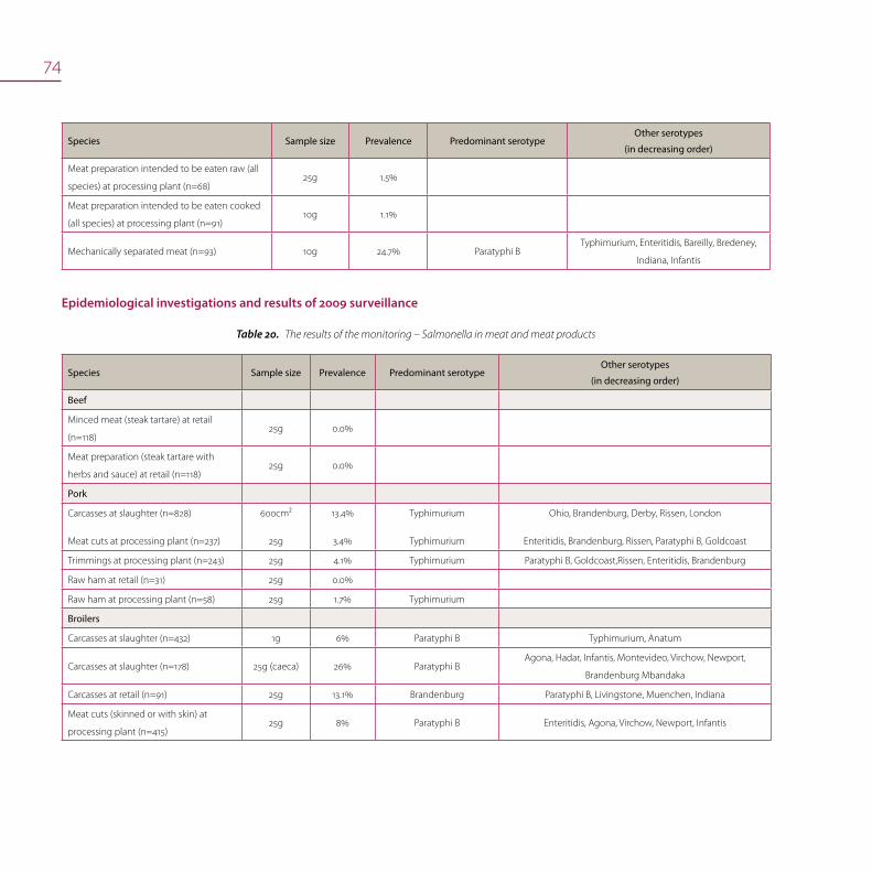
74
Species Sample size Prevalence Predominant serotypeOther serotypes
(in decreasing order)
Meat preparation intended to be eaten raw (all
species) at processing plant (n=68)25g 1.5%
Meat preparation intended to be eaten cooked
(all species) at processing plant (n=91)10g 1.1%
Mechanically separated meat (n=93) 10g 24.7% Paratyphi BTyphimurium, Enteritidis, Bareilly, Bredeney,
Indiana, Infantis
Epidemiological investigations and results of 2009 surveillance
The results of the monitoring – Salmonella in meat and meat productsTable20.
Species Sample size Prevalence Predominant serotypeOther serotypes
(in decreasing order)
Beef
Minced meat (steak tartare) at retail
(n=118)25g 0.0%
Meat preparation (steak tartare with
herbs and sauce) at retail (n=118)25g 0.0%
Pork
Carcasses at slaughter (n=828)
Meat cuts at processing plant (n=237)
600cm²
25g
13.4%
3.4%
Typhimurium
Typhimurium
Ohio, Brandenburg, Derby, Rissen, London
Enteritidis, Brandenburg, Rissen, Paratyphi B, Goldcoast
Trimmings at processing plant (n=243) 25g 4.1% Typhimurium Paratyphi B, Goldcoast,Rissen, Enteritidis, Brandenburg
Raw ham at retail (n=31) 25g 0.0%
Raw ham at processing plant (n=58) 25g 1.7% Typhimurium
Broilers
Carcasses at slaughter (n=432) 1g 6% Paratyphi B Typhimurium, Anatum
Carcasses at slaughter (n=178) 25g (caeca) 26% Paratyphi BAgona, Hadar, Infantis, Montevideo, Virchow, Newport,
Brandenburg Mbandaka
Carcasses at retail (n=91) 25g 13.1% Brandenburg Paratyphi B, Livingstone, Muenchen, Indiana
Meat cuts (skinned or with skin) at
processing plant (n=415)25g 8% Paratyphi B Enteritidis, Agona, Virchow, Newport, Infantis

75
Species Sample size Prevalence Predominant serotypeOther serotypes
(in decreasing order)
Meat cuts (skinned or with skin) at retail
(n=58)25g 1.7%
Layers
Carcasses at slaughter (n=350) 1g 36% Enteritidis Virchow, Infantis, Typhimurium, Indiana, Paratyphi B
Carcasses at slaughter (n=171) 25g (caeca) 43% EnteritidisParatyphi B, Typhimurium, Livingstone, Corvallis, Havana,
Virchow
Carcasses at retail (n=60) 25g 11.6% Typhimurium Paratyphi B, Livingstone, Enteritidis
Poultry
Meat cuts (with skin) at retail (n= 89) 25g 5.6% Paratyphi B Mbandaka
Meat cuts (without skin) at retail (n= 91) 25g 2.2%
Minced meat at retail (n=66) 10g 12.1% Paratyphi B Agona
Meat preparation at retail (n=60) 10g 16.7% Paratyphi B Agona, Enteritidis, Typhimurium
Meat preparation at processing plant
(n=58)10g 15.5% Typhimurium Agona, Enteritidis, Paratyphi B, Virchow
Meat products intended to be eaten
cooked at processing plant (n=37)10g 0.0%
Meat products intended to be eaten
cooked at retail (n=55) 10g 1.8% Paratyphi B
Meat and meat products
Minced meat intended to be eaten raw
(all species) at retail (n=120)25g 4.1% Typhimurium
Minced meat intended to be eaten
cooked at retail (all species) (n=120)10g 1.7% Typhimurium
Meat preparation intended to be eaten
raw (all species) at retail (n=40)25g 2.5% Typhimurium
Meat preparation intended to be eaten
cooked (all species) at retail (n=79)10g 1.3% Typhimurium
Meat preparation intended to be eaten
raw (all species) at processing plant
(n=77)
25g 1.3% Typhimurium

76
Species Sample size Prevalence Predominant serotypeOther serotypes
(in decreasing order)
Meat preparation intended to be eaten
cooked (all species) at processing plant
(n=77)
10g 3.9% Typhimurium Paratyphi B
Mechanically separated meat (n=116) 10g 15.5% TyphimuriumParatyphi B, Enteritidis, Infantis, Derby, Kentucky, Rissen,
Virchow, Heidelberg
The contamination rate of different products of pork, broilers and
layers is mentioned in the next table xx for the period 2005–2009.
The Salmonella contamination of pig carcasses increased in 2007
and decreased slightly successively in 2008 and 2009.
Evolution of the food Salmonella prevalence, 2005-2009Table21.
Samples
Samplin
g level
2005
2006
2007
2008
2009
PorkCarcasses
Cutting meats
600cm²
25g
9.3%
7.3%
7.1%
2.4%
16%
4.1%
14.6%
5.7%
13.4%
3.4%
Trimmings 25g 7.3% 2.4% 4.1% 4.1% 4.1%
Broilers Carcasses 1g 5.7% 1.4% 10.3% 7% 6%
Cutting meats 25g 14.2% 13.3% 7.4% 7.2% 8%
Layers Carcasses 1g 22.6% 35.6% 45.4% 23.4% 36%
Salmonella in other food of animal origin
The results of the national monitoring program of milk and dairy
products for 2008 are as follows:
at retail: nursing bottles (n=117), cheese of pasteurised milk •
(n=137), cheese of raw milk (n=89), butter of raw milk (n=20),
milk desserts (n=40), ice cream (n=62)
at processing plant: cheese of raw milk (n=41), cheese of pas-•
teurised milk (n=99), milk powder (n=21)
at farm: cheese of raw milk (n=92), butter (n=49), cream (n=20), •
ice cream (n=30).
All results were negative.
The results of the national monitoring program in other foods of
animal origin for 2008 are as follows:
at retail: fresh fish (n=62), cooked molluscs (n=62), cooked •
crustaceans (n=31), meat salad (n=47), chicken salad (n=46),
crustacean salad (n=45), bakery products with cream (n=76),
live bivalve molluscs (n=61), raw crustaceans (n=54), choco-
late (n=30), confectionary with chocolate (n=30), table eggs
(n=129), liquid egg products (n=54), salty preparation based on
raw eggs (n=105), dessert based on raw eggs (n=118)
at processing plant: gelatine (n=10), cooked crustaceans and •
molluscs (n=108), sandwich spreads (crustacean, meat, chicken)
(n=137), chocolate (n=15) and confectionery (n=16), bakery
products with cream (n=40), egg products (n=125).
All results were negative, except for raw crustaceans at retail
(14.8% positive) and egg products at processing plant (0.8% posi-
tive).
77
The results of the national monitoring program of milk and dairy
products for 2009 are as follows:
at retail: dried follow-on formula (n=86), nursing bottles (n=115), •
cheese of pasteurised milk (n=116), cheese of raw milk (n=76),
butter of raw milk (n=11), milk desserts (n=23), ice cream (n=61)
at processing plant: cheese of raw milk (n=40), cheese of pas-•
teurised milk (n=97), milk desserts (n=23), milk powder (n=45),
ice cream (n=30)
at farm: cheese of raw milk (n=94), butter (n=51), cream (n=20).•
All results were negative, except for milk desserts at retail (4.3%
positive).
The results of the national monitoring program in other foods of
animal origin for 2009 are as follows:
at retail: fresh fish (n=62), cooked molluscs (n=60), cooked •
crustaceans (n=22), meat salad (n=45), chicken salad (n=45),
crustacean salad (n=59), bakery products with cream (n=59),
live bivalve molluscs (n=95), raw crustaceans (n=131), chocolate
(n=30), confectionary with chocolate (n=30), table eggs (n=118),
liquid egg products (n=15), salty preparation based on raw
eggs (n=21), dessert based on raw eggs (n=90)
at processing plant: gelatine (n=4), cooked crustaceans and •
molluscs (n=154), sandwich spreads (crustacean, meat, chicken)
(n=149), chocolate (n=15) and confectionery (n=16), bakery
products with cream (n=31), egg products (n=76).
All results were negative, except for live bivalve molluscs at
retail (0.6% positive) and egg products at processing plant (1.3%
positive).
Salmonella in humans
Surveillance programme and methods used
Data about human salmonellosis cases and human isolates were
obtained from 160 clinical laboratories. All isolates were serotyped
by slide agglutination with commercial antisera following the
Kauffmann-White scheme. When necessary, additional biochemi-
cal tests were performed to confirm the identification or to dif-
ferentiate between the subspecies. Phage typing and antimicro-
bial susceptibility testing were performed on isolates randomly
sampled from the four serotypes Enteritidis, Typhimurium, Hadar
and. All isolates of S. Brandenburg, S. Derby, S. Infantis, S. Newport, S.
Typhi and S. Paratyphi were selected and tested for their antimi-
crobial susceptibility.
The objective of the national surveillance programme is to docu-
ment the occurrence and trends of serotypes, to detect local,
regional, national or even international outbreaks, to find and
eliminate the source and to suggest preventive actions to the
FASFC. This national Salmonella surveillance also intended to rap-
idly interact at the international level via electronic communica-
tion (with the Food and Water Diseases international surveillance
network, ECDC) and helped detecting outbreaks and targeting
preventive strategies.
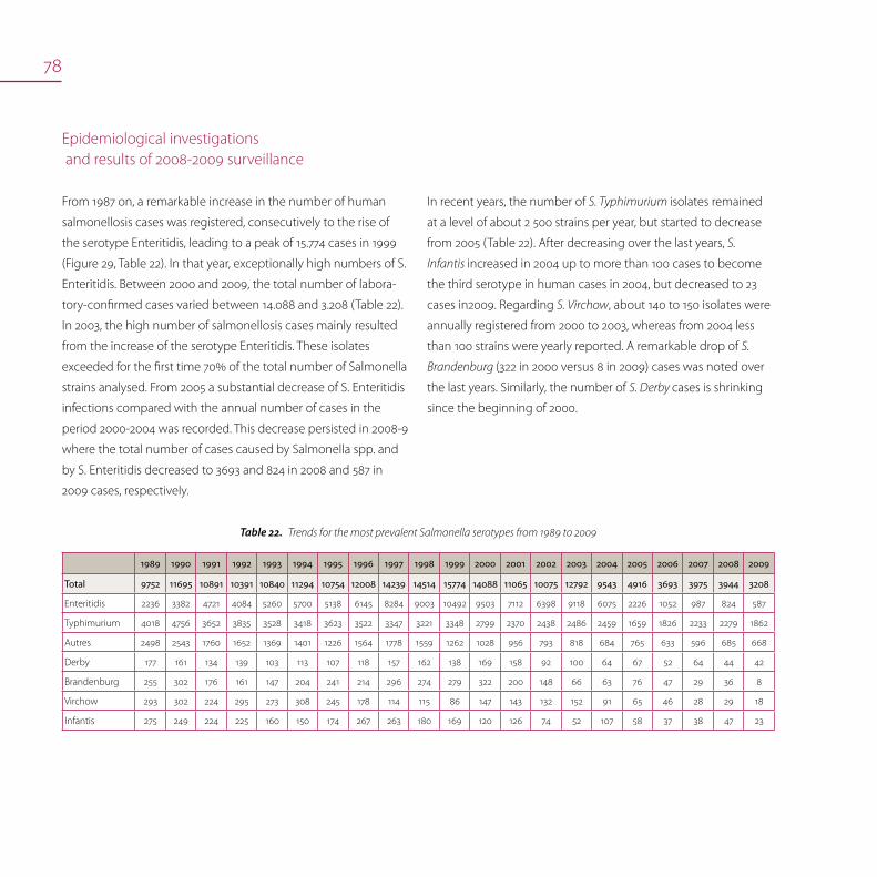
78
Epidemiological investigations and results of 2008-2009 surveillance
From 1987 on, a remarkable increase in the number of human
salmonellosis cases was registered, consecutively to the rise of
the serotype Enteritidis, leading to a peak of 15.774 cases in 1999
(Figure 29, Table 22). In that year, exceptionally high numbers of S.
Enteritidis. Between 2000 and 2009, the total number of labora-
tory-confirmed cases varied between 14.088 and 3.208 (Table 22).
In 2003, the high number of salmonellosis cases mainly resulted
from the increase of the serotype Enteritidis. These isolates
exceeded for the first time 70% of the total number of Salmonella
strains analysed. From 2005 a substantial decrease of S. Enteritidis
infections compared with the annual number of cases in the
period 2000-2004 was recorded. This decrease persisted in 2008-9
where the total number of cases caused by Salmonella spp. and
by S. Enteritidis decreased to 3693 and 824 in 2008 and 587 in
2009 cases, respectively.
In recent years, the number of S. Typhimurium isolates remained
at a level of about 2 500 strains per year, but started to decrease
from 2005 (Table 22). After decreasing over the last years, S.
Infantis increased in 2004 up to more than 100 cases to become
the third serotype in human cases in 2004, but decreased to 23
cases in2009. Regarding S. Virchow, about 140 to 150 isolates were
annually registered from 2000 to 2003, whereas from 2004 less
than 100 strains were yearly reported. A remarkable drop of S.
Brandenburg (322 in 2000 versus 8 in 2009) cases was noted over
the last years. Similarly, the number of S. Derby cases is shrinking
since the beginning of 2000.
Trends for the most prevalent Salmonella serotypes from 1989 to 2009Table22.
1989 1990 1991 1992 1993 1994 1995 1996 1997 1998 1999 2000 2001 2002 2003 2004 2005 2006 2007 2008 2009
Total 9752 11695 10891 10391 10840 11294 10754 12008 14239 14514 15774 14088 11065 10075 12792 9543 4916 3693 3975 3944 3208
Enteritidis 2236 3382 4721 4084 5260 5700 5138 6145 8284 9003 10492 9503 7112 6398 9118 6075 2226 1052 987 824 587
Typhimurium 4018 4756 3652 3835 3528 3418 3623 3522 3347 3221 3348 2799 2370 2438 2486 2459 1659 1826 2233 2279 1862
Autres 2498 2543 1760 1652 1369 1401 1226 1564 1778 1559 1262 1028 956 793 818 684 765 633 596 685 668
Derby 177 161 134 139 103 113 107 118 157 162 138 169 158 92 100 64 67 52 64 44 42
Brandenburg 255 302 176 161 147 204 241 214 296 274 279 322 200 148 66 63 76 47 29 36 8
Virchow 293 302 224 295 273 308 245 178 114 115 86 147 143 132 152 91 65 46 28 29 18
Infantis 275 249 224 225 160 150 174 267 263 180 169 120 126 74 52 107 58 37 38 47 23
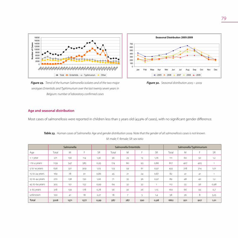
79
02000400060008000
1000012000140001600018000
198019
8119
8219
8319
8419
8519
8619
8719
8819
8919
9019
9119
9219
9319
9419
9519
9619
9719
9819
9920
0020
0120
0220
0320
0420
0520
0620
0720
0820
09
Num
ber o
f Cas
es
Total Enteritidis Typhimurium Other
Trend of the human Salmonella isolates and of the two major Figure29.
serotypes Enteritidis and Typhimurium over the last twenty seven years in
Belgium: number of laboratory confirmed cases
Age and seasonal distribution
Most cases of salmonellosis were reported in children less than 5 years old (43,9% of cases), with no significant gender difference.
Human cases of Salmonella: Age and gender distribution 2009. Note that the gender of all salmonellosis cases is not known. Table23.
M: male; F: female; SR: sex ratio
Salmonella Salmonella Enteritidis Salmonella Typhimurium
Age Total M F SR Total M F SR Total M F SR
< 1 year 271 156 114 1,36 36 23 13 1,76 111 60 50 1,2
1 to 4 years 1139 547 585 0,93 174 80 93 0,86 817 407 405 1
5 to 14 years 636 321 309 1,03 123 59 61 0,97 433 218 214 1,01
15 to 24 years 169 78 91 0,86 45 21 24 0,87 82 41 41 1
25 to 44 years 270 138 130 1,06 71 35 36 0,97 89 48 40 1,2
45 to 64 years 305 151 152 0,99 64 32 32 1 112 55 56 0,98
≥ 65 years 318 139 178 0,78 56 30 26 1,15 160 66 93 0,7
unknown 100 41 18 2,27 18 7 5 1,4 58 26 8 3,25
Total 3208 1571 1577 0,99 587 287 290 0,98 1862 921 907 1,01
Seasonal Distribution 2005-2009
0100200300400500600700
Jan Feb Maa Apr Mei Jun Jul Aug Sep Oct Nov Dec
2005 2006 2007 2008 2009
Seasonal distribution 2005 – 2009Figure30.

80
Antimicrobial resistance
Antimicrobial resistance in isolates from living animals
Methods used
The NRL for Salmonella, Animal Health, performed the antibiotic
susceptibility testing of Salmonella strains from livestock. Suscep-
tibility tests were done by means of the disk diffusion test, using
Neo-Sensitabs (Rosco). Tests and interpretation were done ac-
cording to the manufacturers guidelines using the methodology
as described by CLSI. Internal control was performed with quality
control strain E. coli ATCC25922. Results were only accepted when
results with the QC strain were within the limits as proposed by
Rosco. In order to reduce bias due to multiple strains from the
same origin at the same sampling time and belonging to the
same serotype, only one isolate per serotype and per origin was
selected for susceptibility testing. Therefore, strains were likely to
be independent from each other.
Animal Salmonella: list of antimicrobials tested - For all suscep-Table24.
tibility tests Neo-Sensitabs from Rosco were used according to the providers
instructions
Antimicrobial Amount of antimicrobial Breakpoints (mm)
Ampicillin 33μg 17 – 19
Ceftiofur 30μg 20 – 22
Streptomycin 100μg 23 – 25
Neomycin 120μg 20 – 22
Gentamicin 40μg 20 – 22
Tetracycline 80μg 20 – 22
Sulphonamides 240μg 20 – 22
Trimethoprim - Sulphonamides 5.2μg + 240μg 27 – 31
Nalidixic acidid 130μg 21 – 24
Enrofloxacin 10μg 20 – 22
Chloramphenicol 60μg 21 – 24
Florfenicol 30μg 15 - 18
Epidemiological investigations
and results of 2008-2009 surveillance
The susceptibility of 1 125 and 1 128 Salmonella isolates was tested
in 2008 and 2009, respectively. A total of 706 (62.6% in 2008) and
613 (54.3% in 2009) of Salmonella isolates was fully susceptible
to all antimicrobial drugs tested. The apparent lower proportion
of susceptible isolates may be due to the lower proportion of S.
Enteritidis strains tested in 2009.

81
In 2008, 22.8% of S. Paratyphi B (n=31) strains were susceptible to
tha antimicrobials tested. Resistance was mainly observed against
St (74.2%), Ap and Nal (both 54.8%) and against Su (48.4%). Also
resistance against Cef was frequently observed (38.7%). However,
5.1% of 2009 S. Paratyphi B (n=78) isolates were fully sensitive. The
most abundant profile was Ap Nal (69.2%). Almost half of the
strains were found Cef resistant.
Only 22% to 23% of S. Typhimurium isolates (252 tested in 2008
and 289 in 2009) were found susceptible. Pentaresistance Ap St
Tc Su Cm was encountered in 21.8% and 17.3% of the 2008 and
2009 isolates, respectively. Ff resistance was detected in 11.4%
and 13.5% of the strains, whereas Cef resistance in 2.4% and 0.3%,
respectively.
In 2008 and 2009, 22 and 13 S. Virchow isolates were tested, and
about one quarter was found susceptible to the antimicrobials
used. As in former years, most resistance was found against Ap
(both 68.2% in 2008, 69.2% in 2009) and Nal (68.2% in 2008 and
53.8% in 2009). Cef resistance was remarkably high: 45.5%. in 2008
and 38.5 in 2009.
In 2009, resistance was mainly found against Ap (37.6%), Su
(32.1%), Tc (28.2%), St (23.9%), but also against TSu (21.8%), which
are similar figures as for 2008. Noteworthy however, is the resist-
ance against Nal (13.3%) and against Cef (7.1%), both considerably
higher than in 2008 (8.7% and 3.0%, respectively). Resistance
against Ne, Gm and Enr (all 0.4%), and against Cm and Ff (7.6%
and 3.5%, respectively) was similar to that of 2008.
Eighty to ninety percent of S. Agona isolates were found fully sus-
ceptible for all antimicrobials tested: 92.9% of 42 strains in 2008
and 90.9% of 11 strains in 2009.
A limited number of S. Blockley isolates were tested (11 in 2008 and
7 in 2009). In 2008, all were resistant against Ap, Su and Nal, 90.9%
against TSu and 81.8% against Tc. All 2009 isolates were from
poulty and had the resistance profile Ap Tc Su Tsu Nal.
Around 60.0% of S. Derby strains (50 isolates tested in 2008 and 26
in 2009) were sensitive, although 25 to 30% of strains were resist-
ant against Tc and Su.
As for the 14 S. Dublin isolates tested in 2008, 28.6% were found
completely susceptible. Resistance against Cm (57.13%), Su and
Nal (both 50.0%) and St (14.3%) was noticed. In 2009, about half of
20 isolates were found completely susceptible.
Both in 2008 and 2009, about 95% of S. Enteritidis isolates (185 and
115 tested) were susceptible.
Only four S. Hadar strains were tested in 2008 and all were found
resistant against St Tc Nal (100%). Two strains were Enr resistant. In
2009, 22 S. Hadar strains were tested and only one (from poultry)
was found sensitive. Resistance profiles Ap Tc Nal and Tc Nal were
most often demonstrated (45.5% and 40.9%, respectively).
All the S. Indiana strains (16 tested in 2008 and 17 in 2009) were
multi-resistant and most showed profile Ap St Su TSu.
More than 80% of the S. Infantis isolates (n=42 in 2008 and 47
in 2009) were susceptible to the antibiotics tested. Two or three
strains per year were found to be Cef resistant.

82
Antimicrobial resistance in strains isolated from meat and meat products
Surveillance programme and method used
During 2008 and 2009, respectively, 718 and 682 strains of Salmo-
nella enterica isolated during the zoonosis monitoring program
were sent to IPH for serotyping and determination of antimi-
crobial resistance. Different food matrices were sampled, mainly
poultry (carcasses from broilers and spent hens, chicken parts and
meat preparations) and pork (carcasses and cut meats). Other
matrices where Salmonella was isolated were ready-to-eat meals,
meat, meat preparations, bovine carcasses, frog’s legs, pudding,
liquid egg product, ham and dry sausage.
Minimum Inhibitory Concentrations (MIC) were determined by
the broth dilution method using Sensititre, as recommended
by the CRL antibiotic resistance. The antimicrobials reported are
listed in the table below, as well as the breakpoints used for the
interpretation of the results. Interpretation was according to CLSI
and using epidemiological cut-off values from EUCAST. Quality
control was performed by using an Escherichia coli ATCC 25922
strain.
Salmonella from food, list of antimicrobials tested with their Table25.
breakpoints
Antimicrobial Breakpoints (µg / ml)
Ampicillin 4
Cefotaxim 0.5
Ceftazidim 2
Chloramphenicol 16
Ciprofloxacin 0.06
Colistin 16
Florfenicol 16
Gentamicin 2
Kanamycin 8
Nalidixic acid 16
Streptomycin 32
Sulphamethoxazole 256
Tetracycline 8
Trimethoprim 2
The level of resistance of Salmonella isolates from poultry and
pork is influenced by the serotype distribution in the correspond-
ing matrix; results are summarized in the next table. The presence
of highly resistant serotypes as Hadar, Virchow, Paratyphi B and
Typhimurium contributed mainly to the high resistance levels in
some matrices. In 2008, 65 different serotypes were identified,
whereas in 2009 only 48. S. Typhimurium was the most preva-
lent serotype on pork (44%/56%-2008/2009) while S. Enteritidis
was dominantly found on carcasses of spent hens (57%/58%-
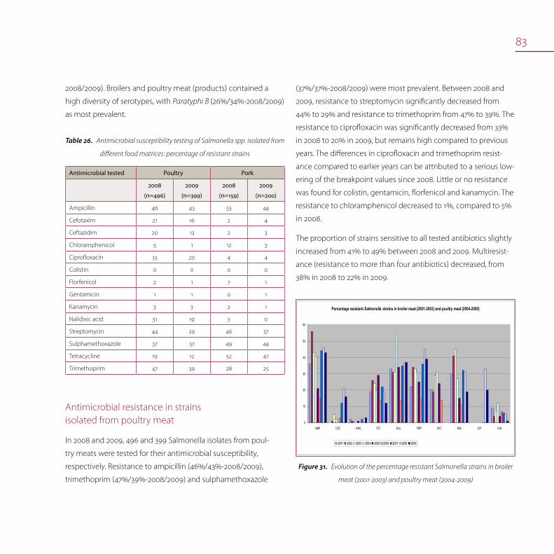
83
2008/2009). Broilers and poultry meat (products) contained a
high diversity of serotypes, with Paratyphi B (26%/34%-2008/2009)
as most prevalent.
Antimicrobial susceptibility testing of Salmonella spp. isolated from Table26.
different food matrices: percentage of resistant strains
Antimicrobial tested Poultry Pork
2008
(n=496)
2009
(n=399)
2008
(n=159)
2009
(n=200)
Ampicillin 46 43 53 44
Cefotaxim 21 16 2 4
Ceftazidim 20 13 2 3
Chloramphenicol 5 1 12 3
Ciprofloxacin 33 20 4 4
Colistin 0 0 0 0
Florfenicol 2 1 7 1
Gentamicin 1 1 0 1
Kanamycin 3 3 2 1
Nalidixic acid 31 19 3 0
Streptomycin 44 29 46 37
Sulphamethoxazole 37 37 49 44
Tetracycline 19 12 52 47
Trimethoprim 47 39 28 25
Antimicrobial resistance in strains isolated from poultry meat
In 2008 and 2009, 496 and 399 Salmonella isolates from poul-
try meats were tested for their antimicrobial susceptibility,
respectively. Resistance to ampicillin (46%/43%-2008/2009),
trimethoprim (47%/39%-2008/2009) and sulphamethoxazole
(37%/37%-2008/2009) were most prevalent. Between 2008 and
2009, resistance to streptomycin significantly decreased from
44% to 29% and resistance to trimethoprim from 47% to 39%. The
resistance to ciprofloxacin was significantly decreased from 33%
in 2008 to 20% in 2009, but remains high compared to previous
years. The differences in ciprofloxacin and trimethoprim resist-
ance compared to earlier years can be attributed to a serious low-
ering of the breakpoint values since 2008. Little or no resistance
was found for colistin, gentamicin, florfenicol and kanamycin. The
resistance to chloramphenicol decreased to 1%, compared to 5%
in 2008.
The proportion of strains sensitive to all tested antibiotics slightly
increased from 41% to 49% between 2008 and 2009. Multiresist-
ance (resistance to more than four antibiotics) decreased, from
38% in 2008 to 22% in 2009.
Percentage resistant Salmonella strains in broiler meat (2001-2003) and poultry meat (2004-2009)
0
10
20
30
40
50
60
AMP CEF KAN TET SUL TMP SXT NAL CIP CHL
2001 2002 2003 2004 2005 2006 2007 2008 2009
Evolution of the percentage resistant Salmonella strains in broiler Figure31.
meat (2001-2003) and poultry meat (2004-2009)

84
Compared to these general results, higher resistances were
observed in chicken meat for cooked consumption and chicken
parts (no carcasses) with, in 2009, 98% and 61% of the isolates
resistant to trimethoprim, 83% and 53% to sulphamethoxazole,
and 96% and 69% to ampicillin, respectively. Especially in chicken
meat for cooked consumption the resistance to these three
antibiotics drastically increased, compared to 2008 (78%, 62%
and 70%, respectively). However, multiresistance significantly
decreased among these isolates to 42% and 33%, respectively
(compared to 68% and 65% in 2008). On the other hand, Salmo-
nella isolates from spent hens showed little antibiotic resistance,
with only 6% and 9% showing multiresistance in 2008 and 2009,
respectively.
Antimicrobial resistance in strains isolated from pork
In total, 159 and 200 Salmonella strains from pork were tested for
their antibiotic susceptibility in 2008 and 2009, respectively. This
included strains from carcasses and cut meats. High resistance
was observed to tetracycline (52%/47%-2008/2009), ampicillin
(53%/44%-2008/2009), sulphamethoxazole (49%/44%-2008/2009)
and streptomycin (46%/37%-2008/2009). Resistance to four or
more antibiotics slightly decreased from 41% to 35% of the tested
isolates, while sensitivity to all antibiotics increased from 28% to
39% between 2008 and 2009. All strains were sensitive to colistin
and gentamicin. Low resistance was observed for cefotaxime
(2%/4%-2008/2009), ceftazidim (1%/3%-2008/2009), kanamycin
(3%/1%-2008/2009), ciprofloxacin (4%/4%-2008/2009) and flor-
fenicol (7%/1%-2008/2009). Compared to 2008, overall resistance
has slightly decreased in 2009.
S. Typhimurium was the most dominantly isolated serotype
(70%/56%-2008/2009) from pork. The observed trends are similar
as described above, with high resistance to ampicillin (70%/69%-
2008/2009), tetracycline (63%/61%-2008/2009), sulphameth-
oxazole (70%/60%-2008/2009) and streptomycin (59%/54%-
2008/2009). However, it is clear that Typhimurium strains are more
resistant than other Salmonella strains found on pork, with only
9% and 22% of all Typhimurium strains sensitive to all antibiotics
in 2008 and 2009, respectively.
Compared to previous years, ampicillin and tetracycline resist-
ance has increased. This can be explained by the lowering of the
breakpoint values used to assess resistance.
Evolution of the percentage resistant Salmonella strains Figure32.
in pork meat
Percentage resistant Salmonella strains in pork meat
0
10
20
30
40
50
60
AMP CEF KAN TET SUL TMP SXT NAL CIP CHL
2001 2002 2003 2004 2005 2006 2007 2008 2009
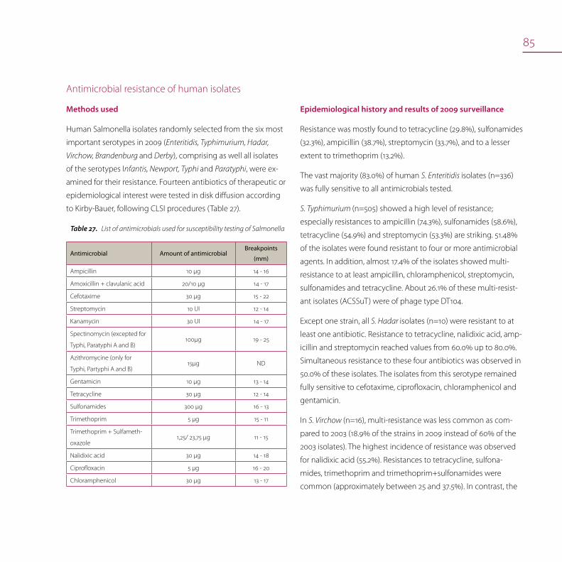
85
Antimicrobial resistance of human isolates
Methods used
Human Salmonella isolates randomly selected from the six most
important serotypes in 2009 (Enteritidis, Typhimurium, Hadar,
Virchow, Brandenburg and Derby), comprising as well all isolates
of the serotypes Infantis, Newport, Typhi and Paratyphi, were ex-
amined for their resistance. Fourteen antibiotics of therapeutic or
epidemiological interest were tested in disk diffusion according
to Kirby-Bauer, following CLSI procedures (Table 27).
List of antimicrobials used for susceptibility testing of SalmonellaTable27.
Antimicrobial Amount of antimicrobialBreakpoints
(mm)
Ampicillin 10 µg 14 - 16
Amoxicillin + clavulanic acid 20/10 µg 14 - 17
Cefotaxime 30 µg 15 - 22
Streptomycin 10 UI 12 - 14
Kanamycin 30 UI 14 - 17
Spectinomycin (excepted for
Typhi, Paratyphi A and B)100µg 19 - 25
Azithromycine (only for
Typhi, Partyphi A and B)15µg ND
Gentamicin 10 µg 13 - 14
Tetracycline 30 µg 12 - 14
Sulfonamides 300 µg 16 - 13
Trimethoprim 5 µg 15 - 11
Trimethoprim + Sulfameth-
oxazole1,25/ 23,75 µg 11 - 15
Nalidixic acid 30 µg 14 - 18
Ciprofloxacin 5 µg 16 - 20
Chloramphenicol 30 µg 13 - 17
Epidemiological history and results of 2009 surveillance
Resistance was mostly found to tetracycline (29.8%), sulfonamides
(32.3%), ampicillin (38.7%), streptomycin (33.7%), and to a lesser
extent to trimethoprim (13.2%).
The vast majority (83.0%) of human S. Enteritidis isolates (n=336)
was fully sensitive to all antimicrobials tested.
S. Typhimurium (n=505) showed a high level of resistance;
especially resistances to ampicillin (74.3%), sulfonamides (58.6%),
tetracycline (54.9%) and streptomycin (53.3%) are striking. 51.48%
of the isolates were found resistant to four or more antimicrobial
agents. In addition, almost 17.4% of the isolates showed multi-
resistance to at least ampicillin, chloramphenicol, streptomycin,
sulfonamides and tetracycline. About 26.1% of these multi-resist-
ant isolates (ACSSuT) were of phage type DT104.
Except one strain, all S. Hadar isolates (n=10) were resistant to at
least one antibiotic. Resistance to tetracycline, nalidixic acid, amp-
icillin and streptomycin reached values from 60.0% up to 80.0%.
Simultaneous resistance to these four antibiotics was observed in
50.0% of these isolates. The isolates from this serotype remained
fully sensitive to cefotaxime, ciprofloxacin, chloramphenicol and
gentamicin.
In S. Virchow (n=16), multi-resistance was less common as com-
pared to 2003 (18.9% of the strains in 2009 instead of 60% of the
2003 isolates). The highest incidence of resistance was observed
for nalidixic acid (55.2%). Resistances to tetracycline, sulfona-
mides, trimethoprim and trimethoprim+sulfonamides were
common (approximately between 25 and 37.5%). In contrast, the

86
vast majority of S. Brandenburg (n=8) and S. Derby (n=42) isolates
remained sensitive to the vast majority of tested antibiotics: 75.0%
and 66.7% sensitive, respectively.
S. Infantis (N= 23) displayed in general a low level of multi-resist-
ance.
The vast majority of S. Paratyphi B (n=73) and S. Newport (n=25)
isolates remained sensitive to the vast majority of tested antibiot-
ics: respectively 56.2% and 92% were fully sensitive to all anti-
microbials tested. However, two isolates of S. Newport displayed
resistance to at least 5 antibiotics but remained sensitive to
cefotaxime and ciprofloxacin.
No tendency could be observed from the results on S. Typhi. That
could be due to the fact that most of isolates are travel-associated
and that the origins (country/region) of the isolates were differ-
ent.
In general, resistance patterns and levels of Salmonella isolated in
2009 were comparable to those from 2002-2008.

87
Phagetyping of human isolates
In 2009, a total of 336 human S. Enteritidis isolates were phage
typed. Of these, 16.0% were PT 21 and 19% were PT 4. In addition,
505 S. Typhimurium isolates were phage typed and most prevalent
types were DT104 (5.5%), DT120 (32.5%), DT193 (15.8%), and U302
(13.4%).
Antimicrobial resistance in human Salmonella of serotypes Enteritidis, Typhimurium, Brandenburg, Derby, Hadar, Virchow, Infantis, Table28.
Typhi, Newport, Paratyphi B and A isolated in 2009
Total N Amp Amx Ctx Tet Nal Cip Azy Spe Gen Kan Chl Stp Tmp Sul Stx
Enteritidis 587 336 6,0 0,6 0,9 2,1 12,5 0,0 ND 1,5 0,3 0,3 0,0 1,8 0,9 1,8 1,2
Typhimurium 1862 505 74,3 0,8 1,0 54,9 2,8 0,0 ND 23,4 1,4 2,6 13,1 53,3 20,2 58,6 20,4
Derby 42 42 14,3 0,0 0,0 21,4 4,8 0,0 ND 7,1 0,0 2,4 0,0 19,0 16,7 21,4 19,0
Hadar 12 10 80 10 0 80 60 0 ND 0 0 10 0 60 0 10 0
Infantis 23 23 4,3 0,0 0,0 21,7 30,4 0,0 ND 17,4 4,3 8,7 4,3 17,4 21,7 21,7 17,4
Virchow 18 16 12,5 0 0 25 43,75 0 ND 6,25 12,5 12,5 6,25 6,25 25 37,5 25
Brandenburg 8 8 0 0 0 25 0 0 ND 0 0 0 0 0 0 0 0
Newport 25 25 8 0 0 8 11,5 0 ND 0 0 0 4 4 4 8 4
Paratyphi B 73 73 24,7 2,7 11,0 16,4 15,1 0,0 11,0 ND 0,0 1,4 4,1 30,1 21,9 19,2 21,9
Typhi 26 25 24 0 0 12 56 16 8 ND 0 0 16 24 16 24 16
Dublin 8 8 0 0 0 0 12,5 0 ND 50 0 0 50 50 0 50 12,5
Paratyphi A 5 5 0 0 0 0 100 0 80 ND 0 0 0 0 0 0 0

88
89
Zoonotic tuberculosis (Mycobacterium bovis)
Mycobacterium bovis in cattle
Mycobacterium bovis in wildlife
Mycobacterium bovis in humans
Human tuberculosis (Mycobacterium tuberculosis)
TuberculosisMaryse Fauville-Dufaux, Marc Govaerts, Luc Vanholme, Maryse Wanlin
Zoonotic tuberculosis (Mycobacterium bovis)
Tuberculosis in humans caused by Mycobacterium bovis is rare.
In regions where M. bovis infections in cattle are largely eliminat-
ed, only few residual cases occur either among elderly persons as
a result of the reactivation of dormant M. bovis within old lesions
and among migrants from high-prevalence countries. Agricultural
workers may get infected by M. bovis by inhaling aerosols from
coughing infected cattle and may subsequently develop typical
pulmonary or genito-urinary tuberculosis. Such patients may
infect cattle through cough or urine. Evidence for human-to-hu-
man transmission of zoonotic tuberculosis is only rarely reported.
In developing countries, where M. bovis is largely prevalent
among cattle, some studies reported that 3-6% of all tubercu-
losis cases are due to M. bovis and that mostly young people
get infected through the ingestion of contaminated raw milk.
Also occupational contacts should be regarded as a risk factor
for transmission to humans, although companion animals can
provide a less common indirect route of infection.

90
In humans, the disease caused by M. bovis is clinically indis-
tinguishable from that caused by M. tuberculosis. Pulmonary
tuberculosis is frequently observed and cervical lymphadenopa-
thy, intestinal lesions, chronic skin tuberculosis and other non
pulmonary forms are particularly common.
Mycobacterium bovis in cattleBelgium is officially free from bovine tuberculosis (Mycobacte-
rium bovis) since 25 June 2003 (Commission Decision 2003/467/
EC establishing the official tuberculosis, brucellosis and enzootic
bovine leucosis free status of certain Member States and regions
of Member States as regards bovine herds).
Surveillance programme
The control of tuberculosis is based on Council Directive 64/432/
EEC, which is implemented and adapted in the national legisla-
tion since 1963 and was last amended by Royal Decree of 17
October 2002.
The control implies:
Skin testing of animals at purchase (mandatory),•
In case of a positive reactor, skin testing of all the animals of the •
holding and skin testing of all contact animals (tracing on and
tracing back),
Systematic post mortem examinations at the slaughterhouse. •
In case a suspected lesion is identified, a sample is sent to the
NRL for analysis.
The FASFC is informed about any doubtful or positive result of
the skin test and may decide to re-examine (additional tests)
the animals or to kill them (test slaughter, additional tests). If M.
bovis is isolated as a consequence of post mortem examinations
or of mandatory test-slaughter, all animals in the herd of origin
are skin tested and a complete epidemiological investigation is
performed.
An animal is defined as infected with bovine tuberculosis if the
skin testing is positive or if M. bovis is isolated by culture or
confirmed by laboratory testing (PCR). A holding is defined as in-
fected if M. bovis was isolated or detected by PCR from an animal
of the holding.
Isolation of M. bovis and biochemical testing is exclusively per-
formed in the NRL where also IFN-gamma and molecular typing
by means of IS6110 RFLP, spoligotyping and MIRU-VNTR are done.
In Belgium, vaccination against tuberculosis is prohibited.
Epidemiological investigations and results of 2008-2009 surveillance
At the slaughterhouse, tissue samples from individual animals
were taken. The samples originated from animals suspected of
being infected with M. bovis, i.e. skin test reactors, animals that
had been in contact with M. bovis infected animals or animals
that showed suspicious lesions at meat inspection. The samples
were submitted to the NRL where culture, PCR and confirmatory
tests were done. M. bovis was only detected in animals belonging
to the outbreak herds.
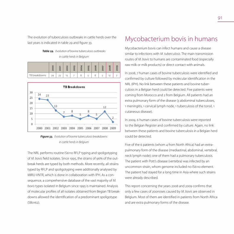
91
The evolution of tuberculosis outbreaks in cattle herds over the
last years is indicated in table 29 and figure 33.
Evolution of bovine tuberculosis outbreaks Table29.
in cattle herds in Belgium
2000
2001
2002
2003
2004
2005
2006
2007
2008
2009
TB breakdowns 24 23 13 7 8 5 8 5 12 2
Evolution of bovine tuberculosis breakdowns Figure33.
in cattle herds in Belgium
The NRL performs routine IS6110 RFLP typing and spoligotyping
of M. bovis field isolates. Since 1995, the strains of 96% of the out-
break herds are typed by both methods. More recently, all strains
typed by RFLP and spoligotyping were additionally analysed by
MIRU-VNTR, which is done in collaboration with IPH. As a con-
sequence, a comprehensive database of the vast majority of M.
bovis types isolated in Belgium since 1995 is maintained. Analysis
of molecular profiles of all isolates obtained from Begian TB break-
downs allowed the identification of a predominant spoligotype
(SB0162).
Mycobacterium bovis in humansMycobacterium bovis can infect humans and cause a disease
similar to infections with M. tuberculosis. The main transmission
routes of M. bovis to humans are contaminated food (especially
raw milk or milk products) or direct contact with animals.
In 2008, 7 human cases of bovine tuberculosis were identified and
confirmed by culture followed by molecular identification in the
NRL (IPH). No link between these patients and bovine tuber-
culosis in a Belgian herd could be detected. Five patients were
coming from Morocco and 2 from Belgium. All patients had an
extra pulmonary form of the disease (3 abdominal tuberculoses,
1 meningitis, 1 cervical lymph node, 1 tuberculosis of the tonsil, 1
cutaneous disease).
In 2009, 6 human cases of bovine tuberculosis were reported
to the Belgian Register and confirmed by culture. Again, no link
between these patients and bovine tuberculosis in a Belgian herd
could be detected.
Five of the 6 patients (whom 4 from North Africa) had an extra-
pulmonary form of the disease (mediastinal, abdominal, vertebral,
neck lymph node); one of them had a pulmonary tuberculosis.
The patient with Pott’s disease (vertebra) was infected by an
uncommon strain, whom genome included no IS6110 element.
The patient had stayed for a long time in Asia where such strains
were already described.
This report concerning the years 2008 and 2009 confirms that
only a few cases of zoonoses caused by M. bovis are observed in
Belgium. Most of them are identified in patients from North Africa
and are extra pulmonary forms of the disease.

92
Human tuberculosis (Mycobacterium tuberculosis)The incidence of human tuberculosis shows little variation over
the last years. In 2001, 2002, 2003, 2004, 2005, 2006, 2007, 2008
and 2009 respectively 1321, 1309, 1128, 1226, 1144, 1127, 1028, 1006
and 1020 new notified cases of active human tuberculosis were
detected. Over the 60% were male patients. In 2009, 49% of the
tuberculosis cases were foreigners.
Groups at risk are persons with a marginal existence, asylum
seekers and refugees. Alcoholism and a co-infection with HIV
are known as specific risk factors. Human tuberculosis cases are
mainly concentrated in urban populations.
93
Yersinia enterocolitica
Yersinia enterocolitica in food
Yersiniosis in humans
YersiniosisKatrien Beullens, Michel Delmée, Lieven De Zutter, Geneviève Ducoffre, Luc Vanholme
Yersinia enterocoliticaThe genus Yersinia comprises three main species that are known
to cause human infections: Yersinia enterocolitica, Y. pseudotuber-
culosis and Y. pestis (plague which is believed to no longer exist in
Europe). Specific types of Y. enterocolitica and Y. pseudotuberculosis
cause foodborne infections. At the EU level Yersiniosis is the third
common zoonotic pathogen with 8348 reported human cases in
2008. Since most human infections are caused by Y. enterocolitica,
this chapter deals mainly with Y. enterocolitica infections.
Only certain biotypes of strains of Y. enterocolitica cause illness
in humans. Infection with Y. enterocolitica most often causes
diarrhoea and abdominal pain and occurs most often in young
children. Common symptoms in children are fever, abdominal
pain and diarrhoea, which is often bloody. Symptoms typically
develop 4 to 7 days after exposure and may last 1 to 3 weeks or
longer. In older children and adults, right-sided abdominal pain
and fever may be the predominant symptoms and may be con-
fused with appendicitis. In a small proportion of cases, complica-
tions such as skin rash, joint pains, and/or bacteraemia may occur.

94
Pigs are the primary reservoir for the human pathogenic Y.
enterocolitica types. In infected pigs the bacteria are most likely to
be found in the tonsils. However other animal species, e.g. cattle,
sheep, deer, small rodents, cats and dogs may also carry patho-
genic biotypes. Clinical disease in animals is uncommon.
Infection is most often acquired by eating contaminated food,
particularly raw or undercooked pork. Since the microorganism
is able to grow at + 4°C the risk for the development of human
infection may increase when contaminated refrigerated food
with a relatively long shelf life is consumed. Drinking contaminat-
ed unpasteurised milk or untreated water can also transmit the
organism. On rare occasions, transmission may occur by direct
contact with infected animals or humans.
Within Y. enterocolitica, the majority of isolates from food and en-
vironmental sources belongs to non-pathogenic types. It is, there-
fore, most important to investigate which isolates are pathogenic
for humans. Biotyping of the isolates is essential to determine
whether or not isolates are pathogenic to humans, and this typ-
ing is ideally complemented by serotyping.
Infection caused by Y. pseudotuberculosis shows many similarities
with the disease pattern of Y. enterocolitica. Infections are caused
by the ingestion of the bacteria from raw vegetables, fruit, other
foodstuffs or via contaminated water or by direct contact with
infected animals.
Yersinia enterocolitica in food
Monitoring programme
The FASFC organised a monitoring of meat since 1997, which
showed a very low prevalence of Yersinia enterocolitica in pork,
beef and poultry. In 2008 and 2009, the monitoring programme
concentrated on one matrix, i.e. minced meat containing pork
meat.
Monitoring Yersinia enterocolitica in pork meatTable30.
Sample
Qua
ntity an
alysed
Percen
tage
of
pos
itive
sam
ple
s
2008
Percen
tage
of
pos
itive
sam
ple
s
2009
Minced meat (containing pig
meat) intended to be eaten raw
at retail
1g0.0%
(n=129)
0.0%
(n=184)
Minced meat (containing pig
meat) intended to be eaten
cooked at retail
1g0.0%
(n=125)
0.0%
(n=179)
Yersiniosis in humansIn 2008 and 2009, the Sentinel Laboratory Network registered 273
and 240 cases respectively, corresponding to a national incidence
estimated at 2.6 and 2.2 per 100,000 inhabitants.
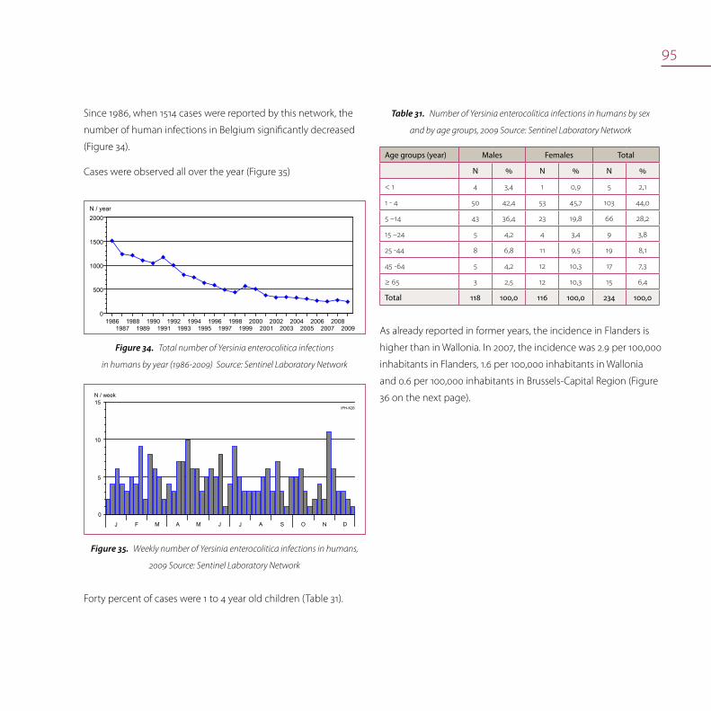
95
Since 1986, when 1514 cases were reported by this network, the
number of human infections in Belgium significantly decreased
(Figure 34).
Cases were observed all over the year (Figure 35)
19861987
19881989
19901991
19921993
19941995
19961997
19981999
20002001
20022003
20042005
20062007
20082009
0
500
1000
1500
2000N / year
Total number of Yersinia enterocolitica infections Figure34.
in humans by year (1986-2009) Source: Sentinel Laboratory Network
N / week
J F M A M J AJ S O N D
IPH-K26
0
15
5
10
Weekly number of Yersinia enterocolitica infections in humans, Figure35.
2009 Source: Sentinel Laboratory Network
Number of Yersinia enterocolitica infections in humans by sex Table31.
and by age groups, 2009 Source: Sentinel Laboratory Network
Age groups (year) Males Females Total
N % N % N %
< 1 4 3,4 1 0,9 5 2,1
1 - 4 50 42,4 53 45,7 103 44,0
5 –14 43 36,4 23 19,8 66 28,2
15 –24 5 4,2 4 3,4 9 3,8
25 -44 8 6,8 11 9,5 19 8,1
45 -64 5 4,2 12 10,3 17 7,3
≥ 65 3 2,5 12 10,3 15 6,4
Total 118 100,0 116 100,0 234 100,0
As already reported in former years, the incidence in Flanders is
higher than in Wallonia. In 2007, the incidence was 2.9 per 100,000
inhabitants in Flanders, 1.6 per 100,000 inhabitants in Wallonia
and 0.6 per 100,000 inhabitants in Brussels-Capital Region (Figure
36 on the next page).
Forty percent of cases were 1 to 4 year old children (Table 31).
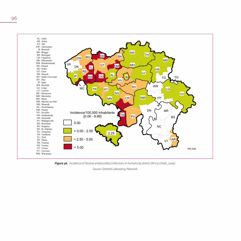
96
AL AR AT
AW B
BG BS CR DK DM DN EK GT HS HV HY
IP KR LG LV MC MH MN MR MS NC NM NV OD OS PV RS SG SN TG TH TL TN TR VR VT VV WR
: Aalst : Arlon : Ath : Antwerpen : Brussels : Brugge : Bastogne : Charleroi : Diksmuide : Dendermonde : Dinant : Eeklo : Gent : Hasselt : Halle-Vilvoorde : Huy : Ieper : Kortrijk : Liège : Leuven : Mouscron : Mechelen : Mons : Marche-en-Fam. : Maaseik : Neufchâteau : Namur : Nivelles : Oudenaarde : Oostende : Philippeville : Roeselare : Soignies : St.-Niklaas : Tongeren : Turnhout : Tielt : Thuin : Tournai : Veurne : Virton : Verviers : Waremme
0.00
2.25
Incidence/100,000 inhabitants[0.00 - 9.89]
> 0.00 - 2.50
> 2.50 - 5.00
> 5.00
VR
BG
DK TLGT
SN
ALOD
MH
HV
LV
AWTH
MS
HS
TG
NV
MRDN
CR
NC
AR
MC TR
KR
SG
IP
TN PV
VT
BS
VV
LG
HY
WR
TG
MN NM
B
DM
EK
RS
AT
OS
IPH-K26
Incidence of Yersinia enterocolitica infections in humans by district (N/105 inhab., 2009) Figure36.
Source: Sentinel Laboratory Network

97
Bio-serotyping was performed by the NRLs.
In 2008, 73.8% of the 397 isolates tested belonged to the patho-
genic bio-serotypes (including 6 Yersinia pseudotuberculosis) with
serotype O:3 / biotype 4 accounting for 68.7% of the total. The re-
maining 104 strains (26.2%) belonged to non-pathogenic Yersinia
enterocolitica bio-serotypes or related species.
In 2009, 72% of the 359 isolates tested belonged to the pathogen-
ic bio-serotypes (including 12 Yersinia pseudotuberculosis) with
serotype O:3 / biotype 4 accounting for 66,3% of the total. The
remaining 101 strains (28%) belonged to non-pathogenic Yersinia
enterocolitica bio-serotypes or related species.
The number of non-pathogenic Yersinia strains did not vary mark-
edly during the last years, in contrast to the obvious decrease of
the pathogenic strains.
trends and sources
viral diseases

99
Avian influenzaBénédicte Lambrecht, Annick Linden, Thierry van den Berg, Didier Vangeluwe, Françoise Wuillaume
GeneralA total of more or less 100,000 wild birds, from 27 Member
States of the European Union was tested during the 2008 and
2009 active avian influenza (AI) surveillance. In contrast to 2007,
where cases in wild birds were reported from four Member
States (Germany, France, the Czech Republic and Hungary), in
2008 and 2009 H5N1 HPAI were reported from only two Member
States, i.e. United Kingdom and Germany. In 2008, the incident
was found following the testing of Mute Swans and a Canada
Goose that were found dead or euthanized on welfare grounds.
The H5N1 HPAI viruses were identified in a total of 11 birds over
approximately six weeks in January and February 2008. In January
2009, an outbreak was demonstrated in one of 39 wild ducks shot
during a hunt near the town of Starnberg in the German state of
Bavaria. Finally in 2009 the virus was also detected in a wild duck
on Lake Sempach, near Lucerne in Switzerland, during a regular
detection programme. The common pochard that tested positive
showed no clinical signs of the disease at the time of sampling.
The other 200 birds caught in the same time and place were
tested negative. The role of wild birds in the spread of virus has
been subject to extensive international debate and uncertainty,
but the likelihood is that both wild birds and activities associated
with domestic poultry are responsible for the H5N1 spread.
General
Monitoring in birds
Influenza in humans: monitoring

100
An evolution of the H5N1 epidemiology in the poultry industry in
Europe: was observed during 2006, 33 domestic poultry out-
breaks were reported in five affected Member States and were
preceded by the positive identification of virus in wild birds in
the vicinity of the index case, giving a strong epidemiological link
to the source of infection. During 2007, a decrease of number of
reported outbreaks in domestic poultry have been observed (14)
with no obvious epidemiological link to wild bird infection in the
majority of cases. Since the end of 2007, however the virus contin-
ued to be detected in wild bird populations, one isolated case
was reported in clinically healthy ducks in a mixed poultry hold-
ing in Saxony, Germany (2008). Surveillance of apparently healthy
wild birds has not provided early warning of likely infection for
the poultry industry, whereas searches for and reports of dead
birds (‘‘passive’’ surveillance) have provided evidence of environ-
mental presence of the virus, but not necessarily its source.
Monitoring in birdsIn Belgium, like in other EU Member-States, a large survey has
been implemented since autumn 2005 including passive (dead
birds) and active wild birds surveillance (swabs), exclusion diag-
nosis in the professional sector (following an abnormal mortality
rate or treatment set-up) and active serological surveillance in
poultry (H5 and H7 specific HI tests). This important monitoring
is organised by the FASFC in close coordination with VAR. The
active wild bird surveillance is a close cooperation between the
Royal Belgian Institute of Natural Science, the Veterinary Faculty of
Liege and VAR. All tests were performed at VAR.
Passive monitoring of dead wild birds
A Belgian expert group had determined the criteria for passive
monitoring and further analysis of dead birds. These criteria are
related to the number of dead birds, the finding place and the
conditions in which the dead birds are found, in order to avoid
an overload of samples to be sent to the laboratory. Species
included each single dead swan, 5 waterfowls, 20 gulls or star-
lings. According to the findings during the spring and summer
in Germany, grebes (single) were added to the initial list. During
2008-2009, a total of 62 suspicions complying with these criteria
were analysed by Real Time RT-PCR and/or viral isolation, all with
negative results.
Active monitoring of wild birds
Cloacal swabs (Figure 37; c) were taken systematically but oral
swabs (Figure 37; b) were also taken on waterfowl during the high
risk season (autumn migration period), as it is now well accepted
that H5N1 is mostly excreted by the respiratory tract. As in previ-
ous years, the bird species taken into consideration were mallard,
common teal, common shelduck, northern pintail, Canada goose,
Egyptian Goose, coot, golden plover, lapwing, black headed gull,
herring gull, terns and raptors. Sampling was organised in the
whole territory of Belgium with a logically greater pressure on areas
where waterfowl density is the highest. Three groups were target-
ed: birds wintering in or migrating through Belgium and potentially
originating from regions where H5N1 occurs, birds-eating raptors
susceptible to be good indicators of virus contamination and feral
waterfowl representing a very important part of the biomass of
Anatidae in Belgium, particularly during breeding season.

101
The active monitoring of wild birds was increased in 2008-2009
(Figure 37).
Number of cloacal (c) and buccal/oral (b) swabs analysed in Figure37.
2005-2009
During these two last years, a total of 8305 swabs were taken
during ringing activities of target wild birds and 1200 samples
were taken from hunted waterfowls. No HP H5N1 was detected
during this active surveillance program but an increased number
of low pathogenic avian influenza have been isolated in 2008 and
in 2009, corresponding to a viroprevalence of about 0.5% (Figure
38). In Northern Europe, the H6 and H4 were the most frequently
detected subtypes, followed by H7, H3, H11, H1, H2, and H5 sub-
types, mainly Anseriformes. In Belgium a relatively large number
of similar viruses were obtained from wild birds, principally from
ducks (anas plathyrynchos, Tadorna tadorna and swan). It is worth
to note that the H13 and H16 subtypes reported essentially from
gulls and other marine birds have also been detected in 2008
during this active surveillance.
0
1
2
3
4
5
6
H1 H2 H3 H4 H5 H6 H7 H8 H9H10 H11 H12 H13 H14 H15 H16
Hemagglutinin subtype
Num
ber o
f iso
late
d st
rain
s
2009
2008
Number of isolated strains per H-subtype for 2008-2009Figure38.
Surveillance of professional poultry flocks (syndromic surveillance)
In case of any abnormal symptoms in a domesticated poultry
flock (chickens, ducks, geese and turkeys), the owner has to
inform his veterinarian who is obliged to examine clinical symp-
toms and to evaluate the possible suspicion. In case of suspicion,
samples are taken for further analysis. In 2008 and 2009, 77 and
106 possible cases were recorded and examined, respectively. The
peak of sampling observed in 2007 (110 cases) was maintained
during these two last years. But contrary to surveillance 2007
when LP AI H6N8 was demonstrated in a turkey, no LP AI have
been detected during these last two years.
102
Serological screening of housed poultry flocks
Serological screening for antibodies against H5 and H7 viral
antigens in poultry holdings was performed during the years
2008 and 2009. In total, sera from 11.630 birds were analysed by
HI test, following standard procedures. In the case that flocks
were found positive for H5 or H7, cloacal swabs were taken to
identify possible active shedders. At the end of December 2008,
two geographically distant holdings were found positive for H5.
Subsequently, cloacal swabs were analysed by RRT-PCR and egg
inoculation. A low pathogenic avian influenza virus of subtype
H5N2 was isolated in the first holding and a H5 virus identified by
real time PCR in the second holding. There were neither clinical
symptoms nor mortality. The first farm housed both ornamental
birds and poultry and the second one was a free range breeding
geese farm. Control measures were taken as foreseen in Coun-
cil Directive 2005/94/EC with notification in ADNS and to OIE.
Stamping out of ducks, geese, chickens, pheasants and turkeys
was done in the first farm whereas the other birds were isolated
and tested again after 1 month. The quarantined birds were nega-
tive in this analysis. In the second farm, all the geese were culled
and as precaution measures, this holding will be repopulated
only after at least 6 months. These two farms are good examples
of « at risk » farms as they were mixed population flocks and/or
free range animals
Influenza in humans: monitoring In 2008, the number of notifications for suspected cases of hu-
man A/H5N1 infections dropped significantly. The IPH duty service
was only called twice during the year for a total three persons
who developed a flu syndrome at their arrival in Belgium. One
tested negative and the two other ones, a couple coming from
Cambodia, were diagnosed with Influenza B.
In 2009, the A/H1N1v pandemic drawn the media and public
attention, putting to the side A/H5N1 avian flu. In Belgium, the
first case has been diagnosed on 12 Mei 2009. During the sum-
mer period, the circulation of the new variant virus was low to
moderate, and most cases were imported from foreign countries.
The epidemic developed from beginning of October to end of
November, with a peak at the end of October. Children below 15
years were the most attacked by the new variant, while persons
aged 65 and more showed low incidence rates.

103

104

105
Hantaviruses Alexandre Dobly, Geneviève Ducoffre, Paul Heyman, Luc Vanholme
Hanta diseaseWild (or laboratory) rodents are the reservoir for hantaviruses
worldwide; humans are accidental hosts. The infection is chronic
and apparently asymptomatic in host animals. A hantavirus
serotype is hosted by a specific rodent species. According to
the infectious agent and its region, hanta-viral diseases present
with different level of severity, from mild infections to severe
hemorrhagic fever with renal syndrome (HFRS). HFRS shows as
an acute onset of fever, lower back pain, hemorrhagic manifesta-
tions and renal involvement. Hantavirus pulmonary syndrome
(HPS) was also described as an infection predominantly involving
the respiratory system. Outbreaks of HFRS and HPS are generally
observed during years with dense rodent populations resulting
from favorable climatic and environmental conditions and when
this population is heavily infected by the virus. Human activities,
such as rodent trapping, farming, cleaning rodent-infested areas,
camping and hunting, are also associated with the occurrence of
hantavirus disease.
Hanta disease
Hantaviruses in animals
Hantaviruses in humans

106
Hantavirus is excreted through urine, faeces or saliva of rodents.
The transmission of hantaviruses to humans mainly occurs via
inhalation of infected excretions. Person-to-person transmission is
rare. The virus can survive hours or days in the environment.
Strategies to prevent hanta-viral infections consist in controlling
rodents in and around the houses, and cleaning houses with
bleach. Preventive measures in endemic areas rely essentially on
information campaigns and rodent control.
Hantaviruses in animalsRodents and insectivores are currently the only known reservoirs
of hantaviruses. Other small mammals (e.g. rabbits, foxes, dogs,
cats, moose, wild boar, bats, etc) can be infected as well, but they
figure as dead-end hosts i.e. they do not transmit the virus to
other animals or humans. It is not known whether other mammal
species than humans show clinical symptoms after infection, but
recent reports demonstrated impaired breeding success and
decreased survival success in the carrier rodents.
A sero-epidemiological study of the presence of hantaviruses in
domestic dogs and cats was recently performed by VAR. Sera of
410 dogs and 124 cats were obtained from private laboratories
and tested by IgG ELISA. The positive results were confirmed with
IFA. Hantavirus antibodies were found in both species but with a
higher seroprevalence in cats than in dogs (17% vs. 5%)(Figure 39).
The high seroprevalence in cats could be linked to their ability
to catch small rodents. In Wallonia, more positive cats are mainly
found in forested areas. In Wallonia, dogs have a higher seroprev-
alence than in Flanders.
The prevalence in cats and dogs is positively influenced by the
presence of forests, as observed in humans. Companion animals
could be candidates as bio-indicators for this cyclic zoonosis.
0%10%20%30%40%50%60%70%80%90%
100%
Flanders Wallonia Flanders Wallonia
Sero
prev
alen
ce (%
)
Dogs Cats
Seroprevalence in dogs and cats per geographical regionFigure39.
Other scientific research programs to monitor the seroprevalence
of various animal species are still running.
Hantaviruses in humansIn 2008 and 2009, the Sentinel Laboratory Network and the NRL
reported respectively 336 and 187 cases of hantavirus infection.
This report indicates an increase of cases in 2008 as compared to
2007 (N=298) but not so high than in 2005 (N=372) (Figure 40).
Classically, hantavirus infections in Belgium display a seasonal
peak in spring and summer and a periodic resurgence every 2
to 3 years. High seasonal peaks were reported in Belgium during
the springs-summers of 1996, 1999, 2001, 2003, specially 2005 and
2008 (Figure 41).
107
1996 1997 1998 1999 2000 2001 2002 2003 2004 2005 2006 2007 2008 20090
100
200
300
400N / year
Total number of Hantavirus infections in humans by year Figure40.
(1996-2009). Sources: Sentinel Laboratory Network and NRL
0
25
50
75N /4 weeks
IPH-K53
1996 1997 1998 1999 2000 2001 2002 2003 2004 2005 2006 2007 2008 2009
Distribution of Hantavirus infections in humans (N/4 weeks), Figure41.
1996–2009 Sources: Sentinel Laboratory Network and NRL
Generally, the majority of cases are male adults over 19 years. In
2009, there were 54% of females and 46% of males (Table 32).
Number of cases of Hantavirus infections in humans by sex and Table32.
age groups, 2009. Sources: Sentinel Laboratory Network and NRL
Age groups (year) Males Females Total
N % N % N %
< 1 0 0,0 2 2,0 2 1,1
1 - 4 1 1,2 1 1,0 2 1,1
5 –14 0 0,0 7 7,0 7 3,8
15 –24 7 8,3 12 12,0 19 10,3
25 -44 37 44,0 36 36,0 73 39,7
45 -64 25 29,8 27 27,0 52 28,8
≥ 65 14 16,7 15 15,0 29 15,8
Total 84 100,0 100 100,0 184 100,0
Among the cases reported in 2008, 73% (N=242) resided in
Wallonia, 11% (N=38) in Flanders and 15% (N=51) in Brussels (5 un-
known). The highest incidence rates are reported in Brussels and
in the districts of Thuin (N=46), Liège (N=26) and Arlon (N=26).
Two of these areas are known to be endemic for the disease, but
cases in the district of Liège are only reported since 2003 on and
those in Brussels since 2008 on (Figure 42 on the next page).

108
AL AR AT
AW B
BG BS CR DK DM DN EK GT HS HV HY
IP KR LG LV MC MH MN MR MS NC NM NV OD OS PV RS SG SN TG TH TL TN TR VR VT VV WR
: Aalst : Arlon : Ath : Antwerpen : Brussels : Brugge : Bastogne : Charleroi : Diksmuide : Dendermonde : Dinant : Eeklo : Gent : Hasselt : Halle-Vilvoorde : Huy : Ieper : Kortrijk : Liège : Leuven : Mouscron : Mechelen : Mons : Marche-en-Fam. : Maaseik : Neufchâteau : Namur : Nivelles : Oudenaarde : Oostende : Philippeville : Roeselare : Soignies : St.-Niklaas : Tongeren : Turnhout : Tielt : Thuin : Tournai : Veurne : Virton : Verviers : Waremme
IPH-K53
0.00
3.15
incidence/100.000 inhabitants[0.00 - 46.11]
> 0.00 - 2.50
> 2.50 - 5.00
> 5.00
VR
BG
DK TLGT
SN
ALOD
MH
HV
LV
AWTH
MS
HS
TG
NV
MRDN
CR
NC
AR
MC TR
KR
SG
IP
TN PV
VT
BS
VVLG
HY
WR
TG
MN NM
B
DM
EK
RS
AT
OS
Incidence of Hantavirus infections in humans by district (N/105 inhab., 2008) Figure42.
Sources: Sentinel Laboratory Network and NRL
Among the cases reported in 2009, 66% (N=123) resided in
Wallonia, 11% (N=20) in Flanders and 18% (N=34) in Brussels (10
unknown). The highest incidence rates are reported in Brussels
and in the districts of Liège (N=19) and Arlon (N=15) (Figure 43).
109
AL AR AT
AW B
BG BS CR DK DM DN EK GT HS HV HY
IP KR LG LV MC MH MN MR MS NC NM NV OD OS PV RS SG SN TG TH TL TN TR VR VT VV WR
: Aalst : Arlon : Ath : Antwerpen : Brussels : Brugge : Bastogne : Charleroi : Diksmuide : Dendermonde : Dinant : Eeklo : Gent : Hasselt : Halle-Vilvoorde : Huy : Ieper : Kortrijk : Liège : Leuven : Mouscron : Mechelen : Mons : Marche-en-Fam. : Maaseik : Neufchâteau : Namur : Nivelles : Oudenaarde : Oostende : Philippeville : Roeselare : Soignies : St.-Niklaas : Tongeren : Turnhout : Tielt : Thuin : Tournai : Veurne : Virton : Verviers : Waremme
IPH-K53
0.00
1.75
Incidence/100,000 inhabitants[0.00 - 26.60]
> 0.00 - 2.50
> 2.50 - 5.00
> 5.00
VR
BG
DK TLGT
SN
ALOD
MH
HV
LV
AWTH
MS
HS
TG
NV
MRDN
CR
NC
AR
MC TR
KR
SG
IP
TN PV
VT
BS
VVLG
HY
WR
TG
MN NM
B
DM
EK
RS
AT
OS
Incidence of Hantavirus infections in humans by district (N/105 inhab., 2009) Figure43.
Sources: Sentinel Laboratory Network and NRL
Part of the increase observed since 2005 could be due to a
greater awareness among health professionals and to a higher
hantavirus testing. However, under-diagnosing of hantavirus
infections remains a problem.

110

111
RabiesSteven Van Gucht, Luc Vanholme
RabiesRabies is a zoonotic viral disease caused by a rhabdovirus of the
genus Lyssavirus. The animal reservoir are carnivores (typically
foxes) and bats. Other animals may be infected also, but do not
play a role in the maintenance of the disease.
The genus Lyssavirus, within the Rhabdoviridae family, is subdi-
vided into several genotypes based on RNA sequencing:
genotype 1 – ‘Classic’ rabies virus, worldwide spread
genotype 2 – Lagos bat virus, Africa
genotype 3 – Mokola virus, Africa
genotype 4 – Duvenhage virus, Africa
genotype 5 – European bat lyssavirus 1 (EBLV-1), Europe
genotype 6 – European bat lyssavirus 2 (EBLV-2), Europe
genotype 7 – Australian bat lyssavirus, Australia.
‘Classic’ rabies virus (RABV), genotype 1, causes an acute viral
encephalomyelitis of warm blooded animals (e.g. foxes, dogs,
cats, wildlife) and humans.
Rabies
Rabies in animals

112
Rabies is transmitted to other animals and humans through close
contacts with saliva from infected animals, especially via bites or
scratches, or less frequently via licks on injured skin or on mucous
membranes. The incubation period is usually from 4 to 8 weeks,
but may range from 10 days to as long as one year or more. Once
symptoms of the disease develop, rabies is fatal to both animals
and humans. In humans, initial symptoms may include anxiety,
headaches and fever. In a later phase, the effects of the encepha-
litis intensify. The inability to swallow liquids has given the disease
the name of hydrophobia. Respiratory failure finally leads to
death. Therefore it is important for any person who has been bit-
ten by a ‘suspected’ animal (abnormal behaviour) to seek medical
attention and start the necessary treatment consisting of wound
treatment, passive immunization and vaccination. Some people
may die despite post-exposure treatment using modern vac-
cines and/or rabies immunoglobulins. Pre-exposure vaccination
should be offered to persons at risk, such as laboratory workers,
veterinarians, animal handlers, international travellers. Currently
available vaccines are safe and effective against both the classic
rabies virus and the bat lyssaviruses.
Lyssaviruses and rabies in European bat species.
Over one thousand species of bats are known worldwide. Bats
are listed as endangered and protected animals across Europe.
Rabies that may be detected in bats in some European countries
is caused by two independent Lyssa virus genotypes 5 and 6 (EBL-
1 and EBL-2) that are related to the Classical rabies virus. Some
but not all the bat species carry the viruses. Bat rabies is a public
health concern: after infection e.g. due to a bat bite, the disease is
fatal in humans. Post-exposure vaccination and treatment follow-
ing a bat bite or after exposure to bats is highly recommended.
Education and recommendations should be given to travellers in
order to reduce the risk of infection. Although dogs represent a
more serious threat in many countries, the risk of rabies infection
by bat bites should not be underestimated.
In July 2001, Belgium has obtained the official status of rabies-
free country according to the OIE guidelines and the WHO
recommendations. The last indigenously acquired case of rabies
occurred in Belgium in a bovine in July 1999. Unfortunately, the
official rabies-free status of Belgium was suspended at the end
of October 2007 due to the detection of a rabid dog illegally
imported from Morocco. The clinical diagnosis was confirmed
by laboratory testing after euthanasia of the animal. Finally 32
persons and 18 pet owners with possible contact with the rabic
animal were detected. Medical information and follow-up by
experts of the IPH of all ‘contact’persons was realised. Belgium
regained its official free rabies status on 28 October 2008.
No indigenous cases of human rabies have been reported since
1923 although imported cases are diagnosed from time to time.
Rabies in animals
Surveillance programme and methods used
Food animals with nervous symptoms are suspect for rabies and
therefore should be notified to the FASFC. Affected animals are
killed and their brain is examined by immunofluorescence and
virus cultivation in neuroblastoma cells at the NRL. The remaining
nervous tissue of rabies-negative animals is afterwards transmit-
ted to the NRL for TSE diagnosis.
113
Wildlife found dead or shot is transferred to the clinical veterinary
laboratories for autopsy. In case of suspected behaviour or le-
sions, brain samples are examined at the NRL.
Vaccination policy
Since there were no more cases of rabies for the last years,
vaccination of foxes by baits was stopped by the end of 2003. Vac-
cination started in 1989. Vaccine baits (Raboral, Rhône-Mérieux)
were dispersed for the oral vaccination of foxes. Twice a year, in
April and October, a zone of approximately 1.800 km2 along the
German border was covered by spreading 32 000 baits by means
of a helicopter (17.78 baits per km2).
In the south of the country, below the rivers Sambre and Meuse,
vaccination of dogs is compulsory.
Epidemiological investigations and results of 2008-2009 surveillance
Passive surveillance of rabies
In 2008 and 2009, a total of respectively 713 and 582 brain
samples were examined for rabies virus at the NRL. The majority
of samples originated from wildlife (n=367 and 324), especially
foxes (n=245 and 183), cattle (n=214 and 181) and sheep and goats
(n=108 and 116). Respectively 25 and 29 dead-found bats were
also examined. The high number of cattle and small ruminants
analysed is in consequence of the surveillance system for
transmissible spongiform encephalopathy (TSE). In these species
all suspected cases were first examined for rabies. Rabies must
be considered in the differential diagnosis of TSE, although the
course of the disease is usually shorter.
None of the samples was found positive.
Number of rabies analyses 2004-2009Figure44.
Number of rabies analyses of cattle 2004-2009Figure45.

114
Surveillance of wildlife
Wildlife found dead or shot for signs of illness and/or aggression
are autopsied by the network of wildlife surveillance. In addition,
brain samples are transmitted to the NRL. In 2008 and 2009, the
network has transmitted 367 and 324 samples of wild animals
(foxes, wild cervids, badgers, mink and raccoon) to the NRL. All
cases were negative (immunofluorescence and virus cultivation).
Number of rabies analyses of foxes 2004-2009Figure46.

115
West Nile virusBénédicte Lambrecht, Olivier Poncin, Thierry van den Berg, Didier Vangeluwe, Marjan Van Esbroeck
West Nile virusThe West Nile virus (WNV) is a zoonotic mosquito-transmitted
arbovirus of the genus Flavivirus in the family Flaviviridae with a
wide geographical range. Transmission occurs mainly through
the bite of ornithophilic mosquitoes of the Culex genus but the
virus has been occasionally isolated from other arthropods, such
as ticks (Figure 47).
Transmission scheme of WNVFigure47.
West Nile virus
West Nile virus in animals
West Nile virus in humans
116
Migratory birds are involved in the transmission cycle of this virus
as reservoir and amplifying hosts of the virus. Humans and horses
are considered to be accidental dead-end hosts. Migratory birds
wintering in or passing through WNV endemic areas could allow
the dissemination of the virus from Africa to the temperate zones
of Europe and Asia during spring migrations. Infected mammals
like humans and horses are incidental hosts, unable to transmit
the virus, the viremia being weak and of short duration. These
hosts do not contribute to the transmission cycle. In humans, the
majority of WNV infections cause a non-symptomatic or a mild
flu-like illness. However some infections can cause encephali-
tis which may lead to death, particularly in elder patients. The
incubation period is 3-6 days. In human infections, transmission
by direct contact does not occur. However, virus transmission
by transfusion of blood and blood products as well as by organ
transplantation and breastfeeding has been observed. Most
equine infections are subclinical or unapparent, as approximately
10% of the infected horses develop clinical neuro-invasive dis-
ease. The fatality rate of clinically affected horses can reach 40%.
Until the end of the 1990s, WN disease was considered as a minor
risk for humans and horses because it only appeared sporadically.
Since a large outbreak in Romania in 1996 and the emergence
of WNV in America in 1999, WN fever has become a major public
health and veterinary concern in Europe. Based on this recent
spread of the disease, it can not ruled out that the disease will
show up in our country, knowing that the vector mosquitoes and
the ecological conditions are present.
West Nile virus in animals
Early warning system
The feasibility of an epidemiological surveillance program in wild
birds and horses has been analysed in Belgium in 2008-2009. This
programme was based on passive surveillance of abnormal bird
mortalities (corvids and raptors) and equine encephalitis, as well
as on active serological surveillance among wild birds (corvids)
and poultry sentinels (free range chicken, ducks and goose).
The surveillance, in collaboration with the Royal Belgian Insti-
tute of Natural Sciences, also targeted the circulation of WNV in
migratory birds, by virological investigation of Silvidae, captured
during spring when they come back from sub-Saharan Africa. In
this approach, there was need to improve serological (ELISA test)
and virological tools (seroneutralisation and RT-PCR) to detect
WN infection and its related immune response as well as to set
up capture and detection systems for migratory passerines and
corvids.
The collection of wild bird carcasses was done in collaboration
with 2 non-profit organizations running a bird hospital. Specific
baited trapping cages were built to catch Corvidae in rural area
as well as in the centre of Brussels and sera were collected. For
the active monitoring, trapping of Sylviidae (Figure 48) in spring
migration from the Mediterranean basin or sub-Saharan Africa
was performed (April and May 2008-2009) and oral swabs (b)
were collected.

117
Trapping of Sylviidae in spring migrationFigure48.
The serological tests include competition ELISA (ID Screen West
Nile Competition, ID VET) and seroneutralisation assay as confir-
mation test for positive ELISA reactions. RNA was extracted from
the swabs and RT-PCR targeting the NS2A region was performed.
For passive surveillance, 32 and 28 corvid and raptor brains re-
spectively were also tested negative by RT-PCR. For active surveil-
lance, a total of 144 buccal swabs were obtained from 11 Sylviidae
species and tested by RT-PCR with negative results (table 33).
Sera (s) and buccal swabs (b) from different corvid species tested
negative in serology and virology, respectively (Table 34).
In conclusion, the results indicate that there is no evidence of
WNV circulation in Belgium to date. The sampling of wild birds
will be extended. This first monitoring will start in 2010 by the
FASFC in close coordination with VAR. As is the case for the Avian
Influenza monitoring, the active and passive wild bird surveillance
will be a close cooperation between the Royal Belgian Institute of
Natural Science and VAR.
Active surveillance of WNV by buccal (b) swabs Table33.
of Sylviidae species in 2008-2009
Year 2008 2009Total
Species b b
Acrocephalus arundinaceus 1 1
Acrocephalus palustris 2 2
Acrocephalus scirpaceus 21 54 75
Acrocephalus shoenobaenus 1 1
Muscicapa striata 2 2
Phylloscopus collybita 1 1
Phylloscopus trochilus 1 1 2
Sylvia atricapilla 44 44
Sylvia borin 1 10 11
Sylvia communis 1 2 3
Sylvia curruca 1 1 2
Total 70 74 144
Active surveillance of WNV by sera (s) and buccal (b) swabs of Table34.
Corvid species in 2008-2009
Year 2008 2009
Espèce Type b s Total b s Total
Corvus corone 10 95 105 217 290 507
Corvus monedula 4 50 54 32 1 33
Pica pica 2 2
Total 14 145 159 249 293 542

118
West Nile virus in humans
In the ITM (NRL), a total of 135 and 128 human sera have been ex-
amined for the presence of antibodies to West Nile virus by ELISA
in 2008 and 2009 respectively.
Most of the positive serological results can be attributed to cross-
reactions with dengue antibodies. In 2008 one possible WNV case
has been detected

119
trends and sources
parasitic diseases

121
Cryptosporidiosis
Cryptosporidiosis in animals
Cryptosporidiosis in humans
CryptosporidiosisLeen Claes, Geneviève Ducoffre, Marjan Van Esbroeck, Luc Vanholme
CryptosporidiosisCryptosporidiosis is a parasitic disease caused by the protozoan
Cryptosporidium spp. Many species of Cryptosporidium can
infect humans and a wide range of animals. Cryptosporidium
parvum and C. hominis are the most prevalent species causing
disease in humans. Infections by C. felis, C. meleagridis, C. canis
and C. muris have also been reported. This parasite can affect the
intestines of all mammals. It is spread by the fecal-oral route. It is
probably one of the most common waterborne diseases caused
by recreational and drinking water worldwide.
People get infected after ingestion of the parasite. It is typically
an acute, self-limiting short-term infection of the intestines in
persons with intact immune systems. Symptoms appear from
two to ten days after infection and last up to two weeks. The most
common symptom is watery diarrhea. Other symptoms are stom-
ach pains or cramps, nausea, vomiting, dehydration, weight loss
and a mild fever. Treatment by oral or intravenous fluid therapy
is primarily supportive to rehydrate infected persons. Some
individuals are asymptomatic after infection but are nevertheless
active shedders of sporulated oocysts of the parasite. Even after
symptoms have subsided, a person may still be infective for some
weeks.
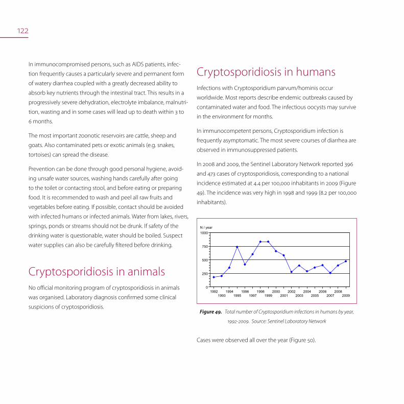
122
In immunocompromised persons, such as AIDS patients, infec-
tion frequently causes a particularly severe and permanent form
of watery diarrhea coupled with a greatly decreased ability to
absorb key nutrients through the intestinal tract. This results in a
progressively severe dehydration, electrolyte imbalance, malnutri-
tion, wasting and in some cases will lead up to death within 3 to
6 months.
The most important zoonotic reservoirs are cattle, sheep and
goats. Also contaminated pets or exotic animals (e.g. snakes,
tortoises) can spread the disease.
Prevention can be done through good personal hygiene, avoid-
ing unsafe water sources, washing hands carefully after going
to the toilet or contacting stool, and before eating or preparing
food. It is recommended to wash and peel all raw fruits and
vegetables before eating. If possible, contact should be avoided
with infected humans or infected animals. Water from lakes, rivers,
springs, ponds or streams should not be drunk. If safety of the
drinking water is questionable, water should be boiled. Suspect
water supplies can also be carefully filtered before drinking.
Cryptosporidiosis in animalsNo official monitoring program of cryptosporidiosis in animals
was organised. Laboratory diagnosis confirmed some clinical
suspicions of cryptosporidiosis.
Cryptosporidiosis in humansInfections with Cryptosporidium parvum/hominis occur
worldwide. Most reports describe endemic outbreaks caused by
contaminated water and food. The infectious oocysts may survive
in the environment for months.
In immunocompetent persons, Cryptosporidium infection is
frequently asymptomatic. The most severe courses of diarrhea are
observed in immunosuppressed patients.
In 2008 and 2009, the Sentinel Laboratory Network reported 396
and 473 cases of cryptosporidiosis, corresponding to a national
incidence estimated at 4.4 per 100,000 inhabitants in 2009 (Figure
49). The incidence was very high in 1998 and 1999 (8.2 per 100,000
inhabitants).
19921993
19941995
19961997
19981999
20002001
20022003
20042005
20062007
20082009
0
250
500
750
1000N / year
Total number of Cryptosporidium infections in humans by year, Figure49.
1992-2009. Source: Sentinel Laboratory Network
Cases were observed all over the year (Figure 50).
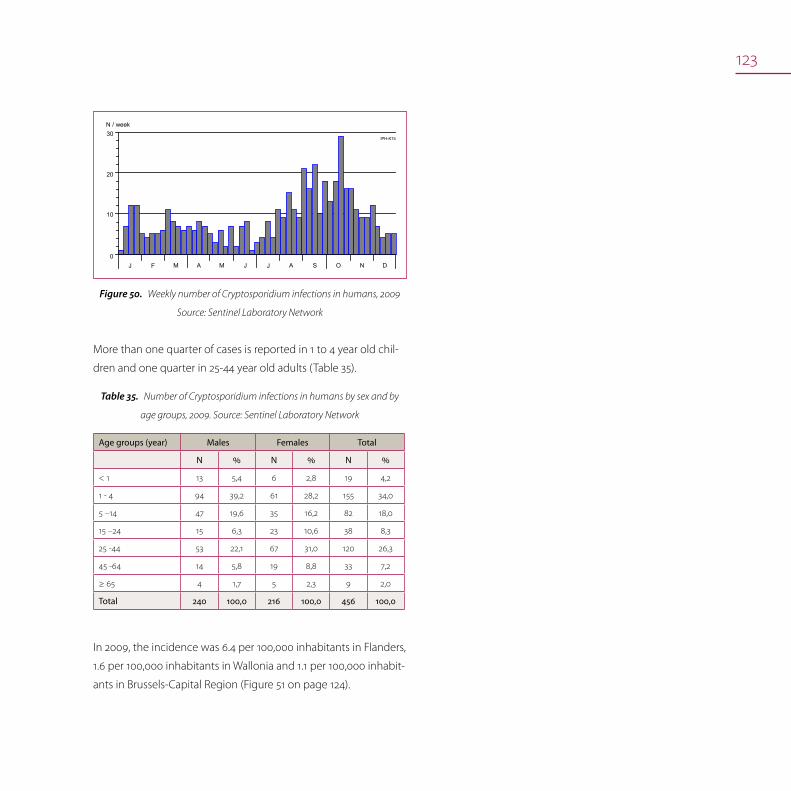
123
N / week
J F M A M J AJ S O N D
IPH-K15
0
10
20
30
Weekly number of Cryptosporidium infections in humans, 2009 Figure50.
Source: Sentinel Laboratory Network
More than one quarter of cases is reported in 1 to 4 year old chil-
dren and one quarter in 25-44 year old adults (Table 35).
Number of Cryptosporidium infections in humans by sex and by Table35.
age groups, 2009. Source: Sentinel Laboratory Network
Age groups (year) Males Females Total
N % N % N %
< 1 13 5,4 6 2,8 19 4,2
1 - 4 94 39,2 61 28,2 155 34,0
5 –14 47 19,6 35 16,2 82 18,0
15 –24 15 6,3 23 10,6 38 8,3
25 -44 53 22,1 67 31,0 120 26,3
45 -64 14 5,8 19 8,8 33 7,2
≥ 65 4 1,7 5 2,3 9 2,0
Total 240 100,0 216 100,0 456 100,0
In 2009, the incidence was 6.4 per 100,000 inhabitants in Flanders,
1.6 per 100,000 inhabitants in Wallonia and 1.1 per 100,000 inhabit-
ants in Brussels-Capital Region (Figure 51 on page 124).

124
AL AR AT
AW B
BG BS CR DK DM DN EK GT HS HV HY
IP KR LG LV MC MH MN MR MS NC NM NV OD OS PV RS SG SN TG TH TL TN TR VR VT VV WR
: Aalst : Arlon : Ath : Antwerpen : Brussels : Brugge : Bastogne : Charleroi : Diksmuide : Dendermonde : Dinant : Eeklo : Gent : Hasselt : Halle-Vilvoorde : Huy : Ieper : Kortrijk : Liège : Leuven : Mouscron : Mechelen : Mons : Marche-en-Fam. : Maaseik : Neufchâteau : Namur : Nivelles : Oudenaarde : Oostende : Philippeville : Roeselare : Soignies : St.-Niklaas : Tongeren : Turnhout : Tielt : Thuin : Tournai : Veurne : Virton : Verviers : Waremme
0.00
4.43
Incidence/100,000 inhabitants[0.00 - 18.28]
> 0.00 - 2.50
> 2.50 - 5.00
> 5.00 IPH-K15
VR
BG
DK TLGT
SN
ALOD
MH
HV
LV
AWTH
MS
HS
TG
NV
MRDN
CR
NC
AR
MC TR
KR
SG
IP
TN PV
VT
BS
VVLG
HY
WR
TG
MN NM
B
DM
EK
RS
AT
OS
Incidence of Cryptosporidium infections in humans by district (N/105 inhab., 2009) Figure51.
Source: Sentinel Laboratory Network

125
In the ITM (NRL), respectively 2048 and 2017 human faecal sam-
ples have been examined for Cryptosporidium after concentra-
tion and staining according to Heine in 2008 and 2009.
In 2008, Cryptosporidium was detected in 20 samples. Ten
samples were from patients consulting the outpatient clinic of
the ITM and 10 samples have been sent to the NRL by external
laboratories. Twelve patients (60%) were male. The age of the
patients ranged from 0 to 62 years, with a median age at 27.8
years. No travel history is known from the patients whose sample
was sent to the reference laboratory by external laboratories. Half
of the patients who consulted at the outpatient clinic of the ITM,
travelled abroad before the start of their illness.
In 2009, Cryptosporidium was detected in 15 samples. Seven sam-
ples were from patients consulting the outpatient clinic of the
ITM and 8 samples have been sent to the NRL by external labo-
ratories. Eight patients (53%) were male. The age of the patients
ranged from 0 to 49 years, with a median age at 20.4 years. No
travel history is known from the patients whose sample was sent
to the NRL by external laboratories. Three of the 7 patients who
consulted at the outpatient clinic of the ITM, travelled abroad
before the start of their illness.

126

127
CysticercosisLeen Claes, Luc Vanholme
CysticercosisCysticercus bovis in muscular tissue of cattle is the larval stage of
the tapeworm, Taenia saginata, a parasitic cestode of the human
gut (taeniasis). The risk factor for bovine cysticercosis infection in
cattle is the ingestion of feed contaminated with T. saginata eggs
shed in human faeces. Cattle can become infected when grazing
contaminated pastures. Free access of cattle to surface water, the
flooding of pastures and the proximity of wastewater effluent
have been identified as risk factors for bovine cysticercosis.
Humans contaminate themselves by the ingestion of raw or un-
dercooked beef containing the larval form (cysticerci). Usually the
pathogenicity for humans is low. The tapeworm eggs contami-
nate the environment directly or through surface waters. Human
carriers should be treated promptly. Strict rules for the hygienic
disposal or sanitation of human faeces with a method that inac-
tivates T. saginata eggs should be developed. The spreading of
human excrement on land should not be allowed.
Cysticercosis
Cysticercosis in cattle

128
Macroscopic examination is routinely done in adult cattle as
well as in calves and sheep in the slaughterhouse. Serological
examination is possible and confirmation of the lesions by PCR
can be done. The introduction of serological techniques for the
detection of cysticerci antigens in the serum of cattle should be
developed. This would allow the detection of more cases than
visual inspection of carcasses at the slaughterhouse, which has a
low sensitivity.
Although Cysticercus ovis in sheep is not transmissible to humans,
its presence causes total rejection of the carcass. No sheep were
found to be infected over the last years.
The Belgian pig population is free of Cysticercus cellulosae. Taenia
solium is not autochthonous in Belgium.
Cysticercosis in cattlePost-mortem, macroscopic examination of carcasses is routinely
done in the slaughterhouse. In 2008 and 2009, respectively
522.557 and 480.068 adult cattle and 301.102 and 319.188 veal
calves were examined.
Figures 52 and 53 from the FASFC show that in 2008 and 2009,
18 and 9 carcasses of adult cattle were rejected for generalised
cysticercosis. In addition, the meat of 2.356 and 1.811 adult cattle
was treated by a 10 days freezing before human consumption.
Cysticercosis: detection of lightly contaminated Figure52.
bovine carcasses at slaughterhouse
Cysticercosis: detection of heavily contaminated Figure53.
bovine carcasses at slaughterhouse

129

130

131
EchinococcosisYves Carlier, Leen Claes, Steven Van Gucht, Luc Vanholme
EchinococcosisHuman echinococcosis, known as hydatid disease, is caused by
the larval stages of the small tapeworms of the species Echino-
coccus granulosus or Echinococcus multilocularis of the genus
Echinococcus.
Echinococcus granulosus, the agent of cystic echinococcosis, pro-
duces unilocular human hydatidosis. E. granulosis is a small tape-
worm (6 mm) that lives in the small intestine of dogs, foxes and
other canids which are the definitive hosts. The adult tapeworm
releases eggs that are passed in the faeces. Sheep, goats, pigs,
cattle, horses and wild boar serve as intermediate hosts in which
ingested eggs hatch and release the larval stage (oncosphere)
of the parasite. Humans also can acquire infection by acciden-
tal ingestion of typical taeniid eggs, which are excreted in the
faeces of infected dogs and foxes. When eggs are ingested by the
intermediate hosts or by humans, the larval stages (oncospheres)
liberated from the eggs migrate via the bloodstream to the liver,
lungs and other tissues to develop hydatid cysts. These cysts may
develop unnoticed over many years, and ultimately rupture. Clini-
cal symptoms and signs of the disease depend on the location of
the cysts and are often similar to those induced by slow growing
tumours. Within these cysts brood capsules and protoscoleces
develop. Each protoscolex is a potentially infective organism for
canids. The definite hosts become infected by ingestion of these
cyst-containing organs (of the infected intermediate hosts).
Echinococcosis
Echinococcus in food animals
Echinococcus in wildlife (foxes)
Echinococcus in humans

132
Indigenous unilocular hydatidosis in man has been sporadically
reported in Belgium. Recommendations for basic risk-mitigation
actions are destruction of contaminated viscera found at the
slaughterhouse in order to avoid the infection of dogs.
Echinococcus multilocularis has a similar life cycle as E. granulo-
sus. Foxes and to a lesser extent dogs are the definitive hosts of
this parasite. Small rodents and voles are the intermediate hosts.
Foxes are infected by ingestion of contaminated rodents. E. mul-
tilocularis is the agent of highly pathogenic alveolar (multilocular)
echinococcosis in humans. Humans can be infected by ingestion
of eggs. Alveolar echinococcosis is of particular public health rel-
evance as it results in a chronic cancer-like liver disease. Ingestion
of the eggs by humans can result in the development of invasive
cysts in the liver. Most untreated cases in humans are fatal. In
the intermediate hosts, the larval form of the parasite remains
in the invasive proliferative stage in the liver, thus invading the
surrounding tissues. With regards to domestic animals, cats have
been ruled out as hosts of E. multilocularis, since the parasite does
not fully develop in their intestine.
Possible risk factors include contact with dogs hunting for game,
and ingestion of contaminated water or contaminated unwashed
fresh products (in particular raspberries and strawberries) and
vegetables. Chewing grass is another practice to be associated
with alveolar echinococcosis. Contamination of the hands during
gardening, through contact with contaminated soil, may also
carry some risk.
Recommendations to improve the protection of public health are
the use of good general hygiene practices such as washing fruit
and vegetables before consumption, cooking berries or mush-
rooms (washing alone is not sufficient, neither does freezing at
-18°C!), hand-washing after gardening and before the consump-
tion of meals. Also hand-washing after contact with dogs, espe-
cially if they have direct contact with wildlife or if they live in areas
where wildlife, in particular, foxes, rodents or voles, is abundant.
Planned treatment of dogs with taenicides and subsequent hygi-
enic disposal of their faeces in endemic areas is recommended.
Echinococcus in food animalsPost mortem macroscopic examination is done at the slaughter-
house in the Echinococcus domestic intermediate hosts: cattle,
sheep, horses and pigs. Whole carcasses or parts are rejected in
case cysts are found.
Echinococcus in wildlife (foxes) E. multilocularis is known to be endemic in a wide area of central
Europe. This area extends as far as the south of Belgium (Wal-
lonia), the southeast of the Netherlands, the east of France and
Switzerland.
Fox tapeworm (Source: IPH)Figure54.

133
Intestinal scraping technique (Source: IPH)Figure55.
In Wallonia, the cestode seems to maintain itself stably in the fox
population. Between 2003 and 2004, 25% of 990 examined foxes
tested positive (Hanosset et al., 2008). The highest prevalences
(41 to 62%) are found in the Ardennes and Fagne-Famenne, areas
which are rather densely forested and located at relatively high
altitudes between 400 to 700 meters above sea level. The musk
rat seems to be an important intermediate host.
In Flanders and Brussels, the parasite remains rare and there is no
proof of a recent increase in prevalence. In 2007-2008, 187 foxes
form the regions of Brussels (n =56) and Flanders (n = 131) were
examined by the IPH with the intestinal scraping technique. In
Flanders, most foxes were healthy specimens that were shot dur-
ing the hunting season. Storage of cadavers and sampling of the
intestines was done by the Research Institute for Nature and For-
est (INBO). In Brussels, most examined foxes were traffic victims
that were deposited by police officers. Sampling was intensified
in the south of Flanders and the southwest of Brussels.
Indeed, in the southwest of Brussels 3 positive foxes were found
during a former study in 1996-1999 (Vervaeke et al., 2003). None
of the examined foxes from 2007-2008 carried E. multilocularis
cestodes.
The possible reasons for the difference in prevalence between
the north and south are not known, but may include differences
in altitude, climate, fox diet or low abundance of suitable interme-
diate hosts.
Distribution of the foxes examined for E. multilocularis in Flanders Figure56.
and Brussels between 2007 and 2008.
Each circle represents one to thirteen foxes. The red circles represent foxes examined
between 2007 and 2008. All foxes were negative. The blue dots indicate the location of
the four foxes which were found positive in a prevalence study in the same study area
between 1996 and 1999.
134
Echinococcus in humansTill the end of 2003, 8 autochthonous cases of alveolar echino-
coccosis have been diagnosed. It concerned patients between
36 and 90 years old, 4 women and 4 men. Two cases presented
a rapid evolution due to immunosuppression. Two other cases
were lethal due to chirurgical complications.
In 2004, a serological study among 115 forest guards working in
Belgium did not identify any suspected case of echinococcosis in
this specific risk group.
Since 2004, the NRL performs on average 250 serological analy-
ses/year for antibodies against Echinococcus multilocularis. Inter-
estingly, a slight tendency to an increased demand is observed.
In screening tests using a crude antigenic extract, 3 to 7% of the
samples are positive, i.e. 7 to 12 patients/year. More specific tests
(in collaboration with the Institute for Parasitology of the Faculty
of Medicine, Berne, Switzerland) show that most of these positive
cases correspond in fact to cross reactions between Echinococ-
cus multilocularis and Echinococcus granulosus, i.e. most of these
patients suffer from hydatidosis.
In 2006 and 2007, 2 new autochthonous cases of alveolar echino-
coccosis were confirmed in patients of 64 and 82 years old.
In 2008 and 2009, no new autochthonous cases of alveolar echi-
nococcosis were confirmed by the NRL.
135
Sarcosporidiosis Leen Claes, Luc Vanholme
Sarcosporidiosis The following species are of zoonotic importance: Sarcocystis
bovihominis (man final host, bovine intermediate host), Sarcocystis
suihominis (man final host, pig intermediate host). Man is infected
with Sarcocystis spp. by ingesting undercooked infected meat.
Sarcocystis spp. infections are mostly asymptomatic but may
cause mild aspecific gastrointestinal symptoms like nausea and
diarrhoea.
Sarcosporidiosis in animalsSurveillance programme in food animals
Carcasses are partially or entirely condemned when myositis
eosinophilica (green colouring spots of the carcass) is observed
at post-mortem examination in the slaughterhouse. Myositis
eosinophilica may be linked with sarcosporidiosis, although the
association is not unequivocally proven.
Sarcosporidiosis
Sarcosporidiosis in animals

136

137
ToxoplasmosisLeen Claes, Stephan Decraeye
Toxoplasma gondii
Toxoplasma gondii is an obligate intracellular organism that can
be found worldwide. The final hosts are the felidea (more com-
monly cats); humans and almost all warm-blooded animals are
intermediate hosts. The sexual cycle takes place exclusively in the
intestines of felidea. As a result, millions of oocysts are shed into
the environment with the cat’s faeces within the first two weeks
after infection. These oocysts sporulate and are very resistant to
environmental damage and can persist for several years. Oral
ingestion of oocysts by a seronegative host leads to toxoplas-
mosis. The infection has an acute and a chronic phase. The latter
characterised by the persistent presence of tissue cysts in the
host (in muscle, brain, heart …). Carnivorous ingestion of infected
tissues by a seronegative host (final or intermediate) will lead to
development of the disease.
Toxoplasma gondii
Toxoplasmosis in animals
Toxoplasmosis in humans

138
Toxoplasmosis in animals
The majority warm blooded animals are indiscernible carriers of
tissue cysts. The prevalence depends on the feeding of the ani-
mal: herbivores are more exposed to oocysts and carnivores more
exposed to tissue cysts.
There is a need for efficient serological and molecular biologi-
cal methods for both indirect and direct detection of T. gondii in
animals and food. Veterinary samples can be screened serologi-
cally to see if the animals have been infected. The presence of
tissue cysts can be detected by PCR, but bio-assay is still needed
to demonstrate the presence of viable and thus infectious cysts
in meat products. Screenings are not routinely done; there is no
data on the status of toxoplasmosis in the livestock in Belgium.
The European Food Safety Authority has recommended the
initiation of monitoring programmes in the pre-harvest sector on
sheep, goats, pigs and game.
The consequences of veterinary toxoplasmosis depend on the
animal species, its feeding habit and the T. gondii strain. In sheep
and goats, primary infection during gestation leads to abortion
or stillbirth, with important economic losses. In general, more
than 70% of sheep and goats are seropositive. The outcome of
toxoplasmosis in cattle is less clear: although 30-50% is seroposi-
tive, there is less association with transmission to humans. The
prevalence in pigs (and other animals) depends on the produc-
tion system, with a higher infection rate in animal friendly farms
(“free run”). In general animals with outdoor access have a higher
prevalence of toxoplasmosis.
The presence of cats and rodents on the farm is a risk factor. Limi-
tation of these factors are important as preventive measures. The
seroprevalence in cats varies between 25 to 70%. A serological
survey on house cats demonstrated an overall chance of 6% for a
seronegative cat to become infected within the next life-year and
actively shed oocysts (NRL for Toxoplasmosis at IPH).
Animal sera tested for diagnosis of toxoplasmosis during 2008-Table36.
2009 at the NRL for Toxoplasmosis at IPH
Species IgM positive IgG positive
Sheep 14/14 14/14
Goat 6/31 13/31
Cats 39/266 114/266
Dogs 124/318 153/318
Toxoplasmosis in humansHumans are mostly infected with T. gondii by oral route: ingestion
of oocysts excreted by cats (e.g. in cat litter trays or not properly
washed raw vegetables) or by ingestion of tissue cysts present in
inadequately cooked or raw meat.
The prevalence of toxoplasmosis increases with age as the risk
that an individual is exposed to the parasite increases in time.
About 50% of the Belgian population is seropositive. More than
80% of infections with T. gondii are asymptomatic. However, mild
(flu-like) to moderate clinical symptoms are possible (lymphad-
enopathy, chronic fatigue, fever, retinochoroiditis). The majority of
adult persons have acquired immunity to re-infection, but remain
carriers for life.
139
Immunocompromised patients (HIV, organ transplantation) are
much more susceptible to the disease, with possible severe medi-
cal complications like pulmonary toxoplasmosis and encephalitis.
In 37% of the cases the consequences are fatal. When T. gondii
seropositive patients have to take immunosuppressive drugs
(for example transplant patients), the parasite present in tissue
cysts reactivates. The disease can also be transmitted via infected
donor tissues to seronegative patients.
When a primo-infection with T. gondii occurs during pregnancy,
the parasite can cross the placental barrier and cause foetal infec-
tion. The pathological consequences for the foetus depend on
the time of infection during pregnancy and the virulence of the
strain. The earlier the infection (first trimester), the higher the risk
of severe complications (ocular disorders, severe mental retarda-
tion, hydrocephalus, intra-uterine death). Infections at the end of
the pregnancy are often without direct consequences. However,
the child can be carrier of latent tissue cysts which can reactivate
later in life and cause symptomatic toxoplasmosis (e.g. ocular
disease). The costs associated with a congenital T. gondii infection
have been estimated and in some cases price can be very high.
The disease burden of toxoplasmosis is comparable to that of
other foodborne diseases such as salmonellosis or campylobac-
teriosis.
There is a whole battery of tests available to diagnose toxoplas-
mosis. As the disease is generally asymptomatic, diagnosis relies
mostly on serological tests (mostly ELISA). In case of immuno-
compromised patients or congenital toxoplasmosis, more direct
tests like PCR and bio-assay are needed to evaluate the severity of
the illness.
During 2008-2009 the NRL for Toxoplasmosis at IPH tested 277 se-
rum samples in the context of possible congenital toxoplasmosis.
Two hundred thirteen sera tested positive in Sabin- Feldman test
and 36 tested positive for IgM. Compared to the high number of
positive serum samples, only 24 out of 842 tissue and body fluids
samples (placenta, amniotic fluid, blood, cerebrospinal fluid…)
tested positive in bio-assay.
Only a very limited number of drugs can be used to control the
infection: macrolides (mainly spiramycine) and inhibitors of folate
metabolism (pyrimethamine and sulfamides). In addition, these
are only active on the free form of the parasite, not on the cysts
formed in the tissues. The treatment takes a long time, does not
prevent the formation of new tissue cysts and is not without
adverse effects. However, the effectiveness of antibiotic treatment
in the case of congenital toxoplasmosis has been questioned. For
this reason preventive measures are very important for high-risk
patients. Efforts are made to avoid a primary T. gondii infection
during pregnancy. The mode of acquiring toxoplasmosis from
meat, cat faeces and contaminated soil is so circumscribed that
simple but effective measures should be recommended during
pregnancy: regular hand-washing, especially after contact with
cats, meat, soil and water. Freezing meat (at -20°C for minimum
48 hours) before consumption or adequate heating of meat (in-
ternal temperature of 60°C) during preparation are other effective
measures. Cleaning the cat litter should be avoided.

140

141
TrichinellosisLeen Claes, Luc Vanholme
TrichinellaTrichinellosis is a zoonotic disease caused by parasitic nematodes
of the genus Trichinella. In the EU, infections are mainly caused
by the species T. spiralis, T. nativa, T. britovi and in a few cases by T.
pseudospiralis. It is transferred to humans by the consumption of
contaminated raw or undercooked meat or meat products from
an infested animal contaminated with infectious larvae.
The clinical signs of acute trichinellosis in humans are charac-
terised in a first phase by nausea, vomiting, diarrhoea, fatigue,
fever and abdominal cramps. In a second phase, symptoms may
include pains, headaches, fevers, eye swelling or facial oedema,
aching joints, chills, cough, itchy skin, diarrhoea or constipation. In
more severe cases, difficulties with coordination of movements as
well as heart and breathing problems may occur.
Trichinella
Trichinella in food animals
Trichinella in wildlife
142
The parasite has a wide range of host species, mostly mammals.
Trichinella undergo all stages of the life cycle, from larva to adult,
in the body of a single host.
Trichinella is an intestinal parasite whose larvae can be present in
the muscles of different animal species. Particularly, the following
animals represent a risk for humans:
game, in particular wild boar and carnivorous hosts such as the •
bear and fox;
backyard pigs and pigs with extensive outdoor access includ-•
ing pigs from organic farms;
horses.•
Therefore, pork, wild boar and horse meat should always be
examined before marketing. Carcasses found positive for the
presence of Trichinella are declared unfit for consumption.
Commission Regulation (EC) No 2075/2005 imposes systematic
Trichinella examination of all pig carcasses intended for export
and all horses, wild boar and other susceptible wildlife animals.
Trichinella has not been detected in carcasses of pigs and horses
destined for human consumption in Belgium for many years.
Improvements in the monitoring and the reporting of Trichinella
in wildlife should be considered.
It is recommended to travellers not to import raw meat of sus-
ceptible animals, e.g. sausages or bear meat. Also the consump-
tion abroad of meat of unknown quality should be avoided.
Trichinella in food animalsSurveillance programme and methods used
Pig carcasses intended for intra community trade or export,
except when frozen, all locally slaughtered horses and wild boars
placed on the market were checked for Trichinella.
The analysis is done by artificial digestion: the magnetic stirrer
method of pooled 100 gram sample as described in Commission
Regulation (EC) No 2075/2005, 1 gram per fattening pig, 2 grams
per breeding sow or boar and 5 grams per horse or wild boar.
Serology may be done in live pigs and for epidemiological studies
on wildlife.
Notification to the FASFC is compulsory.
Results of the 2008-2009 surveillance
In 2008 and 2009, a total of respectively 11.547.720 and 11.677.883
pigs, 9.173 and 8.711 solipeds (mainly horses) and 15.177 and 10.744
wild boars were examined. All samples were negative.
Trichinella in wildlifeIn 2008 and 2009, respectively 61 and 142 foxes were analysed for
Trichinella, all tested negative.
An important measure to avoid spreading of trichinellosis among
wildlife is not to leave offal of animal carcasses in the field after
skinning of hunted animals.

143
trends and sources
prion diseases

145
TSEPatrick Cras, Sophie Quoilin, Stefan Roels
Transmissible spongiform encephalopathiesTransmissible spongiform encephalopathies (TSEs) known as
‘prion’ diseases, are caused by an infectious agent whose molecu-
lar properties have not been fully determined. The animal TSEs
include the archetype – scrapie in domestic sheep and goats –
and animal diseases much more recently recognized, including
transmissible mink encephalopathy (TME) and feline spongiform
encephalopathy (FSE); chronic wasting disease (CWD) of deer and
elk; and bovine spongiform encephalopathy (BSE).
Public health protection is covered by the testing of all animals
in the slaughterhouse of 30 months of age and more in 2008 and
48 months of age and more in 2009, suspected animals (in farm
and slaughterhouse) and fallen stock of 24 months and more in
2008 and 48 months and more in 2009. Additionally, all organs
and tissues that can be infectious are routinely removed at the
slaughterhouse, destroyed and therefore excluded from the (hu-
man and animal) food chain. This last measure is better known as
the removal of specific risk material (SRM).
TSE
TSE in animals
Humans

146
TSE in animalsBSE became a notifiable disease in Belgium in 1990. In 1997, a
Royal Decree described the regulations for the epidemiologi-
cal surveillance for ruminant TSE in Belgium, including the herd
slaughter and compensation policy. In the beginning of 2001, this
‘passive’ surveillance was supplemented with an ‘active’ surveil-
lance (based on EU Regulation (EC) No 999/2001) controlling
slaughtered animals and the fallen stock.
For the moment the NRL uses 5 tests for diagnosis, i.e. the ‘rapid’
ELISA test, histopathology, immunohistochemistry, electronmi-
croscopic detection of scrapie associated fibrils (SAFs) and west-
ern blotting. In Belgium, all private and regional animal health
laboratories (primary ‘active’ screening) and the NRL are accred-
ited (ISO 17025:2005) and the whole epidemiological surveillance
is coordinated by the FASFC.
Number of animals controlled in Belgium between 2001 and 2009Table37.
Year Animal At the slaughterhouse (healthy)
Suspected Animals:
Herd screening / farm, slaughter, autopsies
(with symptoms)
Fallen stock
2001Cattle
Small ruminants
360.948
0
3.522 / 379
11 / 45
13.060
0
2002Cattle
Small ruminants
410.379
2.195
3.277 / 377
428 / 85
36.386
780
2003Cattle
Small ruminants
357.389
2 447
1.126 / 250
205 / 52
33.691
499
2004Cattle
Small ruminants
358.120
39
172 / 254
333 / 170
35.322
1.650
2005Cattle
Small ruminants
325.302
703
15 / 234
8 / 86
41.729
1.588
2006Cattle
Small ruminants
320.541
8.076
8 / 185
81 / 246
44.066
3.064
2007Cattle
Small ruminants
313.570
7.291
0 / 240
111 / 155
46.099
2.584
2008Cattle
Small ruminants
317.781
0
0 / 311
0 / 117
49.188
3.512
2009Cattle
Small ruminants
199.621
0
0 / 217
0 / 121
26.202
1.633
Total Cattle
Small ruminants
2.972.660
20.751
8.120 / 2.475
1.177 / 993
325.743
15.310

147
In the next table 38 the total number of positive TSE cases is indicated and subdivided by category and place of detection.
Positive TSE cases in cattle and sheep in Belgium until 2009Table38.
Year Cattle Sheep
1992 0 5 (First case) / 5C
1993 0 0
1994 0 0
1995 0 0
1996 0 0
1997 1 (First case) / C 2 / C
1998 6 / C 11 / 3C – 8Sc
1999 3 / C 12 / 2C – 10Sc
2000 9 / C 0
2001 46 / 28S – 10C – 7F – 1Sc 0
2002 38 / 17S – 5C – 16F 25 (1 atypical case) / 1S - 2C - 2F – 20Sc
2003 15 / 10S – 5F 2 / F
2004 11 / 6S – 3C – 2F 13 (1 atypical case) / 1S – 3F – 9Sc
2005 2 / 1S – 1C 2 (2 atypical cases) / F
2006 2 / S 3 (3 atypical cases) / 2F – 1S
2007 0 3 (2 atypical cases) / S
2008 0 0
2009 0 0
Total
133 cases
(64 slaughterhouse / 38 clinical cases /
30 fallen stock / 1 second case in a farm)
78 cases
(25 primary cases - 9 atypical cases) /
(6 slaughterhouse / 14 clinical cases /
11 fallen stock / 47 second cases in a farm)
S = slaughterhouse control / C = suspected clinical / F = fallen stock / Sc = additional case in a herd
Laboratory and epidemiologic studies provided strong circumstantial evidence for a causal link between vCJD and the epizootic bo-
vine spongiform encephalopathy (BSE) in cattle with the most likely route of primary human infection being through dietary exposure
to highly infected bovine tissues.

148
Atypical BSE prion strains
While in scrapie strain variations were found, BSE showed homo-
geneity of the phenotype. Indeed, the incubation time, the lesion
localisation (“lesion profile”) and the biochemical signature of the
prions were identical for all cases. This uniform aspect of the BSE
prions during the epidemic was probably due to the contamina-
tion of the food chain by a single strain in United Kingdom.
However, due to the extensive active surveillance and new
research techniques, rare variants of BSE have been recently
reported. The analysis of their features showed that these emerg-
ing types represent different strains of BSE prions. Two atypical
forms of BSE prions have been described. Western blot studies
showed that, in comparison to the classic BSE prion (C-type),
they are characterised by a higher or lower molecular weight of
the unglycosylated PrPres. Therefore, they were named H-type
and L-type BSE (L-type is also called bovine amyloid spongiform
encephalopathy (BASE)). They also show a lower proportion of
diglycosylated PrPres than C-type.
Such atypical types are very rare; only 42 cases were described
worldwide. They were detected in old bovines (at least eight
years in age) and reported on different continents. Indeed,
atypical prions might correspond to natural “sporadic” forms of
BSE, not originating from the contamination of the food chain.
It is essential for the surveillance programmes to determine the
prevalence of these atypical types on a global level.
In order to determine the prevalence of the two types of atypi-
cal BSE in Belgium, a retrospective analyses was carried out on
samples from the Belgian collection of BSE-diagnosed animals
originating from cattle of at least 7-years-old, using the latter
criteria and techniques. Dobly et al. 2010 showed that till now, no
atypical cases were present in the Belgian BSE collection.
The emergence of atypical types of BSE is partially due to a better
knowledge of prion strains and more efficient diagnostic tech-
niques. However the area in the brain where the PrPres is depos-
ited differs drastically between the types and the atypical types
are more susceptible to a proteinase treatment. It is therefore
essential to ascertain that the routine sampling techniques and
analyses are adapted to these new types. As these new strains
seem more virulent than classic types, at least in mice models,
they represent one of the next challenges in the field of prions.
Differential diagnosis with Listeria monocytogenes
In the field of differential diagnosis of BSE cases, a retrospective
study was performed on the occurrence of Listeria monocytogenes
infections in BSE suspected cattle (Roels et al. 2009). L. monocy-
togenes is an important foodborne pathogen both in humans and
animals. In order to determine the presence and importance of
this zoonotic bacterial agent in Belgian cattle, we have reviewed
brain tissue specimens for the presence of histopathological lesions
pathognomonic for L. monocytogenes meningoencephalitis. Ad-
ditional Listeria-specific immunohistochemistry was performed in
order to confirm the diagnosis of these cases.
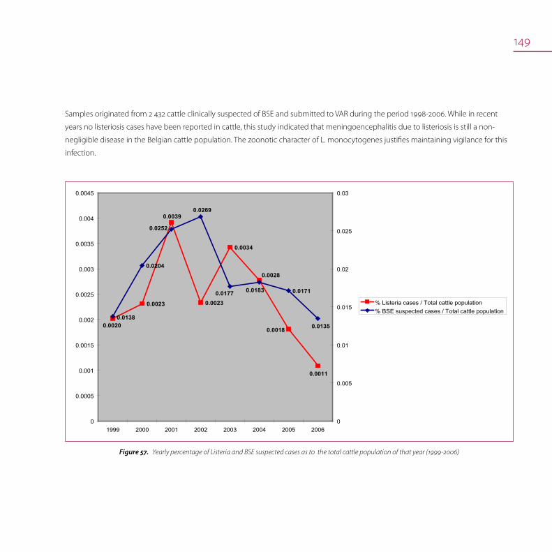
149
0.0020
0.0039
0.0028
0.0018
0.0011
0.0023 0.0023
0.0034
0.0177
0.0138
0.0252
0.0269
0.0183
0.0135
0.0204
0.0171
0
0.0005
0.001
0.0015
0.002
0.0025
0.003
0.0035
0.004
0.0045
1999 2000 2001 2002 2003 2004 2005 20060
0.005
0.01
0.015
0.02
0.025
0.03
% Listeria cases / Total cattle population% BSE suspected cases / Total cattle population
Yearly percentage of Listeria and BSE suspected cases as to the total cattle population of that year (1999-2006)Figure57.
Samples originated from 2 432 cattle clinically suspected of BSE and submitted to VAR during the period 1998-2006. While in recent
years no listeriosis cases have been reported in cattle, this study indicated that meningoencephalitis due to listeriosis is still a non-
negligible disease in the Belgian cattle population. The zoonotic character of L. monocytogenes justifies maintaining vigilance for this
infection.

150
TSE in humansCreutzfeldt-Jakob disease
The disease of Creutzfeldt-Jakob (CJD) is a transmissible spongi-
form encephalopathy (TSE), which is characterised by the fast
deterioration of the cerebral functions. Prion diseases are rare
when compared with other neurodegenerative diseases such as
Alzheimer’s disease. On the other hand, prion diseases are unique
because they are transmissible, coming on the international
public health stage when a new expression of CJD (variant CJD
or vCJD) was diagnosed and linked to the consumption of beef
contaminated by BSE (bovine spongiform encephalopathy) prion.
A Belgian CJD surveillance network was created in 1998 in order
to follow the trends of the disease and the potential occurrence
of variant CJD cases. The network is built on the collaboration of
the seven academic reference centres of neurology/neuropathol-
ogy and the IPH.
Academic reference centres offer a support for diagnosis of CJD
by communicating to IPH data on patients with a probable or
neuropathological confirmed diagnosis. Data are analysed and
interpreted by IPH allowing an epidemiological follow up of the
disease in the country. A procedure was set up in order to deal
with possible public health consequences in case a variant CJD
was diagnosed. Incidence data are shared with other European
countries in a Collaborative Study Group (EuroCJD) and figures
are communicated to ECDC.
Several forms of CJD are described according to their transmis-
sion way: sporadic, genetic and acquired including a.o. the
iatrogenic and variant forms. The diagnosis of CJD is based on
clinical presentation, evolution of the symptoms and laboratory
tests but can only be confirmed by neuropathological investiga-
tion. Added to age of onset and disease duration, these elements
allow making a differential diagnosis.
In Europe, the estimated incidence of sporadic CJD is around 1 to
2 cases per million-year. After 10 years surveillance, the observed
incidence rate in Belgium is 1.39 cases by 1.000.000 inhabitants
per year. The median number of cases per year in Belgium is 15
(range 3-22) with a total 174 reported cases on a 12 years period.
About 97% of cases were sporadic, 2.5% genetic and only one is
classified as iatrogenic. There was no preponderance in the sex
ratio although year-to-year variations were observed. The median
age at onset was 66 year (range 31-88). The mean disease dura-
tion was 6 month.
0,0
0,5
1,0
1,5
2,0
2,5
1998 1999 2000 2001 2002 2003 2004 2005 2006 2007 2008 2009
Year
Inci
de
nce
/1.0
00
.00
0
Incidence of CJD in Belgium, 1998-2009. Figure58.
(Report of the surveillance network, IPH, 2009)

151
Since 1995, 215 patients died of variant CJD in 11 countries of
which 7 are European. UK is most affected with 79% of the pa-
tients deceased of this disease. In Belgium no case of variant CJD
has ever been diagnosed. Considering that the population has
been exposed to BSE and that bordering countries are affected
(France, the second most affected country and The Nether-
lands), the probability to diagnose a case in Belgium can not
be excluded but the risk to face a major public health problem
lessens. This assumption is illustrated by the European situation.
After a peak in 2000, a continuous decline in the number of vCJD
cases is observed in UK unlike other countries where the majority
of the new cases are diagnosed since 2005 but in a limited extent
(n=46).
0
5
10
15
20
25
30
1995 1996 1997 1998 1999 2000 2001 2002 2003 2004 2005 2006 2007 2008 2009
Year
Nu
mb
er o
f ca
ses
UK
Else whe re
Number of vCJD in UK and outside UK, 1995 to 2009 Figure59.
Source: IPH
Despite the declining incidence, vCJD remains a public health
concern because prions could be present in the population thus
highlighting today’s risk of secondary transmission through medi-
cal acts (e.g. blood transfusion, surgery, ocular, ORL, dental care).
Introduced as a foodborne disease 15 years ago, vCJD has the
potential to become a more significant public health issue due to
new transmission ways associated with medical care!
trends and sources
foodborne outbreaks
153
Foodborne outbreaksNadine Botteldoorn, Sarah Denayer, Katelijne Dierick, Jean-Yves Michelet, Maria Naranjo
In Belgium different authorities have competences related to
FBO’s.
The Federal Agency for Safety of the Food Chain (FASFC) deals
with safety of foodstuffs, epidemiological investigation of FBOs
and related animal health issues.
The Communities (Flemish, French and German speaking Com-
munity) which deal with person related matters as human health,
can start an epidemiological investigation by its Public Health
medical inspectors in case of a FBO.
At the IPH, the Scientific Service of Food Pathogens, which is NRL
for FBO’s, analyses the suspected food samples, coordinates the
analyses of human samples, collects all data on FBOs and gives
scientific support to the FASFC officers and the Public Health
Inspectors.
Foodborne outbreaks
Major etiological agents
Foodborne outbreaks 2007
Working group on foodborne outbreaks

154
A national ‘Platform for foodborne infections and intoxications
and zoonoses transmitted by food’, approved by the National
Conference of Ministers of Public Health, was created to advance
data exchange between different competent authorities on food
safety, animal health and public health. Furthermore in 2007, for a
better communication, a protected web- application was made
available to exchange outbreak data and laboratory results in
“real time” between the different authorities dealing with FBOs. In
this web-application a common file is created for each individual
outbreak, and the data and laboratory results are shared between
food inspectors and human health inspectors.
Foodborne outbreaks in humansA foodborne outbreak is defined as an incident, observed under
given circumstances, of two or more human cases of the same
disease and/or infection, or a situation in which the observed
number of human cases exceeds the expected number and
where the cases are linked, or are probably linked, to the same
food source (Directive 2003/99/EC, Article 2(d)). Data are collected
from FASFC, the communities (French, Flemish, Brussels and Ger-
man), the sentinel laboratories network for human clinical micro-
biology, and the Federal Reference Centres for FBOs, Salmonella
and Shigella, Listeria and C. botulinum. The reporting includes
both general and household outbreaks.
The causative agents covered are Salmonella spp., Shigella spp.,
Campylobacter spp., Verotoxinogenic E.coli, Listeria monocy-
togenes, Clostridium botulinum, Staphylococcus aureus, Bacillus
cereus, Clostridium perfringens, Yersinia enterocolitica, Giardia,
Norovirus, Hepatitis A virus, toxins of Staphylococcus aureus and
Bacillus cereus, histamine and shellfish biotoxins.
Major etiological agents
Foodborne bacteria
Salmonella enterica
Although the number of human salmonelloses has drasti-
cally decreased in Belgium since 2005, it remains an important
reported pathogen in FBOs. The onset time varies between 6
and 48 hours after ingestion of the contaminated food. Nausea,
vomiting, abdominal cramps, diarrhoea, fever and headache are
the symptoms in an acute outbreak and last for 1-2 days or longer.
In case of an outbreak human samples (stool) and suspected food
samples are tested for Salmonella. If Salmonella is detected, Pulse
Field Gel Electrophoresis (PFGE) typing can confirm the clonal
relationship between the human isolates and those isolated from
food products. Raw or undercooked meat, poultry meat, eggs,
shrimps, cream-filled desserts and chocolate are frequently as-
sociated with food borne Salmonella outbreaks. The food can be
at the origin of contamination or can transmit the infection from
a contaminated food handler.
Campylobacter jejuni and C. coli
Since 2005, Campylobacter is the most frequently reported illness
of food borne origin in humans in Belgium. Campylobacter jejuni
and C. coli infections cause diarrhoea, which may be watery or
sticky and can contain blood. Other symptoms often observed
are fever, severe abdominal pain, nausea, headache, malaise and
muscle pain. Rarely, it can cause constipation leading to a misdi-
agnosis of appendicitis. The illness usually occurs 2-5 days after
ingestion of the contaminated food or water and generally lasts
155
7-10 days, but relapses are not uncommon (about 25% of cases).
Some rare but feared complications are acute colitis, reactive
arthritis and Guillain-Barré syndrome.
Campylobacter frequently contaminates raw poultry meat and
raw pork. Raw milk and cheeses made from raw milk are also
sources of infections.
Yersinia enterocolitica
Yersiniosis is frequently characterised by symptoms as gastro-
enteritis with diarrhoea and/or vomiting; however, fever and
abdominal pain are typical symptoms. Yersinia infections can also
cause pseudo-appendicitis and reactive arthritis. Illness onset is
usually between 24 and 48 hours after ingestion of food or water,
which are the usual vehicles of infection. Contaminated and
undercooked pork is a common source of infection, but also ice-
cream has been reported as the source of infection.
Clostridium perfringens
The common form of Clostridium perfringens poisoning is charac-
terised by intense abdominal cramps and diarrhea which begin
8-22 hours after consumption of food contaminated with large
numbers of vegetative C. perfringens cells capable of produc-
ing the food poisoning toxin. Toxin production in the human
digestive tract is associated with sporulation. The illness is usually
self-limited within 24 hours but less severe symptoms may persist
in some individuals for 1 or 2 weeks. In most instances, the actual
cause of poisoning by C. perfringens is temperature abuse of
prepared foods. Indeed, small numbers of the spores are often
present after cooking and multiply to food poisoning levels
during cooling and storage of prepared foods at too elevated
temperatures under anaerobic conditions (e.g. fat layer on stock).
Meat, meat products, and gravy are the foods most frequently
implicated.
Staphylococcus aureus
Some Staphylococcus strains are capable of producing highly
heat-stable enterotoxins in the food, which cause illness in hu-
mans. The onset of symptoms in staphylococcal food poisoning
is usually rapid and in many cases acute, depending on individual
susceptibility to the toxin, the amount of contaminated food
eaten, the amount of toxin in the ingested food, and the general
health status of the victim. The most common symptoms are
nausea, vomiting and abdominal cramping. Recovery gener-
ally takes two days. Foodstuffs at risk for staphylococcal food
poisoning are those that require considerable handling during
preparation and that are kept at slightly elevated temperatures
after preparation. Contamination occurs by infected food handler
or by the food itself (e.g. milk).
Bacillus cereus
Two types of food poisoning, an emetic and a diarrhoeal type,
can be observed. For the emetic type, a heat-stable emetic toxin
named cereulide, preformed in the food, is responsible for the
symptoms similar to those of Staphylococcus aureus intoxication,
and is characterised by a short incubation period. This type is
probably the most dangerous since it has been associated with
life-threatening acute conditions like acute liver failure. Heat-
unstable enterotoxins, produced in the gut by vegetative cells
cause the diarrhoeal type, with symptoms similar to those of the
C. perfringens food poisoning, with a 6 to 24h incubation period.
The emetic type is frequently associated with the consumption
156
of food rich in carbohydrates such as rice and pasta whereas the
diarrhoeal type is often associated with cooked meat and meat
products.
Foodborne viruses
Viruses cannot grow in or on food but may be present on fresh
products by contact with polluted water in the growing area or
during processing. Unhygienic handling during distribution or
final preparation is also reported as a cause of contamination.
People can be infected without showing symptoms. Person
to person transmission is common and the high frequency of
secondary cases following a FBO results in amplification of the
problem.
Although numerous faecal-orally transmitted viruses exist, the risk
of foodborne transmission is highest for Hepatitis A virus and No-
rovirus. European data show that oysters are frequently reported
as a main source of contamination, but water, fruits and food
handler contamination are also reported. Increased awareness
towards viral infections is raised thanks to improved molecular
detection methods. In particular real-time polymerase chain reac-
tion (RT-PCR) allows quantification and has made diagnosis and
outbreak management easier.
Noroviruses are among the most important causes of gastro-
enteritis in adults and often occur as outbreaks which may be
foodborne. They are the most common cause of non-bacterial
FBOs recognised in Europe and United States and have been
diagnosed worldwide. Noroviruses can be transmitted from
person-to-person, or indirectly via food or water contaminated
with faeces or vomit. They are responsible of mild and usually self-
limiting gastroenteritis. Considering the high attack rates and the
fact that Norovirus can cause disease in all age groups, the socio-
economical burden of Norovirus infection is very important.
Marine biotoxins
Marine biotoxin poisoning in humans is caused by ingestion of
shellfish containing algae toxins. Bivalve molluscs like mussels,
oysters and scallops feed with phytoplankton. Some kinds of phy-
toplankton produce, under favorable climatic and hydrographic
circumstances, natural toxins which are consequently absorbed
by the bivalve molluscs. According to the effects that they cause,
biotoxins are classified in different groups, the 3 main groups
being the paralytic shellfish poisoning toxins (PSP), the diarrhoeic
shellfish poisoning toxins (DSP) and the amnesic shellfish poison-
ing toxins (ASP).
The effects of these toxins are generally observed as acute in-
toxications: PSP are causing paralysis in human which in extreme
cases results in death. These toxins are accumulated by shellfish
grazing on algae producing these toxins. Symptoms of human
PSP intoxication vary from a slight tingling or numbness to
complete respiratory paralysis. In fatal cases, respiratory paralysis
occurs within 2 to 12 hours of consumption of the PSP contami-
nated food. The responsible toxins are produced by worldwide
present dinoflagellates.
DSP are characterised by the diarrhoea they cause in human,
unpleasant but not lethal. Symptoms include diarrhoea, nausea,
vomiting and abdominal pain starting 30 minutes to a few hours af-
ter ingestion. Complete recovery occurs within three days. Here also,
worldwide present dinoflagellates are responsible for the production
of the toxins. Europe and Japan seem to be the most affected areas.

ASP have been detected at the American and Canadian east-
coast. The symptoms of the intoxication include abdominal
cramps, vomiting, disorientation and memory loss (amnesia).
A permanent loss of memory is possible. In extreme cases, for
older people, a lethal result has been reported. These toxins are
produced by a diatom and the main ASP toxin is domoic acid.
Parasites
Giardia lamblia
Giardia lamblia is a protozoan that may cause diarrhea within 1
week of ingestion of the cyst, which is the environmental survival
form and infective stage of the organism. Illness often lasts for 1 to
2 weeks, but there are cases of chronic infections lasting months to
years. Illness is most frequently associated with the consumption
of contaminated water, contaminated vegetables that are eaten
raw, or food contamination by infected or infested food handlers.
Cool moist conditions favor the survival of the organism.
Foodborne outbreaks in 2008 and 2009Reported outbreaks in 2008 and 2009
During 2008 and 2009, a total of 209 outbreaks of foodborne
infections and intoxications were recorded in Belgium. More than
1753 people became ill and at least 55 were hospitalized. In 2009
one person died from Listeriosis.
The geographic distribution of all foodborne outbreaks is shown
in the Figure 60 for 2008 and in Figure 61 for 2009.
Geographical distribution of FBOs in Belgium – 2008Figure60.
Geographical distribution of FBOs in Belgium – 2009Figure61.

158
Causative agents in 2008 and 2009
Foodborne viruses stayed the most frequently detected agents
in FBOs. In total 14 Norovirus outbreaks were reported in 2008 and
2009 and 3 outbreaks were caused by Hepatitis A. For Norovirus
in 11 outbreaks the agent was confirmed in the human faecal
samples and in 3 outbreaks the agent could also be detected in
the food (meat, filling of the sandwiches or a witness meal).
Salmonella was the causative agent in 8 outbreaks. In 4 outbreaks
S. Enteritidis was detected in food products based on raw eggs
(tiramisu, chocolate mousse). S. Ohio was found in an outbreak
caused by pork that was contaminated at the slaughterhouse.
The slaughterhouse was closed during several weeks and disin-
fected. It took several weeks to get rid of the contamination. S.
Brandenburg was found in an outbreak where the origin could
not be detected.
Coagulase positive Staphylococcus spp. caused 4 outbreaks.
Toxine A was produced by most of the strains. Spaghetti with
sauce, pasta with ham, hamburger and beans were the identified
vehicles.
Thermotolerant Campylobacter was responsible for 10 outbreaks.
The agent was almost always detected in the human samples. In
one outbreak it was very clear that undercooked stuffed quails
were the cause of the outbreak.
Bacillus cereus was the causative agent in six outbreaks. Four of
them were caused by B. cereus enterotoxin and 2 by the emetic
toxin cereulide. Cold dishes, spices and sauces were the food
vehicles.
Verotoxinogenic E. coli was detected in 3 outbreaks. One out-
break affected 6 patients of a psychiatric hospital. The cause was
minced meat that had been consumed raw.
In many outbreaks no causative agent could be identified. An
important reason for this was the absence of leftovers of the meal
in many of those outbreaks.

159
FBOs in humans in Belgium in 2008 and 2009Table39.
Outbreaks 2008 Outbreaks 2009
Causative agent NHuman
casesDeaths
Hospitalisa-
tions N Human cases Deaths
Hospitalisa-
tions
Bacillus cereus 2 10 0 3 4 53 0 1
Campylobacter 6 31 0 6 4 8 0 unknown
Clostridium perfringens 1 4 43 0 1
Verotoxinogenic E. coli 3 11 0 6 1 4 0 4
Hepatitis A Virus 1 49 0 2 10 1
Norovirus 7 439 7 91 0 0
Listeria 1 2 0 1 2 4 1 2
Other agents 4 3 0 0 0
Parasites 1 10 0 0 3 6 0 0
Salmonella 3 39 0 3 5 68 0 2
Staphylococcus 2 32 0 10 2 24 0 0
Histamine 0 1 11 0 2
Unknown 73 68 533 0 7
Total 104 841 0 35 105 912 0 20
Source of the foodborne outbreaks
Most FBOs were due to the consumption of meals composed of
different ingredients (42% in 2008 and 17% in 2009). In 2008 and
2009 meat and meat based products were responsible for 22 % of
the outbreaks. Bakery products, including preparations with raw
eggs such as tiramisu and chocolate mousse, dairy products and
crustaceae were each responsible for 5% of the outbreaks.
05
1015202530354045
meat
mixed
bake
ryoth
er
crusta
ceae
unkn
owndri
nks
vege
tables
dairy
fis
h
2008 2009
Food vehicles responsible for FBO in 2008 and 2009Figure62.

160
Setting of the foodborne outbreaks
In 94% of FBOs, the place of exposure could be traced back.
Restaurants (36%) were the most frequently associated with FBOs,
18% occurred at home and about 12% of the outbreaks occurred
by an institutional catering. The place of exposure reported to the
NRL FBO was almost identical during the two different years.
05
10152025303540
at h
ome
at w
ork
farm
inst
itutio
nal
cate
ring
mas
sga
ther
ing
recr
eatio
npl
ace
shop
unkn
own
Res
taur
ant
or s
imila
rfa
cilit
y
othe
r
2008 2009
Place of exposure in FBOs 2008-2009Figure63.
Description of outbreaks of special interest
In the summer of 2008, 4 patients of a psychiatric institution were
hospitalized with the symptoms of bloody diarrhea. Two of them
developed an hemolytic uremic syndrome (HUS) and, as specific
biochemical characteristic, the strains were all urease +. PFGE
revealed that consumption of raw minced meat was at the origin
of the outbreak
In 2009, there has been a significant increase in registration of
Salmonella enterica serovar Ohio infections in the Belgian popula-
tion (p<0.01). During the period from the 1st of August to the 31th
of October 2009, 34 strains of S. Ohio isolated in clinical laborato-
ries had been reported to the NRCSS. Most human strains caused
self-limiting gastroenteritis, but two of the enquêted patients
had to be hospitalized. One terminal cancer patient died due to
the Salmonellosis and the other hospitalized case suffered from
kidney insufficiency. With regard to the population, both sexes
(18 males and 16 females) and all age groups (3 children aged < 5
years, 22 adults 15-64 years and 9 adults >65 years) were infected.
A cluster of patients was identified around the city of Liège. At
the same time, an increase of this serovar was also observed in
the Salmonella isolates sampled during the monitoring program
of the Food Agency for the Safety of the Food Chain. The samples
containing S. Ohio were of pork origin suggesting that this spe-
cies was responsible for the outbreak of the disease. PFGE typing
confirmed the clonal relationship between the human isolates
and those isolated from pork products. Further epidemiological
investigations showed that one slaughterhouse was involved. In
that slaughterhouse the carcasses were contaminated during the
evisceration process by contaminated equipment and uncon-
trolled environmental conditions.
161
Prevention of foodborne outbreaksSince the most frequent causes of FBOs are disruption of cold
chain, insufficient heating of the food, lack of personal hygiene,
bad hygiene in the kitchen, long delay between preparation and
consumption and raw materials of poor microbiological quality,
outbreaks can be prevented by the application of simple hygienic
rules like adequate refrigeration of the food, hand washing before
and during preparation, clean surfaces and materials in the
kitchen, separation of raw and cooked food and sufficient heating
during preparation.
Working group on foodborne outbreaksThe working group was created in 1995 by IPH and brings togeth-
er, on a voluntary basis, the main actors in the field of foodborne
infections and intoxications in Belgium.
Since its final reform in 1993, Belgium consists of Communi-
ties and Regions, each with their specific responsibilities and
competences. Since food and food hygiene is a federal issue and
matters related to human such as illness are the competence
of the communities (Flemish, French or German), data on FBOs
are dispersed. As a consequence, there was a need for a working
group that assures the coordination, the streamlining of policy
and the harmonization of the approach between the different
partners implicated in outbreaks.
The group is composed of delegates representing
the FPS - Public Health, Food Chain Safety and Environment, •
the FASFC, •
the Health Inspection Services of the Communities, •
the Brussels Community Coordination Commission, •
the Anti-poison Centre, •
the Food microbiology laboratory of the University of Ghent, •
the NRL for food microbiology at the University of Liège, •
the VAR.•
The IPH houses the working group and is represented by the
Epidemiology section, the Reference centres for Salmonella and
Shigella, for Listeria and for FBOs.
The main goals of the working group are to exchange field infor-
mation on detection, epidemiological investigation, controlling
and reporting of outbreaks and eventually of sporadic cases of
foodborne infections in the country. Significant effort has been
put on the improvement of outbreak data collections and case-
control studies. The working group also provides scientific sup-
port to the mandatory annual Belgian Zoonoses Report to EFSA.
Since 2004, the Belgian authorities recognized the working group
as ‘Platform for Foodborne infections and intoxications and food
related zoonoses’ reporting to the National Conference of Minis-
ters of Public Health.

162
Tables
Table 1. Evolution in the total human population 2005-2009 16
Table 2. Total number of herds and animals in the period 2005 – 2009 17
Table 3. Total number of herds and animals of poultry in the period 2005–2009 19
Table 4. Number of slaughtered animals in the period 2005–2009 20
Table 5. Evolution of individual serological tests and testing of pools of bulk milk samples 25
Table 6. Zoonosis monitoring programme – Campylobacter in food 31
Table 7. Evolution of the Campylobacter prevalence of pork carcasses 2005-2009 31
Table 8. Campylobacter in meat and meat products: list of antimicrobials tested and breakpoints used 32
Table 9. Antimicrobial susceptibility testing of Campylobacter in food: percentage of resistant strains 32
Table 10. Number of Campylobacter infections in humans by sex and by age groups, 2009 35
Table 11. Resistance of Campylobacter in Belgium fecal isolates, trend from 2001 till 2009 37
Table 12. Repartition by biotype of invasive Campylobacter in humans, 2009 38
Table 13. Zoonosis monitoring programme - E. coli O157, 2008 -2009 41
Table 14. E. coli: evolution in number of isolates in humans, 1998-2009 42
Table 15. Zoonosis monitoring programme - Listeria monocytogenes in food, 2008-2009 48
Table 16. Number of Listeria monocytogenes infections in humans by sex and by age group, 2009 50
Table 17. NRL alayses for Q-fever 58
Table 18. DGZ/ARSIA analyses for Q-fever 59
Table 19. The results of the monitoring – Salmonella in meat and meat products 72
Table 20. The results of the monitoring – Salmonella in meat and meat products 74
Table 21. Evolution of the food Salmonella prevalence, 2005-2009 76
Table 22. Trends for the most prevalent Salmonella serotypes from 1989 to 2009 78
Table 23. Human cases of Salmonella: Age and gender distribution 2009 79
Table 24. Animal Salmonella: list of antimicrobials tested 80
Table 25. Salmonella from food, list of antimicrobials tested with their breakpoints 82
Table 26. Antimicrobial susceptibility testing of Salmonella spp. isolated from different food matrices: percentage of resistant strains 83
Table 27. List of antimicrobials used for susceptibility testing of Salmonella 85
163
Table 28. Antimicrobial resistance in human Salmonella of serotypes Enteritidis, Typhimurium, Brandenburg,
Derby, Hadar, Virchow, Infantis, Typhi, Newport, Paratyphi B and A isolated in 2009 87
Table 29. Evolution of bovine tuberculosis outbreaks in cattle herds in Belgium 91
Table 30. Monitoring Yersinia enterocolitica in pork meat 94
Table 31. Number of Yersinia enterocolitica infections in humans by sex and by age groups, 2009 95
Table 32. Number of cases of Hantavirus infections in humans by sex and age groups, 2009 107
Table 33. Active surveillance of WNV by buccal (b) swabs of Sylviidae species in 2008-2009 117
Table 34. Active surveillance of WNV by sera (s) and buccal (b) swabs of Corvid species in 2008-2009 117
Table 35. Number of Cryptosporidium infections in humans by sex and by age groups, 2009 123
Table 36. Animal sera tested for diagnosis of toxoplasmosis during 2008-2009 at the NRL for Toxoplasmosis at IPH 138
Table 37. Number of animals controlled in Belgium between 2001 and 2009 146
Table 38. Positive TSE cases in cattle and sheep in Belgium until 2009 147
Table 39. FBOs in humans in Belgium in 2008 and 2009 159

164
Figures
Figure 4. Evolution of the total number of cattle herds, period 2005–2009 18
Figure 1. Evolution of the total number of pig herds, period 2005–2009 18
Figure 3. Evolution of the total number of fattening pigs, period 2005–2009 18
Figure 5. Evolution of the total number of bovines, period 2005–2009 18
Figure 2. Evolution of the total number of breeding pigs, period 2005–2009 18
Figure 6. Gallus gallus, evolution number of poultry herds, period 2005–2009 19
Figure 7. Gallus gallus, evolution total number of poultry, period 2005–2009 19
Figure 8. Evolution of slaughtered cattle 2005–2009 20
Figure 9. Evolution of slaughtered calves 2005–2009 20
Figure 10. Evolution of slaughtered pigs 2005–2009 21
Figure 12. Evolution of slaughtered broilers and layers 2005–2009 21
Figure 11. Evolution of slaughtered sheep & goats 2005–2009 21
Figure 13. Evolution of individual serological tests for bovine brucellosis, 2005–2009 25
Figure 14. Evolution of pools of bulk milk tests for bovine brucellosis, 2005–2009 25
Figure 15. Number of notified abortions, 2005–2009 26
Figure 16. Number of tests of Brucella melitensis, 2005 – 2009 27
Figure 17. Evolution of the percentage resistant Campylobacter jejuni strains in poultry 33
Figure 18. Evolution of the percentage resistant Campylobacter coli strains in poultry 34
Figure 19. Evolution of the percentage resistant Campylobacter coli strains isolated in pork 34
Figure 20. Total number of Campylobacter infections in humans by year (1986-2009) 35
Figure 21. Weekly number of Campylobacter infections in humans, 2009 35
Figure 22. Incidence of Campylobacter infections in humans by district (N/105 inhab., 2009) 36
Figure 23. Number of Listeria monocytogenes infections in humans by year (1994-2009) 50
Figure 24. Weekly number of Listeria monocytogenes infections in humans, 2009 50
Figure 25. Incidence of Listeria monocytogenes infections in humans by district (N/105 inhab., 2009) 51
Figure 26. Number of analysed sera and probable human cases, 2007 – 2009 60
Figure 27. Evolution of the percentages of the principal Salmonella serotypes isolated from poultry between 1998 and 2009 69

165
Figure 28. Evolution of the percentage of the three principal Salmonella serotypes isolated from pigs between 1998 and 2009 71
Figure 29. Trend of the human Salmonella isolates and of the two major serotypes Enteritidis and Typhimurium
over the last twenty seven years in Belgium: number of laboratory confirmed cases 79
Figure 30. Seasonal distribution 2005 – 2009 79
Figure 31. Evolution of the percentage resistant Salmonella strains in broiler meat (2001-2003) and poultry meat (2004-2009) 83
Figure 32. Evolution of the percentage resistant Salmonella strains in pork meat 84
Figure 33. Evolution of bovine tuberculosis breakdowns in cattle herds in Belgium 91
Figure 34. Total number of Yersinia enterocolitica infections in humans by year (1986-2009) 95
Figure 35. Weekly number of Yersinia enterocolitica infections in humans, 2009 95
Figure 36. Incidence of Yersinia enterocolitica infections in humans by district (N/105 inhab., 2009) 96
Figure 37. Number of cloacal (c) and buccal/oral (b) swabs analysed in 2005-2009 101
Figure 38. Number of isolated strains per H-subtype for 2008-2009 101
Figure 39. Seroprevalence in dogs and cats per geographical region 106
Figure 40. Total number of Hantavirus infections in humans by year (1996-2009) 107
Figure 41. of Hantavirus infections in humans (N/4 weeks), 1996–2009 107
Figure 42. Incidence of Hantavirus infections in humans by district (N/105 inhab., 2008) 108
Figure 43. Incidence of Hantavirus infections in humans by district (N/105 inhab., 2009) 109
Figure 44. Number of rabies analyses 2004-2009 113
Figure 45. Number of rabies analyses of cattle 2004-2009 113
Figure 46. Number of rabies analyses of foxes 2004-2009 114
Figure 47. Transmission scheme of WNV 115
Figure 48. Trapping of Sylviidae in spring migration 117
Figure 49. Total number of Cryptosporidium infections in humans by year, 1992-2009 122
Figure 50. Weekly number of Cryptosporidium infections in humans, 2009Source 123
Figure 51. Incidence of Cryptosporidium infections in humans by district (N/105 inhab., 2009) 124
Figure 52. Cysticercosis: detection of lightly contaminated bovine carcasses at slaughterhouse 128
Figure 53. Cysticercosis: detection of heavily contaminated bovine carcasses at slaughterhouse 128

166
Figure 54. Fox tapeworm 132
Figure 55. Intestinal scraping technique 133
Figure 56. Distribution of the foxes examined for E. multilocularis in Flanders and Brussels between 2007 and 2008. 133
Figure 57. Yearly percentage of Listeria and BSE suspected cases as to the total cattle population of that year (1999-2006) 149
Figure 58. Incidence of CJD in Belgium, 1998-2009 (Report of the surveillance network, IPH, 2009) 150
Figure 59. Number of vCJD in UK and outside UK, 1995 to 2009, IPH 151
Figure 60. Geographical distribution of FBOs in Belgium – 2008 157
Figure 61. Geographical distribution of FBOs in Belgium – 2009 157
Figure 62. Food vehicles responsible for FBO in 2008 and 2009 159
Figure 63. Place of exposure in FBOs 2008-2009 160
Editor in charge:
Gil Houins, FAVV-AFSCA
CA-Botanique, Food Safety Center
Bd du Jardin botanique 55
1000 Bruxelles
© FAVV-AFSCA, 2010
Graphic design: FAVV-AFSCA Communication service
Print: XXXX
Printed on FSC compliant paper
D/2010/10.413/7


• Federal Agency for the Safety of the Food Chain (FAVV-AFSCA)
• Scientific Institute of Public Health (WIV-ISP)
• Veterinary and Agrochemical Research Centre (CODA-CERVA)