trematode s 1
DESCRIPTION
proTRANSCRIPT

Vector Intro
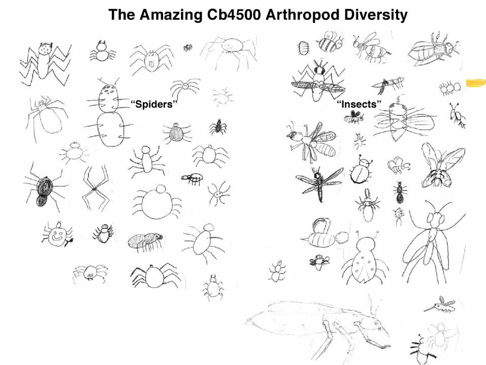
Arthropods are animals with an exoskeleton, articulated legs and segmented body plans
Two groups of major medical importance: chelicerata (in particular ticks & mites) and insects
Insects: capud, thorax & abdomen, 6 legs, two pairs of wings, 2 large complex eyes, open circulation, trachea

Platyhelminthes (flat worms) as human parasites I
Know your worm, anatomy & physiology of trematodes
Complex life cycles and adaptations to parasitism
Human intestinal, liver and lung flukes
Dr. Moreno will focus for an entire lecture next week on Schistosoma the medically most important flat worm


Flat worms, a rough classification
Planaria Aspidobothrea Digenea Monogenea Cestoda
Platyhelminthes
Trematoda
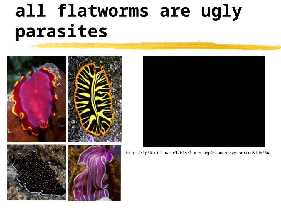
Pretty planaria, not all flatworms are ugly parasites
http://ip30.eti.uva.nl/bis/limno.php?menuentry=soorten&id=284

Flat worms, a rough classification
Planaria Aspidobothrea Digenea Monogenea Cestoda
Platyhelminthes
Trematoda

trematodes or flukes - when they say ‘flat’ worms they mean it
All digenea are parasitic Small dorso-ventrally
flattened worms with simple anatomy and without segmentation
No coelom (secondary body cavity lined by mesoderm), but animals are filled with mesodermal parenchyma
No blood vessels, simple ladder nervous system

trematodes or flukes - know your worm
Digenea posses two suckers (oral and ventral acetabulum) which they use to attach within the host
Oral sucker contains the mouth
Muscular pharynx permits the worm to pump food into the blind ending gut
Most trematodes are hermaphrodites (they are male and female, and cross as well as self-fertilization occurs)

trematodes or flukes - know your worm
The gut of trematodes is blind ending but can be quite extensive and highly branched (here shown in living Fasciola (liver) flukes, the dark staining is due to bile)
Smooth muscle fibers (longitudinal and cross) run under the tegument and around all the organs (the gut is shown in this picture)

trematodes or flukes - know your worm
The gut is not the only organ these worms use for food uptake
The tegument (“skin”) is highly active in nutrient uptake
The epidermis is essentially a single cell (a syncytium formed by fusion of multiple cells)
The tegument’s cell bodies and nuclei underlie the two muscle layers
Actin spines are found in many species and help the worms to anchor themselves

trematodes or flukes - know your worm
Platyhelminths have a simple protonephridial excretion (kidney) system
A protonephridium consists of flame cell and tubule cell
Both cells interdigitate forming a micro filter and cilia beating within this cage act as the mechanical pump
Excretes are collected from protonephridia through small ducts which merge to a central duct which opens into the excretion porus (usually towards the end of the animal)
There is a nice little animation of this process a http://www.biology.ualberta.ca/facilities/multimedia/uploads/zoology/Excretion.swf
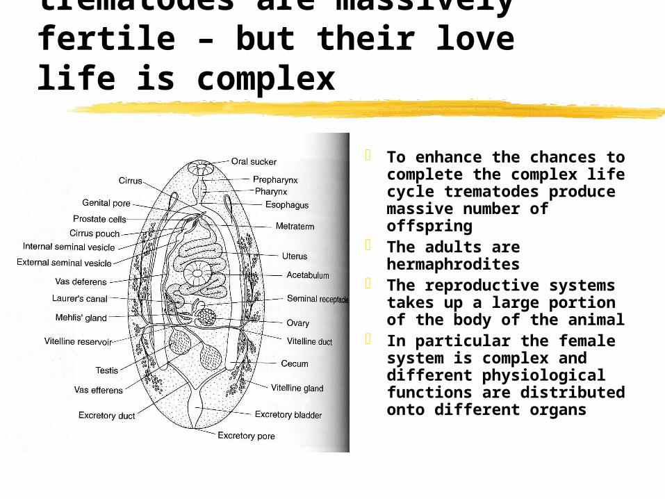
trematodes are massively fertile – but their love life is complex
To enhance the chances to complete the complex life cycle trematodes produce massive number of offspring
The adults are hermaphrodites
The reproductive systems takes up a large portion of the body of the animal
In particular the female system is complex and different physiological functions are distributed onto different organs

trematodes or flukes - know your worm
Trematodes form complex ectolecithal “eggs”
Oocytes (developing in the ovary) meet with vitelline or yolk cells (from the vitelary, which carry the bulk of the nutrients for the embryo) in the ootype which is surrounded by the Mehlis gland
An egg shell forms from secretions of the vitelline cells (the contribution of the gland to the shell is unclear)

trematodes or flukes - know your worm
Worm eggs travel through the uterus to the genital pore (tens of thousands a day)
On this way the proteinacous egg shell is hardened by quinone tanning
The amino acid tyrosine is modified into a highly reactive quinone in several enzymatic steps
The quinone then cross links free amino groups of adjacent proteins generating a very stable shell
The tanning process can be visualized by following the progressive darkening of the egg shells along the uterus

Trematode life cycles
Trematodes produce an enormous number of offspring by combining sexual and asexual reproduction cycles
Asexual reproduction occurs in germinal balls. These areas are home to omnipotent (stem cell-like) progentior cells that can initiate the development of embryos without fertilization
All have at least two hosts of which one is a snail
Not all stages are found in the life cycle of all species
Miracidia and cercariae are infective (invasive) stages
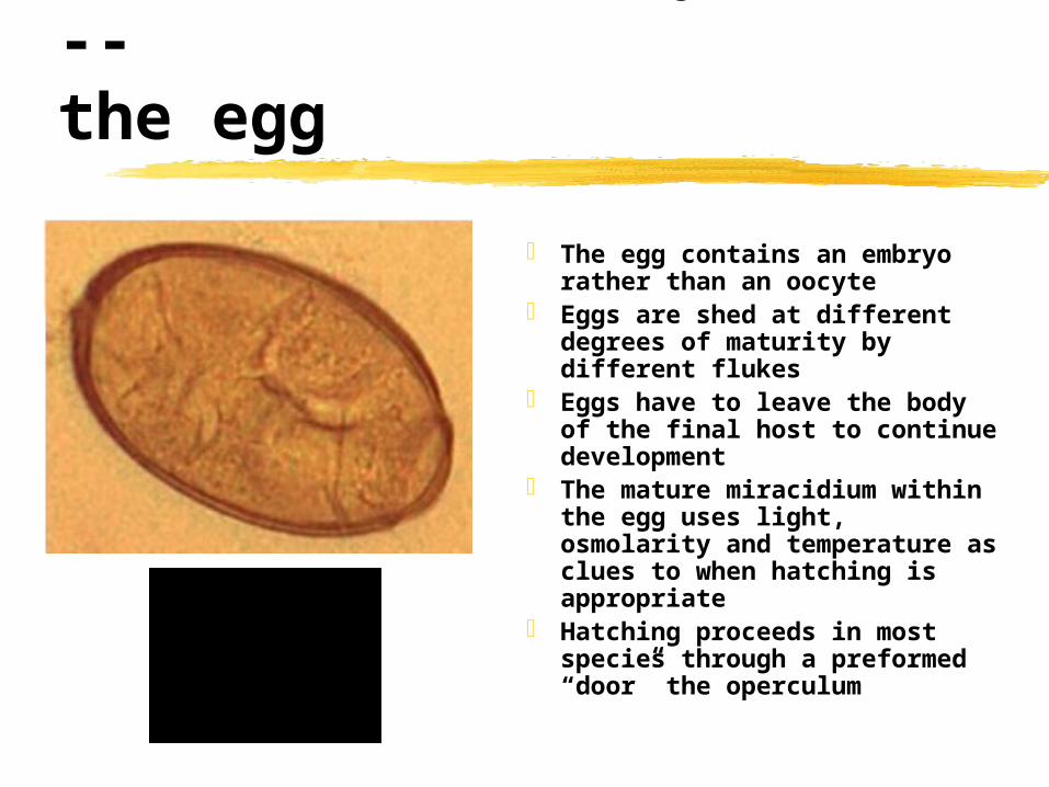
Trematode life cycles --the egg
The egg contains an embryo rather than an oocyte
Eggs are shed at different degrees of maturity by different flukes
Eggs have to leave the body of the final host to continue development
The mature miracidium within the egg uses light, osmolarity and temperature as clues to when hatching is appropriate
Hatching proceeds in most species through a preformed “door” the operculum

Trematode life cycles --the miracidium
The miracidium is highly motile due to the cilia on its surface
Miracidia have simple eyes (they avoid light) and several chemical and mechanical receptors which they use to find the intermediate snail host
Penetration glands secrete proteases and other lytic enzymes on contact with appropriate host
Miracidia of flukes with land snails as intermediate host will hatch upon ingestion by the snail and penetrate the gut epithelium

Trematode life cycles --the miracidium
Dept. Biology, Univ. of Alberta, Canada

Trematode life cycles --the sporocyst
After penetration the miracidum undergoes metamorphosis into the sporocysts
This stage has most organ systems reduced to the bare minimum and acts as a germinal sac
The sporocyst takes up nutrients only over its tegument and the germinal mass expands and develops into daughter sporocysts, redia or cercaria
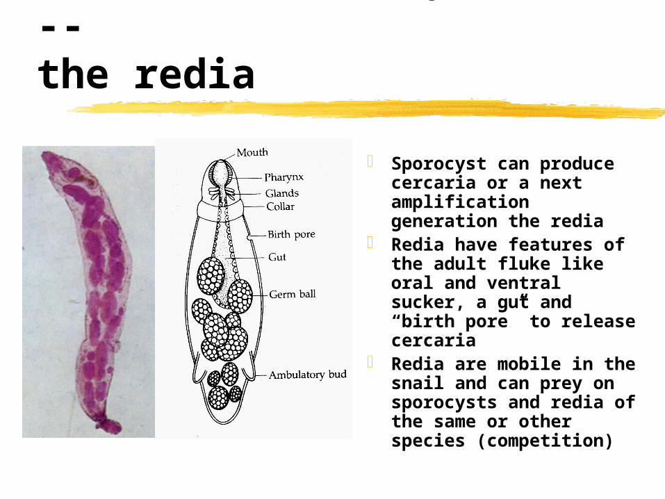
Trematode life cycles --the redia
Sporocyst can produce cercaria or a next amplification generation the redia
Redia have features of the adult fluke like oral and ventral sucker, a gut and “birth pore” to release cercaria
Redia are mobile in the snail and can prey on sporocysts and redia of the same or other species (competition)

Trematode life cycles --the cercaria
Cercaria are the stages that leave the intermediate host and infect the final host
There can be many consecutive waves of “shedding” from the snail
Cercaria already show many anatomical features of the adult fluke
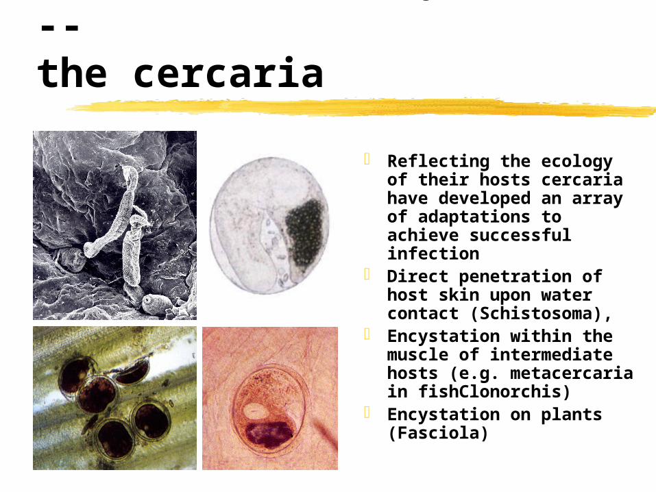
Trematode life cycles --the cercaria
Reflecting the ecology of their hosts cercaria have developed an array of adaptations to achieve successful infection
Direct penetration of host skin upon water contact (Schistosoma),
Encystation within the muscle of intermediate hosts (e.g. metacercaria in fishClonorchis)
Encystation on plants (Fasciola)

Trematode life cycles --enhance transmission
Dicrocoelium dendriticum the lancet fluke
One metacercaria becomes the ‘brain worm’ and lodges into the central ganglia of the end
The brain worm manipulates the behavior of the ant. In the evening when the temperature drops they experience spasms of their manidibles
Trematode life cycles --enhance transmission
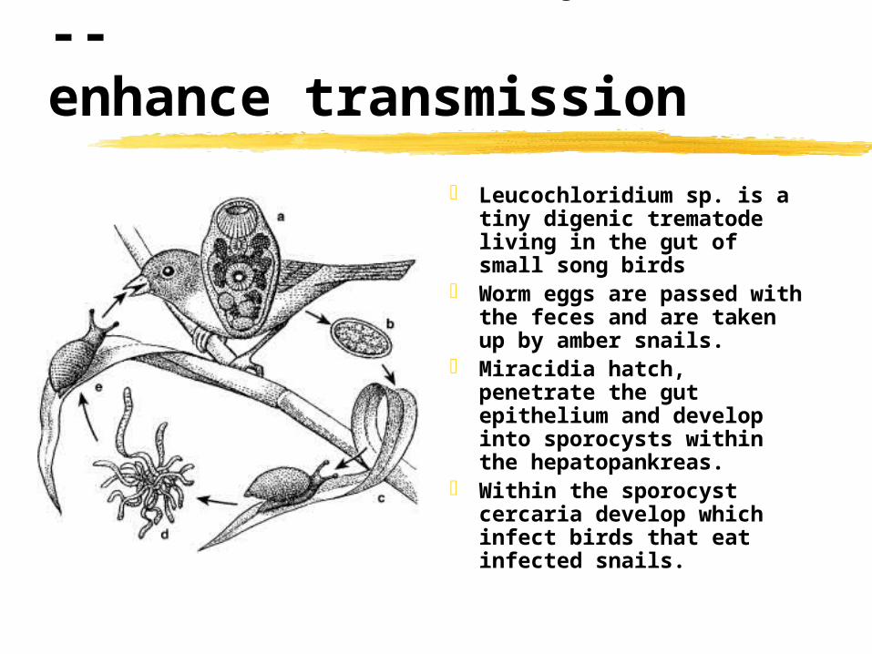
Leucochloridium sp. is a tiny digenic trematode living in the gut of small song birds
Worm eggs are passed with the feces and are taken up by amber snails.
Miracidia hatch, penetrate the gut epithelium and develop into sporocysts within the hepatopankreas.
Within the sporocyst cercaria develop which infect birds that eat infected snails.

Trematode life cycles --enhance transmission
Amber snails (uninfected, upperpanel and infected, lower panel) andLeucochloridium sp. sporocyst dissected from a snail (lower right)

Trematode life cycles --enhance transmission
Dr. Oldrich Nedved, Univ. South Bohemia

Trematodes of medical importance
Schistosoma, blood flukesClonorchis & Opistorchis, liver flukes with
metacercaria in fishParagonimus, lung flukes with
metacercaria in crabs Fasciolopsis, Fasciola, Dicrocoelium,
intestinal and liver flukes with metacercaria on plants
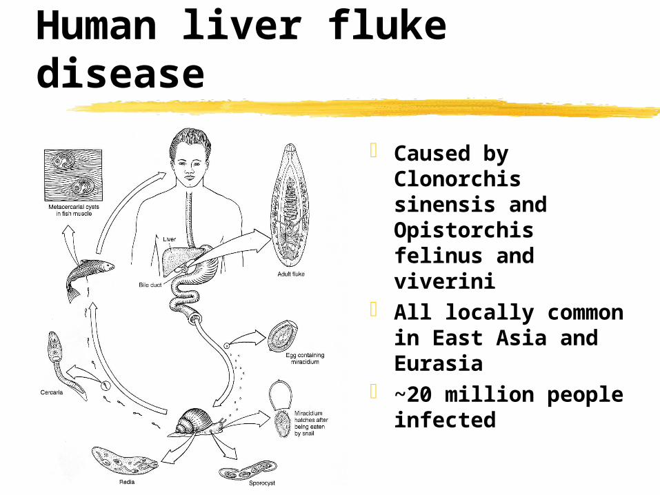
Human liver fluke disease
Caused by Clonorchis sinensis and Opistorchis felinus and viverini
All locally common in East Asia and Eurasia
~20 million people infected

Human liver fluke disease
Clonorchis and Opistorchis are quite similar causing similar disease

Human liver fluke disease
Metacercaria are found in many fish especially various carp related species
Raw or undercooked fish dishes are a source of human infection
Fertilization of ponds with untreated night soil boost infection in fish
Cats, dogs and other carnivores can be additional hosts and reservoirs

Human liver fluke disease
Pathology depends on worm burden, generally infections are light and free of major symptoms
Heavy infections Flukes residing in the bilary ducts can chronically iritate the epithelium resulting in hyperplasia of the epithelium and fibrosis around the ducts (pipe stem fibrosis)
Blockage of bile ducts and impairment of liver function, liver swelling
Human liver fluke disease
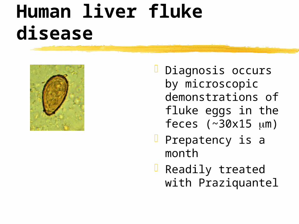
Diagnosis occurs by microscopic demonstrations of fluke eggs in the feces (~30x15 m)
Prepatency is a month
Readily treated with Praziquantel
Human lung fluke disease
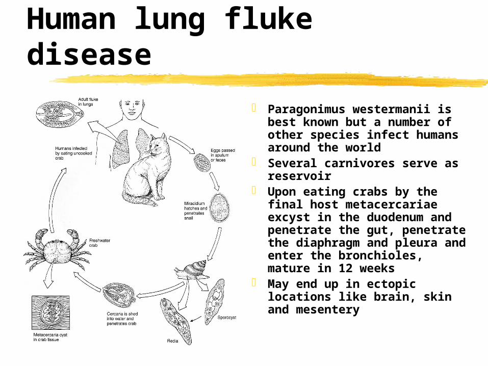
Paragonimus westermanii is best known but a number of other species infect humans around the world
Several carnivores serve as reservoir
Upon eating crabs by the final host metacercariae excyst in the duodenum and penetrate the gut, penetrate the diaphragm and pleura and enter the bronchioles, mature in 12 weeks
May end up in ectopic locations like brain, skin and mesentery

Human lung fluke disease

Human lung fluke disease
Adults are encapsulated in a granuloma (often two at a time)
Cyst rupture can result in cough and increase sputum, and chest pain
Chronic high worm burden can result in chronic bronchitis and dyspnea and increasing fibrosis -- symptoms can be very similar to pulmonary tuberculosis
Cerebral paragonimiasis produces headaches, fever, nausea, visual disturbances and convulsive seizures

Fasciola & Fasciolopsis
Important parasite of livestock, ocasionally infects humans
Symptoms similar to Clonorchis but Fasciola is much bigger
Fasciolopsis buski the human intestinal fluke has similar ecology
Usually asymptomatic if not heavy burden
Fasciola
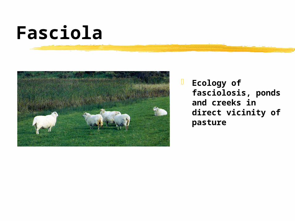
Ecology of fasciolosis, ponds and creeks in direct vicinity of pasture