treatment of symptomatic abnormal skin scars with ... · treatment of symptomatic abnormal skin...
TRANSCRIPT

practice!
J O U R N A L O F WO U N D C A R E VO L 1 9 , N O 1 0 , O C TO B E R 2 0 1 0 4 4 7
Treatment of symptomatic abnormal skin scars with electrical stimulation" Objective: To evaluate the effect of non-invasive biofeedback electrical stimulation on symptomatic abnormal skin scars." Method: Thirty patients with over 140 scars with long-term pain and itch were recruited into the study. Patients monitored the intensity of symptoms (pain and itching) on a numerical rating scale. In addition, a modi!ed Manchester scar scale was used to objectively assess digital photographs of each scar in terms of colour, contour, distortion and texture, while a non-invasive spectrophotometric intracutaneous analysis was used to monitor the scars’ physical characteristics. " Results: The electrical stimulation device resulted in a clinically and statistically signi!cant (p<0.05) reduction of symptoms and scar scores. Pain and itch scores were both reduced to a median score of 0 by 2 months, from a baseline of 7 and 6 respectively. Scar scores were reduced from a baseline of 14 to a median score of 11 by 2 months. " Conclusion: These results give a preliminary indication of the potential role of non-invasive biofeedback electrical stimulation in the management of chronic scar pain and itch. However, further large scale controlled studies are warranted to elucidate its overall ef!cacy and mechanistic action." Con!ict of interest: Funding was provided from Fenzian Ltd for this study.
itch; pain; sensitive scars; pruritus; abnormal skin scarring
!A ll cutaneous scarring has the potential to affect quality of life.1,2 With nearly 100 million people acquiring skin scars every year in the developed world alone,3–5 !nding an effective treatment
poses a signi!cant challenge. Current management options are often unacceptable to the patient, and minimally invasive procedures, such as steroid injections, or radical interventions, such as scar excision, can cause further pain. A promising area of development in the management of problematic soft tissue conditions is electrical stimulation.
Undamaged human skin has an endogenous elec-trical potential and a transcutaneous current poten-tial (10–60mV),6 generated by the inward move-ment of sodium ions through Na+/K+ ATPase pumps in the epidermis.7 When an injury affects epidermal integrity, an overall "ow of current through the wound pathway generates a lateral electrical !eld, both within and beneath the epidermis; this is known as the ‘current of injury’ or the ‘skin battery’ effect.8 As the wound heals, the current of injury returns to its baseline level.9 Therefore, the current of injury is thought to be signi!cant in triggering biological repair; indeed, it is absent in some chron-ic wounds.8,10,11
In the past few years, there has been greater recog-nition of the role played by electrical !elds in cellu-lar behaviour and motility.12,13 Studies have demon-strated that electrical stimulation can enhance tissue
healing by promoting the migration of keratinocytes and macrophages,14 encouraging angiogenesis,15 stimulating !broblasts, and increasing adenosine tri-phosphate and protein synthesis.16 Further evidence suggests it may have antimicrobial effects.17
In 2009, Poltawski and Watson reviewed the evi-dence on microcurrent therapy, which applies elec-trical current levels similar to those produced by the body during normal tissue repair, and concluded that it can promote healing in skin lesions and may have a potential role to play in wound care.18
More recently, a novel in vitro model for testing the effects of precisely de!ned types of electrical stimulation on collagen expression in normal and keloid human skin !broblasts was developed at the senior author’s (AB) laboratory.19 Both cell types were electrically stimulated with alternating current, direct current or degenerate waves (the wave form generated by the biofeedback electrical stimulation device investigated in this paper). Following 12 hours of exposure to degenerate waves, keloid !brob-lasts (which show excessive collagen production) were found to have a statistically signi!cant decrease in collagen I expression. This indicates that electrical currents, in particular degenerate waves, are a prom-ising, novel therapeutic strategy for suppressing excessive collagen I formation in keloid disease.
Meanwhile, there is growing acknowledgement of the link between the skin’s electrical impedance pat-terns (surface readings of electrical !eld gradi-
D. Perry,1,2 BSc (Hons);J. Colthurst,3 FRCS;P. Giddings,3 Dip HE;D.A. McGrouther,2 MD FRCS;J. Morris,2,4 MSc;A. Bayat,1,2 PhD MRCS;1 Plastic and Reconstructive Surgery Research, School of Translational Medicine, University of Manchester, UK2 Manchester Academic Health Science Centre, Department Plastic & Reconstructive Surgery, University Hospital of South Manchester, UK3 Eumedic Ltd, Hungerford, UK4 Medical Statistics, Education and Research Centre, University Hospital of South Manchester, UKEmail: [email protected]
!"#$%&$%'$()'$*+,,-./011222((3 '34%'45'%'222%67'3

practice
J O U R N A L O F WO U N D C A R E VO L 1 9 , N O 1 0 , O C TO B E R 2 0 1 04 4 8
ents20,21) and underlying clinical conditions, such as asthma.22,23 It is thought these electrical impedence patterns represent a modi!cation of cell behaviour which may also be represented at the central nerv-ous system level. Interactions between the central nervous system and the skin involve neuropeptides, cytokines, hormones and other effector molecules.24
It has been proposed that there is an interrelation-ship between the skin, endocrine, immune and cen-tral nervous systems, which has been termed the neuro-immuno-cutaneous-endocrine model. According to this theory, electrical stimuli at the skin surface can in"uence all of these systems at both a local and central level.25 For example, trans-cutaneous electrical nerve stimulation activates opi-oid receptors in the central nervous system, as dem-onstrated in basic science studies using both high and low frequencies.26,27 In mild asthma, electrical stimulation appears to facilitate neurological adjust-ment of mast cell sensitivity.28
The Fenzian treatment systemAn emerging adjuvant therapy is the Fenzian bio-feedback electrostimulation treatment system (Eumedic, UK, Fig 1), which delivers a low-intensity transcutaneous electrostimulation current to specif-ic skin areas. It follows the theory that the electrical potential of skin forms a global electrical network, and that any changes in skin impedance re"ect underlying neurological activity.29 The mechanism by which this body-wide electrical network might stimulate a healing response is not yet fully under-stood. However, the disruption to these body-wide potential patterns during injury is a likely trigger for tissue repair, in addition to the release of hormones and numerous chemical medicators.30,31
The Fenzian system detects the skin’s electrical impedance using a microcurrent generator. The out-going transformer signal is measured across a con-centric electrode, and a biofeedback impulse is applied (this comprises a sequence of electrical impulses, the sizes of which depend on alterations in skin response).32 The user is guided to optimal bio-feedback sites by a numerical depiction of the outgo-ing signal characteristics. When this shows that bio-feedback is complete (by reaching an unchanging electrical state), an audible bell sounds and the device is then moved to another site, or the treat-ment may be complete (depending on the protocol).
Fenzian is applied to a patient’s skin by a specially
trained medical practitioner (a doctor, nurse or phys-iotherapist) in a protocol that depends on the indi-vidual patient. Patients are treated while sitting or (very occasionally) lying down. This microcurrent electrical stimulation uses currents that are in the microampere range, which are a thousand times low-er than transcutaneous electrical nerve stimulation (TENS). Pulse widths are also different (average 0.5 seconds), typically 2,500 times longer than a TENS unit, and often below the sensation threshold.33 The device has a 45 x 22mm electrode, which is brushed or physically held in contact with the skin for the duration of treatment. Impulses are of short duration (~10µs) and of relatively high amplitude (80V).
Background to the evaluationMany scars have a chronic in"ammatory compo-nent, either with erythematous colouration34 or as a result of acute sensitisation of nociceptors and/or activation of puriceptors,35 although the exact underlying mechanism is not fully understood.36
Previous retrospective case note reviews32 and controlled pilot studies28 of the Fenzian system have demonstrated a persistent pattern of improved symptoms across a wide range of conditions, includ-ing asthma and traumatic cutaneous injuries.
We conducted an open-label observational study to assess the subjective bene!ts and objective changes in symptomatic, raised, dermal scars treat-ed with the Fenzian system. This is the !rst study to formally evaluate its use in cutaneous scars. Prob-lematic scarring was chosen because of the extent of Fig 1. The Fenzian treatment system
Table I. Basic demographics (n=19)
Sex (male/female) 3/16
Age (years) ""#25 7""26–35 3""36–45 4""#46 & over 5
Ethnicity: Caucasian/other 14/5
Fitzpatrick skin scale ""I–III 14""IV–VI 5
Positive abnormal family scar history 2
Positive previous history of abnormal scarring 4
Past medical history conditions ""Respiratory 3""Dermatological 4""Other 5
References1 Rhee, P., Brown, C., Martin, M. et al. QuikClot use in trauma for hemorrhage control: case series of 103 documented uses. J Trauma. 2008; 64: 4, 1093–1099.2 Brown, B.C., Moss, T.P., McGrouther, D.A., Bayat, A. Skin scar preconceptions must be challenged: Importance of self-perception in skin scarring. J Plast Reconstr Aesthet Surg. 2010; 63: 6, 1022–1029.3 Bayat, A., McGrouther, D.A., Ferguson, M.W. Skin scarring. BMJ. 2003; 326: 7380, 88–92.4 Sund, B. (ed). New Developments In Wound Care. PJB Publications, 2000.5 Gangemi, E.N., Gregori, D., Berchialla, P. et al. Epidemiology and risk factors for pathologic scarring after burn wounds. Arch Facial Plast Surg. 2008; 10: 2, 93–102.6 Foulds, I.S., Barker, A.T. Human skin battery potentials and their possible role in wound healing. British J Dermatol. 1983; 109: 5, 515–522.7 McGinnis, M.E., Vanable, J.W. Jr. Voltage gradients in newt limb stumps. Prog Clin Biol Res. 1986; 210: 231–238.8 Barker, A.T., Jaffe, L.F., Vanable, J.W. Jr. The glabrous epidermis of cavies contains a powerful battery. Am J Physiol. 1982; 242: 3, R358–366.9 Jaffe, L.F., Vanable, J.W. Jr. Electric !elds and wound healing. Clinics in dermatology. 1984; 2: 3, 34–44.10 Ojingwa, J.C., Isseroff, R.R. Electrical stimulation of wound healing. J Invest Dermatol. 2003; 121: 1, 1–12.11 Nuccitelli, R. A role for endogenous electric !elds in wound healing. Curr Top Dev Biol. 2003; 58: 1–26.12 Zhao, M., Song, B., Pu, J. et al. Electrical signals control wound healing through phosphatidylinositol-3-OH kinase-gamma and PTEN. Nature. 2006; 442: 7101, 457–460.13 Song, B., Gu, Y., Pu, J. et al. Application of direct current electric !elds to cells and tissues in vitro and modulation of wound electric !eld in vivo. Nat Protoc. 2007; 2: 6, 1479–1489.
!"#$%&$%'$()'$*+,,-./011222((8 '34%'45'%'222%67'3
practice!
J O U R N A L O F WO U N D C A R E VO L 1 9 , N O 1 0 , O C TO B E R 2 0 1 0 4 4 9
pain and pruritic symptoms endured by some patients, and the poor range of non-invasive man-agement options currently available.
Materials and methodPatients attending the specialist scar service clinic at the University Hospital of South Manchester (UHSM) NHS Foundation Trust between January 2009 and June 2009 were eligible for recruitment. Inclusion criteria were:" Patients with one or more cutaneous scars that had not responded to previous treatment, such as steroid injections, surgical excision and silicone gel therapy (based on unsatisfactory scar appearance and/or symptoms), or for which the patient had requested further non-invasive management" Patients with any problematic scar type, such as keloid, hypertrophic, or history of scarring such as trauma, surgery, acne.
Exclusion criteria were: " Patients taking medication that reduce electrical activity of the skin, such as antibiotics and steroids)37
" Patients with implanted electrical devices, such as pacemakers and cochlear implants" For cautionary reasons, patients who were preg-nant or planning to conceive
There were no exclusion criteria relating to sex, age or past medical history.
The interventionThe biofeedback electrical stimulation therapy was administered by a single therapist as part of the patient’s routine care. The battery-operated Fenzian system is both CE approved and US FDA 510(k) reg-istered, and it passed the UHSM Trust Medical Engi-neering requirements for clinical usage.
As the device was used as indicated by its CE mark, ethics committee approval was not required. However, all patients gave written informed con-sent for the image/photographic monitoring of their scars.
Treatment was administered according to stand-ardised local (scar location) or global (whole body) protocols, depending on the anatomical site affect-ed and the physiological systems linked with the individual’s scar history (for instance, targeting low-er abdominal hormonal sites in relation to acne scarring).
Treatment times were dictated by the device via its biofeedback electrical mechanism (average 20 minutes in duration) and administered by a single clinician. For the !rst 3–4 weeks, treatments were administered twice weekly. Further treatments and review appointments continued for up to 6 months, on a monthly basis.
Patients did not receive any other forms of scar therapy during the course of bioelectrical stimula-tion treatment.
AssessmentSubjective and objective outcome measures were recorded by a single unblinded therapist (!rst author, DP) and then evaluated by the senior author (AB). " At every visit, we recorded the patient’s subjective rating of perceived pain and intensity of itch over the past 24 hours, using the validated 11-point numerical rating scale (NRS, where 0 = no pain/itch, 10 = worst possible pain/itch). We also noted wheth-er the symptom was constant or intermittent" Digital photographs were taken and the scar sites were clinically evaluated at all treatment visits using a modi!ed Manchester Scar Score (mMSS).38 This includes assessment of scar colour, contour, distor-tion and texture. Each parameter is scored on a lin-ear scale of 1–4, with increasing scar severity scoring more highly. This also records each scar’s matte or shiny appearance, with 1 = matte and 2 = shiny. Scores are totalled and range from 5 (clinically well-healed scar) to 18 (clinically poor scar).38
" At multiple time points throughout treatment and on consistently selected scar site areas, objective spectrophotometric intracutaneous analysis (SIA) or
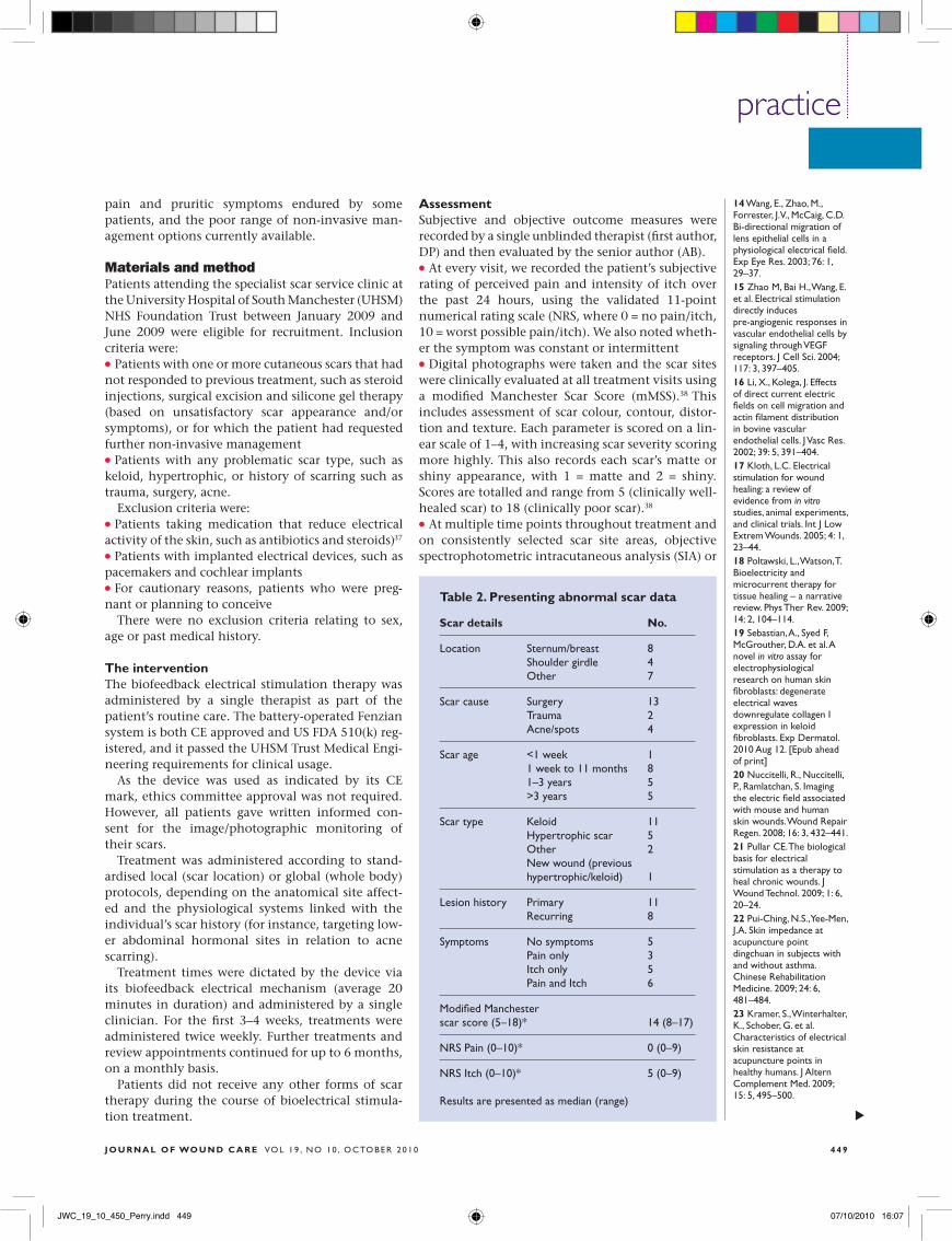
Table 2. Presenting abnormal scar data
Scar details No.
Location Sternum/breast 8 Shoulder girdle 4 Other 7
Scar cause Surgery 13 Trauma 2 Acne/spots 4
Scar age <1 week 1 1 week to 11 months 8 1–3 years 5 >3 years 5
Scar type Keloid 11 Hypertrophic scar 5 Other 2 New wound (previous hypertrophic/keloid) 1
Lesion history Primary 11 Recurring 8
Symptoms No symptoms 5 Pain only 3 Itch only 5 Pain and Itch 6
Modi!ed Manchester scar score (5–18)* 14 (8–17)
NRS Pain (0–10)* 0 (0–9)
NRS Itch (0–10)* 5 (0–9) Results are presented as median (range)
14 Wang, E., Zhao, M., Forrester, J.V., McCaig, C.D. Bi-directional migration of lens epithelial cells in a physiological electrical !eld. Exp Eye Res. 2003; 76: 1, 29–37.15 Zhao M, Bai H., Wang, E. et al. Electrical stimulation directly induces pre-angiogenic responses in vascular endothelial cells by signaling through VEGF receptors. J Cell Sci. 2004; 117: 3, 397–405.16 Li, X., Kolega, J. Effects of direct current electric !elds on cell migration and actin !lament distribution in bovine vascular endothelial cells. J Vasc Res. 2002; 39: 5, 391–404.17 Kloth, L.C. Electrical stimulation for wound healing: a review of evidence from in vitro studies, animal experiments, and clinical trials. Int J Low Extrem Wounds. 2005; 4: 1, 23–44.18 Poltawski, L., Watson, T. Bioelectricity and microcurrent therapy for tissue healing – a narrative review. Phys Ther Rev. 2009; 14: 2, 104–114.19 Sebastian, A., Syed F, McGrouther, D.A. et al. A novel in vitro assay for electrophysiological research on human skin !broblasts: degenerate electrical waves downregulate collagen I expression in keloid !broblasts. Exp Dermatol. 2010 Aug 12. [Epub ahead of print] 20 Nuccitelli, R., Nuccitelli, P., Ramlatchan, S. Imaging the electric !eld associated with mouse and human skin wounds. Wound Repair Regen. 2008; 16: 3, 432–441.21 Pullar CE. The biological basis for electrical stimulation as a therapy to heal chronic wounds. J Wound Technol. 2009; 1: 6, 20–24.22 Pui-Ching, N.S., Yee-Men, J.A. Skin impedance at acupuncture point dingchuan in subjects with and without asthma. Chinese Rehabilitation Medicine. 2009; 24: 6, 481–484.23 Kramer, S., Winterhalter, K., Schober, G. et al. Characteristics of electrical skin resistance at acupuncture points in healthy humans. J Altern Complement Med. 2009; 15: 5, 495–500.
!"#$%&$%'$()'$*+,,-./011222((& '34%'45'%'222%67'3

practice
J O U R N A L O F WO U N D C A R E VO L 1 9 , N O 1 0 , O C TO B E R 2 0 1 04 5 0
SIAscopy (Siascope, Astron Clinica Ltd, Cambridge, UK) was performed. Using a non-invasive light-based technology probe, the quantities of light remitted by the skin at different wavelengths are determined,39 providing a photographic pigmentary status and quantitative numerical values for the constitutional elements of the !rst 2mm of skin (melanin, haemoglobin and collagen).40
Additionally, basic demographic data were col-lected from the patient notes for cross analysis pur-poses. If patients presented with more than one scar, the most problematic scars were selected for objective monitoring. Patients were also monitored for any adverse reactions.
Statistical analysisDescriptive statistics were used to document trends between demographic characteristics and variables where group size prevented inferential statistical testing. Only one scar per subject (de!ned by the highest pain, itch and scar scores) was selected for inclusion in the statistical analysis. Non-parametric Wilcoxon signed rank tests were applied to assess dif-ferences in pain, itch, scar score and chromophores
between time points. The statistical package SPSS version 15.0 was used, and all analyses were carried out using the conventional 5% signi!cance level.
ResultsDemographicsThirty patients with more than 140 (52 evaluated) scars were treated with the biofeedback electrical stimulation system (the test treatment). Patient demographic details are given in Table 1. Eleven participants were excluded from the basic statistical analysis due to either failing to complete a basic course of treatment (minimum !ve sessions) or because they started medication known to reduce the therapeutic effects of the test treatment. This resulted in an observational case series sample of 19 patients with 31 monitored scars, of which 19 were included in the statistical analysis.
Patients had a mean age of 37 years (range 15–85), a modal Fitzpatrick skin classi!cation of type II (fair skin, burns easily and tans poorly) and 84% were female. Co-existing dermatological conditions were common, and included eczema, psoriasis and acne.
Presenting complaintThe 19 individuals included presented with a range of abnormal skin scars (Table 2), primarily due to surgery. Acne keloid scarring or a strong propensity for keloid disease accounted for the majority of self-de!ned ‘problematic’ scars. Most scarring was kel-oid in nature, affecting the sternum/breast, and was over 3 years in duration (median 2 years, range one week to 30 years).
On entry into the study, most individuals had a primary abnormal scar lesion (not previously excised). However, one new wound also received treatment, following repeat debulking surgery.
At the start of treatment, !ve patients had no pain or itch, !ve complained of itch only, three of pain only, and six of itch and pain together. At baseline, the median NRS pain score was 0 (range 0–9) and the median NRS itch score was 5 (range 0–9). In gen-eral, a high proportion of scars were rated ‘clinically poor’ using the mMSS (median 14, range 8–17).
Therapeutic outcomesThe 19 patients received a median of nine (range 5–16) treatments over a median 70-day period (range 27–138).
Outcome data were selected and grouped into speci!c one-week, and one, two and three-month time point ranges for simpli!cation. The latter time point was omitted from statistical analysis as only 11 patients had follow-up data at this time.
Symptomatic outcomePatient-perceived symptomatic outcomes are dis-played in Table 3. Of the nine patients with pain at
Table 3. Symptomatic response to treatment
Pain Itch
No. No.
Initial symptom
0 10 8
1–3 0 1
4–6 4 5
7–10 5 5
Response at 1 week (with score "1 at baseline)
Increased 2 2
Decreased 4 6
Same 3 3
Overall response to biofeedback electrical stimulation by 2 months (with score "1 at baseline)
Increased 0 0
Decreased but continued symptom 4 4
Same 0 0
Symptom resolved 5 7
24 O’Sullivan, R.L., Lipper, G., Lerner, E.A. The neuro-immuno-cutaneous-endocrine network: relationship of mind and skin. Arch Dermatol. 1998; 134: 11, 1431–1435.25 Brazzini, B., Ghersetich, I., Hercogova, J., Lotti, T. The neuro-immuno-cutaneous-endocrine network: relationship between mind and skin. Dermatol Ther. 2003; 16: 2, 123–131.26 Ainsworth, L., Budelier, K., Clinesmith, M. et al. Transcutaneous electrical nerve stimulation (TENS) reduces chronic hyperalgesia induced by muscle in$ammation. Pain. 2006; 120: 1–2, 182–187.27 Sluka, K.A., Deacon, M., Stibal, A. et al. Spinal blockade of opioid receptors prevents the analgesia produced by TENS in arthritic rats. J Pharmacol Exp Ther. 1999; 289: 2, 840–846.28 Cooper, C.B., Boscardin, W.J., Colthurst, J.R., Kleerup, E.C. Treatment of mild persistent asthma by cutaneous electronic stimulation. Eur Respir J. 2009; 34: 2, 515–517.29 Becker, R.O. Some observations indicating the possibility of longitudinal charge-carrier $ow in the peripheral nerves. In: Bernard, E.E., Kare, M.R. (eds). Biological Prototypes And Synthetic Systems. Plenum, 1962.30 Frank, C.B., Szeto, A.Y. A review of electromagnetically enhanced soft tissue healing. IEEE Eng Med Biol Mag. 1983; 2: 4, 27–32.31 Watson, T. Electrical stimulation for wound healing: a review of current knowledge. In: Kitchen, S. (ed). Electrotherapy: Evidence-Based Practice. Churchill Livingstone, 2002.32 Colthurst J, Giddings P. A retrospective case note review of the Fenzian electrostimulation system: a novel non-invasive, non-pharmacological treatment. The Pain Clinic. 2007; 19: 1, 7–14.33 Mercola, J.M., Kirsch, D.L. The basis for microcurrent electrical therapy in conventional medical practice. J Adv Med. 1995; 8: 2, 107–120.
!"#$%&$%'$()'$*+,,-./011222()' '34%'45'%'222%67'3

practice!
J O U R N A L O F WO U N D C A R E VO L 1 9 , N O 1 0 , O C TO B E R 2 0 1 0 4 5 1
baseline, four (44%) reported decreased pain at one week. Symptoms more frequently decreased in itch sufferers, with six (55%) of the 11 with itch at base-line reporting improvement. In all, four scars had an initial exacerbation of symptoms (three by one NRS point and one by two NRS points). For those patients in whom pain and itch improved, median reductions of four (range 2–6) and 2.5 (range 1–7) NRS points were observed for pain and itch respec-tively, following initial treatment.
By one month, of the nine patients with pain at baseline, three had no pain and the remaining six had reduced pain. Of these six patients, two had no pain by 2 months. Therefore, resolution of pain was achieved by 2 months for !ve (56%) of the nine patients with pain at baseline, after a median of six treatments (range: 5–8). Of the four patients with continuing pain, pain scores had reduced by a medi-an of 2.5 (range: 1–6).
Of the 11 patients with itch at baseline. Five had no itch by one month, and the remaining six had reduced symptoms. By 2 months, resolution of itch was achieved in a total of seven (64%) patients after a median of six treatments (range: 5–14). Of the remaining four patients, itch scores had reduced by a median of 3.5 (range: 2–6).
Median scores and ranges are displayed in Table 4, together with Wilcoxon test results, which show that statistically signi!cant changes from baseline occurred at one and 2 months for both pain and itch scores.
Observed scar score outcomesA positive response was observed in 24 of the 31 scars affecting the 19 patients. Only two patients had no observed alteration in their scar characteris-tics during adjuvant therapy.
Overall, the mMSS had reduced by a median of three (range: 0–5) at two months. We observed sta-tistically signi!cant reductions in total scar scores at one week, one month and two months (see Table 4).
Scar score reductions recorded using the mMSS were largely due to objective reduced scores in the ‘colour’ and ‘texture’ categories, but also in the ‘mat-te/shiny’ appearance. Figs 2–4 display example plain photographs and colour, haemoglobin, melanin and collagen SIAmetric images of three scars before ini-tial Fenzian application and after treatment.
Objective melanin, haemoglobin and collagen chromophore analysisNo known widespread normative quantitative val-ues of melanin, haemoglobin and collagen chromo-phores exist in new wounds or abnormal scarring. Hence, data were examined for cumulative and sig-ni!cant patterns within individuals. No statistically signi!cant changes in haemoglobin levels, collagen and melanin were observed (see Table 4).
Fig 2. Four month old raised facial scar before treatment (a) and 6 weeks’ post-initial treatment (b). SIAmetrics chromophore images (ordered plain photograph, haemoglobin, melanin and collagen) taken from the scar region highlighted depict changes before (i–iv) and after (v–viii).
a
i ii iii
vi vii
iv
v viii
b
Fig 3. Eight month old recurring keloid sternal scar before treatment (a) and 2 months post-initial treatment (b). SIAmetrics chromophore images (ordered plain photograph, haemoglobin, melanin and collagen) taken from the scar region highlighted depict changes before (i–iv) and after (v–viii).
a b
i
v vi vii
ii iii iv
viii
!"#$%&$%'$()'$*+,,-./011222()% '34%'45'%'222%67'3

practice
J O U R N A L O F WO U N D C A R E VO L 1 9 , N O 1 0 , O C TO B E R 2 0 1 04 5 2
Other effects A number of additional ‘side-effects’, not formally evaluated, were observed during the treatment proc-ess. Three individuals with increased skin oil pro-duction and scars caused by acne/spots41 appeared to have a reduction in symptoms in the upper trunk region following 1–6 treatments. Three participants described a non-painful tingling sensation at the treatment site for approximately 2 hours post-thera-py. Furthermore, two patients with high-intensity scar pain noted immediate (within 12–24 hours) relief of symptoms for 4 and 7 days post-initial treat-ment. In one patient this was maintained with fur-ther treatment to eradicate pain, while the other individual’s pain persisted, but at a lower intensity. No adverse events were reported.
DiscussionA positive effect was observed with the biofeedback stimulation adjuvant therapy, in terms of both symptoms and the objective scar parameters moni-tored during treatment. Pain and itch were signi!-cantly reduced (p<0.05) in all participants at the three monthly intervals recorded, and the majority of patients’ symptoms resolved completely during the study period. These results are clinically signi!-cant, as illustrated by the reduction of two NRS points, a requirement for a clinically important dif-ference to be proven.42
Despite their similarly matched baseline NRSs, greater pain relief was observed in keloid scars and greater itch relief was observed in hypertrophic scars. Mast cells, which are involved in the healing process, contain many itch mediators (including histamine and substance P).43 Raised scars have been shown to have higher substance P nerve !bre densi-ties, greater substance P quantities and an increased number of mast cells.44 Substance P is also thought to mediate pain via small, unmylinated C !bres.45 In raised scars, it may contribute to an exuberant neu-roin"ammatory response due to a reduction in its regulatory enzyme, endopeptidase.46 In"amed scars are often typically red and raised. The post-treat-ment reduction in scar score (by objective parame-ters), as well as reduction in pain and itch, indicated a decreased in"ammatory state. However, this would have to be veri!ed histologically.
Signi!cant reductions in scar scores were noted at all time points, and the clearest reductions were observed in hypertrophic and surgical scars. Given that scar score reductions recorded using the mMSS were most frequently associated with a lessening of colouration, and that problematic hypertrophic scars are often persistently erythematous,34 it makes sense that this group had lower scores following therapy. It has been suggested that neurogenic in"ammation stimulates abnormal scarring,47 so normalisation of this response may enhance abnor-mal scar resolution.
We examined the chromophore levels of constitu-
Fig 4. Eighteen-month-old red and partially raised caesarean section scar before treatment (a) and post-3 months after initial treatment (b). SIAmetrics chromophore images (ordered plain photograph, haemoglobin, melanin and collagen) taken from the scar region highlighted depict changes before (i–iv) and after (v–viii)
ba
i
v vi vii
ii iii iv
viii
Table 4. Symptomatic outcomes scores
Variable Baseline 1 week 1 month 2 months
Pain (n=9) 7 (4–9) 5 (0–9) 3 (0–8)** 0 (0–7)**
Itch (n=11) 6 (3–9) 4 (0–8) 2 (0–6)** 0 (0–4)**
Scar score (n=19) 14 (8–17) 13 (8–17)* 12 (6–17)** 11 (6–17)**
Haemoglobin (n=19) 191 (16–224) 196 (14–244) 190 (14–244) 184 (9–236)
Collagen (n=19) 192 (129–226) 190 (135–293) 200 (132–301) 195 (146–233)
Melanin (n=19) 216 (117–842) 202 (139–842) 212 (103–842) 228 (120–842)
Scores are presented as median (range) *p<0.05, **p<0.01For pain and itch scores, only patients with non-zero scores at baseline are included
34 Köse, O., Waseem, A. Keloids and hypertrophic scars: are they two different sides of the same coin? Dermatol Surg. 2008; 34: 3, 336–346.35 Schmelz, M. Itch and pain. Neurosci Biobehav Rev. 2010; 34: 2, 171–176.36 Cheng, B., Liu, H.W., Fu, X.B. et al. Coexistence and upregulation of three types of opioid receptors, mu, delta and kappa, in human hypertrophic scars. Br J Dermatol. 2008; 158: 4, 713–720.37 Emtestam, L., Kuzmina, N., Talme, T. Evaluation of the effects of topical clobetasol propionate by visual score, electrical impedance and laser Doppler $owmetry. Skin Res Technol. 2007; 13: 1, 73–78.38 Beausang, E., Floyd, H., Dunn, K.W. et al. A new quantitative scale for clinical scar assessment. Plast Reconstr Surg. 1998; 102: 6, 1954–1961.39 Moncrieff, M., Cotton, S., Claridge, E., Hall, P. Spectrophotometric intracutaneous analysis: a new technique for imaging pigmented skin lesions. Br J Dermatol. 2002; 146: 3, 448–457.
!"#$%&$%'$()'$*+,,-./011222()5 '34%'45'%'222%67'3
practice
J O U R N A L O F WO U N D C A R E VO L 1 9 , N O 1 0 , O C TO B E R 2 0 1 0 4 5 3
al elements in the epidermis (melanin, haemoglob-in and collagen) for general pattern changes, as no current literature exists to suggest the extent and speci!city of cellular changes following biofeedback electrical stimulation treatment. An electro-thera-peutic, pro-in"ammatory response can be observed initially, as haemoglobin increases, and it has been suggested that this may stimulate chronically in"amed tissue to progress to resolution.28,31 Previ-ous data suggest that low frequency electrical stimu-lation increases blood "ow, causing vasodilation by the release of neuropeptides from the terminal end-ings of excited axons through C !bres.48 Hence, we postulate a mechanistic path for altered scar symp-toms with biofeedback electrical stimulation.
At 3 months, greater improvements were noted in symptoms and scar scores, perhaps signalling resolu-tion of the acute in"ammatory response. No scars grew larger in response to the treatment.49 Fluctua-tions in the scar collagen levels recorded may account for the raised collagen levels in these individuals.
Overall, a chromophore’s trend response to the treatment can only be postulated, due to the lack of published scar data and questions of the suitability of the instrument used in this preliminary study. Additionally, the proposed body-wide mechanism of action of this treatment prevents determination of the extent of any changes, as there is no opportu-nity for a subject to act as his or her own control.
Skin, endocrine and immune system interactions involve a number of neuropeptides, cytokines, hor-mones and other effector molecules.24 Therefore, stimuli at the skin’s surface have in"uence both locally and centrally. A growing body of evidence suggests that interaction between the skin and nerv-ous system contributes greatly to wound healing.50 The nervous system can modulate locally induced in"ammatory responses in the skin through the release of neuropeptides.25
This was a small, open label observational case series study designed to gather preliminary data to help guide future applications of this modality. Obvious study limitations exist. These include the limited follow-up period, due to the new therapeu-tic service studied in this case series, and the possi-ble placebo effect in certain cases. Hence, in order to establish evidence of any long-term, symptomatic and objective bene!ts, future standard follow-up assessments should be completed using a larger sample group, and a RCT or a prospective compara-tive cohort study be performed.
A number of different measurement tools were used in this study. Although the NRS has no intrinsic meaning, it is easy for patients to understand, quick to apply and clinically valid. Hence, the NRS presents a useful research tool in gauging the main focus of symptomatic change. For additional objective moni-toring, the Manchester Scar Score is the only cur-
rently available valid and reliable clinician-rated instrument for use with all scar types. However, some of its rating categories can prove ambiguous in practice, especially the ‘contour’ category, for which ‘keloid’ has the maximum ranking. This categorical rating lacks clinical signi!cance, as some keloid lesions may be morphologically "at and widespread in contrast to extensively raised hypertrophic scars, which would in practice achieve lower scores.
The current study would have bene!ted from independent evaluation and scar scoring of the images taken at treatment appointments, to reduce possible bias. This was not attempted, as our treat-ment facilities were changed part way through the case series, which dramatically affected the lighting of photos and could have led to the interpretation of variances. All treatments and objective data col-lections were completed by the same individual, so we assume that any errors were standardised throughout the study, negating their impact. Given the risk of bias with an unblinded assessor, we used objective scar measurement tools and independent statistical analysis methods.
Our sample group consisted largely of individuals with long histories of problematic scarring and symptomatic distress, in whom previous routine treatments had either ‘failed’ or achieved inade-quate results. Therefore, initial symptom scores were high. Post-surgical scars represented the largest aetiological group, including scars from both pri-mary surgery and previous scar revision. Due to the nature of keloids, these secondary lesions have a high risk of recurrence. It is possible that the rela-tively high representation of these scars in our sam-ple group skewed the statistical analysis. The rela-tively low representation of some other groups restricts other analysis possibilities. For instance, a cross analysis of treatment outcome against propen-sity for scarring (considering family history, or per-sonal scarring history) was not possible. Analysis was further restricted by the strict statistical analysis methods employed, as only one scar could be included from each patient.
ConclusionAll patients with symptomatic scars had a positive outcome. Individuals with keloid scars showed the greatest pain relief, those with hypertrophic scars had the best improved itch symptoms, while scar score reduction was better achieved in hypertrophic and surgical scar cases. Our results suggest that patient age and the number of problematic scars present affects response to biofeedback electrical stimulation. Further controlled studies are warrant-ed. The current study provides encouraging early evidence of the use of biofeedback electrical stimu-laton in the successful management of symptomatic abnormal skin scarring. #
40 Cotton, S.D. A non-invasive imaging system for assisting in the diagnosis of malignant melanoma. PhD Thesis, University of Birmingham, 1998.41 Goodman, G. Acne--natural history, facts and myths. Aust Fam Physician. 2006; 35: 8, 613–616.42 Farrar, J.T., Young, J.P. Jr., LaMoreaux, L. et al. Clinical importance of changes in chronic pain intensity measured on an 11-point numerical pain rating scale. Pain. 2001; 94: 2, 149–158.43 Eishi, K., Bae, S.J., Ogawa, F. et al. Silicone gel sheets relieve pain and pruritus with clinical improvement of keloid: possible target of mast cells. J Dermatolog Treat. 2003; 14: 4, 248–252.44 Van Loey, N.E., Bremer, M., Faber, A.W. et al. Itching following burns: epidemiology and predictors. British J Dermatol. 2008; 158: 1, 95–100.45 Salemi, S., Aeschlimann, A., Reisch, N. et al. Detection of kappa and delta opioid receptors in skin--outside the nervous system. Biochem Biophys Res Commun. 2005; 338: 2, 1012–1017.46 Scott, J.R., Muangman, P.R., Tamura, R.N. et al. Substance P levels and neutral endopeptidase activity in acute burn wounds and hypertrophic scar. Plast Reconstr Surg. 2005; 115: 4, 1095–1102.47 Akaishi, S., Ogawa, R., Hyakusoku, H. Keloid and hypertrophic scar: neurogenic in$ammation hypotheses. Med hypotheses. 2008; 71: 1, 32–38.48 Dusch, M., Schley, M., Rukwied, R., Schmelz, M. Rapid $are development evoked by current frequency-dependent stimulation analyzed by full-!eld laser perfusion imaging. Neuroreport. 2007; 18: 11, 1101–1105.49 Koshihara, Y., Honda, Y. Age-related increase in collagen production in cultured human osteoblast-like periosteal cells. Mech Ageing Dev. 1994; 74: 1–2, 89–101.50 Ansel, J.C., Armstrong, C.A., Song, I. et al. Interactions of the skin and nervous system. J Investig Dermatol Symp Proc. 1997; 2: 1, 23–26.
!"#$%&$%'$()'$*+,,-./011222()9 '34%'45'%'222%67'3