treatment and management of acute ischemic stroke · differentiate transient ischemic attack, acute...
TRANSCRIPT
Treatment and Management of Acute Ischemic Stroke Contact Hours: 2
First Published: February 10, 2012 Course Revised: April 1, 2017
Course Expires: April 30, 2020
Copyright © 2017 by RN.com
All Rights Reserved. Reproduction and distribution of these materials are prohibited without the
express written authorization of RN.com
Acknowledgments RN.com acknowledges the valuable contributions of… …. Suzan R. Miller-Hoover DNP, RN, CCNS, CCRN-K Disclaimer RN.com strives to keep its content fair and unbiased. The author(s), planning committee, and reviewers have no conflicts of interest in relation to this course. Conflict of Interest is defined as circumstances a conflict of interest that an individual may have, which could possibly affect Education content about products or services of a commercial interest with which he/she has a financial relationship. There is no commercial support being used for this course. Participants are advised that the accredited status of RN.com does not imply endorsement by the provider or ANCC of any commercial products mentioned in this course. You may find that both generic and trade names are used in courses produced by RN.com. The use of trade names does not indicate any preference of one trade named agent or company over another. Trade names are provided to enhance recognition of agents described in the course. Note: All dosages given are for adults unless otherwise stated. The information on medications contained in this course is not meant to be prescriptive or all-encompassing. You are encouraged to consult with physicians and pharmacists about all medication issues for your patients. Purpose The purpose of the “Acute Ischemic Stroke Management” course is to provide evidence-based information to help prepare nurses for the challenges the acute ischemic stroke patient may present during their emergency department and hospital stays. Based on the 2013 Guidelines for the Early Management of Patients with Acute Ischemic Stroke: A Guideline for Healthcare Professionals from the American Heart Association/American Stroke Association, this course will offer insight into complex continuum of care that is stroke care (Jauch, Saver, Adams, et al., 2015 & (Powers, Derdeyn, Biller, et al., 2015). Learning Objectives After successful completion of this course, you should be able to:
1. Differentiate transient ischemic attack, acute ischemic stroke, and hemorrhagic stroke 2. Discuss modifiable and non-modifiable risk factors

3. Identify the different cerebral arteries commonly at risk for strokes 4. Compare the diagnostic tests and scans used to diagnose an acute ischemic stroke 5. Delineate risks and benefits of treatment modalities 6. Describe nursing interventions utilized with an acute ischemic stroke
Introduction An acute ischemic stroke can be debilitating and life changing; no matter how significant or insignificant the outcomes. Strokes cost an estimated $33 billion each year; this includes the cost of health care, medicines, and missed days of work (Centers for Disease Control and Prevention (CDC), 2016). Early intervention is the key for increasing survival and decreasing morbidity. It is essential that the public, the emergency medical system, hospitals and healthcare providers know the risks, warning signs and symptoms, diagnostic procedures, and treatment modalities that may help decrease mortality and morbidity. To meet this educational need, the American Heart Institute (AHA) and the American Stroke Institute (ASA) published the Guidelines for the Early Management of Patients with Acute Ischemic Stroke. These guidelines were updated in 2015 (Jauch, et al., 2015). Additionally, the AHA/ASA published a scientific paper Comprehensive Overview of Nursing and Interdisciplinary Care of the Acute Ischemic Stroke Patient: A Scientific Statement from the American Heart Association, to provide guidance to nurses caring for the AIS patient in the emergency department and intensive care units (Summers, Leonhard, Went worth, et al., 2011). This module will address these guidelines and the role of the healthcare worker in recognizing and treating ischemic stroke victims. Stroke Incidence
• Stroke is the fifth leading cause of death • Stroke is the leading cause of serious long-term disability; reducing mobility in more
than half of stroke survivors age 65 and over • Stroke kills more than 137,000 Americans annually (one out of every 20 deaths) • A stroke occurs every 40 seconds and someone dies of stroke every four minutes • 795,000 people annually have a stroke • 610,000 strokes are new strokes • 185,000 strokes are recurrent (one in four) • 87% of all strokes are ischemic strokes • American Indians, Alaskan natives, and Blacks are more likely to have a stroke than
other groups • Women aged 55 to 75 years have a one in five chance of stroke • Men have a one in six chance of stroke • Stroke risks increase with age but can occur at any age
(CDC, 2016; American Heart Association (AHA), 2017; & Go, Mozaffarian, Roger, et al., 2014)
Did You Know?

The chance of having a stroke doubles with each decade of life after the age of 55. Approximately 15,000 people will have their first stroke before the age of 45 years (AHA, 2017). Stroke Risks At least two thirds of Americans have at least one risk factor stroke! These risk factors are:
• High blood pressure: Approximately 34% of adults have high blood pressure • High cholesterol: Approximately 40% of adults have a cholesterol greater than 200
mg/dL • Smoking: Tobacco smoking and second-hand smoking was one of three leading risk
factors for disease in 2010
In addition, to the above most common factors in activity, obesity, and diabetes can increase the risk of stroke (AHA, 2017 & CDC, 2016). Test Your Knowledge Which of the following groups of people have the highest incidence of stroke risk? Choose all that apply:
A. Women B. Men C. Caucasians D. African Americans Rationale: • American Indians, Alaskan natives, and Blacks are more likely to have a stroke than
other groups • Women aged 55 to 75 years have a one in five chance of stroke • Men have a one in six chance of stroke
Signs and Symptoms It is important to know what symptoms indicate that the patient or family member is having a stroke, because during a stroke, every minute counts! Call for help if you recognize these signs and symptoms:
• Sudden numbness or weakness in the face or extremities; especially if the weakness/numbness is one-sided
• Sudden confusion, trouble speaking, or difficulty understanding speech • Sudden trouble seeing in one or both eyes • Sudden trouble walking, dizziness, loss of balance, or lack of coordination • Sudden severe headache with no known cause
(CDC, 2017; Derdeyn, Biller, et al., 2015 & Jauch, Saver, Adams et al., 2013)
Healthcare providers must be vigilant and identify other pathophysiology which may mimic AIS. Some of the differential diagnoses that the healthcare provider may rule out are hemiplegic migraines, seizures (postictal stage), hypoglycemia, syncope and psychogenic disorders. Did You Know? There is an acronym for getting help for stroke victims? Acting F.A.S.T F: Face: Ask the person to smile. Does one side of the face droop? A: Arms: Ask the person to raise both arms. Does one arm drift downward? S: Speech: As the person to repeat a simple phrase. Is the speech slurred or strange? T: Time: If you see any of these changes, call 9-1-1 immediately To get more information regarding the incidence, signs and symptoms, Acting F.A.S.T., or risk factors go to https://www.cdc.gov/stroke/signs_symptoms.htm STROKES Time is Brain “Approximately 2 million neurons are lost during each minute of acute ischemia” (Silver & Silver, 2014). The healthcare providers needs to use their astute assessment skills to find the cause of the neurological deficit and treat it appropriately, because time is brain. All types of strokes will be briefly mentioned; however, ischemic strokes will be the focus of this course. Types of Stroke There are three types of stroke:
• Ischemic: Occur because of an obstruction within a blood vessel suppling blood to the brain and account for 87% of all stroke cases
• Hemorrhagic: Occur when a blood vessel ruptures most commonly from uncontrolled high blood pressure and account for 13% of all stroke cases. These types of strokes may also occur from aneurysms and arteriovenous malformations.
o 10% are an intracerebral hemorrhagic (ICH) – 42% mortality within 30 days of stroke
o 3% are subarachnoid hemorrhagic (SAH) – 28.7% mortality within 30 days of stroke
o Symptoms include a severe headache, nausea, and vomiting • Transient ischemic attack: Occur when a blood vessel supplying blood to the brain is
temporarily occluded by a clot o Precursors to or a warning sign of an acute ischemic stroke o Symptoms mimic as a stroke including hemiparesis, slurred speech, confusion,
difficulty walking and dizziness o Symptoms last anywhere from an hour to 24 hours
(Go et al., 2014 & Gonzalez-Perez, Gaist, Wallander, et al., 2013)
Test Your Knowledge The most common type of stroke is:
A. Transient ischemic attacks B. Hemorrhagic C. Ischemic D. Aneurysmal
Rationale: • Ischemic: Occur because of an obstruction within a blood vessel suppling blood to the
brain and account for 87% of all stroke cases • Hemorrhagic: Occur when a blood vessel ruptures most commonly from uncontrolled
high blood pressure and account for 13% of all stroke cases. These types of strokes may also occur from aneurysms and arteriovenous malformations.
Acute Ischemic Stroke (AIS) Significance of Transient Ischemic Attack Many patients experiencing a TIA do not seek medical help due to the transient nature of the symptoms. However, the community needs to be educated that TIAs are often a warning or precursor of an impending ischemic stroke. The data show that the greatest risk for an ischemic stroke is within one week of a TIA, 15% of TIAs lead to an ischemic stroke within three months, and 12% of these patients will die within the first year (Go et al., 2014). Risk factors that increase the risk of having a AIS after a TIA are: greater than 60 years of age, diabetes mellitus, speech and motor difficulties during the TIA, weakness during the TIA and duration of the TIA lasting longer than 10 minutes. (Silver &Silver, 2014). Risk Factors Risk factors for AIS are divided into two categories:
1. Non-Modifiable Risk Factors: factors that cannot be controlled or changed 2. Modifiable Risk Factors: factors which can be mediated with life style changes
In addition, there may be other risk factors that do not fit within these categories, such as atrial fibrillation. It is not clear if atrial fibrillation (AF) can be prevented; however, it is a major contributor to acute ischemic strokes; causing clot formation at the cardiac valves which, may break loose and travel to the brain vasculature. Paroxysmal, persistent and permanent AF:
• Increases the risk for AIS 5-fold • Is responsible for up to 15-20% of all ischemic strokes • Can be asymptomatic, leaving patients unaware that deadly clots are forming in their
heart (CDC, 2017 & Go et al., 2014)
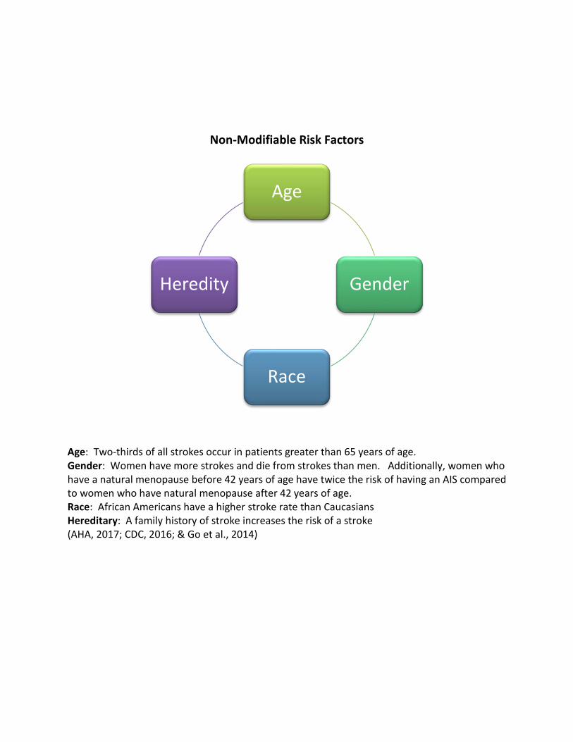
Non-Modifiable Risk Factors
Age: Two-thirds of all strokes occur in patients greater than 65 years of age. Gender: Women have more strokes and die from strokes than men. Additionally, women who have a natural menopause before 42 years of age have twice the risk of having an AIS compared to women who have natural menopause after 42 years of age. Race: African Americans have a higher stroke rate than Caucasians Hereditary: A family history of stroke increases the risk of a stroke (AHA, 2017; CDC, 2016; & Go et al., 2014)
Age
Gender
Race
Heredity
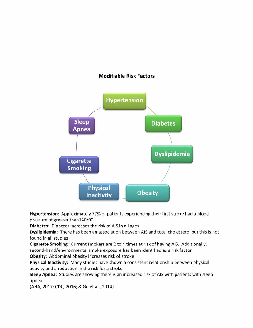
Modifiable Risk Factors
Hypertension: Approximately 77% of patients experiencing their first stroke had a blood pressure of greater than140/90 Diabetes: Diabetes increases the risk of AIS in all ages Dyslipidemia: There has been an association between AIS and total cholesterol but this is not found in all studies Cigarette Smoking: Current smokers are 2 to 4 times at risk of having AIS. Additionally, second-hand/environmental smoke exposure has been identified as a risk factor Obesity: Abdominal obesity increases risk of stroke Physical Inactivity: Many studies have shown a consistent relationship between physical activity and a reduction in the risk for a stroke Sleep Apnea: Studies are showing there is an increased risk of AIS with patients with sleep apnea (AHA, 2017; CDC, 2016; & Go et al., 2014)
Hypertension
Diabetes
Dyslipidemia
ObesityPhysical
Inactivity
Cigarette Smoking
SleepApnea
Test Your Knowledge Which of the following are Modifiable Risk Factors?
A. Atrial fibrillation/atrial flutter/heart block B. Age, gender, family history C. Obesity, hypertension, sleep apnea D. Smoking, race, heart disease
Rationale: Modifiable Risk Factors include: Hypertension: Approximately 77% of patients experiencing their first stroke had a blood pressure of greater than140/90 Diabetes: Diabetes increases the risk of AIS in all ages Dyslipidemia: There has been an association between AIS and total cholesterol but this is not found in all studies Cigarette Smoking: Current smokers are 2 to 4 times at risk of having AIS. Additionally, second-hand/environmental smoke exposure has been identified as a risk factor Obesity: Abdominal obesity increases risk of stroke Physical Inactivity: Many studies have shown a consistent relationship between physical activity and a reduction in the risk for a stroke Sleep Apnea: Studies are showing there is an increased risk of AIS with patients with sleep apnea Classifications While, ischemic strokes are caused by a blockage within the cerebral vasculature, AIS can be further divided into three types:
1. Thrombotic: an abnormal clot formed in the vessel due to atherosclerosis and overtime may grow and occlude the vessel
2. Embolic: an abnormal clot that has broken off from a thrombus that has formed elsewhere in the body and is moving through the vascular system and becomes lodged in a vessel too small to let it pass through
3. Decreased perfusion: an abnormal blood flow to the cerebral vasculature caused by hypotension, severe carotid stenosis, cardiogenic shock after a myocardial infarction or arrhythmia.
(Albertson & Sharma, 2014 & Finnie, 2014) Cerebral Vasculature To understand the effects of an ischemic stroke one must understand the vasculature that supplies the cranial vault with a constant blood supply. The cerebral vasculature contains four cerebral arteries and the carotid arteries. Any disturbance of blood flow in these arteries can cause debilitating injuries. The four cerebral arteries are:
1. Middle Cerebral Artery
2. Anterior Cerebral Artery 3. Posterior Cerebral Artery 4. Vertebral-Basilar Artery
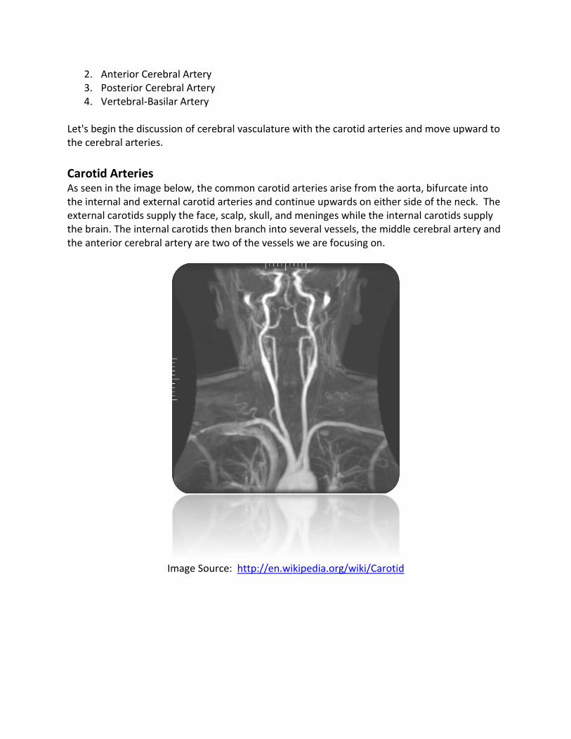
Let's begin the discussion of cerebral vasculature with the carotid arteries and move upward to the cerebral arteries. Carotid Arteries As seen in the image below, the common carotid arteries arise from the aorta, bifurcate into the internal and external carotid arteries and continue upwards on either side of the neck. The external carotids supply the face, scalp, skull, and meninges while the internal carotids supply the brain. The internal carotids then branch into several vessels, the middle cerebral artery and the anterior cerebral artery are two of the vessels we are focusing on.
Image Source: http://en.wikipedia.org/wiki/Carotid
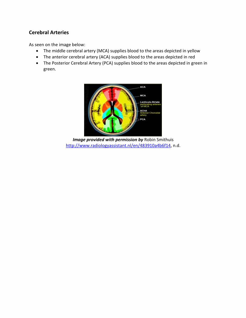
Cerebral Arteries As seen on the image below:
• The middle cerebral artery (MCA) supplies blood to the areas depicted in yellow • The anterior cerebral artery (ACA) supplies blood to the areas depicted in red • The Posterior Cerebral Artery (PCA) supplies blood to the areas depicted in green in
green.
Image provided with permission by Robin Smithuis
http://www.radiologyassistant.nl/en/483910a4b6f14, n.d.
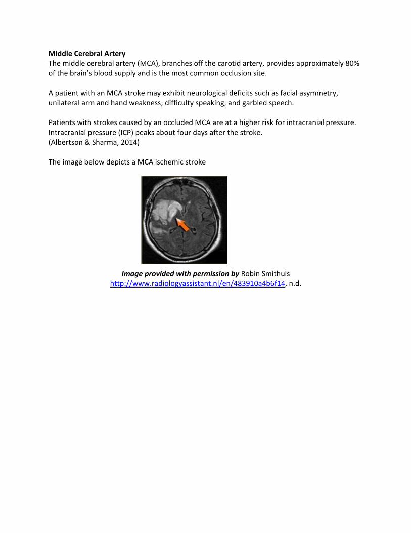
Middle Cerebral Artery The middle cerebral artery (MCA), branches off the carotid artery, provides approximately 80% of the brain’s blood supply and is the most common occlusion site. A patient with an MCA stroke may exhibit neurological deficits such as facial asymmetry, unilateral arm and hand weakness; difficulty speaking, and garbled speech. Patients with strokes caused by an occluded MCA are at a higher risk for intracranial pressure. Intracranial pressure (ICP) peaks about four days after the stroke. (Albertson & Sharma, 2014) The image below depicts a MCA ischemic stroke
Image provided with permission by Robin Smithuis http://www.radiologyassistant.nl/en/483910a4b6f14, n.d.
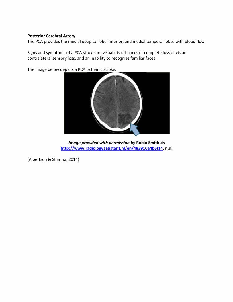
Posterior Cerebral Artery The PCA provides the medial occipital lobe, inferior, and medial temporal lobes with blood flow. Signs and symptoms of a PCA stroke are visual disturbances or complete loss of vision, contralateral sensory loss, and an inability to recognize familiar faces. The image below depicts a PCA ischemic stroke.
Image provided with permission by Robin Smithuis http://www.radiologyassistant.nl/en/483910a4b6f14, n.d.
(Albertson & Sharma, 2014)
Anterior Cerebral Artery The ACA provides the anterior and medial portion of the frontal and parietal lobes with blood flow. The ACA rarely is the primary site for strokes. If a stroke occurs here, assess for signs and symptoms of sensory loss, lower extremity weakness, behavioral abnormalities, and incontinence (Albertson & Sharma, 2014). Vertebral-Basilar Ischemia effecting the vertebral-basilar circulation influences the function of the cerebellum, brain stem or both. These types of strokes are rare and the mortality rate is high. These patients often require intensive care treatment and mechanical ventilation support. Signs and Symptoms: Cerebellar strokes include deficits in balance and coordination, dizziness, nausea, vomiting, headache, and slurred speech Brain stem strokes include deficits such as hemiparesis, quadriplegia, double vision, and abnormal respirations. Test Your Knowledge A patient is admitted to the unit where you are teaching a new staff member about ischemic strokes. In a class on cerebral blood supply you taught that the ______________ artery was the most common site for ischemic strokes because it provides all but 20% of the brain’s blood supply.
A. Vertebral-Basilar B. Posterior cerebral C. Anterior cerebral D. Middle cerebral
Rationale: Middle cerebral artery (MCA): branches off the carotid artery, provides approximately 80% of the brain’s blood supply and is the most common occlusion site. Anterior cerebral arter7 (ACA): provides the anterior and medial portion of the frontal and parietal lobes with blood flow. The ACA rarely is the primary site for strokes. Posterior cerebral artery: provides the medial occipital lobe, inferior, and medial temporal lobes with blood flow. Vertebral-Basilar: Ischemia effecting the vertebral-basilar circulation influences the function of the cerebellum, brain stem or both. These types of strokes are rare and the mortality rate is high. 2013 Stroke Guidelines with 2015 Updates Prehospital Care The AHA/ASA stroke guidelines begin with prehospital care, focusing on limiting delays and transferring patients to a higher-level of care when appropriate. The basics of prehospital care include:
• Stroke education • Call 911
• Prehospital assessment tools • Field management • Rapid transport to stroke center • Prehospital notification
It is important to note that patient should be transported to the closest certified primary or comprehensive stroke center or when not available to the closest facility offering emergency stroke care. Emergency Evaluation and Diagnosis Emergency departments should apply and organize protocol for emergency evaluation of patients with a suspected stroke. A neurologic exam, clinical assessment, utilization of a stroke team, and fibrinolytic treatment within 60 minutes of arrival should be completed. Recommended diagnostic tests include:
• Blood glucose • Oxygen saturation • Electrolytes/renal function tests • Complete blood count • Troponin assessment • Prothrombin time/international normalized ratio (INR) • Activated partial thromboplastin time (aPTT) • ECG
Note: A blood glucose measurement must proceed IV rtPA administration Stroke Scales The guidelines recommend the use of a stroke rating scale; specifically, the National Institutes of Health Stroke Scale (NIHSS) during patient assessment. The NIHSS:
• Assesses level of consciousness, commands, visual fields, facial and limb weaknesses, aphasia, and dysarthria
• It is scored from 0-42; a score of 0 equals no deficits, while a score of 42 exhibits severe deficits
• Should be administered on admission and every 12 for the first 24 hours, and every 24 hours thereafter until discharge.
An example of the NIHSS can be found at: http://www.nihstrokescale.org/docs/HospitalStrokeScales.pdf Thought Provoking Research Stroke scales have been used by healthcare providers for over two decades for estimating the severity of stroke at onset and assessing prognostic information. In a review of the literature
from 1970 through 2013, Ghandehari, 2013) reviewed five of the most common stroke scales in use today. Ghandehari concluded that the National Institute of Health Stroke Scale (NIHSS) continues to be the most utilized scale despite the advantages of the modified National Institute of Health Stroke Scale (mNIHSS) and Scandinavian Stroke Scale (SSS). These advantages include:
• Ease of use • Increased interrater reliability • Higher reliability and validity
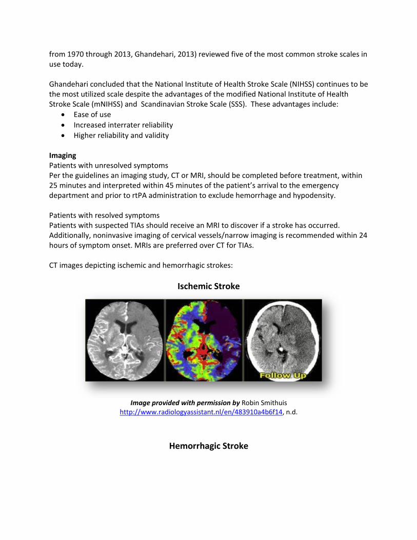
Imaging Patients with unresolved symptoms Per the guidelines an imaging study, CT or MRI, should be completed before treatment, within 25 minutes and interpreted within 45 minutes of the patient’s arrival to the emergency department and prior to rtPA administration to exclude hemorrhage and hypodensity. Patients with resolved symptoms Patients with suspected TIAs should receive an MRI to discover if a stroke has occurred. Additionally, noninvasive imaging of cervical vessels/narrow imaging is recommended within 24 hours of symptom onset. MRIs are preferred over CT for TIAs. CT images depicting ischemic and hemorrhagic strokes:
Ischemic Stroke
Image provided with permission by Robin Smithuis
http://www.radiologyassistant.nl/en/483910a4b6f14, n.d.
Hemorrhagic Stroke
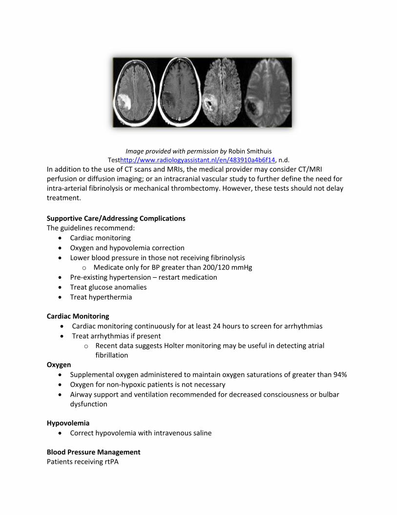
Image provided with permission by Robin Smithuis Testhttp://www.radiologyassistant.nl/en/483910a4b6f14, n.d.
In addition to the use of CT scans and MRIs, the medical provider may consider CT/MRI perfusion or diffusion imaging; or an intracranial vascular study to further define the need for intra-arterial fibrinolysis or mechanical thrombectomy. However, these tests should not delay treatment. Supportive Care/Addressing Complications The guidelines recommend:
• Cardiac monitoring • Oxygen and hypovolemia correction • Lower blood pressure in those not receiving fibrinolysis
o Medicate only for BP greater than 200/120 mmHg • Pre-existing hypertension – restart medication • Treat glucose anomalies • Treat hyperthermia
Cardiac Monitoring
• Cardiac monitoring continuously for at least 24 hours to screen for arrhythmias • Treat arrhythmias if present
o Recent data suggests Holter monitoring may be useful in detecting atrial fibrillation
Oxygen • Supplemental oxygen administered to maintain oxygen saturations of greater than 94% • Oxygen for non-hypoxic patients is not necessary • Airway support and ventilation recommended for decreased consciousness or bulbar
dysfunction Hypovolemia
• Correct hypovolemia with intravenous saline Blood Pressure Management Patients receiving rtPA
• Reduce blood pressure to less than 185/100 mmHg prior to rtPA • Maintain blood pressure after rtPA below 180/105 mmHg for at least 24 hours
Patients not receiving rtPA • Lower blood pressure by 15% during the first 24 hours • Do not treat with medications unless BP is higher than 220/120 mmHg • May restart antihypertensives in stable patients who were hypertensive prior to stoke
after 24 hours Note: As healthcare providers, it is important to note that patients who are experiencing an acute ischemic stroke require a rise in blood pressure due to the opening of the collateral vessels trying to supply blood to the ischemic part of the brain or penumbra. It is imperative that blood pressure is maintained at the recommended levels to help prevent further brain damage due to ischemia. Glucose Anomalies
• Hypoglycemia of less than 60 mg/dL • Blood sugar levels should be maintained to a range of 140-180 mg/dL
Temperature Control
• Hyperthermia should be identified and treated Intravenous Fibrinolysis Timely restoration of blood flow with the use of rtPA is the gold standard treatment for ischemic stroke in the appropriate patient (Powers, et al., 2015). Recent updates show that patients with the following may be considered for rtPA; however, risks should be weighed against benefits.
• Rapidly improving symptoms • Mild stroke deficits • Major surgery within the past three months • Recent myocardial infarctions • Seizures, provided residual symptoms are stroke related and not postictal
Exclusion criteria for 3- to 4.5-hour window
• Older than 80 years of age • On oral anticoagulants • Baseline NIHSS score greater than 25 • History of both stroke and diabetes • Imaging evidence of ischemic damage to more than one third of the MCA
Recommendations
• IV rtPA in eligible patients at up to 4.5 hours; treatment within 60 minutes of presentation to hospital is ideal
• IV rtPA contingent upon BP control (below 185/110 mm Hg) • IV rtPA at a dose of 0.9 mg/kg (maximum dose 90 mg) • Other fibrinolytic or defibrinogenating agents not recommended (Streptokinase) • Sonothrombolysis efficacy not well established • IV rtPA is not recommended if taking direct thrombin inhibitors or direct factor Xa
inhibitors, unless qualified based on lab panel or if medications have not been taken more than 48 hours prior to admission
o Activated partial thromboplastin time (aPTT) o INR o Platelet count o Ecarin clotting time o Thrombin time o Direct factor Xa
Endovasuclar Interventions Patients who are candidates for rtPA should receive rtPA even when endovascular interventions are being considered. Endovascular interventions should be considered for patients who are not candidates for rtPA treatment. New research evidence supports endovascular therapy with a stent to retriever when the patients meet the following requirements:
• Prestroke mRS score 0 to 1 • Acute ischemic stroke receiving intravenous rtPA within 4.5 hours of onset per
guidelines from professional medical societies • Causative occlusion of the ICA or proximal MCA (M1) • Age ≥18 years • NIHSS score of ≥6 • ASPECTS of ≥6 • Treatment can be initiated (groin puncture) within 6 hours of symptom onset
Note: Intra-arterial fibrinolysis with rtPA is not approved by the Food and Drug Administration; however, in carefully selected patients in whom IV rtPA is contraindicated, intra-arterial fibrinolysis might be considered. Therefore, endovascular therapy with stent retrievers is recommended over intra-arterial fibrinolysis as first-line therapy. Anticoagulation There is not enough research evidence to support anticoagulation in acute stroke. Anticoagulation within 24 hours of IV rtPA is not recommended. Antiplatelet Agents
Aspirin within 24-48 of initial onset of symptoms remains the only agent supported by research evidence for use in acute stroke. Although, clinical trials are ongoing for other agents. Aspirin is NOT a substitute for rtPA or other interventions. Volume Expansion, Vasodilators, and Induced Hypertension
• In stroke patients with symptomatic hypotension, vasopressors should be considered. • Induced hypertension and hemodilution efficacy in acute stroke have not been well
established. Neuroprotection and Surgery
• Current data does not support the use of neuroprotective agents in acute stroke • Hyperbaric oxygen treatment is reserved for cases of stroke due to air embolization • Induced hypothermia is not recommended • Urgent carotid endarterectomy in patients with unstable neurological status is not
supported by current data
Test Your Knowledge Which of the following treatments are supported by current data in the treatment of acute stroke?
A. Hyperbaric oxygen B. induced hypothermia C. Anticoagulants D. Aspirin
Rationale: Aspirin within 24-48 of initial onset of symptoms remains the only agent supported by research evidence for use in acute stroke. Although, clinical trials are ongoing for other agents. Hyperbaric oxygen treatment is reserved for cases of stroke due to air embolization
• Induced hypothermia is not recommended • There is not enough research evidence to support anticoagulation in acute stroke.
Anticoagulation within 24 hours of IV rtPA is not recommended. Hospital Admission and Treatment Comprehensive stroke management with standardized order sets in a stoke unit; should be the main stay of acute stoke care. Management should include:
• Swallowing assessment prior to feeding • Deep vein thrombosis prevention with subcutaneous anticoagulation and external
compression devices • Antibiotics for potential or actual pneumonias or urinary tract infections • Nasal gastric or post pyloric feeding tubes preferred over percutaneous endoscopy
feeding (PEG) tubes in patients who cannot take fluids, medications, or food o PEG tubes should be considered after 2-3 weeks if the patient cannot eat or
drink
Treating Neurologic Complications Neurologic complications should be treated on a case by case basis.
• Antiepileptics should be given for recurrent seizures but not for prophylaxis • Treatments to reduce hydrocephalus and cerebral edema should be used:
o Corticosteriods are not recommended o Ventricular drains for acute hydrocephalus o Decompressive surgery if necessary
(Jauch et al., 2013; Stetka & Lutsep, 2013; Powers et al., 2015) Primary Stroke Centers One of the most consistent recommendations by the AHA/ASA is the use of a stroke center to care for the acute stroke patient. In 2002, the Joint Commission (TJC) instituted a Disease-Specific Care certification program. This program offers institutions an opportunity to become certified in nine clinical or procedural areas. In 2003, TJC, in collaboration with the AHA/ASA, developed a stroke certification program. The Joint Commission offers three advanced levels of certification designed to recognize the specific capabilities of hospitals that can treat the most complex stroke cases. These levels are:
• Acute Stroke Ready Hospital: No designated beds for acute care of stroke patients • Primary Stroke Center: Stroke unit or designated beds for acute care of stroke patients • Comprehensive Stroke Center (over 100 certified facilities): Dedicated Neuro Intensive
Care beds for complex stroke patients A complete comparison of the levels of certification can be found at: https://www.jointcommission.org/assets/1/18/StrokeProgramGrid_abbr_AHA-TJC_5-1-15.pdf (The Joint Commission (TJC), 2017) Nursing Considerations In 2009, the AHA/ASA published a scientific paper describing evidence-based nursing and interdisciplinary care for the acute ischemic stroke patient. As new evidence was developed through research, the information was corrected; the last correction was in 2011 (Summers, et al., 2011). The AHA/ASA paper describes a continuum of care occurring in two phases.
• Phase 1: a hyperacute phase encompasses the first 3 to 24 hours after the onset of stroke with a focus on identifying symptoms, infarct location, assessing the patient for risk of acute and long-term complications, and determining treatment plans.
• Phase 2: an acute care phase encompasses from 24 to 72 hours after the on stroke of stroke and focuses on clarifying the cause of stroke, preventing medical complications, preparing the patient and family for discharge and instituting long-term secondary prevention modalities.
Phase 1 The most important nursing goal is to determine the time the patient started exhibiting symptoms of stroke. This is known as ‘the patient was last known to be well time.’ The EMS team focuses on patient stabilization, identifying signs and symptoms of stroke preferably with a tool such as F.A.S.T. described earlier in this course; transporting the patient to the nearest hospital that can care for stroke patients. Their practice is guided by local policies and regulations. These regulations may vary from state to state, so it is important for the nurse in the emergency room to know what this team is authorized to do. For example, some teams are not allowed to do point of care glucose testing in the field. Therefore, the standing orders or protocol in the emergency department (ED) should include point-of-care glucose testing. When the patient arrives in the ED, the nurse using a triage system such as the five-level Emergency Severity Index, recommended by the Emergency Nurses Association and the American College of American physicians. Acute stroke patients are triaged as a level two/needs immediate assessment. Emergency department nurses recognize that time is brain and use specialized checklist protocols and other tools to identify the stroke patients during a rapid assessment with the goal to treat ischemic strokes within 60 minutes with rtPA per the national Institute of neurological disorders and stroke (NINDS) benchmark. The AHA/ASA paper recommends:
• Vital signs are measured frequently, not less than every 30 minutes or as clinically indicated while in the ED
• Maximization of oxygenation • Head of bed up at least 30° to increase cerebral blood flow and help decrease
intracranial pressure if present • When hemiparesis is present the patient should be positioned on the affected side to
prevent aspiration • Evaluation of swallowing ability, to determine the best route for medication
administration • Emergent laboratory specimens should be drawn, labeled STAT, to rule out processes
such as electrolyte imbalance which may mimic stroke o Laboratory specimen collection should be done prior to CT scans to allow for
simultaneous processing and reporting of results • Establish 2 to 3 intravenous sites; designating a single purpose for each site
o Administration of thrombolytic therapy o Administration of medications o Administration of intravenous fluids
• Administration of non-glucose containing intravenous fluids at a rate of 75-100 mL/hr • Insertion of urinary catheter, IV lines, endotracheal tube, or gastric tubes should be
performed prior to thrombolytic therapy to decrease the possibility of hemorrhage • Measure vital signs every 15 minutes for two hours during the administration of
thrombolytic therapy (Summers, et al., 2011)
Did You Know? Hyperthermia is associated with poor outcome in stroke patients; an increase of 1°F is a predictor of poorer patient outcome and is an independent factor in short- and long-term mortality rates. The rationale for this additional injury may be related to increased metabolic demands and free radical production. Immediate treatment of the source of the fever will reduce its duration; therefore, it is important to consider treating any fever >99.6°F (Summers, et al., 2011). Phase 2 The acute care phase should focus on continue stabilization of the patient through frequent monitoring in the intensive care unit. The focus of nursing care should be to prevent complications and deterioration. The nurse should evaluate the patient for complications due to the following:
• Respiratory issues such as smoking • Pneumonia • Hypertension • Hyperglycemia • Dehydration • Malnutrition • Fever • Coronary artery disease • Cerebral edema • Infection • Deep vein thrombosis or pulmonary embolism
Did You Know? Up to 30% of all stroke patients will deteriorate in the first 24 hours Approximately 40-80% of all stroke patients will have elevated blood pressure within the first 24 to 48 hours Elevated glucose occurs in two thirds of AIS patients (Summers, et al., 2011)
The AHA/ASA recommends the use of standardized protocols/standing orders which include parameters to treat fever, blood glucose, blood pressure, and consultation with the multidisciplinary team. Phase 2 nursing care should include:
• Intensive monitoring for: o Bleeding for 24 to 36 hours after thrombolytic administration o Increasing intracranial pressure o Neurological stroke assessment using the NIHSS scale o Blood pressure management; rapid lowering of blood pressure may result in
decreased cerebral perfusion and increased ischemia Maintain blood pressure at less than 180/105 mmHg for 24 hours after
thrombolytic therapy Administration of antihypertensives is not recommended in non-
thrombolytic candidates • Administer antihypertensives ONLY if systolic blood pressure is
greater than 220 mmHg or diastolic pressure is greater than 110 mmHg
o Fever management (see Did You Know) o Cardiac monitoring; ventricular ectopy, tachycardia, and heart blocks have been
associated with AIS o Oxygenation; hypoxemia will increase the risk of neurological deterioration keep
oxygen saturations greater than 92% o Angioedema occurs in 1-2% of patients with AIS o Blood glucose control o Cerebral edema
Peaks 3-5 days after AIS May be significant in the first 24 hours in patients with large cerebellar
infarcts or younger patients • Younger patients usually do not have significant cerebral atrophy;
therefore, there is no room in the cranial fault for swelling o Seizures o Hydrocephalus o Reduction of hospital acquired conditions
Deep vein thrombosis prophylaxis Catheter associated urinary tract infection prevention Central line associated bloodstream infection prevention Ventilator associated event prevention Hospital acquired pressure ulcer prevention
o Aspiration prevention o Nutritional compromise prevention
Use of gastric and post-pyloric feeding tubes when appropriate
An important part of phase 2 is discharge planning. Stroke education should include discussion of risk factors specifically the modifiable risk factors, and compliance with prescribed medication to help prevent secondary stroke. (Summers, et al., 2011) Did You Know? 30% of stroke survivors will recover completely or with minor impairments 40% will require subacute care 10% will require skilled nursing facility care 20% will die soon after stroke onset (Summers, et al., 2011) Test Your Knowledge Discharge planning should include education regarding:
A. Smoking, weight, medications B. Medications, age, gender C. Heredity, weight, smoking D. Race, heredity, gender
Rationale: An important part of phase 2 is discharge planning. Stroke education should include discussion of risk factors specifically the modifiable risk factors, and compliance with prescribed medication to help prevent secondary stroke. The Big Picture Over the past several slides, you have been introduced to the most current care guidelines, a large amount of information over a short period. So, let’s put it all together and care for a patient named Mr. Young. Mr. Young, is a 65-year-old African-American diabetic who is attending a concert-in-the-park with his wife. Near the end of the performance, Mr. Young suddenly slumps in his chair. His wife immediately suspects low blood sugar and calmly administers a quick dissolving piece of candy. However, Mr. Young does not respond to the treatment as he usually does. His wife then notices that his face is droopy and he is drooling. She calls for help. As the EMS team evaluates her husband, she provides the following history:
• Smoker for over 40 years • Type two diabetes for 25 years, usually well controlled • High cholesterol controlled by medication • High blood pressure controlled by medication
The EMS team calls in to report Mr. Young’s condition. You are the healthcare provider on-call and suspect that Mr. Young is experiencing a:
A. Hypoglycemic episode and needs glucagon B. Myocardial infarction C. Stroke
If you chose C, you are correct. Mr. Young has many of the high-risk factors for having a stroke or myocardial infarction. Although a dose of glucagon might help, unless the EMS team can test for capillary glucose, it is an unknown factor if Mr. Young is hypo or hyperglycemic. His symptoms do not support a myocardial infarction. However, his drooling and droopy face may signal a stroke. You know the nearest hospital is a certified Stroke Ready Hospital, a Primary Stroke Center is 5 miles away, and the Comprehensive Stroke Center is a 40-minute air-evac ride away. Which facility will you advise the EMS to transport to?
A. Stroke Ready B. Primary Stroke Center C. Comprehensive Stroke Center
Did you choose C? While this facility is the best prepared to care for the most complex stroke patients and would be ideal, it is a 40-minute ride away which does not include the time it would take to get the air evacuation team to your site, get the patient loaded and unloaded at the facility. This additional time to diagnosis and treatment may not be optimal if a Primary Stroke Center is only 5 miles away and the nearest hospital is a Stroke Ready facility. What else do you need to know to help you chose the correct facility? Did you ask:
A. How long has it been since Mr. Young’s symptoms began? B. What is Mr. Young’s current condition? C. Is he stable or unstable?
YES! You asked these questions and received the following report: Mr. Young’s symptoms began about 4:15pm and it is now 5pm, his vital signs are stable except for a blood pressure of 230/110, he is responsive but lethargic and cannot move his left side. You decide that he is not stable enough for a Stroke Ready Facility as he will need more extensive treatment available to him, depending on the type of stroke. Furthermore, since an hour of the 4.5-hour window is almost gone and he will need definitive treatment soon. Adding the additional time to get him to the Comprehensive Care Center extends the symptom to treatment time. You know TIME is BRAIN, so you advise the EMS team to proceed to the Primary Care Center. When Mr. Young arrives in the Emergency Department his care is transferred to you as part of the Stroke Team. He is placed on routine monitoring. Your assessment reveals:
• An obese African-American male in no acute distress
• Vital signs: BP 230/110, HR 90, RR 20, pulse oximetry 93%, temperature 37.4 • Regular rhythm on the monitor • Left side facial drooping of the eyes and mouth • Left sided weakness of his extremities • No complaint of headache
What would be your next steps?
A. Follow the standardized protocol for stroke patients B. Send Mr. Young to lab and radiology for routine emergency room diagnostics C. Call the Stroke Team to the emergency room
If you selected to follow the standardized protocol for stroke patients, you would be correct. Having a standardized and organized protocol helps reduce the time to treatment. The Stroke Team assembled in the room prior to Mr. Young’s arrival. The NIHSS screen has been completed, blood samples have been drawn, a bedside glucose measurement has been done, and within 20 minutes, Mr. Young is in the CT suite. The blood glucose is 185 mg/dL NIHSS indicates significant neurological deficits The CT scan indicates no evidence of hemorrhage or hypodensity What should be the teams next steps?
A. Administer rtPA immediately B. Treat the hyperglycemia C. Await the results of the remaining labs D. Treat the hypertension E. Treat the hypoxia
Although, Time is Brain, you know you cannot administer the rtPA without reducing Mr. Young’s blood pressure to less than 185/110. The hyperglycemia is not an immediate issue; however, he is hypoxic and could use some supplemental oxygen. It has now been 90 minutes since Mr. Young started experiencing symptoms, and 35 minutes since his arrival in the emergency department. The physician orders an antihypertensive medication and supplemental oxygen. Mr. Young’s oxygen saturations rise to 95%. After 30 minutes, Mr. Young’s blood pressure is 180/90. Should rtPA be administered?
A. No, it has been more than an hour since the patient exhibited his first symptoms B. No, it has been more than an hour since the patient was admitted to the emergency
room
C. Yes, it has been more than an hour but less than 4.5 hours since the patient exhibited his first symptoms
D. Yes, it has been more than an hour but less than 4.5 hours since the patient was admitted to the emergency room
Did you administer the rtPA? Awesome. Although it had been 65 minutes since the patient arrived in the emergency room and because of his blood pressure issue, the ideal window of 60 minutes passed, he was still well within the treatment window at 2 hours since his symptoms began. The next morning, Mrs. Young visits to find her husband smiling with very little residual drooping. You instruct Mr. Young that you will be restarting his antihypertensive and diabetes medication. You have a new staff member orienting and as she reads the chart, she sees that the CT scan was not only negative for hemorrhage but also for ischemic changes. She is concerned that Mr. Young received rtPA for a negative CT scan. How would you answer her concerns? If you replied that it takes 24 hours for ischemic changes to appear on CT scan and that in the absence of hemorrhage or hypodensity; rtPA should be given to symptomatic patients, you would be correct! Mr. Young is discharged home with minimal deficits. Conclusion Time is Brain, and now you know how to save brain tissue and reduce brain damage from acute ischemic strokes. Public education regarding modifiable and non-modifiable high-risk behaviors and family history; signs and symptoms of stroke, and the importance of time to treatment can help reduce the devastating consequences of acute ischemic strokes. Healthcare providers who follow the AHA/ASA guidelines can facilitate rapid assessment and treatment by utilizing a comprehensive treatment protocol.
References Albertson, M. & Sharma, J. (2014). Stroke: Current concepts. Primers in Medicine, South Dakota Medicine 455-464. American Heart Association (AHA). (2017). Heart disease and stroke statistics 2017 at-a-glance. Retrieved from http://www.heart.org Centers for Disease Control and Prevention (CDC). (2016). Stroke facts. Retrieved from:
https://www.cdc.gov/stroke/facts.htm. CDC. (2017). Stroke signs and symptoms. Retrieved from:
https://www.cdc.gov/stroke/signs_symptoms.htm Ghandehari, K. (2013). Challenging comparison of stroke scales. Journal of Research in Medical Sciences. 18, 906-910. Gonzales-Perez, A., Gaist, D., Wallander, M.A., McFeat, G, Garcia-Rodriquez, L.A. (2013). Mortality after hemorrhagic stroke: Data from general practice (The Health Improvement Network). Neurology, 81. 559-65. Go, A. S., Mozaffarian, D., Roger, V. L., Benjamin, E. J., Berry, H. D., Blaha, M. J. …Turner, M. B. (2014). Heart disease and stroke statistics—2014 Update: A report from the American Heart Association. Circulation, 129 (e28-e292). Jauch, E. C., Saver, J.L., Adams, H.P., Bruno, A., Connors, J.J., Demaerschalk, B.M., …Yonas, H. (2013). Guidelines for the early management of patients with acute ischemic stroke: A guideline for healthcare professionals from the American Heart Association/American Stroke Association. Stroke. 44:870-947. Liebesking, D.S. & O’Connor, R.E. (2017). Hemorrhagic Stroke Workup. Retrieved from: http://emedicine.medscape.com/article/1916662-workup Powers, W.J., Derdeyn, C.P., Biller, J., Coffey, C.S., Hoh, B.L., Jauch, E.., Yavagal, D.R.; on behalf of the American Heart Association Stroke Council. (2015). AHA/ASA focused update of the 2013 guidelines for the early management of patients with acute ischemic stroke regarding endovascular treatment: a guideline for healthcare professionals from the American Heart Association/American Stroke Association. Stroke. 46: 3020-3035. Retrieved from: http://stroke.ahajournals.org/content/46/10/3020.long Silver, B. & Silver, R. W. (2014). Diagnosis and management of acute ischemic stroke. Retrieved from: http://www.aafp.org/test/fpcomp/FP-E_420/pt2.html Summers, D., Leonard, A., Wentworth, D……Mitchell, P.H. (2011). Comprehensive overview of nursing and interdisciplinary care of the acute ischemic stroke patient: A scientific statement from the American Heart Association. Stroke. 40:2911-2944.
Stetka, B.S. & Lutsep, H. L. (2013). New stroke management guidelines: A quick and easy guide. Retrieved from: http://www.medscape.com/viewarticle/779968_15. The Joint Commission (TJC). (2016). Facts about Joint Commission stroke certification. Retrieved from: https://www.jointcommission.org/facts_about_joint_commission_stroke_certification/ TJC. (2017). Certification Comprehensive stroke center. Retrieved from: https://www.jointcommission.org/certification/advanced_certification_comprehensive_stroke_centers.aspx Please Read This publication is intended solely for the use of healthcare professionals taking this course, for credit, from RN.com. It is designed to assist healthcare professionals, including nurses, in addressing many issues associated with healthcare. The guidance provided in this publication is general in nature, and is not designed to address any specific situation. This publication in no way absolves facilities of their responsibility for the appropriate orientation of healthcare professionals. Hospitals or other organizations using this publication as a part of their own orientation processes should review the contents of this publication to ensure accuracy and compliance before using this publication. Hospitals and facilities that use this publication agree to defend and indemnify, and shall hold RN.com, including its parent(s), subsidiaries, affiliates, officers/directors, and employees from liability resulting from the use of this publication. The contents of this publication may not be reproduced without written permission from RN.com.