treating navicular syndrome in equine patients* · corticosteroids (e.g., methylprednisolone...
TRANSCRIPT
©Copyright 2011 MediMedia Animal Health. This document is for internal purposes only. Reprinting or posting on an external website without written permission from MMAH is a violation of copyright laws.
Vetlearn.com | January 2011 | Compendium: Continuing Education for Veterinarians® E1
In collaboration with the American College of Veterinary Surgeons
CE Article3 CECREDITS
Treating Navicular Syndrome in Equine Patients*R. Wayne Waguespack, DVM, MS, DACVSR. Reid Hanson, DVM, DACVS, DACVECCAuburn University
Navicular syndrome is a chronic progressive condi-tion affecting the navicular bone and bursa as well as the associated soft tissue structures1 (FIGURE 1).
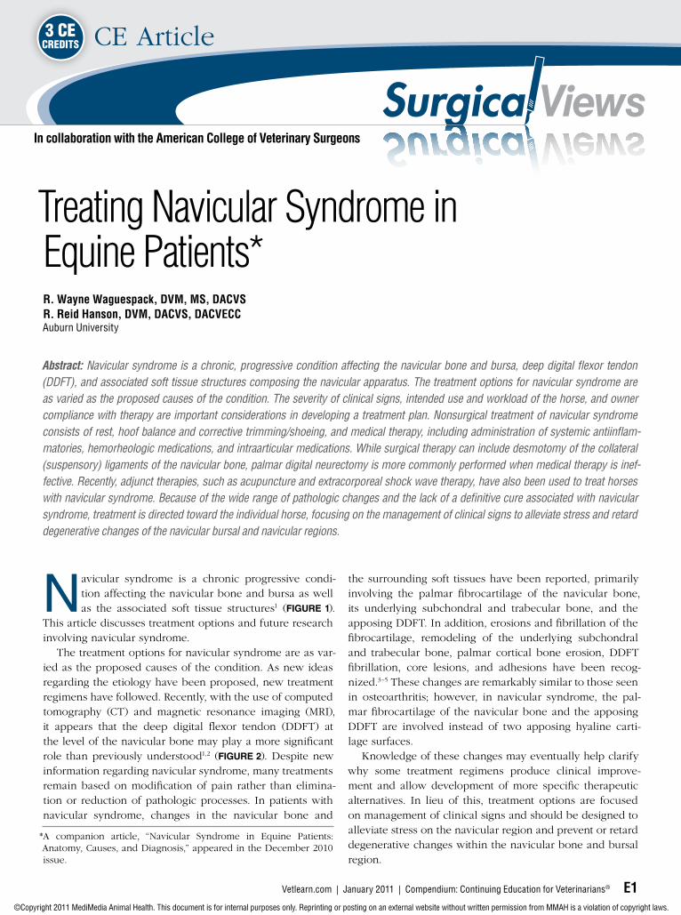
This article discusses treatment options and future research involving navicular syndrome. The treatment options for navicular syndrome are as var-ied as the proposed causes of the condition. As new ideas regarding the etiology have been proposed, new treatment regimens have followed. Recently, with the use of computed tomography (CT) and magnetic resonance imaging (MRI), it appears that the deep digital flexor tendon (DDFT) at the level of the navicular bone may play a more significant role than previously understood1,2 (FIGURE 2). Despite new information regarding navicular syndrome, many treatments remain based on modification of pain rather than elimina-tion or reduction of pathologic processes. In patients with navicular syndrome, changes in the navicular bone and
the surrounding soft tissues have been reported, primarily involving the palmar fibrocartilage of the navicular bone, its underlying subchondral and trabecular bone, and the apposing DDFT. In addition, erosions and fibrillation of the fibrocartilage, remodeling of the underlying subchondral and trabecular bone, palmar cortical bone erosion, DDFT fibrillation, core lesions, and adhesions have been recog-nized.3–5 These changes are remarkably similar to those seen in osteoarthritis; however, in navicular syndrome, the pal-mar fibrocartilage of the navicular bone and the apposing DDFT are involved instead of two apposing hyaline carti-lage surfaces. Knowledge of these changes may eventually help clarify why some treatment regimens produce clinical improve-ment and allow development of more specific therapeutic alternatives. In lieu of this, treatment options are focused on management of clinical signs and should be designed to alleviate stress on the navicular region and prevent or retard degenerative changes within the navicular bone and bursal region.
Abstract: Navicular syndrome is a chronic, progressive condition affecting the navicular bone and bursa, deep digital flexor tendon (DDFT), and associated soft tissue structures composing the navicular apparatus. The treatment options for navicular syndrome are as varied as the proposed causes of the condition. The severity of clinical signs, intended use and workload of the horse, and owner compliance with therapy are important considerations in developing a treatment plan. Nonsurgical treatment of navicular syndrome consists of rest, hoof balance and corrective trimming/shoeing, and medical therapy, including administration of systemic antiinflam-matories, hemorheologic medications, and intraarticular medications. While surgical therapy can include desmotomy of the collateral (suspensory) ligaments of the navicular bone, palmar digital neurectomy is more commonly performed when medical therapy is inef-fective. Recently, adjunct therapies, such as acupuncture and extracorporeal shock wave therapy, have also been used to treat horses with navicular syndrome. Because of the wide range of pathologic changes and the lack of a definitive cure associated with navicular syndrome, treatment is directed toward the individual horse, focusing on the management of clinical signs to alleviate stress and retard degenerative changes of the navicular bursal and navicular regions.
*A companion article, “Navicular Syndrome in Equine Patients: Anatomy, Causes, and Diagnosis,” appeared in the December 2010 issue.

E2 Compendium: Continuing Education for Veterinarians® | January 2011 | Vetlearn.com
Nonsurgical TherapyThe severity of clinical signs, the horse’s intended use and workload, and the owner’s compliance with therapy are important considerations in developing a treatment plan. Young horses and horses with less severe clinical signs are often candidates for conservative therapy. Nonsurgical treatment of navicular syndrome consists of rest, hoof bal-ance and corrective trimming/shoeing, and medical therapy, including systemic antiinflammatories, hemorheologic med-ications, and intraarticular medications.6–8
RestOne of the most overlooked components of treatment is rest. Continued stress on the navicular region leads to structural failure and eventual breakdown. Time is necessary to allow soft tissue inflammation to subside and bone remodeling to take place. The time necessary to reduce inflammation var-ies on an individual basis. Absolute confinement is contrain-dicated; pasture or paddock turnout is sufficient.
Corrective Shoeing and Hoof Balance Careful evaluation of foot conformation and balance is the focal point in nonsurgical treatment of navicular syndrome. Many horses respond to correction of hoof abnormalities.9 The goal is to reduce forces on the navicular region by (1) cor-recting hoof balance and the hoof–pastern axis, (2) allowing the use of all weightbearing structures of the hoof by main-taining the heel mass and protecting the palmar aspect of the foot from concussion, and (3) decreasing the work of the moving foot by either shortening the toe length of the foot to permit an easier breakover or rolling the toe of the shoe. Horses with navicular syndrome often have unbalanced hooves consisting of long toes with low, underrun, and contracted heels (FIGURE 3). One of the first requirements of hoof balance is hoof–pastern alignment. Proper hoof–pastern alignment is assumed if a straight line can be drawn through the dorsal pastern and dorsal hoof wall (FIGURE
3). The foot should be trimmed to maintain heel mass and shorten the toe to facilitate breakover if the hooves are char-acterized by long toes. Use of the so-called four-point or natural-balance trim has recently been favored by some clinicians, but the same principles of breakover can prob-ably be achieved with more traditional trimming if the toe is shortened sufficiently. Radical changes in foot trimming may temporarily increase lameness; therefore, correct foot balance may need to be achieved in stages. In principle, a hoof with proper hoof–pastern alignment has the appropriate amount of tension or force exerted on the navicular bone by the DDFT. In horses with low heels, correct alignment can be established with selective trimming or, more commonly, by elevating the heels with a wedge-heel
Surgical Views is a collaborative series between the American College of Veterinary Surgeons (ACVS) and Compendium.
All Surgical Views articles are peer-reviewed by ACVS diplomates.
To locate a diplomate, ACVS has an online directory that includes practice setting, species emphasis, and research interests (acvs.org/Veterinary Professionals/FindaSurgeon).
The navicular bursa (black arrow), distal interphalangeal joint (blue arrow), deep digital flexor tendon, impar liga-ment, and collateral sesmoidean ligament. (NB = navicular bone; PII = second phalanx; PIII = third phalanx)
FIGURE 1
NBPII
PIII
Vetlearn.com | January 2011 | Compendium: Continuing Education for Veterinarians® E3
shoe.9 In a study of normal Dutch Warmblood horses, heel elevation with a 6° wedge reduced the maximal force on the navicular bone by 24% compared with use of flat shoes.10 In another study, force-plate analysis indicated that lameness sig-nificantly decreased in horses with navicular syndrome that were shod to produce a 3° heel elevation alone or in com-bination with phenylbutazone administration.11 Lateromedial radiographs may be necessary to demonstrate whether a horse needs more or less heel elevation. If the horse’s lame-ness worsens after elevation of the toe using the wedge test, some degree of heel elevation may be beneficial. The hoof should also be maintained in medial-to-lateral
balance. This is determined either by obtaining a dorsopal-mar radiograph of the foot or watching the horse walk to ensure that the medial and lateral aspects of the hoof hit the ground simultaneously. If the hoof cannot be trimmed to establish normal hoof–pastern alignment or lateromedial balance, shoes can be applied to encourage the hoof to grow correctly. Many types of shoes have been recommended for horses with navicular syndrome. Full-bar, egg-bar, natural balance, and wide-web aluminum or steel shoes with or without elevated heels have been used to support the heels and move the weightbearing axis in a palmar direction in horses with low, collapsed, and underrun heels (FIGURE 4). A recent study showed that using egg-bar shoes with no heel elevation in clinically normal Dutch Warmblood horses with well-conformed feet did not reduce the force on the navicular bone compared with using regular flat shoes.10 However, these findings cannot necessarily be translated to lame horses with less-than-ideal foot conformation. In the same study, heel wedges did reduce the force on the navic-ular bone compared with flat shoes.10 In a study evaluat-ing 55 horses with clinically diagnosed navicular syndrome, 53% had permanent relief of lameness 12 to 40 months after application of egg-bar shoes.12 In another study, horses shod with egg-bar shoes showed histomorphometric evidence of altered navicular bone modeling compared with untreated controls.13 In any case, shoes should be set (1) beyond the
Advanced imaging modalities such as MRI and CT have helped veterinarians realize that navicular syndrome involves more than just the navicular bone in horses.
FIGURE 2
(A) MRI of the navicular region. Cross-sectional and longitudinal views are shown. (B) CT scan of the navicular region. Based on the visible asymmetry, an erosive lesion is noted in the navicu-lar bone.
B
A

E4 Compendium: Continuing Education for Veterinarians® | January 2011 | Vetlearn.com
heels to provide heel support and (2) full or wide to allow proper hoof expansion.7,10
Medical TherapyAntiinflammatories Intraarticular MedicationsCorticosteroids (e.g., methylprednisolone acetate, triamci-
nolone acetonide, betamethasone sodium phosphate) have been injected into the navicular bursa and the distal inter-phalangeal joint (DIPJ) to treat navicular syndrome, and the results have varied.7,14 These powerful antiinflammatories are routinely used in combination with sodium hyaluronate to control clinical signs associated with navicular syndrome. Most clinicians medicate the DIPJ when treating navicular syn-
Unbalanced hooves and proper hoof–pastern alignment.FIGURE 3
(A) Unbalanced hooves (long toes and low, underrun heels) of a horse with navicular syndrome. Note the medial-to-lateral imbal-ance in the horse’s feet. One of the first requirements for hoof balance is hoof–pastern alignment. (B) Proper hoof–pastern align-ment is assumed if a straight line can be drawn through the bony column of the phalanges. The dorsal hoof wall should be parallel to the dorsal aspect of the pastern. A line bisecting the third metacarpal bone should reach the ground at the weightbearing part of the heel.
B
A

Vetlearn.com | January 2011 | Compendium: Continuing Education for Veterinarians® E5
drome.15 Medications injected into the DIPJ, which is near the navicular bursa, have been shown to diffuse to the navicular region. A study by Pauwels et al15 showed that clinically effective concentrations of methylprednisolone and triamcinolone diffused between the DIPJ and the navicular bursa. This is also supported by the response to treatment of clinical cases of navicular syndrome. Over time, some horses stop responding to intraarticular therapy of the DIPJ. Intrabursal therapy may provide another option for reducing inflammation in the navicular region of the foot. One study showed that 80% of horses that no longer responded to traditional therapy for navicular syndrome (i.e., corrective shoeing, administration of systemic antiinflam-matories, intraarticular therapy of the DIPJ) were sound 2 weeks after intrabursal injections of corticoster-oids, sodium hyaluronate, and amika-cin.16 The mean duration of soundness in this study was 4.5 months. In recent years, interleukin-1 recep- tor antagonist protein (IRAP) therapy (Vet Arthrex Biosystems, Bonita Springs, FL) has been used increasingly in veterinary practices to treat osteoarthri-tis in horses. Several studies have investigated the effects of IRAP therapy for this purpose.17–19 While the efficacy of IRAP therapy for treating navicular syndrome has not been investigated, some clinicians have noted positive effects from IRAP injections either in the DIPJ or directly in the navicular bursa. IRAP is a naturally occurring protein that works by occupying receptor sites on the membrane of car-tilage cells, preventing the normal cytokine-initiated inflam-matory cascade.19,20 Blockage of matrix metalloproteinases and cytokines (e.g., interleukin-1) reduces their degenerative effects on cartilage, preventing degenerative joint disease and osteoarthritis.18,19,21–23
Systemic AntiinflammatoriesNSAIDs have been the mainstay of navicular syndrome therapy for many years.7,8,11 The analgesic and antiinflamma-tory properties of NSAIDs make them attractive therapeutic agents. Phenylbutazone is the most commonly used NSAID. Flunixin meglumine, diclofenac, carprofen, naproxen, keto-profen, meclofenamic acid, and aspirin are also used in clin-
ical situations. NSAID toxicosis may develop with long-term use of certain NSAIDs because of the nonselective inhibi-tion of cyclooxygenases 1 and 2. In a study comparing oral firocoxib (a highly selective cyclooxygenase-2 inhibitor) and oral phenylbutazone in controlling pain associated with naturally occurring osteoarthritis, there was no statistically significant difference.24
Other drugs with potential antiinflammatory proper-ties that may be useful for treating navicular syndrome are glucosamine–chondroitin sulfate compounds, polysulfated glycosaminoglycans (PSGAGs), and sodium hyaluronate.16,25 Glucosamine–chondroitin sulfate compounds are given orally, sodium hyaluronate can be given intravenously or intrasynovially, and PSGAGs can be given intrasynovially or intramuscularly. Administration of glucosamine–chondroitin sulfate compounds and intramuscular PSGAGs has been shown to reduce clinical signs associated with navicular syndrome.25–27 These medications are effective only while they are administered. Lameness reappears after the therapy is discontinued. The bioavailability of glucosamine–chon-droitin sulfate compounds in horses had been questioned, but it has now been documented.28
FIGURE 4
Full-bar, egg-bar, and wide-web aluminum or steel shoes have been used to support the heels and move the weightbearing axis in a palmar direction in horses with low, collapsed, and underrun heels.

E6 Compendium: Continuing Education for Veterinarians® | January 2011 | Vetlearn.com
Hemorheologic DrugsIsoxsuprine hydrochloride (a β agonist) is a peripheral vasodilator that has been successfully used in relieving the clinical signs of navicular syndrome.29,30 The drug can also decrease blood viscosity and platelet aggregation.31 Isoxsuprine is given orally, and numerous dosages have been recommended. The specific mode of action of isoxsu-prine in treating navicular syndrome is unknown. In a study by Ingle-Fehr and Baxter,30 oral isoxsuprine did not increase blood flow to the equine foot. Isoxsuprine also binds strongly to α adrenoreceptors and, therefore, may be active despite insignificant but measurable levels in plasma.32,33 The drug may also have antiinflammatory and hemorheologic prop-erties, which may be the source of its beneficial effects in horses with navicular syndrome. A clinical trial that evalu-ated the clinical efficacy of isoxsuprine for treating navicular syndrome in horses demonstrated a decrease in lameness.29 The length of the treatment period varies, with some horses showing clinical improvement for months after discontinu-ing therapy. One author noted that some horses that did not respond to an isoxsuprine dosage of 0.6 mg/kg PO q12h did respond satisfactorily to 0.9 mg/kg PO q12h.34 Pentoxifylline and propentofylline are other hemo-rheologic agents that have been used for treating navicular syndrome. In one study on the efficacy of propentofylline
in treating navicular syndrome, the lameness scores of horses receiv-ing propentofylline significantly im- proved.35 In a study evaluating the oral administration of pentoxifylline, therapeutic levels were achieved in horses.36 This was not supported in an earlier study in which oral admin-istration of pentoxifylline had no significant effects on digital blood flow to the equine foot.30 The find-ings in these two studies may be due to the use of different dosages of pentoxifylline.
Bone Metabolism MedicationsBisphosphonates, such as tiludronate, are used to normalize bone metab-olism through inhibition of bone resorption; however, bisphospho-nates approved for horses are not available in the United States. Areas of increased bone resorption and formation are common on radio-graphs of horses with navicular syn-drome. Delguste et al37 showed that tiludronate reduces bone resorption
and prevents long-term osteopenia in healthy horses. Several studies have evaluated the dosages (0.1 mg/kg q24h IV for 10 days versus 1 mg/kg IV in a single dose), bioavailability, and pharmacologic effects of tiludronate in horses.38,39 In a clini-cal trial by Denoix et al,38 horses treated with tiludronate (1 mg/kg IV in a single dose) showed significant improvement in lameness and returned to their normal level of activity within 2 to 6 months after treatment.
Surgical TherapyPalmar Digital NeurectomyPalmar digital neurectomy (PDN) involves disrupting the nerve supply to the palmar one-third of the hoof to alleviate heel pain (FIGURES 5 and 6). PDN is considered a palliative procedure because it only alleviates the pain associated with navicular syndrome and does not prevent or retard progres-sion of the damaging processes. The various PDN meth-ods share the common goal of atraumatically sectioning the nerve to minimize the likelihood of the most common com-plication: neuroma formation.40–45 Research suggests that the simple guillotine technique results in the longest period of desensitization and least chance of neuroma formation compared with epineural capping and carbon dioxide laser division.40,43,44 The guillotine technique can be performed in
Anatomy surrounding the palmar digital nerve.FIGURE 5
(A) The lateral aspect of the phalangeal region of the distal limb. The needle is in the ligament of the ergot, which can be incor-rectly identified as the palmar digital nerve during neurectomy. (B) The needle is in the palmar digital nerve adjacent to the palmar digital artery, which is elevated by the surgi-cal instrument. The ligament of the ergot is just dorsal to the palmar digital artery and nerve.
BA

Vetlearn.com | January 2011 | Compendium: Continuing Education for Veterinarians® E7
several different ways. Some clinicians make a 1- to 2-cm incision over the nerves for each affected foot, just proxi-mal to the collateral cartilages, and remove approximately 1 cm of palmar digital nerve.41 Other clinicians make either a 3- to 4-cm incision per nerve or two 1-cm incisions (one just proximal to the collateral cartilage, and the other just distal to the proximal sesamoid bones)34,42,45 (FIGURE 6). In this method, approximately 3 to 4 cm of each palmar digital nerve is removed.42,45 Postoperative care is extremely important in the success of this procedure. Horses are confined to a stall and hand-walked for 1 month. Support bandages are maintained while the horse is on stall rest. Some clinicians support postopera-tive perineural injection of the proximal transected nerve root with a corticosteroid to reduce postoperative inflam-mation. Limited exercise, bandaging, and an atraumatic sur-gical procedure all have a significant effect on preventing neuroma formation. Clinical interpretation of the long-term resolution of lame-ness after PDN varies, as do reported success rates. Published reports indicate that up to 92% of horses were in work 1 year after PDN.43 In the same study, 77% of horses in work 1 year after surgery were sound.43 In another study, 74% of horses were sound 1 year after PDN; however, this percentage decreased to 63% after 2 years.41 Complications associated with PDN include failure to alleviate lameness due to remain-ing accessory branches of the palmar digital nerve, rupture of the DDFT, subluxation or luxation of the DIPJ, and deep hoof infection; therefore, PDN should not be recommended casually. Recurrence of lameness is most commonly associ-ated with reinnervation or neuroma formation. Development of a painful neuroma may be managed with surgical excision or perineural injections of Sarapin (High Chemical Company, Levittown, PA) and triamcinolone acetate.
Alternative Surgical OptionsNavicular bursoscopy is usually a diagnostic tool but may be used for treatment, depending on the pathology noted on endoscopic examination.46,47 Desmotomy of the collateral (suspensory) ligaments of the navicular bone has been recommended as a surgical treatment for navicular syndrome.48 In a study evaluating 118 horses, 76% of treated horses were sound 6 months after surgery, but only 43% remained sound after 3 years.49 In a study from New Zealand, 12 of 17 horses were sound at least 6 months after surgery.50 Surgical drilling of cyst-like lesions involving the navicu-lar bone via an arthroscopic approach has been described, but no follow-up data are available at this time.51 Some clinicians advocate core decompression of the navicular bone in horses with navicular syndrome.52 No
clinical trials have been performed to date, but anecdotal evidence suggests that this may be a treatment option in horses with severe bone edema identified on MRI. Desmotomy of the accessory ligament of the DDFT has been described as a treatment for navicular syndrome in horses with a markedly upright foot conformation.53
FIGURE 6
Palmar digital neurectomy using Black’s technique, in which the guillotine technique is used for the proximal and distal incisions to remove 3 to 4 cm of the palmar digital nerve.

E8 Compendium: Continuing Education for Veterinarians® | January 2011 | Vetlearn.com
These alternative surgical therapies need further research and clinical evaluation to determine their merit.
Adjunct TherapyTemporary resolution of palmar foot pain can be achieved by chemical ablation of sensory fibers in the palmar digi-tal nerves. Some products (Sarapin [a plant alkaloid that is thought to alter transmission in type-C fibers] and cobra venom35) can cause temporary loss of sensation and are usu-ally injected in combination with a corticosteroid. Liquid nitrogen can be injected percutaneously to freeze the digital nerves.35,54 The combination of nontraditional therapies (e.g., acu-puncture) with traditional ones (e.g., rest, corrective shoe-ing) may be beneficial.55 Several studies have evaluated the efficacy of extracor-poreal shock wave therapy for treating navicular syndrome, and the results have varied.56–58
ConclusionCurrently, there is no standardized treatment for navicular syndrome. The response to various therapies indicates that affected horses most likely experience pain from several different sources, and many of the therapies improve the associated lameness to some degree. Treatment is often directed at pain relief rather than preventing further damage. Focusing on maintaining a balanced hoof through proper trimming and shoeing in combination with improved diag-nostic techniques during the period of soft tissue inflamma-tion may help prevent this devastating condition.
References1. Widmer WR, Buckwalter KA, Fessler JF, et al. Use of radiography, computed to-mography and magnetic resonance imaging for evaluation of navicular syndrome in the horse. Vet Radiol Ultrasound 2000;41:108-116.2. Dyson S, Murray R, Schramme M, et al. Magnetic resonance imaging of the equine foot: 15 horses. Equine Vet J 2003;35:18-26.
3. Wright IM, Kidd L, Thorp BH. Gross, histological and histomorphometric features of the navicular bone and related structures in the horse. Equine Vet J 1998;30:220-234.4. Pool RR, Meagher DM, Stover SM. Pathophysiology of navicular syndrome. Vet Clin North Am Equine Pract 1989;5:109-129.5. Pleasant RS, Baker GJ, Foreman JH, et al. Intraosseous pressure and pathologic changes in horses with navicular disease. Am J Vet Res 1993;54:7-12.6. Honnas C. Navicular syndrome: non-surgical management. Am Coll Vet Surg 1990;233-235.7. Crisman M, Pleasant RS. Treating navicular disease. Vet Med 1997:454-459.8. Trotter G. Therapy of navicular disease. Compend Contin Educ Pract Vet 1991;13:1462-1466.9. Turner TA. Proper shoeing and shoeing principles for the management of navicular syndrome. Proc AAEP 1988:299-305.10. Willemen MA, Savelberg HH, Barneveld A. The effect of orthopaedic shoeing on the force exerted by the deep digital flexor tendon on the navicular bone in horses. Equine Vet J 1999;31:25-30.11. Schoonover MJ, Jann HW, Blaik MA. Quantitative comparison of three common-ly used treatments for navicular syndrome in horses. Am J Vet Res 2005;66:1247-1251.12. Ostblom LC, Lund C, Melsen F. Navicular bone disease: results of treatment using egg-bar shoeing technique. Equine Vet J 1984;16:203-206.13. Ostblom L, Lund C, Melsen F. Navicular bone disease: a comparative histomorpho-metric study. Equine Vet J 1989;21:431-433.14. Verschooten F, Desmet P, Peremans K, et al. Navicular disease in the horse: the ef-fect of controlled intrabursal corticoid injection. J Equine Vet Sci 1990;10:316.15. Pauwels FE, Schumacher J, Castro FA, et al. Evaluation of the diffusion of cortico-steroids between the distal interphalangeal joint and navicular bursa in horses. Am J Vet Res 2008;69:611-616.16. Dabareiner RM, Carter GK, Honnas CM. Injection of corticosteroids, hyaluronate, and amikacin into the navicular bursa in horses with signs of navicular area pain unre-sponsive to other treatments: 25 cases (1999-2002). JAVMA 2003;223:1469-1474.17. Morisset S, Frisbie DD, Robbins PD, et al. IL-1ra/IGF-1 gene therapy modulates repair of microfractured chondral defects. Clin Orthop Relat Res 2007;462:221-228.18. Nixon AJ, Haupt JL, Frisbie DD, et al. Gene-mediated restoration of cartilage matrix by combination insulin-like growth factor-I/interleukin-1 receptor antagonist therapy. Gene Ther 2005;12:177-186.19. Kato H, Ohashi T, Matsushiro H, et al. Molecular cloning and functional expression of equine interleukin-1 receptor antagonist. Vet Immunol Immunopathol 1997;56:221-231.20. Howard RD, McIlwraith CW, Trotter GW, et al. Cloning of equine interleukin 1 re-ceptor antagonist and determination of its full-length cDNA sequence. Am J Vet Res 1998;59:712-716.21. Frisbie DD, McIlwraith CW. Evaluation of gene therapy as a treatment for equine traumatic arthritis and osteoarthritis. Clin Orthop Relat Res 2000:S273-S287.22. Frisbie DD, Ghivizzani SC, Robbins PD, et al. Treatment of experimental equine osteoarthritis by in vivo delivery of the equine interleukin-1 receptor antagonist gene. Gene Ther 2002;9:12-20.23. Frisbie DD, Kawcak CE, Werpy NM, et al. Clinical, biochemical, and histologic ef-fects of intra-articular administration of autologous conditioned serum in horses with experimentally induced osteoarthritis. Am J Vet Res 2007;68:290-296.24. Doucet MY, Bertone AL, Hendrickson D, et al. Comparison of efficacy and safety of paste formulations of firocoxib and phenylbutazone in horses with naturally occurring osteoarthritis. JAVMA 2008;232:91-97.25. Crisman M. Evaluation of polysulfated glycosaminoglycan for the treatment of navicular disease: a double blind study. Proc AAEP 1995:219-220.26. Hanson RR, Hammad TA, Brawner WR. Evaluation of the clinical efficacy of a nutraceutical in the treatment of navicular syndrome: a double-blinded placebo-con-trolled randomized clinical trial. Vet Surg 1997:416-417.27. Carter GK, Dabareiner R. Therapeutic considerations for horses presenting lame-ness from palmar foot pain. Proc AAEP 2006:203-208.28. Du J, White N, Eddington ND. The bioavailability and pharmacokinetics of glu-cosamine hydrochloride and chondroitin sulfate after oral and intravenous single dose administration in the horse. Biopharm Drug Dispos 2004;25:109-116.29. Turner A, Tucker C. The evaluation of isoxsuprine hydrochloride for the treatment of navicular disease: a double blind study. Equine Vet J 1989;21:338.30. Ingle-Fehr JE, Baxter GM. The effect of oral isoxsuprine and pentoxifylline on digi-tal and laminar blood flow in healthy horses. Vet Surg 1999;28:154-160.31. Erkert RS, Macallister CG. Isoxsuprine hydrochloride in the horse: a review. J Vet Pharmacol Ther 2002;25:81-87.32. Belloli C, Carcano R, Arioli F, et al. Affinity of isoxsuprine hydrochloride for the
Key Facts
• Navicularsyndromeisachronic,progressivecondition affecting the navicular bone and bursa, deep digital flexor tendon, and associated soft tissue structures composing the navicular apparatus.
• Becauseofthewiderangeofpathologicchangesassociated with navicular syndrome and lack of a cure for it, treatment is directed toward palliative care.
• Treatmentofnavicularsyndromeshouldfocuson managing clinical signs, alleviating stress on the navicular region, and preventing or retarding degenerative changes in the navicular bursal region.

Vetlearn.com | January 2011 | Compendium: Continuing Education for Veterinarians® E9
treatment of navicular disease: a double blind study. Equine Vet J 2000;32:119.33. Deumer J, de Haan F, Tulp M, et al. Effect of isoxsuprine-resin preparation on blood flow in the equine thoracic limb. Vet Rec 1991;129:427.34. Madison JB, Dyson SJ. Treatment and prognosis of horses with navicular disease. In: Ross MW, Dyson SJ, eds. Diagnosis and Management of Lameness in the Horse. St. Louis: Saunders; 2003:299-304.35. Kirker-Head K. Use of propentofylline for the treatment of equine navicular disease. Proc Eur Coll Vet Surg 1993;2:10.36. Liska DA, Akucewich LH, Marsella R, et al. Pharmacokinetics of pentoxifylline and its 5-hydroxyhexyl metabolite after oral and intravenous administration of pentoxifyl-line to healthy adult horses. Am J Vet Res 2006;67:1621-1627.37. Delguste C, Amory H, Doucet M, et al. Pharmacological effects of tiludronate in horses after long-term immobilization. Bone 2007;41:414-421.38. Denoix JM, Thibaud D, Riccio B. Tiludronate as a new therapeutic agent in the treatment of navicular disease: a double-blind placebo-controlled clinical trial. Equine Vet J 2003;35:407-413.39. Delguste C, Amory H, Guyonnet J, et al. Comparison pharmacokinetics of two in-travenous administration regimens of tiludronate in healthy adult horses and effects on the bone resorption marker CTX-1. J Vet Pharmacol Ther 2008;31:108-116.40. Dabareiner RM, White NA, Sullins KE. Comparison of current techniques for pal-mar digital neurectomy in horses. Proc AAEP 1997:231-232.41. Jackman BR, Baxter GM, Doran RE, et al. Palmar digital neurectomy in horses. 57 cases (1984-1990). Vet Surg 1993;23:285-288.42. Maher O, Davis DM, Drake C, et al. Pull-through techniques for palmar digital neurectomy: forty-one horses (1998-2004). Vet Surg 2008;37:87-93.43. Matthews S, Dart AJ, Dowling BA. Palmar digital neurectomy in 24 horses using the guillotine technique. Aust Vet J 2003;81:402-405.44. Evans LH. Procedures used to prevent painful neuromas. Proc AAEP 1970:103.45. Black JB. Palmar digital neurectomy: an alternative surgical approach. Proc AAEP
1992:429.46. Cruz AM, Pharr JW, Bailey JV, et al. Podotrochlear bursa endoscopy in the horse: a cadaver study. Vet Surg 2001;30:539-545.47. Smith MR, Wright IM, Smith RK. Endoscopic assessment and treatment of lesions of the deep digital flexor tendon in the navicular bursae of 20 lame horses. Equine Vet J 2007;39:18-24.48. Wright I. Navicular suspensory desmotomy in the treatment of navicular disease: technique and preliminary results. Equine Vet J 1986;18:1986.49. Wright IM. A study of 118 cases of navicular disease: treatment by navicular sus-pensory desmotomy. Equine Vet J 1993;25:501-509.50. Bell B, Bridge I, Sullivan S. Surgical treatment of navicular syndrome in the horse using navicular suspensory desmotomy. N Z Vet J 1996;44:26.51. Zierz J, Schad D, Heeb D, et al. Chirurgische moglichkeitenzurversorgung von strahlbeinzysten sowie strukturdefekten im strahlbein. Pferdheilkunde 2000;16:171.52. Schramme MC. Recent advances in the treatment of palmar foot pain. Auburn Uni-versity Equine Fall Conference 2008.53. Turner T. Inferior check desmotomy as a treatment for navicular disease. Proc Int Symp Podotrochlosis 1993.54. Tate LP. Cryoneurectomy in the horse. JAVMA 1980;177:423.55. Schoen AM. Navicular disease. In: Schoen AM, ed. Veterinary Acupuncture: An-cient Art to Modern Medicine. Goleta, CA: American Veterinary Publications; 1994:520-528.56. McClure S, Evans RB, Miles KG, et al. Extracorporeal shock wave therapy for treat-ment of navicular syndrome. Proc AAEP 2004:316-319.57. Brown KE, Nickels FA, Caron JP, et al. Investigation of the immediate analgesic effects of extracorporeal shock wave therapy for treatment of navicular disease in horses. Vet Surg 2005;34:554-558.58. Blum N, Kreling K, Litzke LF. The use of extracorporeal shock wave therapy in horses with navicular disease. Pferdheilkunde 2005;21:29-38.

E10 Compendium: Continuing Education for Veterinarians® | January 2011 | Vetlearn.com
1. Which change(s) has/have been associ-ated with the navicular bone and sur-rounding soft tissue structures in horses with navicular syndrome?
a. erosions and fibrillation of the fibrocartilage
b. remodeling of the underlying subchon-dral and trabecular bone
c. palmar cortical bone erosion d. all of the above
2. Which medical treatment(s) is/are used in horses with navicular syndrome?
a. systemic antiinflammatories b. corrective shoeing c. systemic hemorheologic medications d. all of the above 3. The goal of shoeing a horse with navicu-
lar syndrome is to reduce forces on the navicular region by
a. establishing the correct hoof balance and hoof–pastern axis.
b. permitting an easier breakover of the foot.
c. moving the weightbearing surface of the hoof forward away from the heel.
d. a and b
4. Which statement(s) regarding treatment of navicular syndrome is/are true?
a. Injecting steroids into the DIPJ does not treat the navicular region of the foot.
b. In one study, 80% of horses that no longer responded to traditional therapy for navicular syndrome were sound 2 weeks after intrabursal injections of corticosteroids, sodium hyaluronate, and amikacin.
c. Administration of glucosamine–chondroitin sulfate compounds and intramuscular PSGAGs is reported to alter the clinical signs associated with navicular syndrome.
d. b and c
5. Which medication(s) can be injected into the DIPJ or navicular bursa to con-trol clinical signs in horses with navicu-lar syndrome?
a. corticosteroids b. pentoxifylline c. sodium hyaluronate d. a and c
6. Which medication(s) can be adminis-tered to minimize pain and inflammation associated with navicular syndrome?
a. phenylbutazone b. flunixin meglumine c. carprofen d. all of the above
7. Which medication(s) has/have been shown to decrease the clinical signs associated with navicular syndrome (clinically or in studies)?
a. isoxsuprine hydrochloride b. pentoxifylline c. propentofylline d. a and c
8. Regarding PDN, which surgical technique(s) best minimizes the poten-tial for neuroma formation?
a. the guillotine technique b. epineural capping c. carbon dioxide laser division d. b and c
9. Which potential complication(s) is/are associated with PDN?
a. failure to alleviate lameness due to remaining accessory branches of the palmar digital nerve
b. rupture of the DDFT c. fracture of the third phalanx d. a and b
10. Which product(s) can be used in chemi-cal ablation of sensory fibers in the palmar digital nerves?
a. Sarapin b. liquid nitrogen c. cobra venom d. all of the above
3 CECREDITS CE TEST This article qualifies for 3 contact hours of continuing education credit from the Auburn University College of Veterinary
Medicine. Subscribers must take individual CE tests online and get real-time scores at Vetlearn.com. Those who wish to apply this credit to fulfill state relicensure requirements should consult their respective state authorities regarding the applicability of this program.
©Copyright 2011 MediMedia Animal Health. This document is for internal purposes only. Reprinting or posting on an external website without written permission from MMAH is a violation of copyright laws.