treating intervertebral disc degeneration in an accelerated aging … dicembre/1605... · "a....
TRANSCRIPT

Luigi Aurelio Nasto, M.D.
D i v i s i o n o f S p i n e S u r g e r y
D e p a r t m e n t o f O r t h o p a e d i c s a n d T r a u m a t o l o g y
" A . G e m e l l i " U n i v e r s i t y H o s p i t a l
C a t h o l i c U n i v e r s i t y o f R o m e , I t a l y
Treating intervertebral disc degeneration in an
accelerated aging model with novel therapeutics

Talk Outline
2 / 45
1.The Ercc1-/Δ mouse model, a new model for studying intervertebral disc degeneration
2.Targeting the NF-κB pathway for slowing down intervertebral disc degeneration
3.Targeting mitochondrial-generated reactive oxygen species (ROS) in intervertebral disc degeneration

1.The Ercc1-/Δ mouse model, a new model for studying intervertebral disc degeneration
2.Targeting the NF-κB pathway for slowing down intervertebral disc degeneration
3.Targeting mitochondrial-generated reactive oxygen species (ROS) in intervertebral disc degeneration
Talk Outline
3 / 45

Grade I: normal juvenile disc • nucleus pulposus and anulus fibrosus can clearly be distinguished • the nucleus pulposus has a gel-like appearance and is highly hydrated • anulus fibrosus consist of discrete fibrous lamellae • cartilage endplates are uniformly thick and consist of hyaline cartilage Grade II: normal adult disc • perpheral appearance of white, fibrous tissue in the nucleus pulposus • mucinous material is found between the lamellae of the anulus fibrosus • thickness of the cartilage endplate is irregulare Grade III: early stage of degeneration • consolidated fibrous tissue in the whole nucleus pulposus • demarcation between nucleus pulposus and anulus fibrosus is lost and extensive mucinous infiltration in the anulus fibrosus is observed • cartilage endplated show focal defects
1
2
3
Stages of Intervertebral Disc Degeneration
4 / 45

Grade IV: advanced stage degeneration • clefts in the nucleus pulposus appear, usually parallel to the end- plate • focal disruptions are found in the anulus fibrosus • hyaline cartilage of the endplate is replaced by fibrocartilage; irregularities and focal sclerosis are found in the subchondral bone
Grade V: end stage • typical disc structure may be lost completely • clefts extend through nucleus pulposus and anulus fibrosus • endplates display diffuse sclerosis
Thompson JP, Pearce RH, Schechter MT, Adams ME, Tsang IK, Bishop PB (1990) Preliminary evaluation of a scheme for grading the gross morphology of the human intervertebral disc.
Spine 15:411-415
4
5
Stages of Intervertebral Disc Degeneration
5 / 45
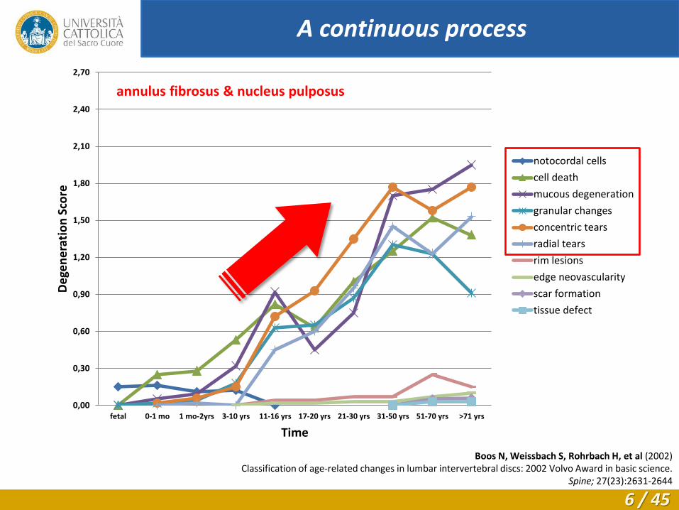
0,00
0,30
0,60
0,90
1,20
1,50
1,80
2,10
2,40
2,70
fetal 0-1 mo 1 mo-2yrs 3-10 yrs 11-16 yrs 17-20 yrs 21-30 yrs 31-50 yrs 51-70 yrs >71 yrs
De
gen
era
tio
n S
core
Time
notocordal cells
cell death
mucous degeneration
granular changes
concentric tears
radial tears
rim lesions
edge neovascularity
scar formation
tissue defect
Boos N, Weissbach S, Rohrbach H, et al (2002) Classification of age-related changes in lumbar intervertebral discs: 2002 Volvo Award in basic science.
Spine; 27(23):2631-2644
A continuous process
annulus fibrosus & nucleus pulposus
6 / 45

Break Make
Young
Balanced (No net loss)
Disc PG homeostasis
Loss of functional cells
PG expression
Core PG, GAG… MMP expression
Break
Make
Old
Perturbed (net PG loss)
Pro-inflammatory cytokines
Anti-inflammatory cytokines
The "central hypothesis" in disc biology
7 / 45

Aging is a major risk factor for IDD
Normal
Degenerated
Construction
quality &
maintenance
GENETICS
(faulty)
MECHANICAL
(trauma)
Excessive loads (sudden)
ENVIRONMENTAL Smoking
occupation
nutrition
Assaults radiation
wind
current
Time-dependent
wears and tears (slow)
AGING (time-dep damage of
macromolecules)
8 / 45
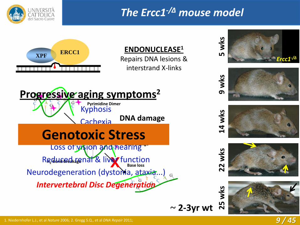
ENDONUCLEASE1
Repairs DNA lesions & interstrand X-links
XPF ERCC1
22
wks
2
5 w
ks
~ 2-3yr wt 1. Niedernhofer L.J., et al Nature 2006; 2. Gregg S.Q., et al DNA Repair 2011;
5 w
ks
14
wks
9
wks
Ercc1-/Δ
The Ercc1-/Δ mouse model
DNA damage
Base change
Pyrimidine Dimer
H2-bond breakage Base loss
Single-Strand Break
Kyphosis
Cachexia
Muscle degeneration
Loss of vision and hearing
Reduced renal & liver function
Neurodegeneration (dystonia, ataxia...)
Intervertebral Disc Degeneration
Progressive aging symptoms2
Genotoxic Stress
9 / 45

Vo N, et al Accelerated Aging of Intervertebral Discs in a Mouse Model of Progeria. J Orthop Res 2010
Ercc1 -/Δ Wt
Agc (green), Actin (red), Nuclei (blue)
Wt (3wks)
Ercc1 -/Δ (18wks)
Wt (2yrs)
X-ray images
Wt (3wks) Ercc1 -/Δ (18wks) Wt (2yrs)
Pe
rce
nt
Dis
c H
eig
ht
0
2
4
6
8
10
12
14
40
45
50
55
60
65
70
75
80
85
wt(4wk) mut(4wk) wt(20wk) mut(20wk) Wt(2.5 yrs)
Agg
reca
n f
luo
resc
en
ce
Wt Ercc1 -/D
3-5 wks
Wt Ercc1 -/D
18-20 wks
Wt
2.5 yrs
*
*
Intervertebral disc degeneration in Ercc1-/Δ
10 / 45
1.The Ercc1-/Δ mouse model, a new model for studying intervertebral disc degeneration
2.Targeting the NF-κB pathway for slowing down intervertebral disc degeneration
3.Targeting mitochondrial-generated reactive oxygen species (ROS) in intervertebral disc degeneration
Talk Outline
11 / 45

The NF-κB transcription factor
NF-κB is the transcription factor most associated with
mammalian aging3
NF-κB is up-regulated in a variety of aged tissues and age-
associated diseases4, 5
Inflammatory diseases
(Rheumatoid arthritis, diabetes, etc.)
Non-inflammatory diseases
(Atherosclerosis, osteoporosis, etc.)
3. Adler AS, et al Genes & Dev 2007; 4. Tilstra JS, et al Aging & Disease 2011; 5. Gosselin D, et al Gerontol 2003; 12 / 45

IκB
Nucleus
Extracellular
DNA
Cytoplasm
P
NF-κB DNA damage
ATM
NEMO
IKK
complex IκB
Nucleus
Extracellular
DNA
Cytoplasm
TNFR-1
TNFα
IKK
complex P
NF-κB
NF-κB is a family of trascription factors inolved
in cellular response to damage.
p50/p65 heterodimer is the most common
factor of the NF-κB family.6
NF-κB is activated in cells by many cellular
stressors; inflammatory, oxidative, genotoxic,
mechanical, and chemical stress are among
them.6
Two separate activation pathways have been
described:
1. Canonical pathway
2. DNA damage pathway
6. Hayden et al., Cell 2008
The NF-κB transcription factor
13 / 45

NF-κB activation in aged human disc nucleus pulpopus7
Aging activates NF-κB transcription factor
7. Nerlich et al., ANYAS 2007;
annulus
nucleus pulposus
14 / 45

NF-kB Promoter element
eGFP
Green cells = NF-kB activation
NF-kB–eGFP reporter mice
Activated NF-kB
• NF-κB activation in aged human disc nucleus pulpopus7
• NF-κB activation in aged mouse discs8
NF-κB is activated in aging mouse discs
7. Nerlich et al., ANYAS 2007; 8. Nasto LA et al., Spine 2012; 15 / 45

NF-kB element
NF-kB gene targets
IL-1β, IL-6, iNOS MMP-1b, MMP-3
qRT-PCR mouse disc RNA
NF-κB is activated in aging mouse discs
7. Nerlich et al., ANYAS 2007; 8. Nasto LA et al., Spine 2012;
• NF-κB activation in aged human disc nucleus pulpopus7
• NF-κB activation in aged mouse discs8
16 / 45

Young Wt (5 mths) Old Wt (2 yrs) Progeroid Ercc1-/D (5mths)
NF-κB is activated in aging mouse discs
7. Nerlich et al., ANYAS 2007; 8. Nasto LA et al., Spine 2012;
• NF-κB activation in aged human disc nucleus pulpopus7
• NF-κB activation in aged mouse discs8
17 / 45
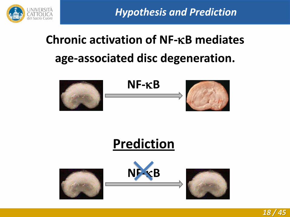
Chronic activation of NF-kB mediates
age-associated disc degeneration.
NF-kB
Prediction
NF-kB
Hypothesis and Prediction
18 / 45

1. Pharmacological inhibition
IKK IkB NF-kB x NBD
Ercc1-/∆ Mice
mNBD / NBD peptide
Approach: Blocking NF-κB activity
19 / 45

-C Transduction Domain GG IKKg Blocking Peptide N- Peptide
8K-NBD: KKKKKKKK-------- GG-------TALDWSWLQTE
8K-mNBD: KKKKKKKK-------- GG-------TALDASALQTE
Bioactivity of 8K-NBD (NEMO Binding Domain) peptide
- Inhibits IL-1β and TNFα induced NF-κB activation in vitro
- Efficacious in animal models of
Inflammatory bowel disease (Dave et al., J Immunology 2007)
Muscolar dystrophy (Acharyya et al., JCI 2007)
Arthritis (Dai et al., J Biol Chem 2004)
The NBD peptide: structure
20 / 45

NBD peptide
decreases NF-kB
expression in
Ercc1-/Δ mice
IκB
Nucleus
Extracellular
DNA
Cytoplasm
IKK
complex P
NF-κB DNA damage
ATM
NEMO
TNFR-1
TNFα
NBD peptide
The NBD peptide: functioning
21 / 45

Wt (untreated)
mtN
BD
N
BD
Ercc1 -/Δ
*
NBD treatment delays the onset of osteoporosis in Ercc1-/Δ mice
22 / 45

NBD treatment improves disc histology in Ercc1-/Δ mice
23 / 45

NBD treatment improves disc matrix content
24 / 45

1. Pharmacological inhibition
IKK IkB NF-kB x NBD
Ercc1-/∆ Mice
p65
p65
p65+/- Ercc1-/∆
Mice
x 2. Genetic reduction
NF-kB dimer
p65 p50 p65
p65 Ercc1-/∆
Mice
Non-target effects
Approach: Blocking NF-κB activity
25 / 45
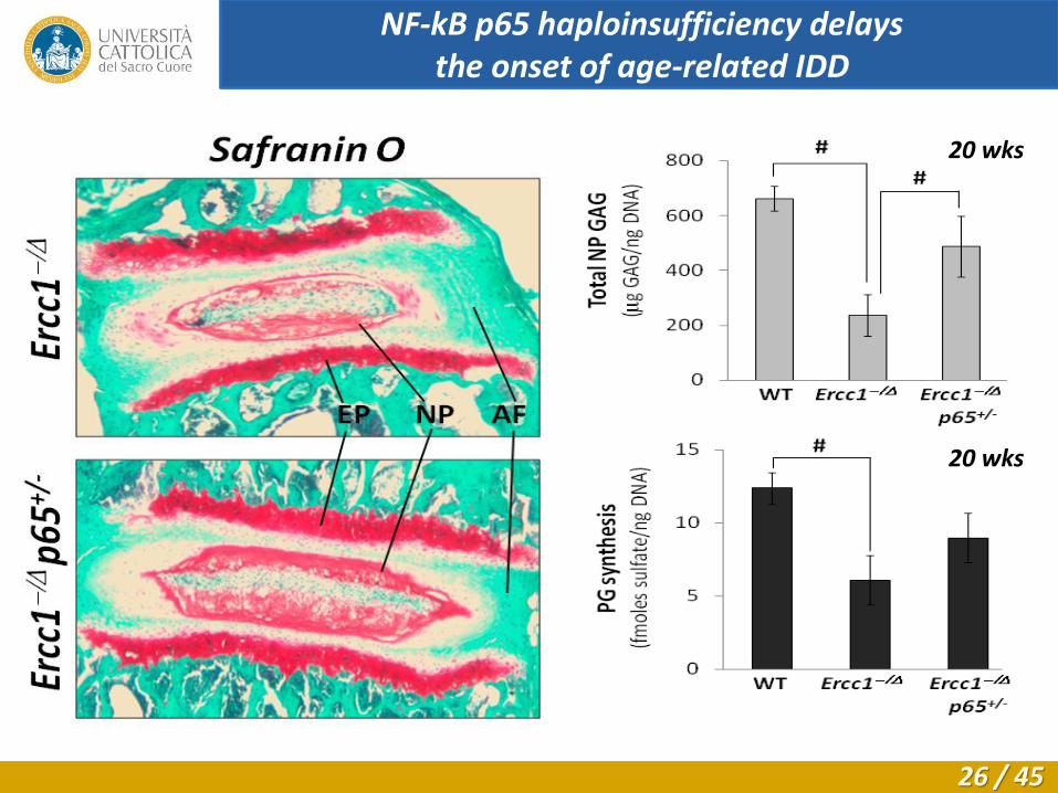
20 wks
20 wks
NF-kB p65 haploinsufficiency delays the onset of age-related IDD
26 / 45

• Increased level of NF-kB activity in disc cells of natural aging (Wt)
and progeroid (Ercc1-/Δ ) mice.
• Systemic treatment of Ercc1-/Δ mice with the NBD peptide (NF-kB
inhibitor) ameliorated age-dependent degenerative changes in
discs/spine.
Disc cellularity
Disc matrix PG content
Disc PG synthesis
Vertebral bone porosity
• Similar protective effects were seen with p65 genetic
haploinsufficiency.
Conclusions
27 / 45

1.The Ercc1-/Δ mouse model, a new model for studying intervertebral disc degeneration
2.Targeting the NF-kB pathway for slowing down intervertebral disc degeneration
3.Targeting mitochondrial-generated reactive oxygen species (ROS) in intervertebral disc degeneration
Talk Outline
28 / 45

Reactive oxygen species (ROS) are chemically reactive molecules (e.g. O2
-, H2O2, OH-). ROS are a natural byproduct of normal oxidative metabolism.
O2-
H2O2
OH-
O2-
O2- H2O2
H2O2
OH-
OH-
Mitochondria are the main source of ROS
within most cells.
Oxidative Stress and ROS
29 / 45
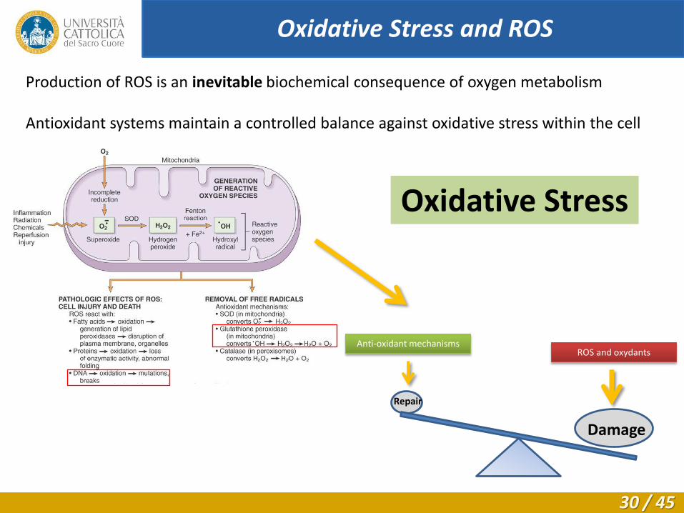
Production of ROS is an inevitable biochemical consequence of oxygen metabolism Antioxidant systems maintain a controlled balance against oxidative stress within the cell
Oxidative Stress
Damage
Repair
ROS and oxydants Anti-oxidant mechanisms
Oxidative Stress and ROS
30 / 45

Numerous studies have shown that oxidative damage increases with
age in many tissues and various organisms.9, 10, 11, 12
9. Bokov A et al., Mech Ageing & Dev 2004; 10. Ali R et al.., Biotech Histochem 2008; 11. Ulrich JA et al., Spine 2007; 12. Kang JD et al., Spine 1997;
Role of oxidative stress in disc degeneration
Neurodegenerative diseases (Patel VP, Int J Clin Exp Pathol 2011)
Lou Gehring’s disease (ALS)
Parkinson’s disease
Alzheimer’s disease
Huntington’s disease
Cardiovascular diseases (Wang JC, Circ Res 2012)
Reperfusion injury following hypoxia
Chronic fatigue syndrome (Nijs J, Man Ther 2006) Gastric cancer (Handa O, Redox Rep 2011)
31 / 45

...but intervertebral disc is hypoxic!
Urban JP, Smith S, Fairbank JC (2004) Nutrition of the intervertebral disc.
Spine; 29 (23):2700-2709
32 / 45

0,00
0,50
1,00
1,50
2,00
fetal 0-1 mo 1 mo-2yrs 3-10 yrs 11-16 yrs 17-20 yrs 21-30 yrs 31-50 yrs 51-70 yrs >71 yrs
cell density
physiolog. vessels
obliterated vessels
cartilage disorg.
cartilage cracks
microfracture
neovascularity
new bone formation
bony sclerosis
scar formation
tissue defect
Adams MA, Clin Biomech 2010
...yes, but not when it’s degenerated!
Boos N, Weissbach S, Rohrbach H, et al (2002) Classification of age-related changes in lumbar intervertebral discs: 2002 Volvo Award in basic science.
Spine; 27(23):2631-2644
Vascular ingrowth
in degenerated
intervertebral discs
cartilage endplate
33 / 45

12000X
12000X
40000X
Wt
Ercc
1 -
/D
Disc cells from 20 wk old Ercc1-/Δ mice show greater
mitochondrial death (arrows) compared to wild-type
littermates by transmission electron microscopy.
9 w
ks
25 w
ks
Ercc1-/∆ mice (DNA repair defect)
Accelerated aging
Mitochondria Dysfunction in Ercc1-/Δ mice
34 / 45

A 5% O2 20% O2 20% O2 + H2O2
Growing human NP
(nucleus pulposus) cells
in vitro increases
ROS production
Effects of oxygen on human disc cells
35 / 45

The XJB-5-131 radical scavenger
XJB-5-131 is a mitochondria-targeted ROS scavenger
XJB-5-131 has been previously shown to be
therapeutic in rodent models of hemorrhagic
shock and sepsis.13
13. Fink MP. Biochem Pharmacol 2007
Dr. Peter Wipf Professor of Pharmaceutical Sciences
Department of Chemistry University of Pittsburgh, USA
36 / 45

Bottom-to-Top Approach
Assessing the effects of oxidative stress in vitro on human disc cell cultures Assessing the effects of oxidative stress in vivo on Ercc1-/Δ mice testing the XJB-5-131 radical scavenger
37 / 45

A B
0
20
40
60
80
100
120
5% O2 20% O2
Re
lati
ve P
G s
ynth
esi
s (%
)
0
100
200
300
400
500
600
700
800
900
Agc ADAMTS4 ADAMTS5 MMP1 MMP3
Re
lati
ve g
en
e e
xpre
ssio
n (
%)
5% 20%
*
Effects of oxygen on human disc cells
High oxygen levels have
catabolic effects on
human disc cells in vitro
catabolic genes
reduced matrix synthesis
38 / 45

A B
0
20
40
60
80
100
120
5% O2 20% O2
Re
lati
ve P
G s
ynth
esi
s (%
)
0
100
200
300
400
500
600
700
800
900
Agc ADAMTS4 ADAMTS5 MMP1 MMP3
Re
lati
ve g
en
e e
xpre
ssio
n (
%)
5% 20%
*
XJB-5-131 rescues in vitro effects of high O2
0
20
40
60
80
100
120
140
20% O2 20% O2 + XJB
Re
lati
ve P
G s
ynth
esi
s (%
) *
0
25
50
75
100
125
150
175
200
20% O2 20% O2 + XJB
Re
lati
ve a
gc e
xpre
ssio
n (
%) *
XJB
XJB
39 / 45

9 w
ks
25 w
ks
Ercc1-/∆ mice (DNA repair defect)
Systemic treatment with XJB in Ercc1-/Δ mice
Accelerated aging
0 4 8 10 12 14 20 Age (weeks)
Treatment start
Treatment end
XJB-5-131 by IP injection 2mg/Kg, 3x/week
Systemic anti-oxidant therapy in Ercc1-/Δ mice to slow down IDD
40 / 45

XJB
-5-1
31
N
eg.
co
ntr
ol
H&E Safranin O
XJB treatment improves disc histology in Ercc1-/Δ mice
41 / 45

XJB treatment improves disc matrix content
0
100
200
300
400
500
600
700
800
900
1000
Tota
l GA
G c
on
ten
t (µ
gGA
G/n
g D
NA
)
NT Oil XJB
Wt Ercc1-/Δ
*
*
0
2
4
6
8
10
12
14
16
18
20
NT Oil XJB
Wt Ercc1-/Δ
PG
Syn
the
sis
(fm
ole
s su
lfat
e/n
g D
NA
) * p < 0.07
42 / 45

• Oxidative stress has been well established as a key driver of aging.
• We showed that ROS were produced in disc cells at high oxygen
tension and this correlates with:
Reduced PG (new matrix) synthesis
Enhanced expression of catabolic factors
• Depleting ROS in disc cells using XJB-5-131 rescued matrix
homeostatic imbalance
• Mitochondria-targeted free radical scavengers could be therapeutic
in delaying the onset of age-related IDD
Conclusions
43 / 45

Acknowledgements
University of Pittsburgh Hillman Cancer Institute
Laura Niedernhofer Paul Robbins Jeremy Tilstra Andria Robison Cheryl Clauson NBD peptide
Ferguson Laboratory of Orthopaedics Research
James Kang (Director) Gwen Sowa (Co-director) Nam Vo (PI) Rebecca Studer, Joon Lee
Hyoung-Yeon Seo Kevin Ngo, Qing Dong Hong Joo Moon, Takashi Yurube, Robert Hartman, Wan Huang, Paulo Coelho, Kevin Bell, Lou
Duerring.
Funding Sources
The Pittsburgh Foundation UPMC Department of Orthopaedics Surgery
Orthopaedic Research and Education Foundation AOSpine North America Young Investigators Research Grant
“BioSpina” Italian Spine Society Research Grant NIH (R21 AG033046), NIH (ES016114)
44 / 45

Thank you
45 / 45