traumatic dental injuries in children and adolescence
TRANSCRIPT

N. V. SHAKOVETS, D. N. NAUMOVICH,
N. D. CHERNYAVSKAYA
TRAUMATIC DENTAL INJURIES
IN CHILDREN AND ADOLESCENCE
Minsk BSMU 2018

1
МИНИСТЕРСТВО ЗДРАВООХРАНЕНИЯ РЕСПУБЛИКИ БЕЛАРУСЬ
БЕЛОРУССКИЙ ГОСУДАРСТВЕННЫЙ МЕДИЦИНСКИЙ УНИВЕРСИТЕТ
КАФЕДРА СТОМАТОЛОГИИ ДЕТСКОГО ВОЗРАСТА
Н. В. ШАКОВЕЦ, Д. Н. НАУМОВИЧ, Н. Д. ЧЕРНЯВСКАЯ
ТРАВМАТИЧЕСКИЕ ПОВРЕЖДЕНИЯ ЗУБОВ
У ДЕТЕЙ И ПОДРОСТКОВ
TRAUMATIC DENTAL INJURIES
IN CHILDREN AND ADOLESCENCE
Учебно-методическое пособие
Минск БГМУ 2018

2
УДК 616.314-001-053.2/.6(075.8)-054.6
ББК 56.6я73
Ш17
Рекомендовано Научно-методическим советом университета в качестве
учебно-методического пособия 18.04.2018 г., протокол № 8
Р е ц е н з е н т ы: канд. мед. наук, доц., зав. курсом детской стоматологии каф. ортопеди-
ческой стоматологии и ортодонтии с курсом детской стоматологии Белорусской медицинской
академии последипломного образования В. А. Андреева; д-р мед. наук, проф., зав. каф. орто-
донтии Белорусского государственного медицинского университета И. В. Токаревич; канд.
филол. наук, доц., зав. каф. иностранных языков Белорусского государственного медицинского
университета М. Н. Петрова
Шаковец, Н. В.
Ш17 Травматические повреждения зубов у детей и подростков = Traumatic dental injuries
in children and adolescence : учебно-методическое пособие / Н. В. Шаковец, Д. Н. Наумо-
вич, Н. Д. Чернявская. ‒ Минск : БГМУ, 2018. – 40 с.
ISBN 978-985-21-0077-9.
Посвящено проблеме тактики врача-стоматолога при оказании помощи детям с травмой временных
и постоянных зубов с незаконченным формированием корней. Изложены вопросы этиологии травматических
дентальных поражений у детей, особенностей проведения диагностики, клинического и рентгенологического
обследования, выбора метода лечения в зависимости от клинической ситуации, прогноз исходов в отношении
пульпы и периодонта, профилактика травмы.
Предназначено для студентов 4–5-го курсов медицинского факультета иностранных учащихся, обучаю-
щихся по специальности 1-79 01 07 «Стоматология» на английском языке.
УДК 616.314-001-053.2/.6(075.8)-054.6
ББК 56.6я73
____________________________________________
Учебное издание
Шаковец Наталья Вячеславовна
Наумович Дарья Николаевна
Чернявская Надежда Дмитриевна
ТРАВМАТИЧЕСКИЕ ПОВРЕЖДЕНИЯ ЗУБОВ У ДЕТЕЙ И ПОДРОСТКОВ
TRAUMATIC DENTAL INJURIES IN CHILDREN AND ADOLESCENCE
Учебно-методическое пособие
На английском языке
Ответственная за выпуск Т. Н. Терехова
Переводчики Н. В. Шаковец, Д. Н. Наумович, Н. Д. Чернявская
Компьютерная верстка Н. М. Федорцовой
Подписано в печать 06.07.18. Формат 6084/16. Бумага писчая «Xerox office».
Ризография. Гарнитура «Times». Усл. печ. л. 2,32. Уч.-изд. л. 2,17. Тираж 150 экз. Заказ 504.
Издатель и полиграфическое исполнение: учреждение образования
«Белорусский государственный медицинский университет».
Свидетельство о государственной регистрации издателя, изготовителя,
распространителя печатных изданий № 1/187 от 18.02.2014.
Ул. Ленинградская, 6, 220006, Минск.
ISBN 978-985-21-0077-9 © Шаковец Н. В., Наумович Д. Н., Чернявская Н. Д., 2018
© УО «Белорусский государственный медицинский
университет», 2018

3
ABRIDGEMENTS AND SYMBOLS
C — Clinical examination
R — Radiographic examination
C* — Clinical and radiographic monitoring until eruption of the permanent
successor
S — Splint removal
+ — for crown fractured teeth with concomitant luxation injury, use the luxation
followup schedule
C++ — clinical and radiographic examination.
S+ — splint removal; S++ — splint removal in cervical third fractures.
C++ — clinical and radiographic examination.
++ — Whenever there is evidence of external inflammatory root resorption, root
canal therapy should be initiated immediately, with the use of calcium hydroxide
as an intra-canal medication.
INTRODUCTION
Traumatic dental injuries (TDIs) occur frequently in children and young
adults, comprising 5 % of all injuries [1]. Twenty‐five percent of all school
children experience dental trauma and 33 % of adults have experienced trauma
to the permanent dentition, with the majority of the injuries occurring before
age 19. Luxation injuries are the most common TDIs in the primary dentition,
whereas crown fractures are more commonly reported for the permanent teeth.
Proper diagnosis, treatment planning and follow‐up are important to assure
a favorable outcome.
It is understood that some of the subsequent treatment may require
secondary and tertiary interventions involving specialists with experience in
dental trauma [1].
4
GENERAL RECOMMENDATIONS
Special considerations for trauma to primary teeth. A young child is
often difficult to examine and treat due to lack of cooperation and because of
fear. The situation is distressing for both the child and the parents. It is important
to keep in mind that there is a close relationship between the apex of the root of
the injured primary tooth, and the underlying permanent tooth germ. Tooth
malformation, impacted teeth, and eruption disturbances in the developing
permanent dentition are some of the consequences that can occur following
severe injuries to primary teeth and alveolar bone. A child’s maturity and ability
to cope with the emergency situation, the time for shedding of the injured tooth
and the occlusion, are all important factors that influence treatment. Repeated
trauma episodes are frequent in children.
Immature versus mature permanent teeth. Every effort should be made
to preserve pulpal vitality in the immature permanent tooth to ensure continuous
root development. The vast majority of TDIs occur in children and teenagers
where loss of a tooth has lifetime consequences. The immature permanent tooth
has considerable capacity for healing after traumatic pulp exposure, luxation
injury and root fractures.
Avulsion of permanent teeth. The prognosis for avulsed permanent teeth
is very much dependent on the actions taken at the place of accident. Promotion
of public awareness of first‐aid treatment for the avulsed tooth is strongly
encouraged. Treatment choices and prognosis for the avulsed tooth are largely
dependent on the vitality of the periodontal ligament (PDL), and the maturity of
the root.
Patient/parent instructions. Patient compliance with follow‐up visits and
home care contributes to better healing following a TDI. Both the patients and
the parents of young patients should be advised regarding care of the injured
tooth/teeth for optimal healing, prevention of further injury by avoidance of
participation in contact sports, meticulous oral hygiene, and rinsing with an
antibacterial such as Chlorhexidine Gluconate 0.1 % alcohol free for 1–2 weeks.
Alternatively, with a young child, it is desirable to apply Chlorhexidine
Gluconate to the affected area with a cotton swab. The use of pacifiers should be
restricted [1].

5
CLASSIFICATION OF DENTAL TRAUMA
ICD-10-CM Diagnosis Codes (2017) [3]
S00-T88 Injury, poisoning and certain other consequences of external causes
S00-S09 Injuries to the head
S02 Fracture of skull and facial bones
S02.5 Fracture of tooth (traumatic)
S02.5XXA…… initial encounter for closed fracture
S02.5XXB…… initial encounter for open fracture
S02.5XXD…… subsequent encounter for fracture with routine healing
S02.5XXG…… subsequent encounter for fracture with delayed healing
S02.5XXK…… subsequent encounter for fracture with nonunion
S02/5XXS…… sequela
S03 Dislocation and sprain of joints and ligaments of head
S03.2 Dislocation of tooth
S03.2XXA…… initial encounter
S03.2XXD…… subsequent encounter
S03. S03.2XXS…… sequela
Classification by Andreasen (1981) [1]
1. Injuries to the dental hard tissue and pulp
crown infraction
crown fracture
uncomplicated crown fracture
– enamel fracture
– enamel dentin fracture without pulp exposure
complicated crown fracture
– enamel dentin fracture with pulp exposure
2. Injuries to the dental hard tissue, pulp and to the alveolar process
crown root fracture
enamel dentin fracture
root fracture
dentin root fracture with pulp exposure
alveolar fracture in connection with tooth trauma
alveolar fracture in the maxilla
alveolar fracture in the mandible
3. Injuries to the periodontium
luxation
concussion (shoсk)
subluxation (loosening)

6
intrusion (central luxation)
extrusion (peripheral luxation)
lateral luxation
total luxation (exarticulation)
TRAUMA OF PERMANENT TEETH
Injuries to permanent teeth tend to occur most often outside the home.
The most common cause of dental injuries is falls, comprising between
26 % and 82 % of all sustained injuries, depending on the subpopulation
investigated. Sports-related injuries are generally the second most common
cause of dental injuries.
The main “peak period” for dental injury for permanent teeth is described
as being between the ages of 7 and 10.
In school-age children, bicycle, skateboard, micro-scooters, fights and road
accidents are the most significant factors, while in adolescence there is another,
although less marked, peak largely due to sports injuries.
Most of these sports injuries result from participation in contact sports such
as American football, rugby, soccer, boxing, wrestling, diving or stick sports.
However, other sports like skiing, skating, cycling, and horse riding, which do
not necessarily involve player contact, may also place the participant at risk.
The Federation Dentaire International (FDI) have recently classified organized
sport into two categories:
1) high-risk sports that include American football, hockey, ice-hockey,
lacrosse, martial sports, rugby, football, and skating;
2) medium-risk sports that include basketball, diving, squash, gymnastics,
parachuting, and waterpolo (FDI 1990).
Gender significance in injuries. For elder children, boys tend to have
more dental injuries than girls. These findings could be a result of typically more
aggressive play from boys and an earlier introduction to competitive sports.
Type of injury. For permanent teeth, uncomplicated crown fractures tend
to be the most common type of dental injury.
Dental location of injury. Epidemiological studies have shown that
the most common tooth to become traumatized is the maxillary central incisor
followed by the maxillary lateral incisor.
Predetermining factors. Because most dental injuries occur to maxillary
central incisors, it is understandable that malocclusion and the lack of natural
protection to these teeth will predispose them to injury. Severe overjet of
the upper central incisors (i.e., overjet greater than 3 mm) will predispose these
teeth to up to five times the risk of trauma compared with a normal overjet.
The natural protection for these teeth involves the adequate coverage by

7
the patient's upper lip. It has been shown that incompetent lip coverage tends to
precipitate more severe injuries to the teeth.
Socioeconomic background may also have some impact. Typically there
are more severe injuries in children of a lower socioeconomic subpopulation.
This may have to do with inadequate supervision, an increase in assault
frequency, and/or the lack of adequate patient education pertaining to
prevention.
Orthodontic appliances may create more soft tissue injuries when trauma
presents, potentially causing increased injury to the gingiva and lips.
Acute medical problems, such as seizures, heart attack, or stroke, may
predispose the patient to falling and traumatically injuring the dentition.
General anesthetic may also predispose the patient to intraoral trauma
from the placement of an endotracheal tube. It has been reported that dental
trauma may occur in as many as 18 % of patients who are intubated [6].
TREATMENT GUIDELINES FOR FRACTURES OF TEETH AND ALVEOLAR BONE
Enamel infraction
Clinical findings. An incomplete fracture (crack) of
the enamel without loss of tooth structure.
Visual signs. A visible fracture line on the surface of
the tooth.
Percussion test. Not tender. If tenderness is observed
evaluate the tooth for a possible luxation injury or a root
fracture.
Mobility test. Normal mobility.
Sensibility pulp test. Usually positive. The test is
important in assesing future risk of healing complications. A
lack of response to the test at the initial examination indicates an increased risk
of later pulp necrosis.
Radiographic findings. No radiographic abnormalities.
Radiographs recommended. A periapical view. Additional radiographs
are indicated if other signs or symptoms are present.
Treatment. In case of marked infractions, etching and sealing with resin to
prevent dislocation of the infraction lines. Otherwise, no treatment is necessary.
Follow-up. No follow-up is generally needed for infraction injuries unless
they are associated with a luxation injury or other fracture types.
Favorable outcome. Asymptomatic. Positive response to pulp testing.
Continuing root development in immature teeth.
Unfavorable outcome. Symptomatic. Negative response to pulp testing.
Signs of apical periodontitis. No continuing root development in immature teeth.
Endodontic therapy appropriate for stage of root development is indicated.

8
Enamel fracture
Clinical findings. A complete fracture of the enamel.
Visual signs. Loss of enamel. No visible sign of
exposed dentin.
Percussion test. Not tender. If tenderness is observed
evaluate the tooth for a possible luxation or root fracture
injury.
Mobility test. Normal mobility.
Sensibility pulp test. Usually positive. The test may be
negative initially indicating transient pulpal damage. Monitor
pulpal response at initial examination until definitive pulpal
diagnosis can be made.
Radiographic findings. Enamel loss is visible.
Radiographs recommended. Periapical, occlusal and eccentric exposures.
They are recommended in order to rule out the possible presence of a root
fracture or a luxation injury. Radiograph of lip or cheek to search for tooth
fragments or foreign materials.
Treatment. If the tooth fragment is available, it can be bonded to the tooth.
Contouring or restoration with composite resin depends on the extent and
location of the fracture.
Follow-up:
– 6–8 weeks C++1;
– 1 year C++.
Favorable outcome. Asymptomatic. Positive response to pulp testing.
Continuing root development in immature teeth. Continue to next evaluation.
Unfavorable outcome. Symptomatic. Negative response to pulp testing.
Signs of apical periodontitis. No continuing root development in immature teeth.
Endodontic therapy appropriate for stage of root development is indicated. 1C++ = clinical and radiographic examination.
Enamel-dentin fracture
Clinical findings. A fracture confined to enamel and
dentin with loss of tooth structure, but not involving
the pulp.
Visual signs. Visible loss of enamel and dentine.
No visible signs of exposed pulp tissue.
Percussion test. Not tender. If tenderness is
observed, evaluate the tooth for possible luxation or root
fracture injury.
Mobility test. Normal mobility.

9
Sensibility pulp test. Usually positive. Test may be negative initially
indicating transient pulpal damage. Monitor pulpal response at initial
examination until definitive pulpal diagnosis can be made.
Radiographic findings. Enamel-dentin loss is visible.
Radiographs recommended. Periapical, occlusal and eccentric exposure
to rule out tooth displacement or possible presence of root fracture. Radiograph
of lip or cheek lacerations to search for tooth fragments or foreign materials.
Treatment. If a tooth fragment is available it can be bonded to the tooth.
Otherwise, perform a provisional treatment by covering the exposed dentin with
glass- Ionomer or a more permanent restoration using a bonding agent and
composite resin, or other accepted dental restorative materials. If the exposed
dentin is within 0.5 mm of the pulp (pink, no bleeding) place calcium hydroxide
base and cover with a material such as a glass ionomer.
Follow-up procedures for fractures of teeth and alveolar bone +1
Follow-up:
– 6–8 weeks C++;
– 1 year C++.
Favorable outcome. Asymptomatic. Positive response to pulp testing.
Continuing root development in immature teeth. Continue to next evaluation.
Unfavorable outcome. Symptomatic. Negative response to pulp testing.
Signs of apical periodontitis. No continuing root development in immature teeth.
Endodontic therapy appropriate for stage of root development is indicated. 1+= for crown fractured teeth with concomitant luxation injury, use
the luxation follow up schedule
Enamel-dentin-pulp fracture (complicated crown fracture)
Clinical findings. A fracture involving enamel and
dentin with loss of tooth structure and exposure of
the pulp.
Mobility test. Normal mobility
Percussion test. Not tender. If tenderness is
observed evaluate for possible luxation or root fracture
injury.
Sensibility pulp test. Exposed pulp is sensitive to
stimuli. The test is important in assessing risk of future
healing complication. A lack of responseat at the initial
examination indicates an increased risk of later pulp necrosis.
Radiographic findings. Enamel-dentin loss visible.
Radiographs recommended. Periapical, occlusal and eccentric exposures,
to rule out tooth displacement or possible presence of root fracture. Radiograph
of lip or cheek lacerations to search for tooth fragments or foreign materials.

10
Treatment. In young patients with immature, still developing teeth, it is
advantageous to preserve pulp vitality by pulp capping or partial pulpotomy.
Also this treatment is the choice in young patients with completely formed teeth.
Calcium hydroxide is a suitable material to be placed on the pulp wound in such
procedures. In patients with mature apical development, root canal treatment
is usually the treatment of choice, although pulp capping or partial pulpotomy
also may be selected. If tooth fragment is available, it can be bonded to
the tooth. Future treatment for the fractured crown may be restoration with other
accepted dental restorative materials.
Follow-up procedures for fractures of teeth and alveolar bone+
Follow-up:
– 6–8 weeks C++;
– 1 year C++.
Favorable and unfavorable outcomes include some, but not necessarily
all, of the following.
Favorable outcome. Asymptomatic. Positive response to pulp testing.
Continuing root development in immature teeth. Continue to next evaluation.
Unfavorable outcome. Symptomatic. Negative response to pulp testing.
Signs of apical periodontitis. No continuing root development in immature teeth.
Endodontic therapy appropriate for stage of root development is indicated.
Crown-root fracture without pulp exposure
Clinical findings. A fracture involving enamel, dentin
and cementum with loss of tooth structure, but not exposing
the pulp. Crown fracture extending below gingival margin.
Percussion test: tender. Coronal fragment mobile. Sensibility
pulp test is usually positive for apical fragment.
Radiographic findings. Apical extension of fracture is
not usually visible. Radiographs recommended: periapical,
occlusal and eccentric exposures. They are recommended in
order to detect fracture lines in the root.
Treatment
Emergency treatment. As an emergency treatment a temporary
stabilization of the loose segment to adjacent teeth can be performed until a
definitive treatment plan is made.
Non-emergency treatment alternatives fragment removal only.
Removal of the coronal crown-root fragment and subsequent restoration of
the apical fragment exposed above the gingival level.
Fragment removal and gingivectomy (sometimes ostectomy). Removal
of the coronal crown-root segment with subsequent endodontic treatment and

11
restoration with a postretained crown. This procedure should be preceded by
a gingivectomy, and sometimes ostectomy with osteoplasty.
Orthodontic extrusion of apical fragment. Removal of the coronal
segment with subsequent endodontic treatment and orthodontic extrusion of
the remaining root with sufficient length after extrusion to support a post-
retained crown.
Surgical extrusion. Removal of the mobile fractured fragment with
subsequent surgical repositioning of the root in a more coronal position.
Root submergence. Implant solution is planned.
Extraction. Extraction with immediate or delayed implant-retained crown
restoration or a conventional bridge. Extraction is inevitable in crown-root
fractures with a severe apical extension, the extreme being a vertical fracture.
Follow-up procedures for fractures of teeth and alveolar bone +
Follow-up:
– 6–8 weeks C++;
– 1 year C++.
Favorable and unfavorable outcomes include some, but not necessarily
all, of the following
Favorable outcome. Asymptomatic. Positive response to pulp testing.
Continuing root development in immature teeth. Continue to next evaluation.
Unfavorable outcome. Symptomatic. Negative response to pulp testing.
Signs of apical periodontitis. No continuing root development in immature teeth.
Endodontic therapy appropriate for stage of root development is indicated.
Crown-root fracture with pulp exposure
Clinical findings. A fracture involving enamel, dentin,
and cementum and exposing the pulp. Percussion test: tender.
Coronal fragment mobile.
Radiographic findings. Apical extension of fracture
usually not visible. Radiographs recommended: periapical
and occlusal exposure.
Treatment
Emergency treatment. As an emergency treatment
a temporary stabilization of the loose segment to adjacent
teeth. In patients with open apices, it is advantageous to
preserve pulp vitality by a partial pulpotomy. This treatment is also the choice in
young patients with completely formed teeth. Calcium hydroxide compounds
are suitable pulp capping materials. In patients with mature apical development,
root canal treatment can be the treatment of choice.
Non-emergency treatment alternatives
Fragment removal and gingivectomy (sometimes ostectomy). Removal
of the coronal fragment with subsequent endodontic treatment and restoration

12
with a post-retained crown. This procedure should be preceded by a gingivectomy
and sometimes ostectomy with osteoplasty. This treatment option is only
indicated in crown-root fractures with palatal subgingival extension.
Orthodontic extrusion of apical fragment. Removal of the coronal
segment with subsequent endodontic treatment andь orthodontic extrusion of
the remaining root with sufficient length after extrusion to support a post-
retained crown.
Surgical extrusion. Removal of the mobile fractured fragment with
subsequent surgical repositioning of the root in a more coronal position.
Root submergence. An implant solution is planned, the root fragment may
be left in situ.
Extraction. Extraction with immediate or delayed implant-retained crown
restoration or a conventional bridge. Extraction is inevitable in very deep crown-
root fractures, the extreme being a vertical fracture.
Follow-up procedures for fractures of teeth and alveolar bone +
Follow-up:
– 6–8 weeks C++;
– 1 year C++.
Favorable and unfavorable outcomes include some, but not necessarily
all, of the following
Favorable outcome. Asymptomatic. Positive response to pulp testing.
Continuing root development in immature teeth. Continue to next evaluation.
Unfavorable outcome. Symptomatic. Negative response to pulp testing.
Signs of apical periodontitis. No continuing root development in immature teeth.
Endodontic therapy appropriate for stage of root development is indicated.
Root fracture
Clinical findings. The coronal segment may be mobile
and may be displaced. The tooth may be tender to percussion.
Bleeding from the gingival sulcus may be noted. Sensibility
testing may give negative results initially, indicating transient
or permanent neural damage. Monitoring the status of
the pulp is recommended. Transient crown discoloration (red
or grey) may occur.
Radiographic findings. The fracture involves the root of
the tooth and is in a horizontal or oblique plane. Fractures that
are in the horizontal plane can usually be detected in the
regular periapical 90o angle film with the central beam through the tooth. This is
usually the case with fractures in the cervical third of the root. If the plane of
fracture is more oblique which is common with apical third fractures,
an occlusal view or radiographs with varying horizontal angles are more likely
to demonstrate the fracture including those located in the middle third.

13
Treatment. Reposition, if displaced, the coronal segment of the tooth as
soon as possible. Check position radiographically. Stabilize the tooth with
a flexible splint for 4 weeks. If the root fracture is near the cervical area of
the tooth, stabilization is beneficial for a longer period of time (up to 4 months).
It is advisable to monitor healing for at least one year to determine pulpal status.
If pulp necrosis develops, root canal treatment of the coronal tooth segment to
the fracture line is indicated to preserve the tooth.
Follow-up procedures for fractures of teeth and alveolar bone
Follow-up:
– 4 weeks S+1, C++;
– 6–8 weeks C++;
– 4 months S++2, C++;
– 6 months C++;
– 1 year C++;
– 5 years C++.
Favorable and Unfavorable outcomes include some, but not necessarily
all, of the following ++3
Favorable outcome. Positive response to pulp testing (false negative
possible up to 3 months). Signs of repair between fractured segments. Continue
to next evaluation.
Unfavorable outcome. Symptomatic. Negative response to pulp testing
(false negative possible up to 3 months). Extrusion of the coronal segment.
Radiolucency at the fracture line. Clinical signs of periodontitis or abscess
associated with the fracture line. Endodontic therapy appropriate for stage of
root development is indicated. 1S+ = splint removal; 2S++ = splint removal in cervical third fractures. 3++ = whenever there is evidence of external inflammatory root resorption,
root canal therapy should be initiated immediately, with the use of calcium
hydroxide as an intra-canal medication.
Alveolar fracture
Clinical findings. The fracture involves the alveolar
bone and may extend to adjacent bone. Segment mobility
and dislocation with several teeth moving together are
common findings. An occlusal change due to misalignment
of the fractured alveolar segment is often noted. Sensibility
testing may or may not be positive.
Radiographic findings. Fracture lines may be located
at any level, from the marginal bone to the root apex.
In addition to the 3 angulations and occlusal film, additional

14
views such as a panoramic radiograph can be helpful in determining the course
and position of the fracture lines.
Treatment. Reposition any displaced segment and then splint. Suture
gingival laceration if present. Stabilize the segment for 4 weeks.
Follow-up procedures for fractures of teeth and alveolar bone
Follow-up:
– 4 weeks S+, C++;
– 6–8 weeks C++;
– 4 months S++, C++;
– 6 months C++;
– 1 year C++;
– 5 years C++.
Favorable and unfavorable outcomes include some, but not necessarily
all, of the following ++
Favorable outcome. Positive response to pulp testing (false negative
possible up to 3 months). No signs of apical periodontitis. Continue to next
evaluation.
Unfavorable outcome. Symptomatic. Negative response to pulp testing
(false negative possible up to 3 months). Signs of apical periodontitis or external
inflammatory root resorption. Endodontic therapy appropriate for stage of root
development is indicated.
TREATMENT GUIDELINES FOR LUXATION INJURIES
Concussion
Clinical findings. The tooth is tender to touch or
tapping; it has not been displaced and does not have increased
mobility. Sensibility tests are likely to give positive results.
Radiographic findings. No radiographic abnormalities.
Treatment. No treatment is needed. Monitor pulpal
condition for at least one year.
Follow-up procedures for luxated permanent teeth
Follow-up:
– 4 weeks C++;
– 6–8 weeks C++;
– 1 year C++.
Favorable and unfavorable outcomes include some, but not necessarily
all, of the following ++
Favorable outcome. Asymptomatic. Positive response to pulp testing.
False negative possible up to 3 months. Continuing root development in
immature teeth. Intact lamina dura.

15
Unfavorable outcome. Symptomatic. Negative response to pulp testing.
False negative possible up to 3 months. No continuing root development in
immature teeth, signs of apical periodontitis. Endodontic therapy appropriate for
stage of root development is indicated.
Subluxation Clinical findings. The tooth is tender to touch or tapping
and has increased mobility; it has not been displaced. Bleeding
from gingival crevice may be noted. Sensibility testing may be
negative initially indicating transient pulpal damage. Monitor
pulpal response until a definitive pulpal diagnosis can be made.
Radiographic findings. Radiographic abnormalities are
usually not found.
Treatment. Normally no treatment is needed, however
a flexible splint to stabilize the tooth for patient comfort can be
used for up to 2 weeks.
Follow-up procedures for luxated permanent teeth
Follow-up:
– 2 weeks S+, C++;
– 4 weeks C++;
– 6–8 weeks C++;
– 6 months C++;
– 1 year C++.
Favorable and Unfavorable outcomes include some, but not necessarily
all, of the following ++
Favorable outcome. Asymptomatic. Positive response to pulp testing.
False negative possible up to 3 months. Continuing root development in
immature teeth. Intact lamina dura.
Unfavorable outcome. Symptomatic. Negative response to pulp testing.
False negative possible up to 3 months. External inflammatory resorption. No
continuing root development in immature teeth, signs of apical periodontitis.
Endodontic therapy appropriate for stage of root development is indicated.
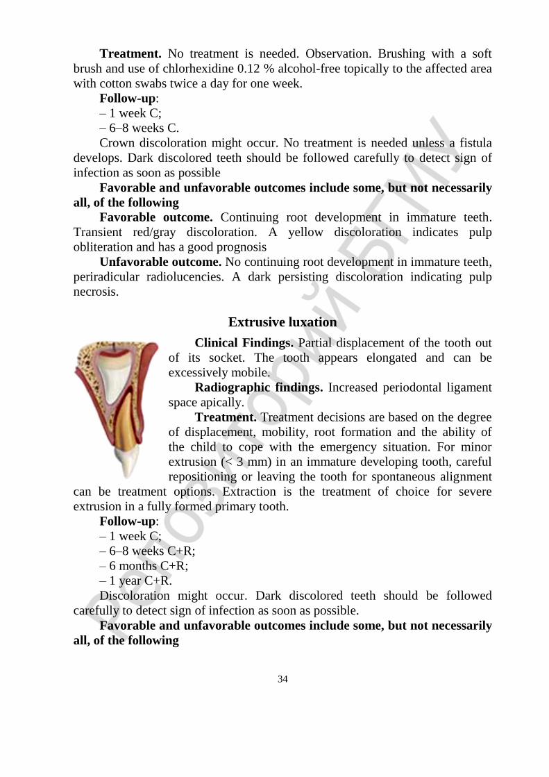
Extrusive luxation Clinical findings. The tooth appears elongated and is
excessively mobile. Sensibility tests will likely give negative
results.
Radiographic findings. Increased periodontal ligament
space apically.
Treatment. Reposition the tooth by gently re-inserting it
into the tooth socket. Stabilize the tooth for 2 weeks using
a flexible splint. In mature teeth where pulp necrosis is

16
anticipated or if several signs and symptoms indicate that the pulp of mature or
immature teeth became necrotic, root canal treatment is indicated.
Follow-up procedures for luxated permanent teeth
Follow-up:
– 2 weeks S+, C++;
– 4 weeks C++;
– 6–8 weeks C++;
– 6 months C++;
– 1 year C++;
– yearly 5 years C++.
Favorable and unfavorable outcomes include some, but not necessarily
all, of the following ++
Favorable outcome. Asymptomatic. Clinical and radiographic signs of
normal or healed periodontium. Positive response to pulp testing (false negative
possible up to 3 months). Marginal bone height corresponds to that seen
radiographically after repositioning. Continuing root development in immature
teeth.
Unfavorable outcome. Symptoms and radiographic sign consistent with
apical periodontitis. Negative response to pulp testing (false negative possible
up to 3 months). If breakdown of marginal bone, splint for an additional
3–4 weeks. External inflammatory root resorption. Endodontic therapy
appropriate for stage of root development is indicated.
Lateral luxation Clinical findings. The tooth is displaced, usually in
a palatal/lingual or labial direction. It will be immobile and
percussion usually gives a high, metallic (ankylotic) sound.
Fracture of the alveolar process present. Sensibility
tests will likely give negative results.
Radiographic findings. The widened periodontal
ligament space is best seen on eccentric or occlusal
exposures.
Treatment. Reposition the tooth digitally or with
forceps to disengage it from its bony lock and gently reposition it into its
original location. Stabilize the tooth for 4 weeks using a flexible splint. Monitor
the pulpal condition. If the pulp becomes necrotic, root canal treatment is
indicated to prevent root resorption.
Follow-up procedures for luxated permanent teeth
Follow-up
– 2 weeks, C++;
– 4 weeks S+, C++;
– 6–8 weeks C++;

17
– 6 months C++;
– 1 year C++;
– yearly for 5 years C++.
Favorable and unfavorable outcomes include some, but not necessarily
all, of the following ++
Favorable outcome. Asymptomatic. Clinical and radiographic signs of
normal or healed periodontium. Positive response to pulp testing (false negative
possible up to 3 months). Marginal bone height corresponds to that seen
radiographically after repositioning. Continuing root development in immature
teeth.
Unfavorable outcome. Symptoms and radiographic signs consistent with
apical periodontitis. Negative response to pulp testing (false negative possible
up to 3 months). If breakdown of marginal bone, splint for an additional
3–4 weeks. External inflammatory root resorption or replacement resorption.
Endodontic therapy appropriate for stage of root development is indicated.
Intrusive luxation Clinical findings. The tooth is displaced axially into
the alveolar bone. It is immobile and percussion may give
a high, metallic (ankylotic) sound. Sensibility tests will
likely give negative results.
Radiographic findings. The periodontal ligament
space may be absent from all or part of the root.
The cemento-enamel junction is located more apically in
the intruded tooth than in adjacent non-injured teeth, at
times even apical to the marginal bone level.
Treatment. Teeth with incomplete root formation.
Allow eruption without intervention. If no movement within few weeks, initiate
orthodontic repositioning. If tooth is intruded more than 7 mm, reposition
surgically or orthodontically.
Teeth with complete root formation:
– allow eruption without intervention if tooth intruded less than 3 mm.
If no movement after 2–4 weeks, reposition surgically or orthodontically before
ankylosis can develop;
– if tooth is intruded 3–7 mm, reposition surgically or orthodontically;
– if tooth is intruded beyond 7 mm, reposition surgically;
– the pulp will likely become necrotic in teeth with complete root
formation. Root canal therapy using a temporary filling with calcium hydroxide
is recommended and treatment should begin 2–3 weeks after repositioning;
– once an intruded tooth has been repositioned surgically or
orthodontically, stabilize with a flexible splint for 4 weeks.

18
Follow-up procedures for luxated permanent teeth
Follow-up:
– 2 weeks, C++;
– 4 weeks S+, C++;
– 6–8 weeks C++;
– 6 months C++;
– 1 year C++;
– yearly for 5 years C++.
Favorable and unfavorable outcomes include some, but not necessarily
all, of the following ++
Favorable outcome. Tooth in place or erupting. Intact lamina dura. No
signs of resorption. Continuing root development in immature teeth.
Unfavorable. Tooth locked in place/ankylotic tone to percussion.
Radiographic signs of apical periodontitis. External inflammatory root
resorption or replacement resorption. Endodontic therapy appropriate for stage
of root development is indicated.
Avulsion
Avulsion of permanent teeth is seen in 0.5–3 % of all dental injuries.
Numerous studies show that this injury is one of the most serious dental injuries
and the prognosis is very much depends on the actions taken at the place of
accident and promptly after the avulsion. Replantation is the treatment of choice
in the most situations, but cannot always be carried out immediately.
An appropriate emergency management and treatment plan are important for
a good prognosis. There are also individual situations when replantation is not
indicated (e.g. severe caries or periodontal disease, noncooperating patient,
severe medical conditions (e.g. immunosuppression and severe cardiac
conditions) which must be dealt with individually. Replantation may
successfully save the tooth, but it is important to realize that some of
the replanted teeth have lower chances of long term survival and may even be
lost or extracted at a later stage.
Guidelines for the emergency management are useful for delivering
the best care possible in an efficient manner. The International Association of

19
Dental Traumatology (IADT) has developed a consensus statement after
updating of the dental literature and discussions in expert groups. Experienced
international researchers and clinicians from various specialties and general
dentistry were included in the groups. In cases in which the data did not appear
conclusive, recommendations were based on the consensus opinion and in some
situations on majority decision among the IADT board members. All
recommendations are not evidence based on a high level. The guidelines should
therefore be seen as the current best evidence and practice based on literature
research and professionals’ opinion.
First aid for avulsed teeth at the place of accident. Dentists should
always be prepared to give appropriate advice to the public about first aid for
avulsed teeth. An avulsed permanent tooth is one of the few real emergency
situations in dentistry. In addition to increasing the public awareness by, e.g.
mass media campaigns, healthcare professionals. Guardians and teachers should
receive information on how to proceed following these severe unexpected
injuries. Also, instructions may be given by telephone to people at
the emergency site. Immediate replantation is the best treatment at the place of
an accident. If for some reasons this cannot be carried out, there are alternatives
such as using various storage media
If a tooth is avulsed, make sure it is a permanent tooth (primary teeth
should not be replanted):
– Keep the patient calm.
– Find the tooth and pick it up by the crown (the white part). Avoid
touching the root.
– If the tooth is dirty, wash it briefly (max 10 seconds) under cold running
water and replace it. Try to encourage the patient/guardian to replant the tooth.
Once the tooth is back in place, bite on a handkerchief to hold it in position.
– If this is not possible, or for other reasons when replantation of
the avulsed tooth is not possible (e.g. an unconscious patient), place the tooth in
a glass of milk or another suitable storage medium and bring with the patient to
the emergency clinic. The tooth can also be transported in the mouth, keeping it
inside the lip or cheek if the patient is conscious. If the patient is very young,
he/she could swallow the tooth – therefor it is advisable to get the patient to spit
in a container and place the tooth in it. Avoid storage in water!
– If there is access at the place of an accident to special storage or transport
media (e.g. tissue culture/transport medium, Hanks balanced storage medium
(HBSS or saline) such media can preferably be used.
– Seek emergency dental treatment immediately.
The poster “Save a Tooth” is written for the public and is available in
several languages: English, Spanish, Portuguese, French, Icelandic, Italian,
Arabic and Turkish and can be obtained at the IADT website: http://www.iadt‐dentaltrauma.org.

20

21
Treatment guidelines for avulsed permanent teeth. Choice of treatment
is related to the maturity of the root (open or closed apex) and the condition of
the periodontal ligament cells. The condition of the cells depends on the storage
medium and the time out of the mouth, especially the dry time is critical for
survival of the cells. After a dry time of 60 minutes or more all PDL cells are
non‐viable. For this reason, the dry time of the tooth, before it was placed
replanted or placed in a storage medium, is very important to assess the patient’s
history.
From a clinical point of view it is important for the clinician to assess
roughly the condition of the cells by classifying the avulsed tooth into one of
the following three groups before starting treatment:
– The PDL cells are most likely viable (i.e. the tooth has been replanted
immediately or after a very short time at the place of an accident).

22
– The PDL cells may be viable but compromised. The tooth has been kept
in storage medium (e.g. tissue culture medium, HBSS, saline, milk or saliva and
the total dry time has been less than 60 min).
– The PDL cells are non‐viable. Examples of this is when the trauma
history tells us that the total extra‐oral dry time has been more than 60 min
regardless of the storage of the tooth in an additional medium or not, or if
the storage medium was non‐physiologic.
Treatment guidelines for avulsed permanent teeth with closed apex.
1a. The tooth has been replanted before the patient’s arrival at the clinic:
– Leave the tooth in place.
– Clean the area with water spray, saline or chlorhexidine.
– Suture gingival lacerations, if present.
– Verify normal position of the replanted tooth both
clinically and radiographically.
– Apply a flexible splint for up to 2 weeks (see Splinting).
– Administer systemic antibiotics (see Antibiotics).
– Check tetanus protection (see Tetanus).
– Give patient instructions (see Patient instructions).
– Initiate root canal treatment 7–10 days after replantation
and before splint removal (see Endodontic considerations).
Follow up. See Follow-up procedures.
1b. The tooth has been kept in a physiologic storage medium or
osmolality balanced medium and/or stored dry, the extraoral dry time has been
less than 60 minutes
Physiologic storage media include e.g. tissue culture medium and cell
transport media. Examples of osmolality balanced media are HBSS, saline and
milk. Saliva can also be used.
– Clean the root surface and apical foramen with a stream of saline and
soak the tooth in saline thereby removing contamination and dead cells from
the root surface.
– Administer local anesthesia.
– Irrigate the socket with saline.
– Examine the alveolar socket. If there is a fracture of the socket wall,
reposition it with a suitable instrument.
– Replant the tooth slowly with slight digital pressure. Do not use force.
– Suture gingival lacerations, if it is present.
– Verify normal position of the replanted tooth both clinically and
radiographically.
– Apply a flexible splint for 2 weeks, keep away from the gingiva.
– Administer systemic antibiotics (see Antibiotics).
– Check tetanus protection (see Tetanus).
– Give patient instructions (see Patient instructions).
23
– Initiate root canal treatment in 7–10 days after replantation and before
splint removal (see Endodontic considerations).
Follow‐up. See Follow‐up procedures.
1c. Dry time longer than 60 minutes or other reasons suggesting non‐viable cell
Delayed replantation has a poor long‐term prognosis. The periodontal
ligament will be necrotic and not expected to heal. The goal of delayed
replantation is an addition to restoring the tooth for aesthetic, functional and
psychological reasons, to maintain alveolar bone contour. However,
the expected eventual outcome is ankylosis and resorption of the root and
the tooth will be lost eventually.
The technique of delayed replantation is:
– Remove attached non‐viable soft tissue carefully e.g. with gauze.
The best way to do it has not yet been chosen (see Future areas of research).
– Tooth treatment of root canal can be carried out prior to replantation or
later (see Endodontic considerations).
– In cases of delayed replantation, root canal treatment should be done
either on the tooth prior to replantation, or it can be done in 7–10 days after it
like in other replantation situations (see Endodontic considerations).
– Administer local anesthesia.
– Irrigate the socket with saline.
– Examine the alveolar socket. If there is a fracture of the socket wall,
substitute it for a suitable instrument.
– Replant the tooth.
– Suture gingival lacerations, if they are present.
– Verify normal position of the replanted tooth clinically and
radiographically.
– Stabilize the tooth for 4 weeks using a flexible splint (see Splinting).
– Administer systemic antibiotics (see Antibiotics).
– Check tetanus protection (see Tetanus).
– Give patient instructions (see Patient instructions).
In order to slow down osseous replacement of the tooth, treatment of
the root surface with fluoride prior to replantation has been suggested (2 %
sodium fluoride solution for 20 min) but it should not be seen as an absolute
recommendation.
Follow‐up. See Follow‐up procedures.
Ankylosis in children and adolescents is frequently associated with
infraposition. Careful follow-up is required and good communication is
necessary to ensure the likely outcome. Decoronation may be necessary later
when infraposition (> 1 mm) is seen. For more detailed information about this
procedure the reader can refer to textbooks.

24
Treatment guidelines for avulsed permanent teeth with an open apex.
2a. The tooth has been replanted before the patient’s arrival at the clinic:
– Leave the tooth in place.
– Clean the area with water spray, saline or chlorhexidine.
– Suture gingival lacerations, if present.
– Verify normal position of the replanted tooth both
clinically and radiographically.
– Apply a flexible splint for up to 2 weeks (see Splinting).
– Administer systemic antibiotics (see Antibiotics).
– Check tetanus protection (see Tetanus).
– Give patient instructions (see Patient instructions).
– The goal for replanting still‐developing (immature) teeth
in children is to allow for possible revascularization of the pulp space. If that
does not occur, root canal treatment may be recommended (see Endodontic
considerations).
Follow‐up. See Follow‐up procedures.
2b. The tooth has been kept in a physiologic storage medium or
osmolality balanced medium and/or stored dry, the extraoral dry time has been
less than 60 minutes.
Physiologic storage media include tissue culture medium and cell transport
media. Examples of osmolality balanced media are HBSS, saline and milk.
Saliva can also be used.
– If contaminated, clean the root surface and apical foramen with a stream
of saline.
– Topical application of antibiotics has been shown to enhance chances for
revascularization of the pulp and can be considered if available (see Antibiotics).
– Administer local anesthesia.
– Examine the alveolar socket.
– If there is a fracture of the socket wall, reposition it with a suitable
instrument.
– Remove the coagulum in the socket and replant the tooth slowly with
slight digital pressure.
– Suture gingival lacerations, especially in the cervical area.
– Verify normal position of the replanted tooth clinically and
radiographically. Apply a flexible splint for up to 2 weeks (see Splinting).
– Administer systemic antibiotics (see Antibiotics).
– Check tetanus protection (see Tetanus).
– Give patient instructions (see Patient instructions).
– The goal for replanting still‐developing (immature) teeth in children is to
allow for possible revascularization of the pulp space. The risk of infection
related root resorption should be weighed up against the chances of
revascularization. Such resorption is very rapid in teeth of children.
25
If revascularization does not occur, root canal treatment may be recommended
(see Endodontic considerations).
Follow‐up. See Follow‐up procedures.
2c. Dry time longer than 60 minutes or other reasons suggesting non‐viable cells.
Delayed replantation has a poor long‐term prognosis. The periodontal
ligament will be necrotic and not expected to heal. The goal in delayed
replantation is to restore the tooth to the dentition for aesthetic, functional and
psychological reasons and to maintain alveolar contour. The eventual outcome
will be ankylosis and resorption of the root.
The technique for delayed replantation is:
– Remove attached non‐viable soft tissue carefully e.g. with gauze.
The best way to this has not yet been decided (See Future areas of research).
– Root canal treatment to the tooth can be carried out prior to replantation
or later (See Endodontic considerations).
– Administer local anesthesia.
– Remove the coagulum from the socket with a stream of saline. Examine
the alveolar socket. If there is a fracture of the socket wall, reposition it with
a suitable instrument.
– Replant the tooth slowly with slight digital pressure. Suture gingival
laceration. Verify normal position of the replanted tooth clinically and
radiographically.
– Stabilize the tooth for 4 weeks using a flexible splint (see Splinting).
– Administer systemic antibiotics (see Antibiotics).
– Check tetanus protection (see Tetanus).
– Give patient instructions (see Patient instructions).
In order to slow down osseous replacement of the tooth, treatment of
the root surface with fluoride prior to replantation (2 % sodium fluoride solution
for 20 min) has been suggested but it should not be seen as an absolute
recommendation
Follow‐up. See Follow‐up procedures.
Ankylosis is unavoidable after delayed replantation and must be taken into
consideration. In children and adolescents ankylosis is frequently associated
with infraposition. Careful follow up is required and good communication is
necessary to ensure the patient and guardian of this likely outcome.
Decoronation may be necessary when infraposition (> 1 mm) is seen. For more
detailed information of this procedure the reader if referred to textbooks.
Anesthetics. Patients and guardians are recommended by us to do
replantation at the place of accident without anesthesia. In the clinic however,
where local anesthetics is available, there is no need not to omit local anesthesia,

26
especially since there are often concomitant injuries. Concern is sometimes
raised whether there are risks of compromising healing by using vasoconstrictor
in the anesthesia. Evidence is weak for omitting vasoconstrictor in the oral &
maxillofacial region and must be further documented before any
recommendations against the use of it can be given (see suggested future areas
of research at the end of this article). Block anesthesia (e.g. infraorbital nerve
block) may be considered as an alternative to infiltration anesthesia in more
severely injured areas and must be related to the clinicians’ experience of such
blocking techniques.
Antibiotics. The value of systemic administration of antibiotics in human
after replantation is still questionable as clinical studies have not demonstrated
its value. Experimental studies have however, usually shown positive effects
upon both periodontal and pulpal healing especially when administered
topically. For this reason antibiotics are in most situations recommended after
replantation of teeth. In addition, the patient’s medical status or concomitant
injuries may warrant antibiotic coverage. For systemic administration
tetracycline is the first choice in appropriate dose for patient age and weight
the first week after replantation. The risk of discoloration of permanent teeth
must be considered before systemic administration of tetracycline in young
patients. In many countries tetracycline is not recommended for patients under
12 years of age. A penicillin phenoxymethylpenicillin (Pen V,) or amoxycillin,
in an appropriate dose for age and weight the first week, can be given as
alternative to tetracycline.
Topical antibiotics (minocycline or doxycycline, 1 mg per 20 ml of saline
for 5 minutes soak) appear experimentally to have a beneficial effect in
increasing the chance of pulpal space revascularization and periodontal healing
and may be considered in immature teeth (2b).
Tetanus. Refer the patient to a physician for evaluation of need for
a tetanus booster if the avulsed tooth has contacted soil or tetanus coverage is
uncertain.
Splinting of replanted teeth. It is considered best practice to maintain
the repositioned tooth in correct position, provide patient comfort and improve
function. Current evidence supports short‐term, flexible splints for splinting of
replanted teeth. Studies have shown that periodontal and pulpal healing is
promoted if the replanted tooth is given a chance for slight motion and
the splinting time is not too long. Given this there is so far no specific type of
splint related to healing outcomes. The splint should be placed on the buccal
surfaces of the maxillary teeth to enable lingual access for endodontic
procedures and to avoid occlusal interference. Replanted permanent teeth should

27
be splinted up to 2 weeks. Various types of acid etch bonded splints have been
widely used to stabilize avulsed teeth because they allow good oral hygiene and
are well tolerated by the patients.
Patient instructions. Patient compliance with follow‐up visits and home
care contributes to satisfactory healing following an injury. Both patients and
guardians of young patients should be advised regarding care of the replanted
tooth for optimal healing and prevention of further injury.
– Avoid participation in contact sports.
– Soft diet for up to 2 weeks. Thereafter normal function as soon as
possible.
– Brush teeth with a soft toothbrush after each meal.
– Use a chlorhexidine (0.1 %) mouth rinse twice a day for 1 week.
Endodontic considerations. If root canal treatment is indicated (teeth with
closed apex), the ideal time to begin treatment is 7–10 days post replantation.
Calcium hydroxide is recommended as an intra‐canal medication for up to
1 month followed by root canal filling with an acceptable material. Alternatively
if an antibioticcorticosteroid paste is chosen to be used as an anti‐inflammatory,
anti‐clastic intra‐canal medicament, it may be placed immediately or shortly
following replantation and left for at least 2 weeks. If the antibiotic in the paste
is dechlortetracycline, there is a risk of tooth discoloration and care should be
taken to confine the paste to the root canal and avoid contact of the paste with
the pulp chamber walls. If the tooth has been dry for more than 60 min before
replantation. The root canal treatment may be done extra‐orally prior to
replantation. In teeth with open apexes, which have been replanted immediately
or kept in appropriate storage media prior to replantation, pulp revascularization
is possible. The risk of infection related root resorption should be weighed up
against the chances of obtaining pulp space revascularization. Such resorption is
very rapid in teeth of children. For very immature teeth root canal treatment
should be avoided unless there is clinical or radiographic evidence of pulp
necrosis.
Follow‐up procedures Clinical control. Replanted teeth should be monitored by clinical and
radiographic control after 4 weeks, 3 months, 6 months, one year and yearly
thereafter. Clinical and radiographic examination will provide information to
determine outcome. Evaluation may include the findings described as follows.
Favorable outcome. Closed apex. Asymptomatic, normal mobility, normal
percussion sound. No radiographic evidence of resorption or periradicular
osteitis: the lamina dura should appear normal.

28
Open apex. Asymptomatic, normal mobility, normal percussion sound.
Radiographic evidence of arrested or continued root formation and eruption.
Pulp canal obliteration is to be expected.
Unfavorable outcome. Closed apex. Symptomatic, excessive mobility or
no mobility (ankylosis) with high‐pitched percussion sound. Radiographic
evidence of resorption (inflammatory, infection‐related resorption, or ankylosis‐related replacement resorption). When ankylosis occurs in a growing patient,
infraposition of the tooth is highly likely leading to disturbance in alveolar and
facial growth over the short, medium and long term.
Open apex. Symptomatic, excessive mobility or no mobility (ankylosis)
with high‐pitched percussion sound. In the case of ankylosis, the crown of
the tooth will appear to be in an infraposition. Radiographic evidence of
resorption (inflammatory, infection‐related resorption, or ankylosis‐related
replacement resorption) or absence of continued root formation. When ankylosis
occurs in a growing patient, infraposition of the tooth is highly likely to occur
leading to disturbance of alveolar and facial growth over the short, medium and
long term.
Loss of tooth. In cases where teeth are lost in the emergency phase or will
be lost later after trauma, discussions with colleagues, where available, who
have expertise with managing such cases is prudent especially in growing
patients. Ideally these discussions should take place before the tooth shows signs
of infraposition. Appropriate treatment options may include decoronation,
autotransplantation, resin retained bridge, denture, orthodontic space closure
with composite modification and sectional osteotomy. Such treatment decisions
are based on a full discussion with the child and parents, clinician’s expertise
and aim to keep all options open until maturity is reached. After growth is
completed implant treatment can also be considered.
INJURIES IN THE PRIMARY DENTITION
The greatest incidence of trauma to the primary teeth occurs at 2 to 3 years
of age, when motor coordination is developing.
Because of where young children tend to spend most of their time, it is not
surprising that injuries to primary teeth tend to happen more at home.
Gender Significance in Injuries. In younger children, there is not much of
a difference in the frequency and severity of dental injuries between the sexes.
This is not surprising because at a younger age, the type of play is very similar
between boys and girls.
Type of Injury. The types of injuries of highest frequency for primary
teeth tend to be luxations. This is not such a surprising finding since primary
teeth are rooted in more resilient and elastic supporting structures. When

29
an injury is sustained to these teeth, there is a tendency for these teeth to become
displaced rather than fractured.
Dental Location of Injury. Epidemiological studies have shown that
the most common tooth to become traumatized is the maxillary central incisor
followed by the maxillary lateral incisor.
Predetermining Factors. Because most dental injuries occur to maxillary
central incisors, it is understandable that malocclusion and the lack of natural
protection to these teeth will predispose them to injury. Severe overjet of
the upper central incisors (i.e., overjet greater than 3 mm) will predispose these
teeth to up to five times the risk of trauma compared with a normal overjet.
The natural protection for these teeth involves the adequate coverage by the
patient's upper lip. It has been shown that incompetent lip coverage tends to
precipitate more severe injuries to the teeth.
Socioeconomic background may also have some impact. Typically there
are more severe injuries in children of a lower socioeconomic subpopulation.
This may have to do with inadequate supervision, an increase in assault
frequency, and/or the lack of adequate patient education pertaining to
prevention.
Special considerations for trauma to primary teeth. A young child is
often difficult to examine and treat due to lack of cooperation and because of
fear. The situation is distressing for both the child and the parents.
It is important to keep in mind that there is a close relationship between
the apex of the root of the injured primary tooth, and the underlying permanent
tooth germ. Primary teeth and their permanent successors must be understood as
interdependent units, where each one of them interacts with and depends upon
the other. This relationship can be altered by mechanical traumas or as
a consequence of tooth decay lesions. The loss of homeostasis from this unit
impacts on the tooth and the surrounding tissues, to different degrees of
intensity. Dental trauma to primary teeth and/or to the jaws may disturb
the development of permanent teeth in 12 to 69 % of cases. These alterations
may occur at the time of accident as consequences of the impact strength itself
over the bone tissue or permanent tooth germ or even as consequences of
mechanical impact of the primary tooth apex over its permanent successor.
Alterations caused at medium- and long-terms from post-traumatic sequels
should also be taken into consideration. There is a direct relationship between
the severity of morphologic and mineralization alterations, which may affect
the permanent germ, with the type of dental trauma to a primary tooth and
degree of development of the permanent successor germ. Intrusive luxation and
avulsion are responsible for the largest disturbances in permanent tooth
development, followed by extrusive luxation and lateral luxation. When a dental
trauma happens at an age ranging from 0 to 4 years old, the percentage of
permanent teeth affected regarding to morphologic and/or mineralization aspects

30
may be higher than 50 %. The frequency of those disturbances diminishes with
aging. Jaw fractures and surgical procedures performed as treatment may also
affect the development of permanent teeth. Tooth malformation, impacted teeth,
and eruption disturbances in the developing permanent dentition are some of
the consequences that can occur following severe injuries to primary teeth
and/or alveolar bone.
A child’s maturity and ability to cope with the emergency situation,
the time for shedding of the injured tooth and the occlusion, are all important
factors that influence treatment. Repeated trauma episodes are frequent in
children.
TREATMENT GUIDELINES FOR FRACTURES OF TEETH AND ALVEOLAR BONE
Enamel fracture
Thirty to forty per cent of children incur at least one
injury to their primary teeth and incidence is not related to
gender (1–4). The teeth most commonly affected in both
primary and permanent dentition are the upper central
incisors, because of their exposed position in the dental arch.
With a statistical gap in incidence, they are followed by
traumatic injuries of the upper and lower lateral incisors and
the upper canines.
Clinical findings. Fracture involves enamel.
Radiographic findings. No radiographic abnormalities.
Treatment. Smooth sharp edges.
Enamel dentin fracture
Clinical findings. Fracture involves enamel and
dentin; the pulp is not exposed.
Radiographic findings. No radiographic
abnormalities. The relation between the fracture and
the pulp chamber will be disclosed.
Treatment. If possible, seal completely the involved
dentin with glass ionomer to prevent microleakage. In case
of large lost tooth structure, the tooth can be restored with
composite.
Follow-up procedures for fractures of teeth and
alveolar bone
Follow-up:
– 1 week C1;
– 6–8 weeks C+R2;
– 1 year C+R.

31
Favorable and unfavorable outcomes include some, but not necessarily
all, of the following:
Favorable outcome. Continuing root development in immature teeth and
a hard tissue barrier.
Unfavorable outcome. Signs of apical periodontitis; no continuing root
development in immature teeth. Extraction or root canal treatment. 1C = Clinical examination. 2R = Radiographic examination.
Crown-root fracture
Clinical findings. Fracture involves enamel, dentin and root structure;
the pulp may or may not be exposed. Additional findings may include loose, but
still attached, fragments of the tooth. There is minimal to moderate tooth
displacement
Radiographic findings. In laterally positioned fractures, the extent in
relation to the gingival margin can be seen. One exposure is necessary to
disclose multiple fragments.
Treatment. Depending on the clinical findings, two treatment scenarios
may be considered:
– Fragment removal only. If the fracture involves only a small part of the
root and the stable fragment is large enough to allow coronal restoration.
– Extraction in all other instances.
Follow-up procedures for fractures of teeth and alveolar bone
In cases of fragment removal only:
– 1 week C;
– 6–8 weeks C+R;
– 1 year C(*)1.
Favorable and unfavorable outcomes include some, but not necessarily
all, of the following
Favorable outcome. Asymptomatic; continuing root development in
immature teeth.

32
Unfavorable outcome. Symptomatic; signs of apical periodontitis; no
continuing root development in immature teeth. 1(C*) = Clinical and radiographic monitoring until eruption of the permanent
successor.
Root fracture
Clinical findings. The coronal fragment may be mobile
and may be displaced.
Radiographic findings. The fracture is usually located
mid-root or in the apical third.
Treatment. If the coronal fragment is not displaced no
treatment is required. If the coronal fragment is displaced,
extract only that fragment. The apical fragment should be left
to be resorbed.
Follow-up procedures for fractures of teeth and
alveolar bone
Follow-up:
– no displacement: 1week C;
– 6–8 weeks C;
– 1 year C+R and C(*) each subsequent year until exfoliation.
Extraction. 1 year C+R and C(*) each subsequent year until exfoliation.
Favorable and unfavorable outcomes include some, but not necessarily
all, of the following
Favorable outcome. Signs of repair between fractured segments.
Continuous resorption of the left apical fragment.
Unfavorable outcome. None.
Alveolar fracture
Clinical findings. The fracture involves the alveolar
bone and may extend to adjacent bone. Segment mobility and
dislocation are common findings. Occlusal interference is
often noted.
Radiographic findings. The horizontal fracture line to
the apices of the primary teeth and their permanent successors
will be disclosed. A lateral radiograph may also give
information about the relation between the two dentitions and
if the segment is displaced in labial direction
Treatment. Reposition any displaced segment and then
splint. General anesthesia is often indicated. Stabilize the segment for 4 weeks.
Monitor teeth in fracture line.

33
Follow-up procedures for fractures of teeth and alveolar bone
Follow-up:
– 1 week C;
– 3–4 weeks S+C+R;
– 6–8 weeks C+R;
– 1 year C+R and C(*) each subsequent year until exfoliation.
Favorable and unfavorable outcomes include some, but not necessarily
all, of the following:
Favorable outcome. Normal occlusion. No signs of apical periodontitis.
No signs of disturbances in the permanent successors.
Unfavorable outcome. Signs of apical periodontitis or external
inflammatory root resorption of primary teeth. Signs of disturbances in
the permanent successors require follow-up until full eruption.
TREATMENT GUIDELINES FOR LUXATION INJURIES
Concussion
Clinical findings. The tooth is tender to touch. It has
normal mobility and no sulcular bleeding.
Radiographic findings. No radiographic abnormalities.
Normal periodontal space.
Treatment. No treatment is needed. Observation.
Follow-up:
– 1 week C;
– 6–8 weeks C.
Favorable and unfavorable outcomes include some,
but not necessarily all, of the following
Favorable outcome. Continuing root development in immature teeth.
Unfavorable outcome. No continuing root development in immature teeth,
periradicular radiolucencies. Crown dark discoloration. No treatment is needed
unless a fistula develops.
Subluxation
Clinical findings. The tooth has increased mobility but
has not been displaced. Bleeding from gingival crevice may be
noted.
Radiographic findings. Radiographic abnormalities are
usually not found. Normal periodontal space. An occlusal
exposure isrecommended in order to screen for possible signs
of displacement or the presence of a root fracture.
The radiograph can furthermore be used as a reference point in
case of future complications.
34
Treatment. No treatment is needed. Observation. Brushing with a soft
brush and use of chlorhexidine 0.12 % alcohol-free topically to the affected area
with cotton swabs twice a day for one week.
Follow-up:
– 1 week C;
– 6–8 weeks C.
Crown discoloration might occur. No treatment is needed unless a fistula
develops. Dark discolored teeth should be followed carefully to detect sign of
infection as soon as possible
Favorable and unfavorable outcomes include some, but not necessarily
all, of the following
Favorable outcome. Continuing root development in immature teeth.
Transient red/gray discoloration. A yellow discoloration indicates pulp
obliteration and has a good prognosis
Unfavorable outcome. No continuing root development in immature teeth,
periradicular radiolucencies. A dark persisting discoloration indicating pulp
necrosis.
Extrusive luxation
Clinical Findings. Partial displacement of the tooth out
of its socket. The tooth appears elongated and can be
excessively mobile.
Radiographic findings. Increased periodontal ligament
space apically.
Treatment. Treatment decisions are based on the degree
of displacement, mobility, root formation and the ability of
the child to cope with the emergency situation. For minor
extrusion (< 3 mm) in an immature developing tooth, careful
repositioning or leaving the tooth for spontaneous alignment
can be treatment options. Extraction is the treatment of choice for severe
extrusion in a fully formed primary tooth.
Follow-up:
– 1 week C;
– 6–8 weeks C+R;
– 6 months C+R;
– 1 year C+R.
Discoloration might occur. Dark discolored teeth should be followed
carefully to detect sign of infection as soon as possible.
Favorable and unfavorable outcomes include some, but not necessarily
all, of the following

35
Favorable outcome. Continuing root development in immature teeth.
Transient red/gray discoloration. A yellow discoloration indicates pulp
obliteration and has a good prognosis.
Unfavorable outcome. No continuing root development in immature teeth,
periradicular radiolucencies. A dark persisting discoloration indicating pulp
necrosis.
Lateral luxation
Clinical findings. The tooth is displaced, usually in a palatal/lingual or
labial direction. It will be immobile.
Radiographic findings. Increased periodontal ligament space apically is
best seen on the occlusal exposure. And an occlusal exposure can sometimes
also show the position of the displaced tooth and its relation to the permanent
successor.
Treatment. If there is no occlusal interference, as is often the case in
anterior open bite, the tooth is allowed to reposition spontaneously. If minor
occlusal interference, slight grinding is indicated. When there is more severe
occlusal interference, the tooth can be gently repositioned by combined labial
and palatal pressure after the use of local anesthesia. In severe displacement,
when the crown is dislocated in a labial direction, extraction is the treatment of
choice.
Follow-up:
– 1 week C;
– 2–3 weeks C;
– 6–8 weeks C+R;
– 1 year C+R.
Favorable and unfavorable outcomes include some, but not necessarily
all, of the following:
Favorable outcome. Asymptomatic. Clinical and radiographic signs of
normal or healed periodontium. Transient discoloration might occur.
Unfavorable outcome. Symptoms and radiographic sign consistent with
periodontitis. Grey persistent discoloration.

36
Intrusive luxation
Clinical findings. The tooth is usually displaced through the labial bone
plate, or can be impinging upon the succedaneous tooth bud.
Radiographic findings. When the apex is displaced toward or through
the labial bone plate, the apical tip can be visualized and appears shorter than its
contra lateral. When the apex is displaced towards the permanent tooth germ,
the apical tip cannot be visualized and the tooth appears elongated
Treatment. If the apex is displaced toward or through the labial bone plate,
the tooth is left for spontaneous repositioning. If the apex is displaced into
the developing tooth germ, extract.
Follow-up:
– 1 week C;
– 3–4 weeks C+R;
– 6–8 weeks C;
– 6 months C+R;
– 1 year C+R and (C*).
Favorable and unfavorable outcomes include some, but not necessarily
all, of the following
Favorable outcome. Tooth in place or erupting. No or transient
discoloration.
Unfavorable outcome. Tooth locked in place. Radiographic signs of apical
periodontitis. Persistent discoloration. Damage to the permanent successor.
Avulsion
Clinical findings. The tooth is completely out of
the socket.
Radiographic findings. A radiographic examination is
essential to ensure that the missing tooth is not intruded.
Treatment. It is not recommended to replant avulsed
primary teeth.
Follow-up:
– 1 week C;

37
– 6 months C+R;
– 1 year C+R and (C*).
Favorable and unfavorable outcomes include some, but not necessarily
all, of the following
Unfavorable outcome. Damage to the permanent successor.
DENTAL TRAUMA PREVENTION
As with other areas of medical disease prevention, the prevention of dental
injuries may be primary, secondary, or tertiary. Primary prevention is
the prevention of circumstances that lead to injury. Secondary prevention is
the prevention or reduction of injury severity in incidents which do happen.
Tertiary prevention is the optimal treatment and rehabilitation of the injured
person to minimize the impact of the injury.
Dentists and pediatricians can advocate for dental injury-preventive
measures as they provide other injury-prevention messages during well-child
visits. Caregivers should be counseled about participation in sports and activities
that are appropriate for the child’s age and development, general household
safety measures such as stairway gates and removal of trip hazards, and adult
supervision of activities that could lead to dental trauma. Although these
measures will not prevent all dental injuries, they can reduce their incidence and
severity. As part of a preparticipation sports physical, physicians should
recommend sports mouth guards to prevent sports-related mouth injuries.
APPLIANCES TO PREVENT DENTAL INJURIES
During sports and other activities, where there is a risk of falling or being
hit by an object, wearing a faceguard and/or mouthguard still seems to be
the only way to prevent or at least significantly reduce the seriousness of dental
injuries. It has been reported that prior to the mandate of wearing face- and
mouthguards in US high school football, facial and oral injuries constituted up
to 50 % of all reported football injuries. Subsequent to that mandate a significant
decrease was noted in reported injuries, down to a small percentage.
Impact to the maxilla and/or mandible during sport is usually by a direct
blow from a fist, elbow, or knee. The injury patterns sustained have led to
the development of mouthguards, protective helmets, and faceguards.
The different functions of mouthguards are:
1. They hold the soft tissues of the lips and cheeks away from the teeth,
preventing laceration or bruising of the lips and cheeks against the hard and
irregular teeth during impact.
2. They cushion the teeth from direct frontal blows and redistribute
the forces that would otherwise cause fracture or dislocation of anterior teeth.

38
3. They prevent opposing teeth from coming into violent contact, reducing
the risk of tooth fracture, or damage to supporting structures.
4. They provide the mandible with resilient support, which absorbs impacts
that might fracture the unsupported angle or condyle of the mandible.
5. They help prevent neurological injury by holding the jaws apart, and act
as shock absorbers to prevent upward and backward displacement of
the mandibular condyles against the base of the skull. Under experimental
conditions they may reduce intracranial pressure and bone deformation due to
impact.
6. They provide protection against neck injuries. It has been demonstrated
on cephaloametric radiographs that repositioning of the mandibular condyle,
cervical vertebrae, and other cervical anatomic structures takes place when
a mouthguard is in place.
7. They are psychological assets to contact sport athletes.
8. They fill the space and support adjacent teeth, so that removable
prostheses can be taken out during contact sports. This prevents possible fracture
of the prostheses and accidental swallowing or inhaling of the fragments.
Criteria for mouthguard construction. The FDI has listed the following
criteria for constructing an effective mouthguard (FDI, 1990):
– The mouthguard should be made of a resilient material which can be
easily washed, cleaned, and readily disinfected.
– It should have adequate retention to remain in position during sporting
activity, and allow for a normal occlusal relationship to give maximum
protection.
– It should absorb and dispense the energy of a shock by: covering
the maxillary dental arch; excluding interferences; reproducing the occlusal
relationship; allow mouth breathing; protecting the soft tissues.
The FDI also recommends that mouthguards should, preferably, be made
by dentists from an impression of the athlete’s teeth.
Another common mechanism of oral injury is traffic accidents. Wearing
seatbelts in a car, and when riding a motorcycle using a helmet with a chin arch,
is in many places mandatory. The most commonly used bicycling helmets do
provide very good protection against head injuries as shown in Victoria,
Australia, when one year after mandatory helmet protection was introduced
there was a 48 % reduction of reported head injuries for bicyclists.
Unfortunately these same helmets do not offer mouth or dental protection.
As the aetiology of dental injuries is multifactorial it is difficult to institute
effective empirical preventive measures. Although there is not much that can be
done about asphalted primary school playgrounds, wooden gymnasia floors, or
steel bicycle handlebars, it is possible to make sure that equipment in pre-school

39
play areas and public parks is constructed to established safety guidelines.
In addition, if has been shown that individuals who take part in contact sports,
and those who have an increased overjet and inadequate lip coverage have
an increased prevalence of dental trauma and injuries also tend to be more
severe. Interestingly, a recent study showed an increased incidence of trauma in
obese children compared to normal counterparts. This was said to be due to less
well-developed protective reflexes while falling. It has also been suggested that
sportswomen may be more susceptible than men to injury as it has not been
traditional for them to wear any form of mouth protection in sports.
REFERENCES
1. Andreasen, J. O. Textbook and color atlas of traumatic injuries to the teeth /
J. O. Andreasen, F. M. Andreasen, L. Andersson. 4th ed. Oxford : Wiley-Blackwell, 2007.
2. Petersson, E. E. Traumatic oral vs non-oral injuries / E. E. Petersson, L. Andersson,
S. Sorensen // Swed Dent J. 1997. Vol. 21. P. 55–68.
3. Glendor, U. Epidemiology of traumatic dental injuries — a 12 year review of
the literature / U. Glendor // Dent. Traumatol. 2008. Vol. 24. P. 603–611.
4. Flores, M. T. Traumatic injuries in the primary dentition / M. T. Flores // Dent.
Traumatol. 2002. Vol.18. P. 287–298.
5. Guidelines for the management of traumatic dental injuries. I. Fractures and luxations
of permanent teeth / M. T. Flores [et al.] // Dent. Traumatol. 2007. Vol. 23. P. 66–71.
7. Guidelines for the evaluation and management of traumatic dental injuries. Part 5 /
M. T. Flores [et al.] // Dent. Traumatol. 2001. Vol. 17. P. 193–198.
8. Traumatic dental injuries : a manual / J. O. Andreasen [et al.]. 3rd ed. Chichester :
Wiley-Blackwell, 2011.

40
ОГЛАВЛЕНИЕ
ABRIDGEMENTS AND SYMBOLS ....................................................................... 3
INTRODUCTION ...................................................................................................... 3
GENERAL RECOMMENDATIONS ....................................................................... 4
CLASSIFICATION OF DENTAL TRAUMA .......................................................... 5
TRAUMA OF PERMANENT TEETH ..................................................................... 6
Treatment guidelines for fractures of teeth and alveolar bone ................................. 7
Enamel infraction .............................................................................................. 7
Enamel fracture.................................................................................................. 8
Enamel-dentin fracture ...................................................................................... 8
Enamel-dentin-pulp fracture (complicated crown fracture) .............................. 9
Crown-root fracture without pulp exposure ...................................................... 10
Crown-root fracture with pulp exposure ........................................................... 11
Root fracture ...................................................................................................... 12
Alveolar fracture ................................................................................................ 13
Treatment guidelines for luxation injuries .................................................................. 14
Concussion......................................................................................................... 14
Subluxation ........................................................................................................ 15
Extrusive luxation .............................................................................................. 15
Lateral luxation .................................................................................................. 16
Intrusive luxation ............................................................................................... 17
Avulsion ............................................................................................................. 18
INJURIES IN THE PRIMARY DENTITION ........................................................... 28
Treatment guidelines for fractures of teeth and alveolar bone ................................. 30
Enamel fracture.................................................................................................. 30
Enamel dentin fracture ...................................................................................... 30
Crown-root fracture ........................................................................................... 31
Root fracture ...................................................................................................... 32
Alveolar fracture ................................................................................................ 32
Treatment guidelines for luxation injuries .................................................................. 33
Concussion......................................................................................................... 33
Subluxation ........................................................................................................ 33
Extrusive luxation .............................................................................................. 34
Lateral luxation .................................................................................................. 35
Intrusive luxation ............................................................................................... 36
Avulsion ............................................................................................................. 36
DENTAL TRAUMA PREVENTION ....................................................................... 37
APPLIANCES TO PREVENT DENTAL INJURIES ................................................... 37
REFERENCES ........................................................................................................... 39

41