transportandmetabolismofdeoxycytidineand1-ß-d...
TRANSCRIPT

[CANCER RESEARCH 38, 978-989, April 1978]
Transport and Metabolism of Deoxycytidine and 1-ß-D-Arabinofuranosyl-
cytosine into Cultured Novikoff Rat Hepatoma Cells,Relationship to Phosphorylation, and Regulationof Triphosphate Synthesis1
Peter G. W. Plagemann,2 Richard Marz,3 and Robert M. Wohlhueter
Department ot Microbiology, University oÃMinnesota Medical School, Minneapolis, Minnesota 55455
ABSTRACT
The zero-trans transport of deoxycytidine and 1-/3-D-arabinofuranosylcytosine was determined in cultured Novikoff rat hepatoma cells that had been depleted of aden-osine 5 -triphosphate by preincubation in glucose-freemedium containing KCN and iodoacetate and thus did notphosphorylate the substrates. Transport of both nucleo-sides was so rapid that the ¡ntracellularconcentrationapproached that in the extracellular fluid within less than1 min. Initial transport velocities were computed frompseudo-first-order time courses of ¡ntracellularsubstrateaccumulation as determined by a rapid mixing-samplingtechnique. The zero-trans Kmwas similar for both nucleo-sides, between 250 and 500 /*M. The rates of transport ofdeoxycytidine and 1-/3-D-arabinofuranosylcytosine intothe cells were 10 to 100 times higher than their rates ofintracellular phosphorylation in untreated cells in whichphosphorylation was not prevented by adenosine 5 -tri-phosphate depletion. Thus phosphorylation rather thantransport was the rate-determining step in their incorporation into the nucleotide pool in these cells. The intracellular phosphorylation of the nucleosides, however, became enhanced 5 to 10 times within minutes of addition of0.1 rriM thymidine or 0.5 to 1 mw hydroxyurea or ofappropriate concentrations of other inhibitors of ribonu-cleotide reducÃase, imidazopyrazole, and 4-methyl-5-amino-1-formylisoquinoline thiosemicarbazone to the medium. Pyrazofurin, an inhibitor of de novo pyrimidinesynthesis, 3-deazauridine, an inhibitor of cytidine 5 -triphosphate synthetase, and alanosine, an inhibitor of theconversion of inosine 5'-phosphate to adenosine 5 -phos
phate, had a similar but more delayed effect. The stimulation of deoxycytidine and 1-/3-o-arabinofuranosylcyto-sine incorporation into the nucleotide pool resulted tovarying extents in their enhanced incorporation into DNAdepending on the degree of inhibition of DNA synthesiscaused by the various treatments.
The results indicate that the intracellular deoxycytidine5 -triphosphate concentration is normally high enough tocause severe feedback inhibition of deoxycytidine kinase.
A decrease in 5 -triphosphate concentration due to inhibition of its de novo synthesis caused by thymidine, 3-deazauridine, or pyrazofurin results in enhancement ofthe salvage pathway. The effect of ribonucleotide reducÃaseinhibitors and of alanosine, on the other hand, maybe related to the depletion of the cells of deoxyadenosine5 -triphosphate.
INTRODUCTION
Previous results by us (36, 37) and other investigators (6,11, 45, 48) have shown that the incorporation4 of dCyd5 byvarious lines of mammalian cells in culture into their nucleotide pool via the salvage pathway (transport into thecell and subsequent phosphorylation) is very slow whencompared to the incorporation of dThd under comparableexperimental conditions but that it is markedly stimulatedby the presence of dThd or hydroxyurea in the medium. Theresults imply that the incorporation of dCyd via the salvagepathway is normally under some regulatory constraint. Onelikely regulatory factor is dCTP derived from de novosynthesis, since dCTP is a potent feedback inhibitor ofdCyd kinase (9, 15, 26). There are several steps in the denovo pathway that might be affected by dThd, hydroxyurea,or other substances resulting in altered steady-state concentrations of dCTP. These include steps in the de novosynthesis of UMP, the conversion of DTP to CTP, and thereduction of CDP to dCDP.
In this study we have investigated the relationship between dCyd transport and phosphorylation and the effect ofvarious substances that inhibit different steps in the denovo formation of dCTP on the incorporation of dCyd inorder to obtain further insight into the regulation of the denovo and salvage pathways. We have also investigatedwhether the metabolism of ara-C is subject to the sameregulation as is dCyd. These questions are of practicalinterest because of the potential of ara-C in cancer therapyand the possibility that other drugs used in combinationtherapy may affect the incorporation of ara-C and thus itsefficacy in inhibiting cell replication. The metabolic path-
1This work was supported by USPHS Research Grant CA 16228 and byContract NOI-CM-43788 from the Division of Cancer Treatment, NationalCancer Institute, NIM, Department of Health, Education, and Welfare.
2To whom requests for reprints should be addressed.3 Recipient of USPHS Research Fellowship CA 00800. Present address:
Institute of Molecular Biology, Austrian Academy of Sciences, BillrothstrasseII, 5020 Salzburg, Austria.
Received September 12, 1977; accepted January 3, 1978.
4 "Incorporation" denotes the transfer of radioactivity from exogenous
labeled substrate to intracellular space or components regardless of metabolic conversions. In this study, we distinguish between incorporation ofnucleosides into total cell material (acid soluble plus acid insoluble) andincorporation into acid-insoluble material (nucleic acid). "Transport" de
notes solely the transfer of unmodified exogenous substrate across the cellmembrane as mediated by a saturable, selective carrier.
•The abbreviations used are: dCyd, deoxycytidine; dThd, thymidine; ara-C, 1-/3-D-arabinofuranosylcytosine; dUrd, deoxyuridine; BM. basal medium.
978 CANCER RESEARCH VOL. 38
on June 27, 2018. © 1978 American Association for Cancer Research. cancerres.aacrjournals.org Downloaded from

dCyd=4=
Transport and Metabolism of dCyd and ara-C
dThd durd
t |
URIDINE
| CELL MEMBRANE
© dThd dUrd URIDINE
Chart 1. Pathways of the formation of certaindeoxynucleotides, postulated regulated metabolicsteps, and effects of inhibitors. In Pathway 1, dCydkinase is feedback inhibited by dCTP and effect isreversed by dTTP (9, 15, 26). In Pathway 2, CDPreduction is inhibited bydATPand by dTTP (19, 28,40). In Pathway 3, purine nucleotide reduction isinhibited by hydroxyurea, imidazopyrazole, andthiosemicarbazone (Refs. 2 and 17; V. H. Bono andD. Abraham, personal communication).5 In Pathway 4, dCMP deaminase is feedback inhibited bydTTP (41). OMP. orotidine 5'-monophosphate.
dCTP «rt
rimiULITIIî1»•
dCDP,:
1—1;
---i-- d TTP<*-»dTMP.
*©tCDP f— CTP«-*
4ldATP\VtdUMP«—
dU DP
f©,-UTP«-»UDP «-»-UIV
dADP
JO
ADP «- AVP
DEAZA-
URIDINE
IMp
•OMP
PYROZAFURIN
_(SYNTHESISDENOVO) INHIBITION
ACTIVATION
SYNTHESISDENOVO
HYDROXYUREAIMIDAZOPYRAZOLETHIOSEMICARBAZONE
IALANOSINE
ways pertinent to these studies and the sites of action ofthe inhibitors used are summarized in Chart 1.
MATERIALS AND METHODS
Cell Culture. Wild-type Novikoff rat hepatoma cells (sub-line N1S1-67), a dThd kinase-deficient subline thereof andmouse L-cells were propagated in Swim's Medium 67 in
suspension culture and enumerated with a Coulter counteras described previously (12, 50). Chinese hamster ovarycells and L1210 leukemia cells were propagated in the samemanner, except that the growth medium was Eagle's mini
mal essential medium for suspension culture supplementedwith 10% (v/v) heat-inactivated (56°; 30 min) fetal calf
serum. Cells to be used in experiments were harvested fromexponential-phase cultures by centrifugation at 400 x g for
1 min and suspended in BM42B (32).Incorporation of Labeled dCyd, dThd, and ara-C. Sus
pensions of cells in BM42B were supplemented with variousconcentrations of [3H]dCyd, dThd, or ara-C as indicated in
the appropriate experiments. Addition of or preincubationswith various inhibitors or nucleosides are also indicatedwhere appropriate. The suspensions were incubated at 37°
on a gyrotory shaker at about 200 rpm and 0.5- or 1.0-
samples of cell suspension were analyzed for radioactivityin total cell material (the cells were washed once in ice-coldbalanced salt solution) or for radioactivity in acid-insoluble
material by washing in cold trichloroacetic acid as described previously (32). The washed pellets were suspendedin 0.2 ml 0.5 N trichloroacetic acid and heated at 70°for 30
min before counting. The counting efficiency was about thesame for both types of samples, and the values are thereforedirectly comparable. The amounts of radioactivity in acid-
soluble material were estimated by subtracting the amountsof radioactivity in acid-insoluble material from those in total
cell material but were also determined directly as follows.Samples of cells were washed in balanced salt solution andthen extracted with 0.4 ml ice-cold 0.5 N trichloroaceticacid, and a 0.2-ml sample thereof was counted. The acid-
soluble pools were also extracted from labeled cells with
cold perchloric acid, and the acid extracts were analyzedby ascending paper chromatography with a solvent composed of 30 ml 1 M ammonium acetate (pH 5) plus 70 ml95% ethanol (Solvent 28) (32). Cellular DMA in the residuewas hydrolyzed in concentrated perchloric acid (32, 36),and the bases were separated by chromatography with asolvent composed of 65 ml isopropyl alcohol, 16.7 mlconcentrated HCI, and H2O to a total volume of 100 ml(Solvent 18). Samples of culture fluid were chromato-graphed with a solvent composed of 84 ml n-butyl alcohol
and 16 ml H2O (Solvent 30) (32).Transport Measurements in ATP-depleted Cells. Cells
were depleted of ATP by preincubation at 37°for 10 min in
glucose-free BM42B (BM42A) supplemented with 5 mwKCN and 5 mM iodoacetate (38). The zero-trans6 transportof dCyd and ara-C was measured by a rapid kinetic tech
nique described in detail elsewhere (51, 52). Samples of 448Ail of suspension of ATP-depleted cells (about 2 x 10⁷ cells/
ml) were rapidly mixed with 61 /¿.Iof labeled substratesolution at short time intervals (1.5 to 10 sec) by means of ahand-operated, dual syringe apparatus. The mixtures were
dispensed directly into 12 tubes mounted in an Eppendorfmicrocentrifuge and containing an oil mixture [Dow No.550 silicone fluid:light mineral oil (84:16, w/w); final density, 1.034 g/ml]. After the last sample had been mixed, thecentrifuge was started, attained 12,000 x g within 5 sec,and was turned off after 6 sec. The cell-free supernatant
was aspirated, 1 ml of H^O was layered over the oil, andthen it was removed with most of the oil. The pellet wassuspended in 0.2 ml of 0.5 N trichloroacetic acid, heated at70°for 30 min, and analyzed for radioactivity. The values
were corrected for substrate trapped in extracellular H/Dspace in the cell pellet, which was estimated by the use of[carooxy/-14C]inulin (51, 52). The extracellular H2O space
represented between 10 and 15% of the total H2O space inthe pellet, which was estimated by the use of [3H]H»O(52).
The corrected uptake values were fit by computer with themethod of least squares to:
•Influx of a labeled substrate into cells in which the concentration of thatsubstrate is assumed initially to be zero; see Ref. 10.
APRIL 1978 979
on June 27, 2018. © 1978 American Association for Cancer Research. cancerres.aacrjournals.org Downloaded from

P. G. W. Plagemannetal.
Si., = S„(1- e~ (A)
where S,., is the intracellular substrate concentration at timef, S,, is the extracellular substrate concentration, and k' isthe pseudo-first-order rate constant. The rationale for thesefits has been discussed elsewhere (51, 52). Initial velocities(v„)were calculated from the estimated k' values accordingto the first derivative of Equation A at f = 0, v„= k'S„(51,52). Apparent zero-trans Kmand Vm:ixvalues were computedby the method of least squares from Lineweaver-Burk plots1/1/0versus 7/S,,). Apparent K,'s for the inhibition of dCyd
and ara-C by other nucleosides were also calculated bycomputer analysis from Lineweaver-Burk plots assumingsimple competitive inhibition (see "Results"). All kinetic
parameters are stated ±S.E. of estimate (square root of thevariance of the given parameter).
Radioactivity Measurements. All samples were mixedwith 8 ml of a modified Bray's solution (32), and the
radioactivity was measured with Packard or Beckman liquidscintillation spectrometers. The counting efficiency withthe 2 instruments was 14 to 15 and 23 to 25%, respectively,for all types of 3H-labeled samples counted.
Materials. Unlabeled nucleosides and nucleotides werepurchased from Sigma Chemical Co., St. Louis, Mo.,[5-3H]dCyd (20 Ci/mmol) and [5-3H]ara-C (15 Ci/mmol) was
purchased from Amersham/Searle, Arlington Heights, III.,[G-3H]dCyd (20 Ci/mmol) and [5,6-3H]ara-C (13 Ci/mmol)
were purchased from New England Nuclear, Boston, Mass.,
and [meffty/-3H]dThd (62 Ci/mmol) was purchased from ICN
Corporation, Irvine, Calif. The following were supplied bythe Division of Cancer Treatment, National Cancer Institute:2,3-dihydroxy-1H-pyrazolo[2,3-a]imidazole (imidazopyra-zole; NSC 51143), 4-methyl-5-amino-1-formylisoquinolinethiosemicarbazone (NSC 246112), pyrazofurin (NSC 143095),3-deazauridine (NSC 126849), and alanosine (NSC 143647).
RESULTSZero-Trans Transport Measurements. Previous results
with cultured Novikoff rat hepatoma cells and other animalcell culture lines (22, 51) have shown that unequivocalnucleoside transport measurements can be made only withcells in which substrate phosphorylation and other types ofmetabolism are prevented. Nucleoside transport is so rapidthat intracellular steady-state concentrations of the substrate are attained in less than 30 sec of incubation at 24°;thereafter phosphorylation is the rate-determining step inthe incorporation of the nucleoside into total cell material(free nucleoside plus nucleotides and nucleic acid derivedtherefrom). Kinetic parameters for nucleoside incorporationbased on velocities estimated from 2-min or longer timepoints of incorporation into total material largely reflectthose of the in vivo phosphorylation of the substrate ratherthan those of the transport system (22, 51).
We have circumvented this problem by the use of mutantsublines that lack the appropriate nucleoside kinase or of
Msec )-
0-CONTROL 00445
•-+560MMaro-C 00292
*-+560uMdThd O.OI44
l/mM DEOXYCYTIDINEChart 2. Kinetics of dCyd transport by ATP-depleted N1S1-67 cells and of inhibition by ara-C and dThd. Samples of a suspension of ATP-depleted cells
were mixed in rapid succession with solutions of [5- 'HjdCyd and centrifugea through an oil layer, and the total radioactivity in the cell pellet was determinedand corrected for substrate trapping in extracellular HO space as described in "Materials and Methods." Final substrate concentrations were 10, 20, 40,80, 160, 320. 640, and 1280 HM [5-3H]dCyd (490 cpm/^l, irrespective of concentration), and the cell density was 1.2 x 10⁷cells/ml. Where indicated, thedCyd solution also contained ara-C or dThd to give a final concentration of 560 MM.The ambient temperature was 24°.Only the equilibrium approach curvesfor an extracellular dCyd concentration of 320 ^M are illustrated (B), k' is the pseudo-first-order rate constant. Intracellular equilibrium space (about 13iil/10" cells; see Refs. 51 and 52) was calculated from the equilibrium values attained. A. Lineweaver-Burk plots of the initial velocities (zero-trans velocities)calculated from individual time courses (see "Materials and Methods"). The apparent kinetic parameters ±S.E. were computed as described in "Materialsand Methods."
980 CANCER RESEARCH VOL. 38
on June 27, 2018. © 1978 American Association for Cancer Research. cancerres.aacrjournals.org Downloaded from

Transport and Metabolism of dCyd and ara-C
ATP-depleted cells. The cells are depleted of ATP and othernucleoside triphosphates by incubation in a glucose-free
basal medium containing 5 mw KCN and 5 rriM iodoacetateat 37°for 10 min, and nucleoside transport is then measuredin the presence of the KCN and iodoacetate at 24°.We have
demonstrated that this treatment reduced the ATP level toless than 1% of the normal concentration (i.e., <50 MM)and that under these conditions no substrate phosphoryla-tion occurs (38). Furthermore, it was shown that the Kmzland V^ax (which zt denotes zero-trans) for the facilitatedtransport of dThd are similar for ATP-depleted and dThdkinase-deficient cells, indicating that ATP depletion and the
KCN:iodoacetate treatment per se have no effect on nucleoside transport. The kinetic analysis of dThd transport wasmade possible only by the development of a rapid mixing-
sampling method that allows uptake measurements in timeintervals as short as 1.5 sec (51, 52). Initial transportvelocities are computed from the overall time course ofaccumulation of the unmetabolized substrate to equilibriumlevels (see "Materials and Methods"). We have applied this
methodology to study the transport of dCyd and ara-C intoATP-depleted Novikoff cells.
Detailed time courses of dCyd or ara-C accumulation inATP-depleted Novikoff cells were determined at 8 concen
trations ranging from 10 to 1280 MM. Chart 2B illustrates atypical time course of dCyd uptake by these ATP-depleted
cells at a concentration of 320 MM (control). Nearly complete equilibrium (90%) between intracellular and extracellular concentration of substrate was attained within about40 sec. Chromatographie analysis of acid extracts preparedfrom labeled cells showed that all intracellular radioactivitywas associated with free dCyd (data not shown). Otherstudies, to be discussed later, have shown that a significantdeamination of dCyd to dUrd does not occur in Novikoffceils. Also, no radioactivity was incorporated into acid-insoluble material (data not shown). Thus no dCyd phos-
phorylation or other metabolism occurred in these cells.Lineweaver-Burk analysis of the initial velocities (v„)esti
mated from the individual time courses of uptake at variousdCyd concentrations (see "Materials and Methods")
showed that dCyd transport followed normal Michaelis-Menten kinetics with an apparent zero-trans Kmof 386 ±60MM and an apparent Vmaxof 25 ±3.6 pmol//ul cell H2O xsec (Chart 2A; control). Similar values were obtained in
repeated experiments and for the transport of ara-C and aresummarized in Table 1. The apparent Km's and Vmax's for
the transport of these substrates were at least 200 and 500times higher, respectively, than the corresponding valuesfor the long-term incorporation of dCyd by untreated wild-
type cells (37) in which dCyd is phosphorylated (apparentKm = 0.8 to 1.6 MM; Vmax= 0.03 to 0.06 pmol//nl cell H,0 x
sec).Chart 2B also illustrates time courses of dCyd uptake at
320 MM in the presence of 560 MM ara-C or dThd. Theinhibition of dCyd transport by ara-C and dThd (Chart 2A)
and by other nucleosides (data not shown) was of a simplecompetitive type. The apparent K,'s are also summarized in
Table 1. ara-C transport was similarly inhibited in a compet
itive manner by dCyd (Table 1). The finding that the Km andVmaxfor dCyd and ara-C transport are similar and that theapparent K,'s for the inhibition of their transport are similarto their transport Km's indicates that the 2 substrates are
transported by the same system with about equal efficiency.Similarly, the apparent K¡for the inhibition of dCyd transport by dThd was similar to the apparent Km for dThdtransport (85 MM; Ref. 51).
dCyd and ara-C Incorporation by Untreated Cells as a
Function of Concentration. The results in Chart 3 illustratethat the absolute amounts of exogenous dCyd or ara-C
incorporated into total cell material (acid soluble plus acidinsoluble) as well as into acid-insoluble material (DNA) by
untreated cells in which both substrates are phosphorylatedincreased with an increase in their concentration in themedium. Chromatographie analyses of acid extracts prepared from these cells showed that the relative distributionof radioactivity among various labeled components depended on the substrates, their concentration in the medium, and the length of time of labeling. Representativechromatograms of acid extracts from 10-min labeled cells
are illustrated in Chart 4, and the overall results are summarized in Table 2. At concentrations of 1/xM dCyd andbelow, at least 50% of the radioactivity in the acid-soluble
pool was associated with nucleotides (mostly triphosphates; see Chart 4/4 and Table 2). The relative proportionof radioactivity associated with nucleotides decreased progressively with increase in dCyd concentration until at 100MM dCyd not more than 7% of the total radioactivity wasrecovered in nucleotides (Chart 4C). The corresponding
Table 1Kinetic parameters for the transport of dCyd and ara-C and their inhibition by other nucleosides
The details of Experiment 1 and of the calculations of the kinetic parameters ±S.E. are described in the legend to Chart 2 and in"Materials and Methods." Experiments 2 to 4 were conducted in the same manner with the inhibitors 560 /J.Mcytidine, uridine, or dThd(Experiment 2); 580 fj.M uridine, cytidine, or inosine (Experiment 3); or 170 pM ara-C (Experiment 4). In Experiment 2 [G-3H]dCyd ratherthan [5-3H]dCyd was used as substrate. Experiment 5 was conducted as described for dCyd transport, except that [5-3H]ara-C (10 to 1280
MM; 350 cpm/Ml, irrespective of concentration) was used as substrate, and dCyd was added where indicated to a concentration of 280MM.
SubstratedCydara-CExperiment12345Km(MM)386
±456±330±349±450
±607824112112V"
max(pmol/Ml
cellH2Oxsec)25
±3.627±4.410±1.520±2.630
±7.2ara-C495453±
125±
44K,
(competitivedCyd
dThd125
±26112±30560
±52inhibition)Urd344
±131±a188(MM)Cyd1012
±147338±15Ino70 ±11
The abbreviations used are: Urd, uridine; Cyd, cytidine; Ino, inosine.
APRIL 1978 981
on June 27, 2018. © 1978 American Association for Cancer Research. cancerres.aacrjournals.org Downloaded from

P. G, W. Plagemann et al.
C DNAHYDROLYSATE[G-3H]dCyd
(fiLi 100-_Hlu
"o
Q.CEOU
•3<Zt-c/>CD
0 30
TIME (MIN)
0 10 20FRACTION NUMBER
Chart 3. Incorporation (INCORP. ) of dCyd (A) and ara-C (B) by untreated N1S1-67 cells as a function of substrate concentration. Samples of a suspensionof 5.6 x 10s N1S1-67 cells/ml were supplemented with 0.03 MM[G-3H]dCyd, (10,000 cpm/pmol) or 0.03 /*M [5,6-3H]ara-C (7,500 cpm/pmol) and unlabeleddCyd or ara-C to 0.125 or 0.13 (A), 1 (A), 10 •),or 100 (O) pM. The suspensions were incubated at 37°,and duplicate 1-ml samples were analyzed forradioactivity in total cell material and acid-insoluble material. All values are averages of the duplicate samples. After equal times of incubation, acid extractswere prepared from samples of 1 x 10⁷labeled cells and analyzed chromatographically (Chart 4), and DNA hydrolysates were prepared from the residue andchromatographed with Solvent 18. C, Chromatographie profile of DNA hydrolysate from cells labeled with 1 ^M [G-3H]dCyd for 60 min. Qua, guanine; Ade.adenine; Cyf, cytosine; Ura, uracil; Thy. thymine.
Chart 4. Chromatograms of acid extracts from cells labeled with various concentrations of [G-3H]dCyd or [5,6-3H]-ara-C. The details of the experiment are described in thelegend to Chart 3. Only Chromatograms of acid extracts fromcells labeled with 0.125 (A). 10 (B). or 100 I¿M(C) [3H]dCydor 0.13 (D), 10 (E), or 100 /xM (F) [3H)ara-C are shown. TheChromatograms were developed with Solvent 28.
o:u.
o
I e
A. O.I25uM dCyd
dCTP dTTP dCMP dCyd
dTMP dThd
°0
B lOuM dCyd
dCTP dCMP dCyd
0©^ <%dTTP dTMP dThd
C lOOpM dCyd
dCTP dCMP dCyd
20 O IO 20 0
DISTANCE FROM ORIGIN (CM)
10 20
982 CANCER RESEARCH VOL. 38
on June 27, 2018. © 1978 American Association for Cancer Research. cancerres.aacrjournals.org Downloaded from
Transport and Metabolism of dCyd and ara-C
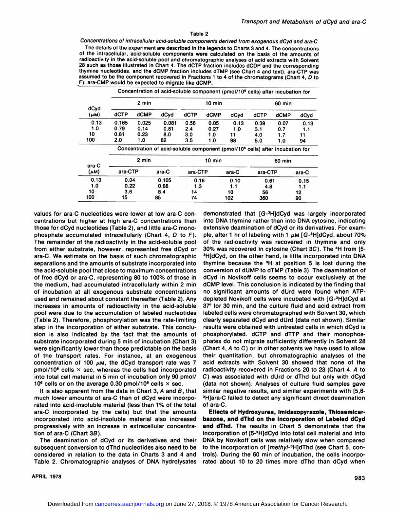
Table 2Concentrations of intracellular acid-soluble components derived from exogenous dCyd and ara-C
The details of the experiment are described in the legends to Charts 3 and 4. The concentrationsof the intracellular, acid-soluble components were calculated on the basis of the amounts ofradioactivity in the acid-soluble pool and Chromatographie analyses of acid extracts with Solvent28 such as those illustrated in Chart 4. The dCTP fraction includes dCDP and the correspondingthymine nucleotides, and the dCMP fraction includes dTMP (see Chart 4 and text). ara-CTP wasassumed to be the component recovered in Fractions 1 to 4 of the chromatograms (Chart 4, D toF); ara-CMP would be expected to migrate like dCMP.
Concentration of acid-soluble component (pmol/106 cells) after incubation for
2 min 10 min 60 mindCyd(MM)0.13
1.010
100dCTP0
002.165
.79
.81
.0dCMP0.025
0.140.23
1.0ConcentrationdCyd0.081
0.818.0
82dCTP0.58
2.43.03.5dCMP0.05
0.271.0
1.0ofacid-soluble componentdCyd0.13
1.011
98(pmol/106dCTP0.39
3.14.0
5.0cells)
afterdCMP0.07
0.71,71.0dCyd0.13
1.111
94incubation
for
2 min 10 min 60 minara-C(MM)0.13
1.010
100ara-CTP0.04
0.223.6
15ara-C0.105
0.888.4
85ara-CTP0.18
1.31474ara-C0.10
1.110
102ara-CTP0.61
4.856
360ara-C0.15
1.11290
values for ara-C nucleotides were lower at low ara-C concentrations but higher at high ara-C concentrations thanthose for dCyd nucleotides (Table 2), and little ara-C mono-
phosphate accumulated intracellularly (Chart 4, D to F).The remainder of the radioactivity in the acid-soluble pool
from either substrate, however, represented free dCyd orara-C. We estimate on the basis of such Chromatographie
separations and the amounts of substrate incorporated intothe acid-soluble pool that close to maximum concentrationsof free dCyd or ara-C, representing 80 to 100% of those in
the medium, had accumulated intracellularly within 2 minof incubation at all exogenous substrate concentrationsused and remained about constant thereafter (Table 2). Anyincreases in amounts of radioactivity in the acid-soluble
pool were due to the accumulation of labeled nucleotides(Table 2). Therefore, phosphorylation was the rate-limiting
step in the incorporation of either substrate. This conclusion is also indicated by the fact that the amounts ofsubstrate incorporated during 5 min of incubation (Chart 3)were significantly lower than those predictable on the basisof the transport rates. For instance, at an exogenousconcentration of 100 /¿M,the dCyd transport rate was 7pmol/106 cells x sec, whereas the cells had incorporated
into total cell material in 5 min of incubation only 90 pmol/106 cells or on the average 0.30 pmol/106 cells x sec.
It is also apparent from the data in Chart 3, A and B, thatmuch lower amounts of ara-C than of dCyd were incorporated into acid-insoluble material (less than 1% of the totalara-C incorporated by the cells) but that the amountsincorporated into acid-insoluble material also increased
progressively with an increase in extracellular concentration of ara-C (Chart 3ß).
The deamination of dCyd or its derivatives and theirsubsequent conversion to dThd nucleotides also need to beconsidered in relation to the data in Charts 3 and 4 andTable 2. Chromatographie analyses of DMA hydrolysates
demonstrated that [G-3H]dCyd was largely incorporated
into DNA thymine rather than into DNA cytosine, indicatingextensive deamination of dCyd or its derivatives. For example, after 1 hr of labeling with 1 /U.M[G-3H]dCyd, about 70%
of the radioactivity was recovered in thymine and only30% was recovered in cytosine (Chart 3C). The 3H from [5-3H]dCyd, on the other hand, is little incorporated into DNAthymine because the 3H at position 5 is lost during the
conversion of dUMP to dTMP (Table 3). The deamination ofdCyd in Novikoff cells seems to occur exclusively at thedCMP level. This conclusion is indicated by the finding thatno significant amounts of dUrd were found when ATP-depleted Novikoff cells were incubated with [G-3H]dCyd at37°for 30 min, and the culture fluid and acid extract from
labeled cells were chromatographed with Solvent 30, whichclearly separated dCyd and dUrd (data not shown). Similarresults were obtained with untreated cells in which dCyd isphosphorylated. dCTP and dTTP and their monophos-
phates do not migrate sufficiently differently in Solvent 28(Chart 4, A to C) or in other solvents we have used to allowtheir quantitation, but Chromatographie analyses of theacid extracts with Solvent 30 showed that none of theradioactivity recovered in Fractions 20 to 23 (Chart 4, A toC) was associated with dUrd or dThd but only with dCyd(data not shown). Analyses of culture fluid samples gavesimilar negative results, and similar experiments with [5,6-3H]ara-C failed to detect any significant direct deaminationof ara-C.
Effects of Hydroxyurea, Imidazopyrazole, Thiosemicar-
bazone, and dThd on the Incorporation of Labeled dCydand dThd. The results in Chart 5 demonstrate that theincorporation of [5-3H]dCyd into total cell material and into
DNA by Novikoff cells was relatively slow when comparedto the incorporation of [mefrty/-3H]dThd (see Chart 5, con
trols). During the 60 min of incubation, the cells incorporated about 10 to 20 times more dThd than dCyd when
APRIL 1978 983
on June 27, 2018. © 1978 American Association for Cancer Research. cancerres.aacrjournals.org Downloaded from

P. G. W. Plagemann et al.
Table 3Distribution of radioactivity in DNA hydrolysates from cells labeled with [G-3H]dCyd or
[5-3H]dCyd in the presence or absence of hydroxyurea or dThd
Samples of a suspension of 1 x 10⁷ N1S1-67 cells/ml of BM42B were supplementedwith 1 fj.M [5-3H]dCyd (250 cpm/pmol) or [G-3H]dCyd (230 cpm/pmol) and where indicatedwith 1 mM hydroxyurea or 0.1 mM dThd. After 60 min of incubation at 37°,samples of 4x 10⁷ cells were collected by centrifugation, the cells were washed, the acid-soluble poolwas extracted, and DNA hydrolysates were prepared from the residue and chromato-
graphed with Solvent 18.[5-3H]dCyd" [G-3H]dCyd
TreatmentNone
1 HIM hydroxyurea0.1 HIM dThdCyt"
(pmol/5x9.03
11.798Thy
10" cells)0.7
0.3<2Thy
(% of total)7
3<2Cyt
(pmol/5x1110
120Thy
106cells)294
10Thy
(% of total)72287
" The abbreviations used are: Cyt, cytosine; Thy, thymine.
Chart 5. Effects of dThd and various inhibitors of DNAreplication on the incorporation (INCORP.) of (A) [5-3H]-dCyd and (B) [3H]dThd into total cell material and acid-insoluble material by Novikoff rat hepatoma cells. Samplesof a suspension of 3 x 10" wild-type N1S1-67 cells/ml ofBM42B were supplemented where indicated with 0.5 mMhydroxyurea (HU), 50 /¿Mara-C, 0.4 mM imidazopyra-zole (IPZ). 75 ¡iM4-methyl-5-amino-1-formylisoquinolinethiosemicarbazone (TSC), or 0.1 mw dThd and 0.025 ^M[5-3H]dCyd (16,000 cpm/pmol) (4) or 0.025 ^M [3H]dThd(10,000 cpm/pmol) (B). The suspensions were incubatedat 37°,and duplicate 1-ml samples were analyzed forradioactivity into total cell material and acid-insolublematerial. Points, averages of the duplicate samples.
u•o
o.CEOuz i
u
cnOD
A[5-3HJDEOXYCYTIDINE
TOTAL IN CELL ACID-INSOLUBLE
—i 1 r
B. [s-3HJ THYMIDINE
TOTAL IN CELL ACID - INSOLUBLE
exposed to an identical substrate concentration in themedium (0.025 ¡J.M),which is well below the apparent Kmforthe respective incorporation systems (0.5 and 1.0 ¡J.M;Ref.37). As shown previously (36, 37), however, the incorporation of [5-3H]dCyd into total cell material was increased 4-to 6-fold almost immediately upon supplementing the medium with 0.5 mM hydroxyurea or 0.1 mM dThd (Chart 5/4).The addition of 0.4 mM imidazopyrazole or 0.075 mM 4-methyl-5-amino-1-formylisoquinoline thiosemicarbazonecaused a stimulation of dCyd incorporation similar to thatcaused by dThd or hydroxyurea, whereas 50 fj.M ara-C wasslightly inhibitory (Chart 5A). The addition of any of the 4drugs resulted in an almost immediate and complete inhibition of DNA synthesis, as indicated by the inhibition of theincorporation of dThd into acid-insoluble material (Chart56, right) in the absence of any effect on the initial rate ofincorporation of dThd into total cell material (Chart 58,left). Any curtailment of DNA replication, however, resultsin a rapid cessation of the net incorporation of dThd intothe nucleotide pool (Ref. 35; Chart 56, left). The maximum
60 0 30
TIME (MIN)
level of radioactivity in dTTP probably reflects the size ofthe dTTP pool because the dTTP pool turns over rapidly(35).
Hydroxyurea seems to inhibit DNA replication by inhibiting the reduction of purine ribonucleotides and thus causing a rapid depletion of intracellular dATP and dGTP (Refs.1, 43, and 49; Chart 1). The reduction of UDP and CDPseems little affected, at least in whole cells (see "Discussion"), since hydroxyurea treatment results in slight in
creases in dCTP and dTTP levels in mouse embryo fibro-blast cultures (43), L^, cells (1), and Chinese hamster ovarycells (49). The higher steady-state level of radioactivity inthe acid-soluble pool of hydroxyurea-treated than ara-C-treated Novikoff cells (Chart 50, left) probably reflectedsuch an elevation in dTTP concentration in these cells (36).
ara-C served as an internal control in these studies sinceit inhibits DNA synthesis without significantly affecting theintracellular levels of deoxynucleoside triphosphates (43). Itis phosphorylated to ara-CTP (Ref. 5 and 42; Chart 4D),which blocks DNA polymerase (13, 14, 25). Thus, as ex-
984 CANCER RESEARCH VOL. 38
on June 27, 2018. © 1978 American Association for Cancer Research. cancerres.aacrjournals.org Downloaded from

Transport and Metabolism of dCyd and ara-C
Chart 6. Effect of dThd on the incorporation (INCORP.)of [5-3H]dCyd by wild-type (WT) and dThd kinase-deficient(TK-) cells. Samples of about 2 x 10s cells of either sublineper ml of BM42B were supplemented with the indicated concentrations of dThd or dCyd and with 0.5 ßM[5-3H]dCyd(600 cpm/pmol). The suspensions were incubated at 37°andmonitored for radioactivity in total cell material and acid-insoluble material.
pected, ara-C also inhibited the incorporation of dCyd intoacid-insoluble material (Chart 5A, right). In contrast, hy-droxyurea, imidazopyrazole, and the thiosemicarbazonecaused slight increases in dCyd incorporation into acid-insoluble material, although to a lesser extent than diddThd (Chart 5A, right). The effects of imidazopyrazole andthe thiosemicarbazone mimicked in all respects those ofhydroxyurea (Chart 5). They most likely were also due to aninhibition of ribonucleotide reducÃase(Chart 1), since suchinhibition has been demonstrated in cell-free preparationsof cultured mammalian cells (Ref. 2; V. H. Bono and D.Abraham, personal communication).7 The slight stimulation
of dCyd incorporation into DNA by the 3 drugs, in spite ofthe inhibition of DNA synthesis, was probably due to amarked increase in specific radioactivity of dCTP, a consequence of the increased incorporation of isotopie dCyd intothe nucleotide pool (Chart 5A, left). However, the stimulation of dCyd incorporation into DNA (2- to 3-fold) was muchless than that of its incorporation into total cell material (upto 7-fold), whereas both incorporations were about equallystimulated by 0.1 mw dThd (about 7-fold).
The incorporation of [G-3H]dCyd into total material
(and into DNA in the case of dThd) was also stimulated byhydroxyurea and dThd, but somewhat less than [5-3H]dCyd
incorporation because the basic incorporation of radioactivity from [G-3H]dCyd was about 4 times higher than thatfrom [5-3H]dCyd (Table 3). This difference was due to the
deamination of dCMP to dUMP by dCMP aminohydrolaseand the further conversion of dUMP to dTMP (Chart 1),whereby the 3Hfrom [5-3H]dCyd is lost, whereas most of thelabel from [G-3H]dCyd is retained and thus recovered in
7 Progress Reports 5 and 7 submitted by Dr. L. L. Bennet, Jr., and Dr. R.W. Brockman of Southern Research Institute, Birmingham, Ala., to Divisionof Cancer Treatment under Contract NOI-CM-43780.
0 20 40
TIME (MIN)
thymine nucleotides. In fact, about the same amounts of[5-3H]dCyd and [G-3H]dCyd were incorporated into DNAcytosine (Table 3). This was also the case in hydroxyurea-and dThd-treated cells. dThd stimulated about 10-fold theincorporation of either [5-3H] or [G-3H]dCyd into DNA cyto
sine while about the same amounts were incorporated inhydroxyurea-treated as in untreated cells, in spite of theinhibition of DNA synthesis by the drug.
The presence of either hydroxyurea or dThd markedlyreduced the incorporation of [G-3H]dCyd into DNA thymine(Table 3). In the case of hydroxyurea, this effect probablyreflected the inhibition of DNA synthesis by the drug,whereas the effect of dThd probably resulted from a feedback inhibition of deamination of dCMP by dTTP (41) and/or from a mass action effect of dTMP on the conversion ofany dUMP formed to dTMP (Chart 1). However, the stimulation of dCyd incorporation into DNA cytosine could notbe accounted for only by an inhibition of the conversion ofdCMP to dTMP. None of the radioactivity from either [5-3H]-or from [G-3H]dCyd was incorporated into RNA (data notshown; see also Ref. 36).
The effects caused by dThd were probably due to anexpansion of the dTTP pool resulting from the incorporation of exogenous dThd by the cells. The dTTP pool size isa function of the dThd concentration in the medium (1, 3,20, 48). That the stimulation of dCyd incorporation by dThdrequires the formation of dTTP is indicated by the findingthat dThd had no stimulating effect on dCyd incorporationin a dThd kinase-deficient subline of Novikoff cells (Chart6B). Hydroxyurea, imidazopyrazole, and the thiosemicarbazone, on the other hand, stimulated dCyd incorporationin dThd kinase-deficient cells to about the same extent asin wild-type cells (data not shown).
The effect of dThd on [5-3H]dCyd incorporation by wild-
APRIL 1978 985
on June 27, 2018. © 1978 American Association for Cancer Research. cancerres.aacrjournals.org Downloaded from

P. G. W. Plagemannetal.
type cells as a function of dThd concentration is shown inChart 6A. A stimulation of dCyd incorporation was observedwith a dThd concentration as low as 0.01 HIM. The stimulation was maximal at 0.1 HIM. The lesser effects at higherconcentrations of dThd were probably due to an inhibitionof dCyd transport by dThd (Table 1). A similar effect wasobserved in dThd kinase-deficient cells (Chart GBJeft). Theinhibition of dCyd incorporation in dThd kinase-deficient
cells by dThd was far less than that caused by addition ofunlabeled dCyd (Chart 60). This was to be expected, sincethe major competition from unlabeled dCyd is at the dCydkinase step (Km = 5 to 15 /¿M;Refs. 7, 15, and 26), while
competition from dThd, which is not a substrate for mammalian dCyd kinase (9), is limited to the nucleoside transport step.
In other experiments it was shown that the incorporationof [5-3H]dCyd into total cell material by mouse L1210 leukemia cells, mouse L-cells, and Chinese hamster ovary cellsduring 60 min of incubation was stimulated about 6-, 3-,and 3-fold, respectively, by the presence of 1 HIM hydrox-
yurea or 0.1 mw dThd in the medium (data not shown).Effects of Pyrazofurin, Deazauridine, and Alanosine on
dCyd Incorporation. Pyrazofurin inhibits the de novo synthesis of pyrimidines (34), most likely by interfering with theconversion of orotidine 5'-monophosphate to UMP (Ref. 46;
Chart 1). Inhibition of cell replication is due exclusively tothis inhibition of pyrimidine synthesis since uridine completely reverses the effect (34). The toxicity of deazauridine,on the other hand, seems to involve the inhibition of theconversion of UTP to CTP, and the inhibition is reversed bycytidine (Refs. 4 and 23; Chart 1). The toxicity of alanosine
is due to an inhibition of the conversion of IMP to AMP andis prevented by the presence of adenine in the medium(Ref. 12; Chart 1). Thus the treatment of cells with pyrazo-
furin results in the depletion of the cells of UTP, CTP, dTTP,and dCTP; treatment with deazauridine of CTP and dCTP;and treatment with alanosine of ATP and dATP.
Pyrazofurin, deazauridine, and alanosine had little or noimmediate effect on dCyd incorporation (Chart 7A), butafter a 4-hr preincubation with the inhibitors, on the otherhand, [5-3H]dCyd incorporation into total cell material aswell as into acid-insoluble material was markedly stimulated(Chart 7B), most likely because of a depletion of intracellu-
lar dCTP in the case of pyrazofurin and deazauridine anddepletion of dATP in alanosine-treated cells. Chromato
graphie analyses showed that the increased radioactivity inthe acid-soluble pool was associated mainly with dCTP and
dCMP (Chart 7C). The lag in the effect of these inhibitorswas not unexpected, since they affect dCTP and dATP concentrations only by inhibiting the formation of their ribo-
nucleotide precursors (Chart 1) that are present in at least100-fold higher concentrations (33, 44, 49).
Effect of Various Drugs on ara-C Incorporation. Theincorporation of ara-C into total cell material was markedlystimulated by hydroxyurea, imidazopyrazole, and 4-methyl-5-amino-1-formylisoquinoline thiosemicarbazone (Chart
BA). Treatment of the cells with pyrazofurin, deazauridine,or alanosine had a similar effect (Chart 86). In fact, thestimulation of ara-C incorporation by the latter 3 drugs
became apparent more rapidly than did that of dCyd incorporation. In each case the increased incorporation into theacid-soluble pool reflected an increased formation of ara-
O-CONTROL
IO
PYRAZOFURIN
IO 20
TIME (MIN) FRACTION NUMBERChart 7. Effect of pyrazofurin, deazauridine, and alanosine on the incorporation (INCORP.} of [5-3H]dCyd by wild-type Novikoff cells. Samples of a
suspension of 2 x 10" cells/ml of BM42B were supplemented where indicated with inhibitors and incubated at 37°.At Ohr (Ai and after 4 hr of incubation at37°(B) samples of each suspension were supplemented with 0.5 MM[5-3H]dCyd (340 cpm/pmol), incubated at 37°,and monitored for radioactivity in total cellmaterial and acid-insoluble material. After 60 min of labeling, acid extracts were prepared from about 1 x 10' cells and chromatographed with Solvent 28.Chromatograms of acid extracts from cells labeled after a 4-hr pretreatment (B) are illustrated in C.
986 CANCER RESEARCH VOL. 38
on June 27, 2018. © 1978 American Association for Cancer Research. cancerres.aacrjournals.org Downloaded from

Transport and Metabolism of dCyd and ara-C
Chart 8. Effect of various inhibitors anddThd on the incorporation of [5-3H]ara-C.Samples of a suspension of 2.6 x 10scells/ml (A) or 2 x 10»cells/ml (B) were
supplemented where indicated with 100 ¿<MdThd, 1 mM hydroxyurea (HU). 200 ^Mimidazopyrazole (IPZ), 75 ^M 4-methyl-5-amino-1-formylisoquinoline thiosemicarba-
zone (TSC), 20 fit» pyrazofurin, 100 ¿iMdeazauridine, or 13.6 ¿iMalanosine. At theindicated times of incubation at 37°,sam
ples of each suspension were supplemented with 0.033 MM [5-3H]ara-C (10,000cpm/pmol), incubated at 37°, and moni
tored for radioactivity in total cell material.Replicate samples were analyzed for radioactivity in acid-insoluble material (data notshown; see text). After 60 min of incubation, acid extracts were prepared from 1 x10⁷ cells and analyzed chromatographically
(C).
2o.<J
Ulu
Q-(£OCJ
O HR
tdThd
CONTROL
OHR
O-CONTROL
•-+20pM
PYRAZOFURINÛ--HOOMM
DEAZAURIDINE
*-+l36pMALANOSINE
4HR
30 60 30 60 30 60
TIME (MIN )
O IO 20
FRACTION NUMBER
CTP (Chart 8C). Pyrazofurin, however, also caused a veryrapid marked increase in ara-C incorporation into DMA.During the first 60-min labeling period, the amount of ara-Cincorporated into DNA was increased to 7000 cpm/2 x 106
cells from 450 cpm for the untreated controls. The presenceof deazauridine had no effect on the ara-C incorporationinto DNA during this labeling period, but the amount incorporated was about twice that of the controls during thesecond labeling period at 4 hr. Alanosine had no effect onara-C incorporation into DNA during either period.
In contrast to its stimulation of dCyd incorporation, dThdhad only little effect on ara-C incorporation into total cellmaterial (Chart 8A), although the amount incorporated intoDNA during 60 min of incubation was increased to 1080cpm/2 x 106 cells from 450 cpm for the untreated control.
Hydroxyurea and imidazopyrazole reduced the incorporation into DNA to 70 cpm and the thiosemicarbazone reduced it to 35 cpm/2 x 106 cells.
Although the amounts of dCyd and ara-C incorporated bythe cells increased with an increase in exogenous substrateconcentration (Chart 3), the extent to which this incorporation could be enhanced was independent of substrateconcentration. The effects of dThd, hydroxyurea, and imidazopyrazole on either [5-3H]dCyd or [5-3H]ara-C incorpo
ration were about the same whether the substrates werepresent in the medium at a concentration of 0.033 or 10 /AM(data not shown).
DISCUSSION
These results support our previous tentative conclusionthat dCyd enters Novikoff cells mainly via a single, common,high-Km, nucleoside-facilitated diffusion system (22, 51, 52)that seems to transport all ribo- and deoxyribonucleosidesincluding ara-C, but with somewhat different efficiency(Ref. 52; R. M. Wohlhueter, R. März,and P. G. W. Plage
mann, (in preparation). Our Km's for dCyd and ara-C trans
port fall in the same range as those reported for dCyd andara-C uptake by ara-C-resistant (dCyd kinase-deficient)Yoshida sarcoma cells (Ref. 30; Km, 400 to 600 /¿M),butour Vmax'sare at least 10 times higher than those reported
for the Yoshida sarcoma cells. We have also not detectedin Novikoff cells a second transport system with a Km ofabout 2 mM reported for Yoshida cells. There is, however,some question as to the validity of the initial transportrates estimated in the study with Yoshida sarcoma cells,since they were based on 2 time points (15 and 120 sec),and it is clear from our more detailed analyses that thereis virtually no net flux of substrate after about 60 sec at25°,even at a substrate concentration of 1.28 mM. Our
results conflict also with those of Kessel and Shurin (16)who reported a Kmof 7.5 mM for dCyd-ara-C transport indCyd kinase-deficient L1210 cells at 0°and who reported
that facilitated transport did not seem to occur at a physiological temperature.
The rates of transport of dCyd and ara-C in Novikoff cellsare much higher than those of their phosphorylation, evenat substrate concentrations as low as 0.025 /¿M,so that theintracellular concentration of free substrate approachesthat in the medium within 2 min of incubation. Thus thephosphorylation step is clearly rate limiting in the overallincorporation of dCyd or ara-C into the nucleotide pool,and incorporation rates estimated from time points ofseveral min clearly reflect the rate of phosphorylation ratherthan that of transport. This pertains also to the kineticparameters of incorporation estimated from such long-termtime points (37, 39). These findings are of some interest inthe chemotherapeutic use of ara-C. These results indicatethat a low efficiency of incorporation of ara-C into thenucleotide pool does not result from slow transport into thecell but rather from a low rate of phosphorylation, mostprobably resulting from feedback inhibition of the dCyd
APRIL 1978 987
on June 27, 2018. © 1978 American Association for Cancer Research. cancerres.aacrjournals.org Downloaded from

P. G. W. Plagemannetal.
kinase (9, 15, 26) by intracellular dCTP formed via thèdenovo synthesis pathway. In fact, the steady-state concentration of dCTP in several lines of cultured mammalian cells,including some used in this study, have been reported to be2 to 4 times higher than those of the other 3 deoxynucleo-side triphosphates, provided the medium does not containany dThd (43, 44, 48, 49), although this has not been auniversal finding (1, 21). The inhibition of dCyd kinase bydCTP involves changes in both Km and Vmax(15). Theamounts of dCyd or ara-C incorporated by the cells, nevertheless, increase with increase in their concentration in themedium and, when present in the /U.Mrange, sufficient ara-C becomes phosphorylated to cause a complete inhibitionof DNA synthesis and cell replication. At lower concentrations, on the other hand, cytotoxicity might be limited dueto the low amounts incorporated into the nucleotide pool.The conversion of dCyd into nucleotides becomes saturatedat concentrations between 1 and 5 ¡¿M(Ref. 37; Table 2),but the incorporation of ara-C into nucleotides (Table 2)and into DNA (Chart 2B) increases in about direct proportion to the ara-C concentration in the medium up to at least10 to 100 /¿M.This difference probably reflects the muchhigher Kmof dCyd kinase for ara-C than for dCyd (18, 26)and might also be a factor in influencing the relativecytotoxicity of ara-C.
The regulatory constraint on the phosphorylation of dCydand ara-C is relieved by treatment of the cells with a numberof substances that affect the synthesis of various nucleosidetriphosphates, particularly of dCTP. As depicted in Chart 1,the intracellular pool of dCTP is fed both by reduction ofCDP to dCDP plus subsequent phosphorylation (de novosynthesis) and by phosphorylation of dCyd, if available(salvage). Thus, the absolute size of the dCTP pool and itsspecific radioactivity in the presence of isotopie dCyd aredetermined by the relative contributions of the cÃenovo andsalvage pathways. The activity of dCyd kinase is inhibitedby dCTP, but this inhibition is reversed by dTTP, whereasthe reduction of CDP to dCDP is inhibited by both dTTPand dATP. The operation of this regulatory scheme in vivoprovides an explanation for most of the phenomena wehave reported here. Thus, dThd probably stimulates dCydincorporation by causing an increase in the intracellularconcentration of dTTP, which in turn inhibits the reductionof CDP to dCDP and thus the de novo synthesis of dCTP(24, 40) but also might further counteract the feedbackinhibition of dCyd kinase by dCTP (15). An increase in dTTPconcentration with a concomitant decrease in dCTP concentration during incubation with dThd has been demonstrated to exist in a number of cell systems (1, 3, 20, 47, 49)and to result in an inhibition of DNA synthesis (3, 20, 24, 29,48).
Similarly, the stimulatory effects of deazauridine andpyrazofurin on dCyd and ara-C incorporation are probablyrelated to a decrease in dCTP production de novo, resultingfrom an inhibition of the UTP to CTP conversion (4, 23) orof de novo pyrimidine synthesis by pyrazofurin (34), respectively. A stimulation of dCyd incorporation by deazauridinehas also been observed in mouse L1210 cells (4).
The mechanism by which hydroxyurea and the otherinhibitors of ribonucleotide reducÃaseused here stimulatedCyd and ara-C incorporation is more uncertain. Hydroxy
urea inhibits the reduction of CDP and of other ribonucleo-tides by cell-free preparations of Escherichia coli (17) andNovikoff cells (27), and imidazopyrazole actually inhibitsthe reduction of CDP more than that of the other ribonu-cleoside diphosphates (V. H. Bono and D. Abraham, personal communication)7 but hydroxyurea treatment of whole
animal cells results in a rapid loss of only dATP and dGTP(1, 43, 49). The reason for this discrepancy between the invivo and in vitro results is not known. It may be related tothe existence of separate enzymes or catalytic subunits of asingle complex for the reduction of purine and pyrimidinenucleotides (8, 31). Nevertheless, an increase in [3H]dCydincorporation in the presence of expanding dCTP and dTTPpools has also been observed in X-irradiated human kidney(45) and Chinese hamster cells (47). This increased dCydincorporation has been attributed to an X-ray inactivationof dCMP aminohydrolase (47), but it is unclear how aninhibition of dCMP aminohydrolase could lead to an expansion of the dCTP pool except if one assumes that normallya large proportion of the dCDP synthesized de novo isdegraded to dCMP and deaminated. Our results indicatethat dThd markedly inhibits the conversion of dCMP todTMP, but the stimulation of [5-3H]dCyd incorporation intothe nucleotide pool by dThd or hydroxyurea cannot beaccounted for by an inhibition of dCMP deamination sinceit is also observed with [G-3H]dCyd. Its deamination andsubsequent conversion products are also part of thedeoxynucleotide pool and are incorporated via dTTP intoDNA.
The stimulation of dCyd and ara-C incorporation byribonucleotide reducÃaseinhibitors could be due to a reversal of the dCTP feedback inhibilion of dCyd kinase by theexpanded dTTP pool concentralion. Insufficienl kinelicdala are available for Ihe syslem lo evalúalethis possibility.It seems more likely, however, thai Ihe effecls of Iheseinhibilors are relaled lo a depletion of Ihe cells of dATP.This conclusion is suggesled by Ihe finding lhal Irealmentof the cells with alanosine also causes a simulation of dCydincorporalion. How a decrease in dATP concenlralion couldcause an enhancemenl of dCyd incorporalion is noi clearsince dATP has no effecl on dCyd kinase per se (15) and isa universal inhibilor of Ihe reduclion of all 4 ribonucleosidediphosphales in bolh mammalian cells (19, 20) and E. coli(40).
The finding lhal various inhibilors of ribonucleolide re-ductase and of the de novo synthesis of pyrimidine nucleotides enhance the incorporalion of ara-C inlo the nucleotidepool may be of practical interesl in cancer chemolherapy.Addilional inleresl arises from Ihe faci lhal dThd, deazauridine, and particularly pyrazofurin markedly slimulate theincorporation of ara-C into DNA. II is conceivable, Ihere-fore, lhal such combined Irealmenls might enhance thetoxicily of ara-C for lumor cells.
ACKNOWLEDGMENTS
The authors thank Marsha Behrens and John Erbe for excellent technicalassistance and Cheryl Thull for competent secretarial work.
REFERENCES
1. Adams, R. L. P., Berryman, S., and Thomson, A. DeoxyribonucleosideTriphosphate Pools in Synchronized and Drug-Inhibited L929 cells.Biochim. Biophys. Acta, 240. 455-462, 1971.
988 CANCER RESEARCH VOL. 38
on June 27, 2018. © 1978 American Association for Cancer Research. cancerres.aacrjournals.org Downloaded from

Transport and Metabolism of dCyd and ara-C
2. Agrawal, K. C., Mooney, P. D., and Sartorelli, A. C. Potential AntitumorAgents. 13. 4-Methyl-5-amino-1-formylisoquinoline Thiosemicarbazone.J. Med. Chem., 19: 970-972. 1976.
3. Bjursell, G., and Reichard, P. Effects of Thymidine on Deoxyribonucleo-side Triphosphate Pools and Deoxyribonucleic Acid Synthesis inChinese Hamster Ovary Cells. J. Biol. Chem., 248. 3904-3909, 1973.
4. Brockman, R. W., Shaddix, S. C., Williams, M., Nelson, J. A., Rose, L.M., and Schabel. F. M., Jr. The Mechanism of Action of 3-Deazauridine
in Tumor Cells Sensitive and Resistant to Arabinosylcytosine. Ann. N. Y.Acad. Sci., 255. 501-521, 1975.
5. Chu, M. Y., and Fischer, G. A. Comparative Studies of Leukemic CellsSensitive and Resistant to Cytosine Arabinoside. Biochem. Pharmacol.,14: 333-341, 1965.
6. Chu, M. Y., Hoovis, M. L., and Fischer. G. A. Effects of 5-Fluorodeoxy-uridine on Cell Viability and Uptake of Deoxycytidine and [3H]CytosineArabinoside in L5178Y Cells. Biochem. Pharmacol., 25. 355-357, 1976.
7. Coleman, C. N., Stoller, R. G., Drake, J. C., and Chabner, B. A.Deoxycytidine Kinase: Properties of the Enzyme from Human LeukemicGranulocytes. Blood, 46. 379-391, 1975.
8. Cori, J. G., and Mansell, M. M. Comparison of the Cytidine 5'-diphos-phate and Adenosine 5'-diphosphate ReducÃase Activities of Mammalian
Ribonucleotide ReducÃase. Cancer Res., 35. 2327-2331, 1975.
9. Durham, J. P., and Ivés,D. H. Deoxycytidine Kinase III. Purification andGeneral Properties of the Calf Thymus Enzyme. J. Biol. Chem., 245:2276-2284, 1970.
10. Eilam, Y., and Stein, W. D. Kinetic Studies of Transport across RedBlood Cell Membranes. Methods Membrane Biol., 2. 283-354, 1974.
11. Gentry, G. A., Morse, P. A., Jr., Ivés,D. H., Gebert, R., and Potter, V. R.Pyrimidine Metabolism in Tissue Culture Cells Derived from Rat Hepa-tomas. II. Thymidine Uptake in Suspension Cultures Derived fromNovikoff Hepatoma. Cancer Res., 25. 509-516, 1965.
12. Graff, J. C., and Plagemann, P. G. W. Alanosine Toxicity in Novikoff RatHepatoma Cells Due to Inhibition of the Conversion of Inosine Mono-phosphate to Adenosine Monophosphate. Cancer Res., 36. 1428-1440,1976.
13. Graham, F. L., and Whitmore, G. F. Studies in Mouse L-cells on theIncorporation of 1-/3-D-Arabinofuranosylcytosine into DNA and on Inhibition of DNA Polymerase by l-^-D-ArabinofuranosylcytosineS'-Triphos-phate. Cancer Res., 30. 2636-2644, 1970.
14. Inagaki, T., Nakamura, T., and Wakisaka, C. Studies on the Mechanismof Action of 1-/3-D-Arabinofuranosylcytosine as an Inhibitor of DNASynthesis in Human Leukemic Leukocytes. Cancer Res., 29: 2169-2176,1969.
15. Ivés,D. H., and Durham, J. P. Deoxycytidine Kinase III. Kinetic andAllosteric Regulation of the Calf Thymus Enzyme. J. Biol. Chem., 245:2285-2294, 1970.
16. Kessel, D., and Shurin, S. B. Transport of Two Non-Metabolized Nucleo-sides, Deoxycytidine and Cytosine Arabinoside, in a Sub-Line of theL1210 Murine Leukemia. Biochim. Biophys. Acta, J63: 179-187, 1968.
17. Krakoff, l. H., Brown, N. C., and Reichard, P. Inhibition of Ribonucleo-side Diphosphate Reductase by Hydroxyurea. Cancer Res., 28: 1559-1565, 1968.
18. Krenitsky, T. A., Tuttle, J. V., Koszalka. G. W.. Chen, I. S., Beacham, L.M., Ill, Rideout, J. L., and Elion, G. B. Deoxycytidine Kinase from CalfLiver. Substrate and Inhibitor Specificity. J. Biol. Chem., 257: 4055-4061, 1976.
19. Larsson, A. Ribonucleotide Reductase from Regenerating Rat Liver. II.Substrate Phosphorylation Level and Effect of Deoxyadenosine Triphosphate. Biochim. Biophys. Acta, 324: 447-451, 1973.
20. Lowe, J. K., and Grindey, G. B. Inhibition of Growth Rate and Deoxynu-cleoside Triphosphate Concentrations in Cultured Leukemia L1210Cells. Mol. Pharmacol., 12: 177-184, 1976.
21. Maley, F., and Maley, G. F. On the Nature of a Sparing Effect ofThymidine on the Utilization of Deoxycytidine. Biochemistry, 7: 847-851,1962.
22. März, R., Wohlhueter, R. M., and Plagemann, P. G. W. Relationshipbetween Thymidine Transport and Phosphorylation in Novikoff RatHepatoma Cells as Analyzed by a Rapid Sampling Method. J. Supramol.Struct.,6: 433-440. 1977.
23. McPartland, R. P., Wang, M. C., Bloch, A., and Weinfeld, H. Cytidine 5'-
Triphosphate Synthetase as a Target for Inhibition by the AntitumorAgent 3-Deazauridine. Cancer Res., 34. 3107-3111, 1974.
24. Meuth, M., and Green, H. Alterations Leading to Increased Ribonucleotide Reductase in Cells Selected for Resistance to Deoxynucleosides.Cell, 3: 367-374, 1974.
25. Momparler, R. L. Kinetic and Template Studies with 1-/3-D-Arabinofura-nosylcytosine S'-Triphosphate and Mammalian DNA Polymerase. Mol.Pharmacol., 8: 362-379, 1972.
26. Momparler, R. L., and Fischer, G. A. Mammalian Deoxycytidine Kinase.I. Deoxycytidine Kinase: Purification, Properties and Kinetic Studies withCytosine Arabinoside. J. Biol. Chem., 243. 4298-4304, 1968.
27. Moore. E. C. The Effects of Ferrous Ion and Dithioerythritol on Inhibitionby Hydroxyurea of Ribonucleotide Reductase. Cancer Res..29: 291-295.
1969.28. Moore. E. C., and Hurlbert, R. B. Regulation of Mammalian Deoxyribo-
nucleotide Biosynthesis by Nucleotides as Activators and Inhibitors. J.Biol. Chem., 247: 4802-4809, 1966.
29. Morris, N. R., and Fischer, G. A. Studies Concerning Inhibition of theSynthesis of Deoxycytidine by Phosphorylated Derivatives of Thymidine.Biochim. Biophys. Acta, 42: 183-184, 1960.
30. Mulder, J. H., and Harrap, K. R. Cytosine Arabinoside Uptake by TumorCells in Vitro. European J. Cancer, 77: 373-379, 1975.
31. Peterson, D. M., and Moore, E. C. Independent Fluctuations of Cytidineand Adenosine Diphosphate Reductase Activities in Cultured ChineseHamster Fibroblasts. Biochim. Biophys. Acta, 432: 80-91, 1976.
32. Plagemann, P. G. W. Nucleotide Pools of Novikoff Rat Hepatoma CellsGrowing in Suspension Culture. I. Kinetics of Incorporation of Nucleo-sides into Nucleotide Pools and Pool Sizes during Growth Cycle. J.Cellular Physiol., 77: 213-240, 1971.
33. Plagemann, P. G. W. Nucleotide Pools in Novikoff Rat Hepatoma CellsGrowing in Suspension Culture. II. Effects of Nucleosides in Medium onLevels of Nucleotides in Separate Nucleotide Pools for Nuclear andCytoplasmic RNA Synthesis. J. Cell Biol., 52: 131-146, 1972.
34. Plagemann, P. G. W.. and Behrens, M. Inhibition of de novo PyrimidineNucleotide and DNA Synthesis and Growth of Cultured Novikoff RatHepatoma Cells and Other Cell Lines by Pyrazofurin (NSC 143095).Cancer Res., 36: 3807-3812, 1976.
35. Plagemann, P. G. W., and Erbe, J. Thymidine Transport by CulturedNovikoff Rat Hepatoma Cells and Uptake by Simple Diffusion andRelationship to Incorporation into Deoxyribonucleic Acid. J. Cell Biol.,55: 161-178, 1972.
36. Plagemann, P. G. W., and Erbe, J. Intracellular Conversions of Deoxy-ribonucleosides by Novikoff Rat Hepatoma Cells and Effects of Hydroxyurea. J. Cellular Physiol., 83: 321-336, 1974.
37. Plagemann, P. G. W., and Erbe, J. The Deoxyribonucleoside TransportSystems of Cultured Novikoff Rat Hepatoma Cells. J. Cellular Physiol.,83. 337-344, 1974.
38. Plagemann, P. G. W.. März, R., and Erbe. J. Transport and Counter-transport of Thymidine in ATP-Depleted and Thymidine Kinase-DeficientNovikoff Rat Hepatoma and Mouse L Cells: Evidence for a High K„Facilitated Diffusion System with Wide Nucleoside Specificity. J. CellularPhysiol.,89: 1-18, 1976.
39. Plagemann, P. G. W., and Richey, D. P. Transport of Nucleosides,Nucleic Acid Bases, Choline and Glucose by Animal Cells in Culture.Biochim. Biophys. Acta, 344: 263-305, 1974.
40. Reichard, P. Control of Deoxyribonucleotide Synthesis in Vivo and inVitro. Adv. Enzyme Regulation, 70. 3-16, 1972.
41. Scarano, E., Gerazi, G., Pulzella, A., and Campanile, E. The EnzymaticAminohydrolysis of 4-Amino Pyrimidine Deoxyribonucleotides. J. Biol.Chem.,238. PC1556-1557, 1963.
42. Schrecker, A. W., and Urshel, M. J. Metabolism of 1-/3-r>Arabinofurano-sylcytosine in Leukemia L1210 Studies with Intact Cells. Cancer Res.,28: 793-801. 1968.
43. Skoog. L.. and Nordenskjöld, B. Effects of Hydroxyurea and 1-/3-D-Arabinofuranosylcytosine on Deoxyribonucleotide Pools in Mouse Embryo Cells. European J. Biochem., 79: 81-89, 1971.
44. Skoog, K. L., Nordenskjöld, B. A., and Bjursell, K. G. Deoxyribonucleoside Triphosphate Pools and DNA Synthesis in Synchronized HamsterCells. European J. Biochem., 33: 428-432, 1973.
45. Smets, L. A. Discrepancies between Precursor Uptake and DNA Synthesis in Mammalian Cells. J. Cellular Physiol., 74: 63-66. 1969.
46. Sweeney, M. J., Davis, F. A., Gutowski, G. E., Hamill, R. L., Hoffman, D.H., and Poore, G. A. Experimental Antitumor Activity of Pyrazomycin.Cancer Res., 33: 2619-2623. 1973.
47. Walters, R. A., Gurley, L. R., Tobey, R. A., Enger, M. D., and Ratliff, R.L. Effects of X-lrradiation on DNA Precursor Metabolism and Deoxyribonucleoside Triphosphate Pools in Chinese Hamster Cells. RadiationRes., 60. 173-201, 1974.
48. Walters, R. A., and Ratliff, R. L. Lack of Specific Correlation of theDeoxycytidine Triphosphate Pool Level with Rate of DNA Synthesis.Biochim. Biophys. Acta, 474: 221-230, 1975.
49. Walters, R. A., Tobey, R. A., and Ratliff. R. L. Cell Cycle DependentVariations of Deoxyribonucleoside Triphosphate Pools in Chinese Hamster Cells. Biochim. Biophys. Acta, 379: 336-347, 1973.
50. Ward, G. A., and Plagemann, P. G. W. Fluctuations of DNA-DependentRNA Polymerase and Synthesis of Macromolecules during the GrowthCycle of Novikoff Hepatoma Cells in Suspension Culture. J. CellularPhysiol., 73: 213-231, 1969.
51. Wohlhueter, R. M., Marz, R., Graff, J. C., and Plagemann, P. G. W. TheApplication of Rapid Kinetic Techniques to the Transport of Thymidineand 3-O-Methylglucose into Mammalian Cells in Suspension Culture. J.Cellular Physiol., 89. 605-612, 1976.
52. Wohlhueter, R. M., Marz, R., Graff, J. C., and Plagemann, P. G. W. ARapid Mixing Technique to Measure Transport in Suspended AnimalCells: Application to Nucleoside Transport in Novikoff Rat HepatomaCells. Methods Cell Biol., 20: 211-236, 1978.
APRIL 1978 989
on June 27, 2018. © 1978 American Association for Cancer Research. cancerres.aacrjournals.org Downloaded from

1978;38:978-989. Cancer Res Peter G. W. Plagemann, Richard Marz and Robert M. Wohlhueter Regulation of Triphosphate SynthesisHepatoma Cells, Relationship to Phosphorylation, and -d-Arabinofuranosylcytosine into Cultured Novikoff Rat
βTransport and Metabolism of Deoxycytidine and 1-
Updated version
http://cancerres.aacrjournals.org/content/38/4/978
Access the most recent version of this article at:
E-mail alerts related to this article or journal.Sign up to receive free email-alerts
Subscriptions
Reprints and
To order reprints of this article or to subscribe to the journal, contact the AACR Publications
Permissions
Rightslink site. Click on "Request Permissions" which will take you to the Copyright Clearance Center's (CCC)
.http://cancerres.aacrjournals.org/content/38/4/978To request permission to re-use all or part of this article, use this link
on June 27, 2018. © 1978 American Association for Cancer Research. cancerres.aacrjournals.org Downloaded from