transplantatoni from deceased donors after circulatory death · · 2018-03-192 executive summary...
TRANSCRIPT
The Voice of Transplantation in the UK
Transplantation from deceased donors after circulatory death
Compiled by a Working Party of The British Transplantation Society July 2013
British Transplantation Society Guidelines
www.bts.org.uk© British Transplantation Society

2
CONTENTS
1 INTRODUCTION 4 1.1 The Need for Guidelines 4 1.2 Process of Writing and Methodology 5 1.3 Editorial Committee 6 1.4 Contributors and Collaborators 7 1.5 Disclaimer 9 1.6 Declarations of Interest 9 1.7 Grading of Recommendations 9 1.8 Definitions and Abbreviations 10
2 EXECUTIVE SUMMARY OF RECOMMENDATIONS 13
PART 1: GENERAL PRINCIPLES
3 CATEGORISATION OF DCD DONORS & DEFINITION OF WARM 21 ISCHAEMIC TIME
3.1 Categorisation of DCD Donors 21 3.2 Nomenclature of Time Periods 22
4 DIAGNOSIS OF DEATH 25 4.1 Professional Frameworks for the Diagnosis and Confirmation of Death 25 4.2 Biological Background to Death that Follows Permanent Loss of Circulatory 26 Function 4.3 Diagnosis of Death 27 4.4 Decision to Withdraw Futile Life Sustaining Treatments 30
5 LAW, ETHICS AND DONOR CONSENT 31 5.1 Ethical Considerations in DCD Transplantation 31 5.2 Patient Autonomy and the Choice to Donate 32 5.3 Donor Distress and Rights after Death 34 5.4 Uncontrolled DCD Donation 35 5.5 Organ Quality and Recipient Risk 36 5.6 Patient Choice 38 5.7 BTS Ethics Committee 38
6 INFORMING THE RECIPIENT 40
7 ORGAN RETRIEVAL 42 7.1 The Procedure of Organ Recovery 43 7.2 Preservation Solutions 49 7.3 Staffing the Retrieval Procedure 50 7.4 Abdominal Organs: Specific Procedures 51 7.5 Thoracic organs: Specific Procedures 56

3
PART 2: ORGAN SPECIFIC DISCUSSION
8 KIDNEY 61 8.1 Introduction 62 8.2 Donor Selection 63 8.3 Organ Preservation 64 8.4 Organ Quality Assessment 65 8.5 Recipient Selection 68 8.6 Immunosuppression 69 8.7 Outcome 71
9 LIVER 76 9.1 Introduction 77 9.2 Donor Selection 80 9.3 Organ Preservation 80 9.4 Organ Quality Assessment 81 9.5 Recipient Selection 82 9.6 Immunosuppression 83 9.7 Outcome 84
10 PANCREAS 91 10.1 Donor Selection 92 10.2 Organ Preservation 94 10.3 Organ Quality Assessment 94 10.4 Recipient Selection 95 10.5 Immunosuppression 95 10.6 Outcome 96
11 PANCREATIC ISLETS 100 11.1 Donor Selection 100 11.2 Organ Preservation 101 11.3 Organ Quality Assessment 102 11.4 Recipient Selection 102 11.5 Immunosuppression 102 11.6 Outcome 102
12 LUNG 105 12.1 Donor Selection 105 12.2 Organ Preservation 108 12.3 Organ Quality Assessment 108 12.4 Recipient Selection 109 12.5 Immunosuppression 109 12.6 Outcome 110
13 HEART 114 13.1 Donor Selection 114 13.2 Organ Retrieval 114 13.3 Organ Preservation 115 13.4 Organ Quality Assessment 115 13.5 Recipient Selection 115 13.6 Immunosuppression 116 13.7 Outcome 116
14 PAEDIATRIC DCD TRANSPLANTATION 117

4
1 INTRODUCTION
1.1 The Need for Guidelines
Transplantation offers patients with end-stage organ failure a cost-effective treatment that
improves quality of life and increases life expectancy. Prior to the introduction of guidance
defining the concept of brain death in the 1970s, all organs for transplantation were donated
after circulatory death (DCD). Following the introduction of brain stem death testing, the
majority of organs for transplantation were donated after brain death (DBD) or, increasingly,
from living donors. However, in the UK, demand for organ transplantation has continued to
rise. By 2012, the mean waiting time for deceased donor kidney transplantation had risen to
over three years, while demand for other organs far outstripped the available supply with the
consequence that many patients died while awaiting an organ transplant.
In an attempt to bridge the gap between supply and demand, a concerted effort has been
made over the last decade to re-evaluate the use of DCD organs, and this has resulted in an
increase in the number of DCD donors. Following a report from the Organ Donor Taskforce
in 2008 (1), 14 initiatives were put in place with the hope of increasing organ donation by
50% over the following five years. Partly as a result, the number of DCD donors increased
from 199 to 373 between 2007 and 2012, an increase of 87%. Overall, this constituted an
increase of 11% of the total deceased donor activity (2).
DCD provides a valuable source of organs for transplantation and helps to address the
shortfall between supply and demand. It also offers an additional donation choice for families
of critically ill patients who are suffering from severe injury or illness that is incompatible with
life but have not deteriorated to brain death. In this situation, DCD can facilitate both patient
and family wishes at the end of life, especially when life-support is to be withdrawn. DCD has
been perceived by some as an attempt to circumvent brain death criteria, but a more
balanced interpretation is that it may play an important role in satisfying the wishes of the
patient and/or family where DBD is not possible.
Despite some historical success with ‘uncontrolled’ DCD kidney programmes, only a small
percentage of DCD donors in the UK are currently uncontrolled, largely because of the
interventions that are necessary to maintain the viability of transplantable organs while
consent/authorisation for donation is sought from the deceased’s next of kin. The majority of
UK DCD donations are ‘controlled’ Maastricht category 3. Efforts to expand donation are
likely to lead to increased ‘uncontrolled’ DCD activity (Maastricht category 2 or even 1).

5
DCD donation results in fewer donated organs per donor than DBD donation (2.6 organs
compared to 4). DCD donors are able to donate kidney, liver, pancreas, lung, and multiple
tissues, and research in the UK is exploring the options for cardiac DCD following the
successful development of such programmes in the USA (3).
Improved immunosuppression, improved organ recovery and implantation, and enhanced
peri- and post-operative care mean that the outcomes from DCD organs compare favourably
with those from DBD organs. Success is dependent upon establishing common practices
and accepted protocols that allow the safe sharing of DCD organs and maximise the use of
the DCD donor pool. Careful recipient selection and optimal donor management are pivotal
to facilitate donation of as many organs as possible and it is essential that the allocation
system accounts for recipient need and organ utilisation to maximise transplant benefit.
Guidelines for transplantation from deceased circulatory death donors were first published
by the British Transplantation Society in 2004 under the title ‘Guidelines relating to solid
organ transplants from non-heart beating donors’ (4). This document extends and updates
the previous guidelines and aims to include recent developments in all aspects of DCD
clinical practice, including outcome data for all relevant organs.
1.2 Process of Writing and Methodology
The document ’Guidelines relating to solid organ transplants from non-heart beating donors’
was commissioned by the BTS in 2002 to develop best practice guidance for clinicians
involved in this area of transplantation. Published in January 2004 (4), the guidelines helped
to establish donation and transplantation after circulatory death as an accepted part of
clinical practice. With the rapid increase in the number of DCD donors over the past five
years and the need to explore the use of DCD organs across all areas of transplantation, the
BTS council agreed that a review and update of the original guidance was necessary.
This document has been written under the auspices of the BTS Standards Committee and
has been significantly enlarged and updated in the light of new data and changing practice.
An important change has been to add guidance regarding the strength of the evidence base
underlying the statements of recommendation, and to ensure that the guidance has been
produced in line with the BTS Clinical Practice Guideline and the recommendations of NHS
Evidence (5). It has been produced with wide representation from UK colleagues and
professional bodies involved in both donor and recipient management, including the BTS

6
Transplant Surgeons Chapter and the Intensive Care Society.
A systematic review of the relevant literature and synthesis of the available evidence was
undertaken by selected clinical experts. This was followed by peer group appraisal and
expert review. Draft proposals were amended by an editorial committee and appropriate
levels of evidence were added to the recommendations at an Editors and Authors’ meeting
held in London in July 2011. Wider discussion with the transplant community was
undertaken through ‘face to face’ consultation at a BTS consensus meeting in York in
October 2011. This was attended by transplant surgeons and physicians, intensivists,
Clinical Leads in Organ Donation (CL-ODs), Specialist Nurses in Organ Donation (SN-ODs),
and representatives of NHS Blood and Transplant (NHSBT). The draft of the document was
placed on the BTS website in March 2013 for a period of open consultation, to which patient
and transplant groups were actively encouraged to contribute. The final document was
posted in July 2013.
Where available, these guidelines are based upon published evidence. With the exception of
descriptive studies, the evidence and recommendations have been graded for strength. A
small number of conference presentations have been included where relevant. With minor
exceptions where relevant results became available, the publication ‘cut off’ date for
evidence was May 2012.
It is anticipated that these guidelines will next be revised in 2018.
1.3 Editorial Committee
Professor Derek Manas MD FCS (SA) FRCS
Professor of Transplantation & Director of the Institute of Transplantation, University of
Newcastle-upon-Tyne and Newcastle NHS Trust
Co-Chair Editorial Group, BTS DCD Guidelines committee
Miss Lisa Burnapp RN MA
Consultant Nurse, Living Donor Kidney Transplantation, Guys & St Thomas NHS Foundation
Trust, London; Lead Nurse - Living Donation, Organ Donation and Transplantation, NHSBT
Co-Chair Editorial Group, BTS DCD Guidelines committee
7
Dr Peter A Andrews MD FRCP
Consultant Nephrologist & Clinical Lead for Transplantation, SW Thames Renal &
Transplantation Unit, St Helier Hospital, Surrey
Co-Chair Editorial Group, BTS DCD Guidelines committee
Chair BTS Standards Committee
Professor John Dark FRCS
Consultant Cardiothoracic Surgeon, Freeman Hospital, Newcastle-upon-Tyne
Chair, Cardiothoracic Advisory Group of NHSBT
Dr Adam Mclean DPhil FRCP
Consultant Nephrologist & Transplant Physician, West London Renal & Transplant Centre,
London
Dr Paul Murphy FRCA
Consultant Intensivist, Leeds Teaching Hospitals NHS Trust, Leeds
NHSBT Lead, Organ Donation
Dr Nicholas Torpey PhD FRCP
Consultant Nephrologist, Addenbrooke’s Hospital, Cambridge
Professor David Talbot
Professor of Surgery, Freeman Hospital, Newcastle-upon-Tyne
Professor Chris Watson
Professor of Transplantation, Transplant Unit, Addenbrooke’s Hospital, Cambridge
President BTS
1.4 Contributors and Collaborators
Mr Murat Akyol, Consultant Surgeon, Edinburgh
Dr Peter Andrews, Consultant Nephrologist, Surrey
Mr Titus Augustine, Consultant Surgeon, Manchester
Prof Andrew Bradley, Professor of Surgery, Cambridge
Dr Daniel Brandhorst, Senior islet Research Scientist, Oxford
Dr Michael Burch, Consultant Cardiologist, Great Ormond Street

8
Ms Lisa Burnapp, Lead Nurse Transplantation, NHSBT
Dr Martin Carby, Consultant Pulmonologist, London
Mr John Casey, Consultant Surgeon, Edinburgh
Mr Marc Clancy, Consultant Surgeon, Glasgow
Prof Paul Corris, Professor of Thoracic Medicine, Newcastle
Prof John Dark, Professor of Cardiothoracic Surgery, Newcastle
Mr Martin Drage, Consultant Surgeon, London
Prof Peter Friend, Professor of Transplantation, Oxford
Mr Paul Gibbs, Consultant Surgeon, Portsmouth
Dr Alexander Gimson, Consultant Hepatologist, Cambridge
Dr Kevin Gunning, Consultant Intensivist, Cambridge
Mr Asif Hasan, Consultant Surgeon, Newcastle
Prof Nigel Heaton, Professor of Liver Transplant and HPB Surgery, London
Dr Mark Hudson, Consultant Hepatologist, Newcastle
Mr Wayel Jassem, Consultant Transplant Surgeon, London
Prof Paul Johnson, Professor of Paediatric Surgery, Oxford
Mr Steve Large, Consultant Surgeon, Papworth
Mr Rajiv Lochan, Consultant Surgeon, Newcastle
Prof Derek Manas, Professor of Transplant Surgery, Newcastle
Dr Stephen Marks, Consultant Paediatric Nephrologist, Great Ormond Street, London
Dr Phil Mason, Consultant Nephrologist, Oxford
Mr David Mayer, Consultant Surgeon, Birmingham
Dr Adam Mclean, Consultant Nephrologist, London
Mr Paulo Muiesan, Consultant Surgeon, Birmingham
Dr Paul Murphy, Consultant Intensivist, Leeds
Mr Vassilios Papalois, Consultant Surgeon, London
Mr Gavin Pettigrew, Consultant Surgeon, Cambridge
Ms Karen Redmond, Consultant Surgeon, Dublin
Mr John Richardson, Lead Nurse Health Informatics, NHSBT
Mr Gourab Sen, Consultant Surgeon, Newcastle
Prof James Shaw, Professor of Regenerative Medicine for Diabetes, Newcastle
Prof David Talbot, Professor of Surgery, Newcastle
Dr Nicholas Torpey, Consultant Nephrologist, Cambridge
Prof Chris Watson, Professor of Transplantation, Cambridge
Prof Steven White, Professor of Surgery, Newcastle
Prof Nizar Yonan, Consultant Surgeon, Manchester

9
1.5 Disclaimer
This document provides a guide to best practice, which inevitably evolves over time. All
clinicians involved in this aspect of transplantation need to undertake clinical care on an
individualised basis and keep up to date with changes in the practice of clinical medicine.
These guidelines represent the collective opinions of a number of experts in the field and do
not have the force of law. They contain information/guidance for use by practitioners as a
best practice tool. It follows that the guidelines should be interpreted in the spirit rather than
to the letter of their contents. The opinions presented are subject to change and should not
be used in isolation to define the management for any individual patient. The guidelines are
not designed to be prescriptive, nor to define a standard of care.
The British Transplantation Society cannot attest to the accuracy, completeness or currency
of the opinions contained herein and do not accept any responsibility or liability for any loss
or damage caused to any practitioner or any third party as a result of any reliance being
placed on the guidelines or as a result of any inaccurate or misleading opinion contained in
the guidelines.
1.6 Declarations of Interest
Editors, authors and contributors have worked to the standards detailed in the BTS Clinical
Practice Guideline accessible at:
http://www.bts.org.uk/MBR/Clinical/Guidelines/Current/Member/Clinical/Current_Guideli
nes.aspx (5).
1.7 Grading of Recommendations
In these guidelines, the GRADE system has been used to rate the strength of evidence and
the strength of recommendations. This approach is consistent with that adopted by KDIGO
in guidance relating to renal transplantation, and also with guidelines from the European
Best Practice Committee, and from the Renal Association (6,7).

10
For each recommendation the quality of evidence has been graded as:
A (high)
B (moderate)
C (low)
D (very low)
For each recommendation, the strength of recommendation has been indicated as one of:
Level 1 (we recommend)
Level 2 (we suggest)
Not graded (where there is not enough evidence to allow formal grading)
These guidelines represent consensus opinion from experts in the field of transplantation in
the United Kingdom. They represent a snapshot of the evidence available at the time of
writing. It is recognised that recommendations are made even when the evidence is weak. It
is felt that this is helpful to clinicians in daily practice and is similar to the approach adopted
by KDIGO (6).
1.8 Definitions and Abbreviations
The following definitions and abbreviations are used in this document:
CIT Cold ischaemic time
CL-OD Clinical lead in organ donation
CNI Calcineurin inhibitor
DBD Donation after brain death
DCD Donation after circulatory death (see below for subdivisions)
DGF Delayed graft function
ECD Expanded (extended) criteria donor
ECMO Extra corporeal membrane oxygenation
ED Emergency department

11
ESLP Ex-situ lung perfusion
EVLP Ex-vivo lung perfusion
FWIT Functional warm ischaemic time
GFR Glomerular filtration rate
HCC Hepatocellular carcinoma
KDIGO Kidney Disease: Improving Global Outcomes
MELD Model for end-stage liver disease
NHBD Non heart beating donor/donation
NHSBT NHS Blood and Transplant
NORS National Organ Retrieval Service
NRP Normothermic regional perfusion
PNF Primary non function
SCD Standard criteria donor
SN-OD Specialist nurse in organ donation
WIT Warm ischaemic time
Controlled DCD
Organ donation which follows circulatory arrest. This may be in the context of withdrawal or
non-escalation of cardio-respiratory treatments that are considered to be no longer in a
patient’s best interests, or occurring in a patient already certified dead by brain stem criteria.
Maastricht Category 3: Awaiting cardiac arrest
Maastricht Category 4: Cardiac arrest in a brain stem dead donor
Uncontrolled DCD
Organ donation from a patient who has suffered an unexpected death that has been
confirmed on cardio-respiratory grounds.

12
Maastricht Category 1: Dead on arrival
Maastricht Category 2: Unsuccessful resuscitation
Maastricht Category 5: Unexpected cardiac arrest in a critically ill patient
References
1. Organs for Transplants: a report from the Organ Donation Taskforce, January 2008.
Accessed at http://www.dh.gov.uk/en/Publicationsandstatistics/Publications/Publicati
onsPolicyAndGuidance/DH_082122
2. NHS Blood and Transplant. Transplant Activity in the UK, Activity Report. Accessed at
https://www.organdonation.nhs.uk/ukt/.../ transplant_activity_report/
3. Boucek M, Mashburn C, Dunn S, et al. Pediatric heart transplantation after declaration
of cardiocirculatory death. N Engl J Med 2008; 359: 709-714.
4. British Transplantation Society Guidelines relating to solid organ transplants from non-
heart beating donors, January 2004. Accessed at www.bts.org.uk/Documents/
Guidelines/Inactive/I5.pdf
5. Andrews PA. BTS Clinical Practice Guideline 2011. Accessed at
http://www.bts.org.uk/MBR/Clinical/Guidelines/Current/Member/Clinical/Current_Guide
lines.aspx
6. Uhlig K, Macleod A, Craig J, et al. Grading evidence and recommendations for clinical
practice guidelines in nephrology. A position statement from Kidney Disease:
Improving Global Outcomes (KDIGO). Kidney Int 2006; 70: 2058-65.
7. Kidney Disease Improving Global Outcomes (KDIGO) Transplant Work Group: KDIGO
clinical practice guideline for the care of kidney transplant recipients. Am J Transplant
2009; 9(S3): S1-157.

13
2 EXECUTIVE SUMMARY OF RECOMMENDATIONS
Categorisation of DCD Donors
Deceased circulatory death donors should be categorised according the Maastricht
classification to aid research, communication and audit. (A1)
The functional (or true) warm ischaemic period starts when the systolic blood
pressure has a sustained (i.e. at least 2 minutes) fall below 50 mmHg and extends up
to the onset of cold in situ perfusion. (B1)
Although donor low oxygen saturation (<70%) is a concern and may well be a
measure of inadequate organ perfusion and poor outcome, prospective evidence is
awaited. The current recommendation is that oxygen saturation below 70% is not
used as an indicator of poor outcome or as a reason for non usage, but that retrieval
teams should keep a record of when oxygen saturation falls below 70% in order to
allow correlation with graft outcome. (C1)
Diagnosis of Death
Death is irreversible and should be regarded as a state in which a patient has
permanently lost the capacity for consciousness and brain stem function. (A1)
Where cardio-respiratory criteria apply, death can be confirmed following five minutes
of continuous cardio-respiratory arrest providing there is no subsequent restoration of
artificial cerebral circulation. (B1)
Where possible, circulatory arrest should be identified by the absence of pulsatile
flow on a correctly functioning arterial line, or by the use of echocardiography if the
expertise is available; or failing that by continuous ECG monitoring. (B1)
DCD organ retrieval protocols should recognise the potential risks around post
mortem interventions that might restore cerebral perfusion. (B1)
The criteria for the diagnosis of death following loss of circulatory function should not
be influenced by the possibility of subsequent organ retrieval. (A1)
Law, Ethics and Donor Consent
All healthcare professionals should be aware of the complex ethical issues that are
associated with donation after circulatory death (DCD) and the transplantation of
donated organs. Such professionals should be familiar with the terminology used to
describe and discuss the ethics of DCD transplantation. (B1)

14
Good ethical practice is integral to efforts to facilitate donation and achieve
transplantation in the context of DCD. This includes decisions about allocation and
consent in relation to both the organ donor and recipient. (B1)
DCD in the United Kingdom is underpinned by definitions of death that are accepted
by society. The principles of donor dignity and non-maleficence must not be
compromised in efforts to facilitate donation and transplantation from DCD donors.
(B1)
Ethical principles integral to the UK controlled DCD programme must extend to any
future uncontrolled DCD programme. (B1)
The BTS Ethics committee is available for guidance and information to support
practice in this complex field. (Not graded)
The UK Donation Ethics Committee is also available to offer guidance and
information in this area. (Not graded)
Informing the Recipient
Providing information, both orally and in writing, for the potential transplant recipient
is a requirement for consent and is the responsibility of the multi-disciplinary
transplant team. This must be updated and reviewed annually and the outcome of
discussions clearly documented in the patient’s medical record. (B1)
Information should be tailored to the requirements of the potential recipient,
recognising that not all patients wish to receive detailed information. However, this
must not preclude engagement with the transplant process. (B1)
The risk-benefit analysis presented to the potential transplant recipient must explain
the relative risk for that recipient of remaining on the transplant waiting list compared
to that of receiving a DCD organ. (B1)
Organ Retrieval
Treatment withdrawal should ideally be planned for a time when the donor HLA type
and virology are known, and the liver and pancreas recipients are in the recipient
hospitals. (C1)
Treatment withdrawal in the operating department is associated with shorter warm
ischaemic times (asystolic periods) than withdrawal on a remote intensive care unit
or ward. (C1)
The retrieval team need to be satisfied about the donor details (blood group, past
medical history, illness leading to death) before treatment is withdrawn. (A1)
15
Retrieval teams should be scrubbed in the operating theatre at the point of treatment
withdrawal. (B1)
Maastricht 4 donors, where death has been established previously by brain stem
criteria, may be given heparin before treatment withdrawal. Death does not need to
be reaffirmed once circulatory arrest has occurred. (A1)
The specialist nurse should keep a record at regular intervals of the donor’s
haemodynamic parameters following treatment withdrawal. (C1)
Death may be confirmed 5 minutes after complete circulatory arrest. There is no
need for a further stand off period following this. (A1)
For controlled donors, retrieval starts by gaining access to a large artery and vein,
typically the right common iliac artery or aorta, and the IVC in the abdomen or right
atrium in the chest. (Not graded)
20 000 units heparin should be added to the first bag of ice-cold preservation solution
to be perfused through the aorta. (Not graded)
A fibrinolytic agent such as streptokinase or recombinant tissue plasminogen
activator may be added to the first bag of preservation solution. (B3)
The kidneys may be removed either individually or en bloc. (Not graded)
The pancreas may be removed either en bloc with the liver, or following removal of
the liver. (Not graded)
Cannulae for preservation fluid should never be placed in the SMV or IMV when the
pancreas is being retrieved. (B1)
The liver should be recovered using a rapid technique which minimises liver
congestion. (Not graded)
Dual perfusion of both artery and portal vein is essential for recovery of DCD livers
for transplantation. (C1)
Viability Testing
Viability testing has a different emphasis depending on the organ being tested.
Kidneys from young controlled DCD donors who have died rapidly after withdrawal of
support, undergone rapid laparotomy, aortic cannulation, perfusion and venous
exsanguination will be expected to work promptly if cold ischaemia is minimised and
the kidneys appear well perfused on retrieval. This is deemed to be sufficient viability
testing in this case. The role of machine perfusion to ‘improve’ the kidney is
controversial. (C2)

16
For uncontrolled DCD or when perfusion of the kidneys on retrieval is poor, using
parameters such as high resistance during machine perfusion or high enzyme levels
within the perfusate may indicate increased cellular damage and an increased risk of
primary non function but this is not universally accepted (C1)
Viability testing for the liver and pancreas has not been fully established. (Not
graded)
Testing the graft function of warm perfused organs prior to transplantation is
considered the ‘gold’ standard. However, warm perfusion of such organs is difficult to
perform and usually impractical. (C2)
Warm viability testing of donor lungs using ex-vivo lung perfusion is the exception.
The test is performed by ventilating the lungs and perfusing them with Steen solution
with or without added red blood cells. The ability of the ventilated lung to oxygenate
the perfusate is assessed together with lung compliance, airway resistance and tidal
volume via the ventilator. (B1)
Immunosuppression
No definitive data suggest significant benefit from any particular immunosuppressive
regimen in the context of DCD organs. (C1)
Following DCD kidney transplantation, there is increased risk of delayed graft
function due to DCD injury. Induction therapy with mono- or polyclonal antibodies
may be used to reduce the risk of clinically unrecognised acute rejection prior to
recovery from DCD injury. (B1)
Induction therapy is often combined with delayed introduction or reduced intensity of
calcineurin inhibition to limit the incidence and duration of delayed graft function. (C2)
Following liver transplantation, consider renal sparing regimens with delayed CNI
introduction. Induction and T cell depletion may also be considered, but risk and
benefit must be balanced with choice of regimen. (C2)
Following heart and lung transplantation, there is no evidence to use a different
regimen from that used in DBD transplantation. (C1)
Kidney
Individuals with advanced or end-stage chronic kidney disease, or with cortical
necrosis demonstrable on biopsy should not be considered as potential kidney
donors. (B1)

17
The use of donors with functional warm ischaemic time >2 hr or absent blood
pressure for 30 minutes should be restricted to (currently experimental) protocols
which attempt to resuscitate organ viability. (B2)
Units undertaking cold machine perfusion of DCD kidney transplants prior to
implantation should collaborate to standardise the prospective collection of data to
enable aggregated analyses of outcomes. (A2)
None of the dynamic characteristics of machine perfusion, perfusate effluent
biochemical analysis, or kidney transplant biopsy scoring systems - alone or in
combination - have sufficient predictive value to mandate organ discard. (A2)
Such assessment may, however, help determine when kidneys should be considered
for dual transplantation. (B2)
Long term outcomes of DCD recipients are similar to those of DBD recipients and the
allocation system for DCD and DBD organs should be similar. Nevertheless, it is
recognised that DCD kidneys appear to be more susceptible to cold ischaemia, and
the proposed national allocation scheme should take this into account. (B2)
The incidence of delayed graft function is increased in DCD recipients and this
should be discussed with the patient prior to transplantation. (A1)
Antibody induction therapy should be considered as part of the initial
immunosuppressive regimen for recipients of DCD kidneys. (B1)
Long-term outcomes for standard criteria donors are equivalent for DCD and DBD
kidney transplants. (A1)
Graft outcome is more closely related to whether a transplant is ECD or SCD than
whether the mode of donation is DCD or DBD. (B2)
Prospective data are required to determine whether the impact of extended criteria
donation (ECD) is different in DCD and DBD donors and whether different thresholds
for organ use may be required. (A1)
Liver
Livers transplanted from Maastricht 3 DCD donors are a useful resource and should
be used where deemed safe. (B1)
Short and medium term outcome appears inferior to livers from DBD donors, with
more PNF and ischaemic cholangiopathy and a higher rate of re-transplantation. (B1)
DCD and DBD subjects transplanted with MELD >30 or on organ-perfusion support
have similar graft survival. However, there is no survival benefit when DCD livers are

18
transplanted in patients with MELD ≤30 or in those not receiving organ-perfusion
support. (B2)
The incidence of biliary stricture is significantly lower when a low viscosity solution is
used to cold flush the aorta. (C2)
DCD liver use should be matched with the need of the recipients on the waiting list,
with allocation to those who will have the greatest transplant benefit. (B2)
The outcome of DCD liver transplantation is improved with short CIT, which is best
kept to under 8hrs. (B1)
A favourable outcome can be expected if an ‘ideal’ DCD liver is transplanted. An
‘ideal’ DCD liver donor is <50 years old, has a functional WIT time <20 min, a shorter
CIT <10 hr, and <10% steatosis. (B1)
Using more restrictive DCD Donor criteria including BMI <29 kg/m2 and a functional
WIT <20 min (SAP <50 mmHg), equivalent 1 and 3-year patient and graft survival
rates can be achieved for both DCD and DBD liver transplants. (B1)
Long FWIT is associated with an increased risk of ischaemic cholangiopathy
Potential recipients of DCD liver grafts should be informed of the potential risk and be
offered the choice to refuse such organs prior to transplant listing. (C2)
DCD grafts are best avoided in recipients of re-transplantation (B1)
DCD liver grafts should be ideally used in younger recipients with age <60 years.
(B1)
Factors predictive of graft failure are: age ≥55 years, male sex, African–American
race, HCV positivity, metabolic liver disorder, transplant MELD ≥ 35, hospitalisation
at transplant, and the need for life support at transplant. Recipient predictors of
mortality are age ≥55 years, hospitalisation at transplant, and re-transplantation.
(B1)
A national audit should be performed to identify factors for non-utilisation of extended
criteria donors to establish the incidence of cholangiopathy under ideal
circumstances, identify more suitable risk factors, identify further subgroups of DCDs
that could potentially be utilised, allow for medical interventions to be evaluated, and
provide qualitative data regarding centre-specific performance. (C1)
Pancreas
Although DCD organs can be used for isolated pancreas transplants in pancreas
transplant alone, pancreas after kidney or simultaneous pancreas kidney (SPK)

19
transplants, available evidence and current practice are increasingly in favour of SPK
recipients. (C2)
Pancreas transplants from DCD donors are at increased risk of reperfusion
pancreatitis and thrombosis and this may be exacerbated by prolonged cold
ischaemia time and higher donor BMI. The issue of donor age is poorly studied but
may constitute a further risk factor. Ideal donors should be <60 years old and have
BMI <30 kg/m2. (C2)
The pancreas team should stand down after a functional warm ischaemia time
(systolic BP <50 mmHg and/or oxygen saturation of 70%) of 60 minutes. (C2)
A primary focus of any DCD pancreas transplant should be to achieve the shortest
possible cold ischaemic time. (B2)
There is little evidence regarding recipient risk factors but it is logical to assume that
higher recipient BMI, age, cardiovascular morbidity, and technical surgical factors
may contribute to poorer outcome. (C2)
Reported outcomes for DCD donor pancreas transplantation are broadly similar to
those of DBD donors, although considerably greater donor selection is likely to have
taken place. (C2)
Graft loss from thrombosis is twice as common in DCD as DBD pancreas
transplants. Particular attention should be paid to measures to prevent thrombosis in
recipients of DCD organs. A DCD organ should not be transplanted into a recipient
with a history of thrombo-embolic disease unless this is monitored and treated. (C2)
Selected transplant centres will have built up a volume of expertise with DCD
extended criteria pancreas grafts and these guidelines should not restrict innovative
but safe practice of pancreas transplantation. (Not graded)
Pancreatic islets
Selection criteria for recipients of islets from DCD donors should be the same as for
DBD donors. (B2)
Organs from DCD donors for islet isolation and transplantation should be allocated
through the National Pancreas Allocation Scheme. (B2)
The long term outcome of islet transplantation from DCD donors has been
satisfactory in the UK but the cohort is small. Further audit and research is required.
(C2)
Satisfactory functional islet preparations can be routinely obtained from DCD donors.
(C2)

20
Lung
The donor selection criteria for lung DCD should be the same as for DBD. (B2)
All patients on the lung transplant waiting list have the potential to receive DCD
lungs. (C1)
DCD lungs should not be regarded as extended or marginal. Transplant outcome and
quality is at least similar to DBD organs. (B1)
Perform antegrade and retrograde flush perfusion at the time of lung retrieval. (B2)
Pre-transplant ex vivo lung perfusion (EVLP) is advised in case of uncertain graft
performance to safely extend donor and procedural criteria (long warm ischaemia,
bad flush, clots). (B1)
Acceptance criteria on EVLP may include measures of pulmonary compliance,
vascular resistance, and gas exchange. (C2)
Heart
In the UK, heart transplantation from DCD donors is currently NOT standard of care.
Because of a number of ethical issues, the use of DCD hearts is not currently
recommended. (Not graded)

21
PART 1: GENERAL PRINCIPLES
3 CATEGORISATION OF DCD DONORS & DEFINITION OF WARM ISCHAEMIC
TIME
Statements of Recommendation
Deceased circulatory death donors should be categorised according the
Maastricht classification to aid research, communication and audit. (B1)
The functional (or true) warm ischaemic period starts when the systolic blood
pressure has a sustained (i.e. at least 2 minutes) fall below 50 mmHg and
extends up to the onset of cold in situ perfusion. (B1)
Although donor low oxygen saturation (<70%) is a concern and may well be a
measure of inadequate organ perfusion and poor outcome, prospective
evidence is awaited. The current recommendation is that oxygen saturation
below 70% is not used as an indicator of poor outcome or as a reason for non
usage, but that retrieval teams should keep a record of when oxygen saturation
falls below 70% in order to allow correlation with graft outcome.
3.1 Categorisation of DCD Donors
Organ donation following circulatory death is classified as either ‘controlled’ or ‘uncontrolled’.
Controlled donation occurs when treatment is withdrawn on an intensive care unit (ICU) or
emergency department (ED) and death follows. Uncontrolled donation refers to potential
donors who suffer an unexpected cardiac arrest and are either brought into hospital dead or
when death is declared in hospital following unsuccessful attempts at cardiopulmonary
resuscitation (CPR). Donation after Circulatory (previously called cardiac) Death (DCD) was
formerly known as Non Heart Beating Donation (NHBD).
DCD donors can be divided into categories based principally on work from the Consensus
meeting held in Maastricht in 1995 (1). Classification is important both for the logistics of
retrieval and analysis of the outcome following transplantation. ‘DCD’ has become the term
of preference as it complements similar terminology such as standard criteria donors (SCD)
and extended criteria donors (ECD). The term NHBD is no longer used.
The following classification is recommended:

22
Maastricht Category 1: Death occurring outside of hospital
Death is confirmed outside a hospital environment. For these individuals to be potential
donors, the moment of sudden death needs to have been witnessed, the time that it
occurred documented, and ‘resuscitation’ continued after death.
Maastricht Category 2: Unsuccessful resuscitation
CPR is started outside of hospital following collapse. Death is confirmed on admission to
hospital.
Both Category 1 and 2 donors are also referred to as ‘Uncontrolled’ DCD donors and
usually present to the emergency department.
Maastricht Category 3: Awaiting cardiac arrest
Death is inevitable but brain stem death criteria are not fulfilled. These patients are cared for
in many areas within hospitals, but are most commonly identified in neurosurgical and
general intensive care units, coronary care units, emergency departments and medical
wards.
Maastricht Category 4: Cardiac arrest in a brain stem dead individual
Death has been diagnosed by brain stem criteria following which the patient suffers a
cardiac arrest. This may be whilst awaiting the donor team or as an intentional arrangement,
depending upon the wishes of the next of kin.
Categories 3 and 4 are also referred to as ‘Controlled’ DCD donors.
Maastricht Category 5: Unexpected cardiac arrest in a hospitalised patient
Category 5 is an ‘uncontrolled’ donor in a hospitalised patient so the warm ischaemic time
(WIT) between cardiac arrest and organ perfusion is likely to be longer than categories 3 or
4 but shorter than categories 1 or 2.
3.2 Nomenclature of Time Periods
The process of organ donation starts with treatment withdrawal, following which the patient’s
vital signs, in particular the blood pressure, deteriorate at varying rates until cardiac activity
ceases (asystole). Following the verification of death, organ retrieval begins with perfusion of
the donor with cold preservation solution. The following time periods have been defined (see
figure 3.2.1):

23
The withdrawal period (sometimes called the agonal period): the time from
treatment withdrawal to circulatory arrest.
The asystolic or acirculatory warm period (also known as the primary warm
ischaemic time): the time from circulatory arrest to the perfusion of the organs with
cold preservation solution in situ.
The functional (or true) warm ischaemic period: starts when the systolic blood
pressure has a sustained (i.e. at least 2 minutes) fall below 50 mmHg and extends up
to the onset of cold in situ perfusion.
Figure 3.2.1 Time points in donation following circulatory death
Time to circulatory death after withdrawal of life-sustaining treatment in potential organ donors (2)
The functional (or true) warm ischaemic period reflects the fact that, even though a
circulation exists, end organ perfusion is poor and the organs suffer a warm ischaemic insult.
It is therefore appropriate to consider this warm ischaemic period when assessing likely
organ damage, rather than the asystolic warm period.
A systolic blood pressure of 50 mmHg has been identified as predicting the onset of warm
ischaemia, although there is little published evidence to support this. Organs from young
donors are likely to tolerate hypotension far better than older donors, and organs from

24
patients who have a history of hypertension are likely to experience significant critical
ischaemia with systolic blood pressures in excess of 50 mmHg. (3)
The time for which the haemoglobin oxygen saturation is below 70% should also be taken
into account, and is used when assessing the suitability of some donor organs; however, it is
not sufficiently robust for universal use as a criterion for functional WIT. NHSBT are
prospectively collecting data in order to inform future clinical practice. As a result, the current
recommendation is that oxygen saturation below 70% should NOT be used as an indicator
of poor outcome or as a reason for non usage, but that retrieval teams should be
encouraged to keep an accurate record of when the oxygen saturation falls below 70% in
order that it may be correlated with graft outcome.
Organs are particularly sensitive to warm ischaemia, since metabolic processes continue but
cells switch from aerobic to anaerobic metabolism. Anaerobic metabolism is heavily
dependent on adenosine triphosphate (ATP) and intracellular ATP stores deplete rapidly.
ATP is essential for the maintenance of membrane-associated ion exchange channels and
as warm ischaemia progresses membrane integrity is lost and cellular dysfunction and cell
death occur. The same processes occur during cold ischaemia, but metabolism is slowed
markedly in the cold so cells and organs are able to survive for longer periods.
References
1. Kootstra G, Daemen JH, Osmen AP. Categories of non-heart-beating donors. Transpl
Proc 1995; 27: 2893-4.
2. Suntharalingam C, Sharples L, Dudley C, Bradley JA, Watson CJ. Am J Transplant
2009; 9: 2157-65.
3. Bernat J, Capron A, Bleck T, et al. The circulatory-respiratory determination of death in
organ donation. Critical Care Medicine 2010; 38: 972-9.

25
4 DIAGNOSIS OF DEATH
Statements of Recommendation
Death is irreversible and should be regarded as a state in which a patient has
permanently lost the capacity for consciousness and brain stem function. (A1)
Where cardio-respiratory criteria apply, death can be confirmed following five
minutes of continuous cardio-respiratory arrest providing there is no
subsequent restoration of artificial cerebral circulation. (B1)
Where possible, circulatory arrest should be identified by the absence of
pulsatile flow on a correctly functioning arterial line, or by the use of
echocardiography if the expertise is available; or failing that by continuous
ECG monitoring. (B1)
DCD organ retrieval protocols should recognise the potential risks around post
mortem interventions that might restore cerebral perfusion. (B1)
The criteria for the diagnosis of death following loss of circulatory function
should not be influenced by the possibility of subsequent organ retrieval. (A1)
4.1 Professional Frameworks for the Diagnosis and Confirmation of Death
Death has clinical, legal and societal elements. As a consequence, although there may be
international professional consensus on the biological features of what can and should be
considered to be a state of death, other factors determine how these elements are
assimilated into the professional and/or legal frameworks that clinicians are required to apply
within a given jurisdiction. Invariably, differences emerge as country-specific criteria are
developed and these become most evident when incorporated into deceased donor organ
retrieval protocols. This is evidenced by the variation in waiting or observation periods from
the onset of loss of vital functions to the confirmation of death across different countries
(1,2).
The World Health Organisation has attempted to develop a consensus of the scientific,
biological and medical aspects of death in a way that supersedes such differences and
which may form the basis of more consistent and globally applicable diagnostic criteria.
26
Through this consensus process, it has become possible to define a number of over-arching
principles:
Death is a biological event and should be diagnosed on such a basis.
The criteria used to diagnose death should be valid regardless of any subsequent
post-mortem intervention (e.g. organ retrieval), and should preserve the integrity of
the ‘dead donor’ rule.
The criteria used to diagnose death should be functional rather than anatomical,
i.e. not referred to as cardiac death or brain death but based upon the loss of vital
organ function.
Death is in essence a neurological event and occurs when there is a permanent loss
of
the capacity for consciousness
all brain stem function
In this context, ‘permanent’ refers to loss of function that cannot be restored
spontaneously and which will not be restored artificially. There is, of course, an
inextricable link between brain and circulatory function, and for this reason it is
recognised that this state can be arrived at in two ways – through permanent loss of
circulatory function, or following a catastrophic brain injury.
The criteria for the diagnosis and confirmation of death within DCD protocols will
need to recognise, where relevant, the possibility of spontaneous return of the
circulation as well as clinical interventions that might restore cerebral perfusion whilst
the brain retains some sensitivity to the restoration of oxygenation.
4.2 Biological Background to Death that Follows Permanent Loss of Circulatory
Function
Neurological and circulatory functions are inextricably linked. Death – i.e. the loss of capacity
for consciousness and all brain stem function – most commonly occurs following the loss of
circulatory function from which a patient should not be or cannot be resuscitated. Some
authorities have used the terms ‘permanent’ (should not be resuscitated) and ‘irreversible’
(cannot be resuscitated) to distinguish between these two groups (3,4), and in this regard
there are clear parallels between controlled DCD (permanent, where cardiopulmonary
resuscitation might be successful but is inappropriate) and uncontrolled DCD (irreversible,
27
where cardiopulmonary resuscitation has been abandoned). Despite these differences, in
both circumstances death can be diagnosed when
1. Asystole has occurred, and
2. Brain function has been lost, and
3. The possibility of spontaneous return of cardiac function has passed, providing that
4. There will be no subsequent intervention that restores cerebral perfusion whilst the
brain remains responsive to such restoration.
The following observations are relevant to the diagnosis of death following circulatory arrest:
All neurological function – including those of consciousness and the brain stem – and
all cortical electrical EEG activity are lost within seconds of circulatory arrest, and will
only return if cerebral blood flow is promptly restored. The minimum duration of
circulatory arrest necessary to ensure that the brain will not respond to subsequent
restoration of the circulation is unknown and likely to be influenced by multiple
variables that are difficult to control, e.g. temperature, pre-existing injury, the duration
of cardiac standstill and the effectiveness of cardiopulmonary resuscitation.
With regards to spontaneous return of the circulation after the onset of asystole.
a. There have been no reported cases of spontaneous return of the circulation
following the withdrawal of life-sustaining treatments (controlled DCD).
b. In patients who continue to be fully monitored after unsuccessful cardiopulmonary
resuscitation has been abandoned (uncontrolled DCD), the longest reported
interval between the onset of asystole and the return of the circulation is around
seven minutes.
ECG activity can persist for several minutes after the onset of mechanical asystole
(i.e. complete absence of effective ventricular contraction).
4.3 Diagnosis of Death
There are important differences in the circumstance in which potential controlled and
uncontrolled DCD donors die, particularly in terms of the likely clinical monitoring that is
available, the risk of spontaneous return of the circulation, and the pitfalls in establishing

28
whether the loss of the circulation is permanent. Furthermore, although guidance on when to
stop cardiopulmonary resuscitation or apply emergency artificial circulatory assist devices for
the purpose of saving a patient’s life are beyond the scope of this document, robust and
objective guidance is essential to support clinicians in the decision-making that may then
generate the potential for DCD.
Table 4.3.1 Characteristics of Potential Controlled and Uncontrolled DCD Donors
Potential Controlled DCD Donors
Setting Planned withdrawal of life-sustaining critical care treatments. Invasive
arterial pressure monitoring, pulse oximetry and continuous surface
ECG monitoring likely.
Permanent
Asystole
Asystole best identified using correctly functioning arterial line or by
transthoracic echocardiography. Reliance on an isoelectric ECG may
unnecessarily extend warm ischaemic injury.
Brain function lost within seconds and no intention to institute
cardiopulmonary resuscitation.
No reported cases of spontaneous return of circulation in literature.
Published recommended observation periods range from 2 to 10
minutes.
Death Confirmed through absence of consciousness, respiration and other
brain stem functions after the agreed period of observation.
Comments Death can be confirmed after the possibility for the spontaneous
return of the circulation has passed providing that interventions that
risk restoration of cerebral perfusion are avoided.

29
Potential Uncontrolled DCD Donors
Setting Abandoned cardiopulmonary resuscitation after unexpected cardiac
arrest. Monitoring likely to be limited to surface ECG, pulse oximetry,
non-invasive arterial pressure monitoring and possibly end-tidal
carbon dioxide. Invasive arterial pressure monitoring unlikely to be in
place. Patient likely to have suffered an ill-defined but significant
period of cerebral ischaemia prior to and during treatment.
Permanent
Asystole
Brain function lost within seconds of cardiac standstill.
Published literature suggests a minimum observation period of seven
minutes from the onset of asystole to allow the possibility of the
spontaneous return of the circulation to be excluded.
Digital palpation of a central pulse may be unreliable, with asystole
most commonly being identified on the basis of an isoelectric ECG.
Transthoracic echocardiography or end-tidal carbon dioxide
monitoring may be of assistance if available.
Death Confirmed through absence of consciousness, respiration and other
brain stem functions after the agreed period of observation, which
should be a minimum of seven minutes.
Comments Death can be confirmed after the possibility for the return of the
circulation has passed providing that interventions that risk restoration
of cerebral perfusion are avoided in circumstances where the brain
might remain responsive to such restoration.
Clinicians should also be aware of circumstances where particular
care over the decision to withdraw cardiopulmonary resuscitation
should be taken, e.g.:
Hypothermia
Cardiac tamponade
Tension pneumothorax
Drug intoxication
AutoPEEP
Massive pulmonary embolus

30
4.4 The Decision to Withdraw Futile Life Sustaining Treatments
The exact definition of death has given rise to significant ethical debate, which is reflected in
the variability of definition between societies (1,2). The key ethical principle is that donation
should proceed only after death has been established and no prospect of spontaneous auto-
resuscitation exists (5). Similarly, the decision to cease attempts at life preserving treatments
should be taken in a manner independent of donation/transplantation considerations and be
based purely on the concept of futility with respect to the prolongation of life. Guidance on
the determination and diagnosis of death can be found in the Academy of Royal Colleges
Code of Practice for the Diagnosis and Confirmation of death (2008) (4). However, there
remains no statutory definition of death in the UK and the working definition ‘the irreversible
loss of the capacity for consciousness combined with irreversible loss of the capacity to
breathe’ put forward by the Department of Health seems to have been adopted as the
pragmatic approach. Ethical aspects related to this area are discussed further in Chapter 5.
References
1. Controversies in the Determination of Death: A White Paper by the President's Council
on Bioethics. US Government Printing Office 2011. ISBN 0160879035,
9780160879036.
2. Dhanani S, Hornby L, Ward R, Shemie S. Variability in the determination of death after
cardiac arrest: A review of guidelines and statements. J Intensive Care Med 2012; 27:
238-52.
3. Bernat J, Capron A, Bleck T, et al. The circulatory-respiratory determination of death in
organ donation. Critical Care Medicine 2010; 38: 972-9.
4. Academy of the Medical Royal Colleges. A Code of Practice for the Diagnosis and
Confirmation of Death. London 2008. Available at www.aomrc.org.uk/reports.aspx
5. Hornby K, Hornby L, Shemie SD. A systematic review of autoresuscitation after
cardiac arrest. Crit Care Med 2010; 38: 1246-53.

31
5 LAW, ETHICS AND DONOR CONSENT
Statements of Recommendation
All healthcare professionals should be aware of the complex ethical issues that
are associated with donation after circulatory death (DCD) and transplantation
of donated organs. Such professionals should be familiar with the terminology
used to describe and discuss the ethics of DCD transplantation. (B1)
Good ethical practice is integral to efforts to facilitate donation and achieve
transplantation in the context of DCD. This includes decisions about allocation
and consent in relation to both the organ donor and recipient. (B1)
DCD in the United Kingdom is underpinned by definitions of death that are
accepted by society. The principles of donor dignity and non-malfeasance
must not be compromised in efforts to facilitate donation and transplantation
from DCD donors. (B1)
Ethical principles integral to the UK controlled DCD programme must extend to
any future uncontrolled DCD programme. (B1)
The BTS Ethics committee is available for guidance and information to support
practice in this complex field. (Not graded)
The UK Donation Ethics Committee is also available for guidance and
information. (Not graded)
5.1 Ethical Considerations in DCD Transplantation
A number of key ethical principles underpin clinical practice in relation to both donors and
recipients in the complex context of DCD and transplantation.
Altruism: the voluntary stated wish of the individual to make the ‘gift’ of donation of their
organs upon death without expectation of reward.
Autonomy: the right of the individual to determine his/her own fate, including that of their
organs after death.

32
Dignity: complex to define, but in this context reflecting the unique and precious status of the
human being and the ethical requirement to treat it respectfully without inflicting harm in both
life and death.
Non-malificence: the ethical principle that healthcare professionals should not cause harm or
distress to their patients.
Futility: the contentious principle that it is unethical to perform interventions which cannot
benefit the individual receiving them; the controversy focusing upon what does or does not
constitute benefit.
Equity: The concept of fairness or justice with respect to the way the organs donated are
allocated and utilised.
This chapter aims to highlight the key areas of ethical significance in DCD practice and to
offer a practical framework for managing these complex situations.
5.2 Patient Autonomy and the Choice to Donate
The organ donor register (ODR) allows accurate determination of an individual’s wish to
donate their organs in the event of his/her death. As defined in the Human Tissue Act (2004)
(1) and the Human Tissue (Scotland) Act (2006) (2), registering to become an organ donor
on the ODR constitutes legal consent or authorisation for the donation of organs and tissues
to proceed. This carries legal primacy over the wishes of the next of kin. However, in the 5
year period from 2005-10, in 331 (7.1%) of all cases of potential deceased donation in the
UK, the next of kin had a full or partial (e.g. one or more organ) objection to donation
proceeding (NHSBT, 2010). The reason for the objection to donation or withdrawal of
authorisation by the next of kin is routinely discussed and documented. In the absence of
their support, despite the recorded legal consent or authorisation, donation does not
proceed.
The stated wish to donate is central to ethical considerations in the context of DCD
transplantation. Where this is clear-cut, judgements regarding the futility of interventions - or
their potential for distress/harm - must take into account the effects on the donation process
and the facilitation of the donor’s stated wish to donate. This wish alone may provide valid
reasons to consider interventions that may otherwise be considered futile or unacceptably
harmful. The UK Donation Ethics Committee (UKDEC) upholds the right of all patients
requiring end of life care and in the absence of a medical contra-indication, to be given the
33
opportunity to donate, regardless of the environment e.g. ICU/ED. Such wishes should be
facilitated and integral to the care of the dying patient (3).
Consent / Authorisation to Organ Donation
As previously highlighted, consent for organ donation is legally defined by the Human Tissue
Act (2004) in England, Wales and Northern Ireland and authorisation for organ donation
defined by the Human Tissue (Scotland) Act (2006) (1,2). Both Human Tissue Acts define
the priority of next of kin to provide authorisation or consent to organ donation in the
absence of the known wishes of an individual. It is important to recognise that permission to
proceed with donation may be required from Her Majesty’s Coroner or Procurator Fiscal in
cases of death which fall within their jurisdiction.
Once an individual’s consent or authorisation is ascertained or, where this is unknown, the
next of kin have indicated that they wish to proceed with donation, the Specialist Nurse in
Organ Donation (SN-OD) documents the decision. As part of the consent/authorisation
process, minimum core information is discussed with the next of kin, which includes:
Which organs could be donated and how they would be used benefit to others.
The time frames associated with the process of organ and/or tissue donation.
Tissues and blood samples that may be taken and stored as part of the donation
process e.g. HLA typing, biopsies, etc.
The option for participation in research (e.g. organs and tissues donated solely for
research, those donated for research if they are unsuitable for donation, participation
in research studies to support and improve donation outcome).
The disposal of organs that are unsuitable for transplantation or discarded following
participation in a research study.
The appearance of an individual following the donation of organs and/or tissues.
The need for virology testing and that results may be discussed with them if a test
result were found to be positive.
The past medical and social history of the potential donor.
Following documentation of consent/authorisation as described above, the organ
donation process can start.
34
5.3 Donor Distress and Rights after Death
Resuscitation methods such as intubation and CPR may cause pain and distress to an
individual. This is usually considered ethically justifiable in attempts to save the individual’s
life but is less clear-cut when possible interventions are futile in terms of the potential for
recovery. The Human Tissue Acts legally sanction the minimum necessary steps to preserve
organs in a state which allows successful donation. The principles of non-malificence and
individual dignity suggest that only the least invasive steps are morally acceptable to
preserve organs after death. Advancing technology may redefine the minimum necessary
steps and the evolution of organ transplantation beyond the current range of organs and
tissues might reflect an increase in this minimum. The process of donation must also respect
the individual’s right to continuity of care, in particular access for relatives and
communication/explanation of events.
The central ethical conundrum that arises from deceased donor transplantation is whether
the facilitation of a stated wish to donate provides an ethical justification for interventions
which may be considered futile in live-saving terms. The autonomy of the individual who has
described a wish to donate should support interventions - even those that may be painful or
undignified - in order to fulfil his/her stated wish. The view that death denies the individual
‘rights’ in the legal sense conflicts with the legal status of the last will and testament; it is
considered morally appropriate to go to some lengths to honour individual’s wishes after
death.
The conflict between donation and dignity in death
Controlled DCD entails a period of time during which supportive treatment, often mechanical
ventilation and inotropic cardiac support, are withdrawn. The basis of such a step is the
futility to continue since recovery has been deemed irreversible.
This is followed by an ‘agonal’ phase (see section 3.2) which varies in length when
respiratory distress, movements consistent with discomfort etc. may be observed. This
situation creates conflicting ethical considerations, including:
Facilitation of the individual’s autonomous wish to donate their organs (see above)
The right (and/or desire) for a dignified death
The societal responsibility to optimise the quality of the organ donated by the individual’s
altruism
35
Best practice must reflect these ethical standpoints and local protocols should provide
appropriate guidance for staff dealing with patients and families in the context of DCD.
5.4 Uncontrolled DCD donation
Uncontrolled DCD raises some unique ethical dilemmas. The majority of these potential
donors either suffer sudden cardiac arrest and are brought into hospital while undergoing
CPR, or they arrest suddenly inside the hospital (Chapter 3). This situation evolves far more
rapidly than DBD or controlled DCD scenarios.
Early approach to the bereaved
The rapid approach to relatives that is necessary to obtain consent or authorisation quickly is
a balance between the autonomy and altruism of a person who may have discussed or
documented their commitment and wish to donate and the potential additional distress that
may be imposed upon newly bereaved next of kin. The experience and expertise of the UK
SN-OD network is central to success in this situation. Experience suggests that an approach
to relatives regarding donation following the death of their loved one is most often a comfort,
and rarely the cause of additional distress (4,5). Experience from The Netherlands and
Spain, where uncontrolled DCD is well established, also supports this (6). The universal
acceptance of this donation pathway in the UK has yet to be confirmed.
Pre-consent preservation measures
Uncontrolled DCD donation can only proceed successfully with external preservation
measures to maintain organ quality i.e. in situ perfusion of organs via a femoral cannula (7).
These must be instigated rapidly and often before relatives are present from whom
consent/authorisation can be sought.
The ODR provides a valuable resource to document the potential donor’s stated wishes and
expression of autonomy; altruism and/or autonomy provide the ethical basis for pre-consent
preservative measures. Where the wishes of the deceased are unknown at the time of
death, the insertion of a femoral cannula protects the right of the individual to donate until the
next of kin can be consulted.
The legal requirement for minimum preservative measures is also ethically applicable in this
situation and such measures must be withheld until death has been certified.

36
Preservation measures and the potential to restore cerebral circulation
The potential for preservative measures to restore the cerebral circulation raises ethical
concerns because it creates a grey area in which the status of the potential donor as ‘truly
dead’ may be in question (8). If the definition of death is robust and all parties involved in
DCD are unequivocally certain that the donor is dead, cerebral reperfusion may be
considered morally acceptable. DBD donors may very rarely retain a cerebral blood supply
despite being unequivocally dead, so cerebral perfusion per se is not ethically incompatible
with death and donation. Protocols must reflect the need for clarity and transparency with
reference to the state of death so that ethical concerns about cerebral reperfusion are
adequately considered.
5.5 Organ Quality and Recipient Risk
Allocation of DCD organs
The unpredictability of events during the agonal phase and the risk of under-perfusion and
prolonged warm ischaemia both increase the risk of poor outcomes from DCD organs,
including intra-operative problems, prolonged post-operative recovery and poor long term
outcomes. If the organ is transplanted into a ‘higher risk’ recipient, the clinical and ethical
complexities are further increased
Balance of donor and recipient risk
Three concepts must be considered in the process of deceased (DBD or DCD) donor organ
allocation:
Equity: all those listed for a transplant have equal opportunity to receive an organ
Efficiency: ensures minimal waste of organs
Utility: distribution of organs maximises benefit to recipients, “the greatest good for
the most people”
With the increasing use of DCD organs and the willingness to operate on recipients who
present a higher risk of graft failure and/or morbidity and mortality, the transplant community
need to consider the implications of the higher risk transplant, rather than the donor or
recipient in isolation. This can generate complex clinical and ethical scenarios. For example,

37
if a DCD organ is given to a low risk recipient, a graft of possibly inferior quality and shorter
life span is allocated to a patient with high expectations regarding the outcome of the
transplant and a longer life expectancy. Subsequent graft dysfunction may result in
transplant failure, sensitisation to future potential grafts, or recipient death. For sensitised
recipients who are listed for re-transplantation, the waiting time on the national transplant list
is increased and many patients will not receive a subsequent transplant. One solution would
be to avoid transplanting lower risk recipients with DCD organs or those from any other
higher risk donor. However, such recipients would then have a clear advantage and,
although it would significantly enhance the chance of a successful transplant for them, the
pool of better quality organs would be limited for all other patients listed for a transplant.
Such an approach would be difficult to justify as ethically acceptable.
Alternatively, if DCD organs were only allocated to higher risk recipients, it might be clinically
justified on the grounds that good organs were not ‘wasted’ on higher risk recipients.
However, it is ethically unacceptable to create a two tier system where a defined cohort of
patients is at greater risk of not achieving a successful transplant from the outset. The
principle of national allocation is that the scheme should not discriminate against individual
groups of patients.
Patients who are listed in the national transplant list will have different expectations based
upon the severity of their clinical conditions, but this does not affect their right to equivalent
access to high quality treatment/organs for transplantation. Organ allocation schemes are
based upon complex algorithms that take into account numerous clinical and patient factors
including predicted outcome, impact of waiting times and scarcity of available organs for
transplantation. The aim is to facilitate equal rights and access for all potential recipients in
the context of a scarce resource. To improve the options for those with already good
chances of success to the detriment of those with less good chances is ethically
unacceptable.
Presentation of risk
Potential recipients have the right to receive information about the risks and benefits of a
more complex transplant so that they have an opportunity to understand the implications of
accepting or not accepting a particular transplant in the context of other available treatment
options. The patients’ suitability for and consent to transplantation must be reviewed
regularly to ensure that the risk-benefit analysis in the context of their medical condition has
not changed and that they remain willing to proceed with a transplant from a DCD donor
and/or from any other source. (See Chapter 6: Informing the Recipient).
38
5.6 Patient Choice
There are few data on patient perspectives on organ allocation. The only notable study is of
128 transplant recipients and 104 dialysis patients in which two hypothetical patients were
selected for deceased donor kidney transplantation, based on eight scenarios (9). Patients in
this study disagreed with several aspects of current allocation systems. Transplant allocation
algorithms do not factor in individual preferences and recipients feel that they are given little
information on donor characteristics. Whilst some recipients may be happy to receive a
higher risk allograft to reduce waiting time on the list, others may choose to hold out for an
organ with more favourable characteristics (e.g. better HLA matching from a younger donor).
To incorporate patient choice in an allocation system, Su et al developed the
UNOS/CHOICE system, a proposed extension to the UNOS policy for allocation of higher
risk allografts, in which patients can define the range of kidneys that would be acceptable to
them for transplantation (9). If adopted, this model would ensure that organs were only
offered to potential recipients who were willing to accept them. The disadvantage would be
that the kidney allocation process would be more complex, and possibly difficult for
recipients to understand. The integration of donor advocates into the preparation for
deceased donation, who provide non-biased professional representation for patients’
interests in the living donor process, may be a possible solution to this (1,2).
5.7 British Transplantation Society (BTS) Ethics committee
The Ethics Committee is a BTS sub-committee of multidisciplinary healthcare professionals
practising in transplantation and its related fields. It consists of elected and appointed
individuals with a specialist interest in ethical issues that are relevant to donation and
transplantation. The committee encourages questions and approaches for advice or help of
any kind in the area of transplantation ethics and may be contacted via [email protected] or
through the officers of the BTS.
The UK Donation Ethics Committee is an analogous resource within the academy of Medical
Royal Colleges. In 2011, UKDEC published ‘An Ethical Framework for Controlled Donation
After Circulatory Death’ (3) which outlines a framework for addressing the key ethical issues
in current controlled DCD practice. UKDEC may be contacted via

39
References
1. Human Tissue Act 2004. London: HMSO; 2011. Accessed at
www.hta.gov.uk/legislationpoliciesandcodesofpractice/legislation/humantissueact.cfm
2. Human Tissue (Scotland) Act 2006. 2011. Edinburgh: Scottish Government. Accessed
at http://www.legislation.gov.uk/asp/2006/4/contents
3. UK Donation Ethics Committee, ‘An Ethical Framework for Controlled Donation After
Circulatory Death’, 2011. Accessed at: http://www.aomrc.org.uk/publications/reports-a-
guidance/
4. Rodrigue JR, Scott MP, Oppenheim AR. The tissue donation experience: a
comparison of donor and nondonor families. Prog Transplant 2003; 13: 258-64.
5. Douglass GE, Daly M. Donor families' experience of organ donation. Anaesth Intensive
Care 1995; 23: 96-8.
6. Dominguez-Gil B, Haase-Kromwijk B, Van LH, et al. Current situation of donation after
circulatory death in European countries. Transpl Int 2011; 24: 676-86.
7. Reznik O, Bagnenko S, Scvortsov A, et al. The use of in-situ normothermic
extracorporeal perfusion and leukocyte depletion for resuscitation of human donor
kidneys. Perfusion 2010; 25: 343-8.
8. Sheth KN, Nutter T, Stein DM, Scalea TM, Bernat JL. Autoresuscitation after asystole
in patients being considered for organ donation. Crit Care Med 2012; 40: 159-61.
9. Su X, Zenios SA, Chertow GM. Incorporating recipient choice in kidney
transplantation. J Am Soc Nephrol 2004; 15: 1656-63.

40
6 INFORMING THE RECIPIENT
Statements of Recommendation
Providing information, both orally and in writing, for the potential transplant
recipient is a requirement for consent and is the responsibility of the multi-
disciplinary transplant team. This must be updated and reviewed annually and
the outcome of discussions clearly documented in the patient’s medical
record. (B1)
Information should be tailored to the requirements of the potential recipient,
recognising that not all patients wish to receive detailed information. However,
this must not preclude engagement with the transplant process. (B1)
The risk benefit analysis presented to the potential transplant recipient must
explain the relative risk for that recipient of remaining on the transplant waiting
list compared to that of receiving a DCD organ. (B1)
Perspective
Valid consent requires that the potential transplant recipient be informed of the risks and
benefits of an intervention, namely transplantation using a DCD donor organ. The NHSBT
and BTS have jointly produced a Guideline for Consent for Solid Organ Transplantation (1).
This provides specific recommendations about the provision of information during patient
consent and reflects the challenges that are unique to transplantation such as diversity of
risk versus benefit depending upon organ type, recipient and donor co-morbidity, timeframes
for decision making, and limited organ supply. The guideline highlights key areas for
consideration to facilitate consent:
Information to be given prior to joining the transplant waiting list
Maintaining consent while on the waiting list
Informing patients about risk
Patient choice and the donor organ
Discussions at the time of an organ offer
Information which the recipient is entitled to know about the donor
Information which the donor family is entitled to know about the recipient

41
Informing the recipient is a complex process and individual patients have different
requirements for information. The method of delivery must be flexible to reflect this and is
best achieved through a multi-disciplinary approach. Specialist nurses/recipient coordinators
often take a lead role in providing education and support for potential recipients, but
engagement across the multi-disciplinary team is vital.
A two-stage consent process is advocated in which the patient is registered on the national
deceased donor transplant waiting list, followed by confirmation of consent on admission for
transplantation. Best practice recommends that consent and accompanying information is
updated annually for recipients who remain on the list (1).
Peer Support also provides a valuable opportunity to involve patients who have previously
experienced transplantation in the support of those who are embarking upon the process.
This complements the approach from healthcare professionals, encourages acceptance of
chronic illness, and supports decision-making (2).
Specific considerations for the recipient of a DCD organ
The key issue for any potential transplant recipient is to understand the risks and benefits of
remaining on the transplant waiting list versus those of accepting a donated organ. In the
context of DCD, there are organ-specific considerations relating to the type of organ that is
required and the characteristics of the individual organ, and recipient considerations such as
the likely length of time to wait for an alternative organ and the risk of death while waiting.
The risks involved from DCD organs, and the risks involved in waiting for a non-DCD organ,
need to be individualised wherever possible, with data from the transplant unit being
supplemented by national data where available. It is pertinent to note that, while the risk of
receiving an organ transplant is correctly highlighted in the consent process, the risk of
remaining on the transplant risk is often significantly underestimated.
References
1. Guidelines for Consent for Solid Organ Transplantation in Adults, NHS Blood and
Transplant/British Transplantation Society, March 2011. Accessed at: www.bts.org.uk/
2. Hughes J, Wood E, Smith G. Exploring kidney patients’ experiences of receiving
individual peer support. Health Expect 2009; 12: 396-406.
42
7 ORGAN RETRIEVAL
Statements of Recommendation
Treatment withdrawal should ideally be planned for a time when the donor HLA
type and virology are known and the liver and pancreas recipients are in the
recipient hospitals. (C1)
Treatment withdrawal in the operating department is associated with shorter
asystolic periods (warm ischaemic times) than withdrawal on a remote
intensive care unit or ward. (C1)
The retrieval team need to be satisfied about the donor details (blood group,
past medical history, illness leading to death) before treatment is withdrawn.
(A1)
Retrieval teams should be scrubbed in the operating theatre at the point of
treatment withdrawal. (B1)
Maastricht 4 donors, where death has been established previously by brain
stem criteria, may be given heparin before treatment withdrawal. Death does
not need to be reaffirmed once circulatory arrest has occurred. (A1)
The specialist nurse should keep a record at regular intervals of the donor’s
haemodynamic parameters following treatment withdrawal. (C1)
Death may be confirmed five minutes after complete circulatory arrest. There is
no need for a further stand off period following this. (A1)
For controlled donors, retrieval starts by gaining access to a large artery and
vein, typically the right common iliac artery or aorta, and the IVC in the
abdomen or right atrium in the chest. (Not graded)
20 000 units heparin should be added to the first two bags of ice-cold
preservation solution to be perfused through the aorta. (Not graded)
A fibrinolytic agent such as streptokinase or recombinant tissue plasminogen
activator may be added to the first bag of preservation solution. (B3)
The kidneys may be removed either individually or en bloc. (Not graded)
The pancreas may be removed either en bloc with the liver, or following
removal of the liver. (Not graded)

43
Cannulae for preservation fluid should never be placed in the SMV or IMV when
the pancreas is being retrieved. (B1)
The liver should be recovered using a rapid technique which minimises liver
congestion. (Not Graded)
Dual perfusion of hepatic artery and portal vein is essential for recovery of
DCD livers for transplantation. (C2)
7.1 The Procedure of Organ Recovery
Timing of treatment withdrawal: general considerations
The timing of treatment withdrawal in the donor should be planned with the intention of
optimising the outcome of the donated organs. There are several screening tests (e.g. donor
virology) and other tests (e.g. histocompatibility testing) that should be completed urgently.
Allocation of kidneys, pancreas and lungs usually require the donor human leucocyte
antigen (HLA) type to be known, therefore donor blood should be sent to the nearest
histocompatibility and immunology laboratory as soon as possible. Additional blood may be
requested by the recipient centres ahead of withdrawal to allow a pre-emptive cross match
to be performed on peripheral blood.
Once the intended recipients have been identified, it is desirable that the lung, liver and
pancreas recipients are all in the recipient hospital when treatment withdrawal in the donor is
planned. In the case of liver transplantation, the recipient should be ready to go to the
operating room as soon as the liver has been declared useable by the retrieving centres.
Standard retrieval interventions
Most controlled DCD protocols allow the retrieval laparotomy and organ perfusion with
cooled crystalloid or colloid solutions as soon as death has been confirmed, although others
require an additional brief period of ‘stand off’ (typically five minutes) after death has been
diagnosed. However, additional precautions may be necessary should interventions risk
restoring the supply of oxygenated blood to the brain.
Continued cardiopulmonary resuscitation
Uncontrolled DCD follows an unexpected cardiac arrest in which a decision is made that
continued resuscitation measures will not succeed in saving the patient’s life. However, there

44
is generally a requirement to resume or continue circulatory support in order to prevent
excessive organ ischaemia prior to organ perfusion/retrieval, and this has generated
anxieties over restoring/maintaining cerebral perfusion, particularly if death has already been
diagnosed. An alternative approach, that has some parallels with controlled DCD, is that
cardiopulmonary support should be continued until the suitability of the patient as a potential
donor has been assessed and the preparations for organ retrieval have been completed,
and that only at this point should those treatments be withdrawn and death subsequently
confirmed.
Preparations for treatment withdrawal
To minimise warm ischaemic injury associated with the time from death to perfusion with ice-
cold preservation solution, treatment withdrawal is best done in the operating theatre
complex wherever possible, although there may be local constraints and family preferences
that prevent this. Three contingencies need to be considered:
1. A clinician must be readily available throughout the period of withdrawal to enable
prompt confirmation of death when it occurs. This clinician must not be a member of
the transplant team.
2. The next of kin must be given the opportunity to be present before and during
treatment withdrawal and until death is confirmed. Their privacy must be respected; a
room should be available into which they may withdraw at the time of death and where
they may remain undisturbed for a period of time following death. Some potential
donors do not die in a manner or time frame conducive to successful organ donation.
3. If the potential donor remains alive beyond the period of time that the retrieval
surgeons deem appropriate (a minimum of 3 hours), it is not suitable to keep him/her
in the operating department. Since death is inevitable, the patient must be transferred
to the most appropriate alternative accommodation, which may be a bed/single room
on an adjacent ward or transfer back to the ICU or ward from whence he/she came.
This possibility must be explained to the next of kin at the time donation is discussed
The retrieval team/s are responsible for reviewing the potential donor’s hospital/medical
notes to satisfy themselves that they have the correct information, in particular with respect
to the donor’s blood group and past medical history.

45
Mode of withdrawal of life sustaining treatment
A decision that continued active management of a patient is futile follows confirmation by
both examination and investigation that he/she has sustained a catastrophic and irreversible
brain injury. At this point, formal testing of brain stem function should be performed to
establish whether the criteria are fulfilled for a diagnosis of brain stem death. This
recommendation is in line with those made in the Organ Donation Task Force report of 2008
(1). If some residual function remains, the supervising ICU or ED clinician may decide that
there is no hope of recovery and that further treatment would be futile. If this opinion is
based on the interpretation of CT or MR images of the head, a written opinion is required
from the neurologist or neurosurgeon involved prior to the declaration of futility (see
Diagnosis of Death, Chapter 4).
Following a diagnosis of futility, the supervising clinician will withdraw life-sustaining
treatment. Typically this includes ventilatory support and inotrope infusions but may also
involve extracorporeal membrane oxygenation (ECMO) support. If consent/authorisation has
been given by the patient in life, or by the next of kin attending the patient at this time, organ
donation may be considered.
To ensure the best transplant outcome from the donated organs, abrupt cessation of
ventilation, extubation, and discontinuation of any inotropic drugs induces the shortest time
to death (withdrawal period) and associated warm ischaemia to the donated organs.
While it may be desirable to administer heparin at the time of treatment withdrawal, as
recommended by the American Society of Transplant Surgeons (2), this is not permitted by
the Human Tissue Act 2004 or the Human Tissue (Scotland) Act 2006 (3,4). However in
Maastricht category 4 controlled donors, in whom death has already been verified, it may be
given. Prior cannulation in Maastricht category 3 controlled donors is also currently
forbidden.
The retrieval teams must be aware of how treatment withdrawal will be performed. The
manner of treatment withdrawal is for the supervising intensivists to decide and must not be
dictated by the transplant team. The retrieval team must check with the recipient centre that
the recipient preparations are sufficiently far advanced for treatment withdrawal to be
undertaken.
Immediately before treatment withdrawal
Before withdrawal of treatment, the following preparations apply:

46
Retrieval teams for all the organs must be present and ‘scrubbed’ in theatre. Adequate
numbers of non-scrubbed staff must be available to allow for rapid transfer of the
donor from bed to operating table.
If the donor is Maastricht type 4 and death has previously been certified by brain stem
criteria, the donor can be heparinised with 20 000 units heparin immediately before
treatment withdrawal. This is not permitted for type 1, 2 or 3 DCD donors. Once
treatment is withdrawn in such donors there is no need to verify death: death has
already been declared. The donor can be moved immediately into the operating
theatre.
The preservation fluid must be run through the giving sets, although the bags
themselves must remain on ice. 20 000 units/litre of heparin must be added to each of
the first two bags of preservation fluid. If both portal vein and aortic perfusion is to be
undertaken, the first portal bag must also contain heparin.
A doctor (usually an anaesthetist) must be available to verify death.
After treatment withdrawal
Following treatment withdrawal, the SN-OD will:
Record serial haemodynamic measurements, i.e. pulse, blood pressure
Measure oxygen saturations every 3 to 5 minutes +/- regular blood gases if available
Record residual urine output from the donor
Once circulatory arrest occurs, five minutes must elapse without any evidence of
resumption of circulation before death may be verified (5). There is no need for any further
“stand off” period once death is verified.
The withdrawal (agonal) phase: the relevance of time to death
Various factors are known to predict the likelihood of death after withdrawal of life supporting
treatment (6-8), but none are fool proof. The haemodynamic changes following treatment
withdrawal vary, with some patients showing an immediate fall in blood pressure while
others have a sustained good blood pressure for many minutes or hours before it eventually
falls (9).
Many authors suggest that organs removed after a fixed withdrawal period, i.e. 30 minutes
for livers, 60 minutes for kidneys, are untransplantable (10), although there is a lack

47
evidence to support this. Indeed there are several reports in support of longer withdrawal
phases producing transplantable organs (6,11).
The most important consideration during the withdrawal phase is the perfusion of the organs;
a prolonged withdrawal phase is unlikely to be harmful as long as the blood pressure is
maintained and urine output continues. To address this, a threshold systolic blood pressure
of 50 mmHg has been proposed below which warm ischaemic injury occurs (12). This has
been adopted widely so that the functional (or true) warm ischaemic period (FWIT) starts
when the systolic blood pressure has had a sustained (at least 2 minutes) fall below
50 mmHg (or the haemoglobin oxygen saturation below 70%) and extends up to the onset of
cold in situ perfusion (see also Chapter 3).
The FWIT reflects the fact that, even though a circulation exists, end organ perfusion is poor
and the organs suffer a warm ischaemic insult. It may be appropriate, therefore, to consider
this warm ischaemic period when assessing likely organ damage, rather than solely the
acirculatory (asystolic) warm period.
Evidence for this threshold blood pressure is limited, particularly for liver transplantation, and
a different BP threshold may be appropriate. Moreover, the threshold may vary with age and
baseline blood pressure, such that a hypertensive 60-year-old will suffer more ischaemic
damage when the systolic pressure is 50 mmHg than will a young normotensive patient. The
surgeon needs to take all these factors into account when evaluating the viability of the
donated organs. More evidence is required before absolute thresholds can be suggested.
Pulse oximetry has also been used to inform the onset of organ hypoxia. However, a
peripherally placed pulse oximeter will only reflect peripheral hypoxia. It is common for
donors to become peripherally shut down as the process of dying proceeds, which reduces
the accuracy of oximetry. For this reason, it has been suggested that oxygen saturation
should no longer be used in the definition of FWIT. In this document, oxygen saturation has
been retained as a marker of FWIT; however, this is an area that may evolve rapidly.
Management on confirmation of death
Individual techniques for organ retrieval vary. The principle is to rapidly cool the organs by a
combination of intra-arterial perfusion with ice-cold preservation solution and application of
topical slush. Once cold, the organs need to be removed quickly but with care to ensure no
inadvertent damage, particularly to anomalous vessels.

48
Once death is confirmed:
The patient is transferred to the operating table.
The chest and abdomen are rapidly cleaned with antiseptic and drapes applied.
A midline incision is made from the suprasternal notch to the symphysis pubis. The
abdomen and chest are opened.
Venous drainage is achieved either by opening the right atrium into the left chest or by
cannulating the inferior vena cava just above the confluence of the common iliac veins
to allow the blood to siphon out to a receptacle. The right common iliac artery or distal
aorta are identified and cannulated; ice-cold, heparinised preservation solution is
flushed into the aorta. A cross clamp is placed either on the descending thoracic aorta
in the chest or on the supra-coeliac aorta in the abdomen; the former is quicker if the
chest is open.
If the liver is being retrieved, the portal vein may be cannulated directly by dividing it
1 cm from the superior mesenteric vein/splenic vein confluence; when the pancreas is
not being retrieved the superior or inferior mesenteric veins may be cannulated. Ice
slush is then placed across the liver, between the bowel loops, in the lesser sac,
behind each kidney, and in the right hemi-thorax on the dome of diaphragm above the
liver. The common bile duct is then divided and flushed with cold saline, and the gall
bladder fundus incised and bile washed out.
Dissection should now wait until the organs are properly perfused and cooled. Allow at
least 2 litres of preservation fluid through the organs before starting final dissection to
remove them.
Caveats:
1. Cannulating the common iliac artery may prejudice its use for reconstructing the
arterial supply to the pancreas so, if it is used, divide it as close to the aortic
bifurcation as possible.
2. Neither the superior nor inferior mesenteric veins should be cannulated when the
pancreas is being retrieved, since the venous perfusion pressure prevents perfusion
of the pancreas.
Following removal of the organs, an extensive search must be made of the abdominal and
thoracic contents for evidence of cancer, in particular of the colon, stomach, pancreas,
oesophagus and lungs. The kidneys should also be exposed by removal of the perinephric

49
fat before cold storage, looking for tumours and checking perfusion. Any abnormal lesions
should be removed and urgent histology obtained before any of the organs are implanted.
7.2 Preservation Solutions
A variety of preservation solutions can be used. There are no randomised controlled trials of
preservation solution in DCD donors. Preservation solutions are further discussed in organ
specific chapters (Chapters 8-13). However, in summary:
Animal work from Holland suggests that UW solution (ViaSpan; SPS-1; Belzer-UW) is
associated with more effective organ cooling in a pig model than Bretschneider’s
histidine-tryptophan-ketoglutarate (Custodiol-HTK) solution (13).
Infusion pressure is limited by the length and diameter of the tubing, the fluid viscosity,
and whether or not the bag is pressurised. The widest bore tubing should be used (e.g.
operating theatre suction tubing) with wide bore cannulae (e.g. 32 Fr) and continuous
pressurisation of the bag.
Static cold storage is the most prevalent method for renal allograft preservation.
Several solutions have been designed to counteract the detrimental effects of cold
ischaemia and reperfusion. A systematic review was carried out by O’Callaghan et al (14)
using delayed graft function (DGF) as a primary end point. Fifteen trials with a total of 3584
kidneys were included. The key findings were as follows and are supported by registry data:
EuroCollins was associated with a higher risk of DGF when compared in two
randomised controlled trials (RCTs) to University of Wisconsin solution (UW), and
when compared in two RCTs to histidine-tryptophan-ketoglutarate (HTK).
UW was associated with an equal risk of DGF compared with Celsior in three RCTs
and compared with HTK in two RCTs.
The choice of preservation solution has an effect on the incidence of DGF, and may
affect long-term outcomes.
Both UW and HTK have lower rates of DGF than EuroCollins.
There is no difference in the incidence of DGF with the use of Celsior, HTK and UW.

50
Heparin / fibrinolytic
Heparin is added to prevent clots forming once preservation solution is infused; solution
alone will cause clot formation. Some units consider the addition of a thrombolytic agent to
be essential, although the evidence for such intervention comes from small animal and
clinical trials (15-19). The effectiveness of agents such as streptokinase in the hypothermic
environment is questionable, although some authors have suggested an initial warm
‘pre-flush’ to overcome this problem.
7.3 Staffing the Retrieval Procedure
The ‘stand alone’ National Organ Retrieval Service (NORS) teams are responsible for
retrieving organs from both DBD donors and controlled DCD donors. Because of the severe
time limitations, uncontrolled donors are outside the remit of the NORS teams and centres
with uncontrolled DCD programmes make their own local staffing arrangements according to
the availability of members of their transplant team.
Currently there are seven abdominal and six cardiothoracic on-call NORS retrieval teams,
which cover the whole of the UK. These teams are commissioned and fully funded by
NHSBT. Each team includes a lead surgeon, an assistant surgeon and a scrub nurse. All the
cardiothoracic and four of the abdominal teams also include a perfusionist. In addition, it is
usual for two SN-ODs to attend a DCD donor, one of whom accompanies the donor family
and liaises with the donor hospital staff whilst the other SN-OD accompanies the retrieval
team and assists with perfusion if required.
In general, the closest available NORS team should attend the donor. If there is doubt about
the suitability of a particular DCD organ for transplant, the recipient centre may send an
individual to join the designated retrieval team and assess the organ in situ provided that this
does not delay the retrieval procedure. DCD lung retrieval is still relatively novel and some
centres have no experience with the procedure; lung recipient centres may retrieve
themselves provided that this does not delay the donor operation and prejudice the chance
of donation occurring.
In the event of a DCD kidney only donor (i.e. consent has only been granted for kidney
retrieval, or all other organs have been offered but declined as unsuitable by all potential
recipient centres), then the local kidney transplant team may retrieve if they are willing and
able to do so without the support of a NORS team. However, if for any reason the local team

51
is unable or unwilling to attend the donor then the designated NORS team is obliged to
retrieve.
7.4 Abdominal Organs: Specific Procedures
Removal of the kidneys
The right colon is reflected upwards to expose the inferior vena cava (IVC), aorta,
ureters and Gerota’s fascia around the kidneys.
The ureters are divided as distal as possible and a marker clip placed on the end of
each.
The left renal vein is divided at its confluence with the IVC leaving the IVC intact to go
with the right kidney; the cut end of the left renal vein is then reflected laterally. The
anterior wall of the aorta is incised in the midline through to the origin of the SMA. The
posterior wall is similarly incised between the origins of the lumbar arteries. A
generous amount of aorta plus underlying tissue is removed with the renal arteries; no
attempt should be made to identify the arteries at this stage, although their ostia
should be identified from within and care taken to ensure that they are on the patch.
The kidneys in their pocket of perinephric fat are then held one by one and dissection
continued posteriorly onto psoas to avoid damage to the arteries. Avoid pulling the
kidney away from the body - intimal arterial tears are readily made, particularly in older
donors.
Alternatively en bloc removal of both kidneys may be performed. Both ureters are
divided as they cross the iliac artery and their ends clipped and held up. The distal
aorta and IVC are also held up by clips and the tissue behind the aorta, cava and
ureters is divided progressively more cranially. Both kidneys come out as a bloc. This
is the preferred technique for retrieval of small paediatric kidneys (under 5 years old)
and may be appropriate if both kidneys are known to be going to the same recipient
centre, or in rare cases of horseshoe kidney. This technique is associated with less
damage to the kidneys.
Removal of the liver
The donor is placed in a supine position, quickly prepped and draped typically with a
large single use light drape to save time.

52
The standard retrieval procedure derives from the super-rapid technique, originally
described by Casavilla et al (20).
A midline laparotomy is performed from the sternal notch to the pubis. There is no
need for diathermy in the absence of a circulation.
The abdomen is kept open using a large self-retaining retractor handed half open (for
speed of action).
The distal ileum, caecum and small bowel is reflected superiorly, exposing the area of
the aorto–iliac bifurcation and just sufficient to rapidly identify and cannulate the distal
aorta or the right common iliac artery.
Cold perfusion is started immediately by gravity with a low viscosity preservation
solution (e.g. Marshall’s) containing 20 000 u/l of heparin. The IVC must be vented
early on to prevent congestion of the liver. This can be via the abdominal IVC or more
preferably the supra-diaphramatic IVC; easily performed by opening the diaphragm or
with a thoracotomy. The abdominal IVC is preferable if the lungs are also being
retrieved.
Copious amounts of saline ice slush are then used in the abdomen and chest for
topical cooling of the liver.
A sternotomy using a Gigli or automated sternal saw can then start and a Finocchietto
retractor be placed. The right atrium can be partially divided to improve venous venting
and both pleura are opened to facilitate drainage into the pleural cavities where two
pool suction tubes are placed to collect the effluent blood and perfusion solution. The
left lung is lifted exposing the descending thoracic aorta and at this time a cross clamp
can be applied. Following aortic cross clamping the perfusion fluid pressure can be
increased to 200 mmHg, applied with a pressure bag to improve perfusion pressure in
the aorta.
In DCD liver retrieval, dual perfusion of the portal vein and aorta is paramount. The
portal vein can be cannulated via the SMV and perfused with 1 litre of UW solution
containing 20 000 u/l of heparin. The SMV is exposed at the root of the mesentery for
cannulation below the head of the pancreas.
In situations of concomitant pancreatic recovery, the portal vein needs to be directly
isolated after division of the common bile duct and cannulated approximately 1 cm
from the edge of the duodenum. The fundus of the gallbladder is secured with a Kelly
clamp and incised over 2 cm. The gallbladder content is aspirated and the lumen
53
flushed with cold normal saline using a bladder syringe. The divided common bile duct
is also directly flushed with cold saline using a 10 ml syringe with a Tibbs cannula.
The procedure now continues with dissection in the cold phase, which is the same as
that used for a rapid retrieval in unstable DBD donors:
The liver is retrieved first, followed by the pancreas and kidneys. One option is to
retrieve the liver and pancreas en bloc and subsequently separate the two organs on
the back bench. Although there is no clear advantage for this, it remains the
preference of some NORS teams and is particularly suitable if both organs are
returning to the same implanting centre since they can be split there, and not at the
donor hospital.
Many centres use a low viscosity solution through the aorta as an initial flush. The
second litre is UW solution. Whichever fluid is used, the first two litres through the
aorta and first litre through the portal vein should each contain 20 000 units heparin.
Usually the flow of UW solution in the portal vein is slowed down after 800 ml to
complete 1 litre of UW portal perfusion in situ. (All steps must be confirmed with the
retrieving surgeon).
Removal of the pancreas
The pancreas can be removed en bloc with the liver, or following removal of the liver.
Removal en bloc has the advantage of allowing identification of accessory or replaced
right hepatic arteries arising from the superior mesenteric artery (SMA).
The duodenum is Kocherised. The gastro-colic ligament divided fully to expose the
body and tail of pancreas.
The short gastric vessels are divided close to the stomach and the tail of the pancreas
mobilised by using the spleen as a handle to lift it medially.
The transverse mesocolon is divided near the colon and the small bowel divided just
beyond the duodenojejunal flexure using a linear stapler; the stomach is similarly
divided just before the pylorus. The small bowel mesentery is then stapled, staying
away from the pancreas in order to avoid damage to the inferior pancreatico-duodenal
artery.
If the pancreas is to be removed en bloc with the liver it is next mobilised; if it is being
removed after the liver has been removed the tail of the pancreas should now be lifted

54
via the spleen and the under surface of the gland dissected away from the underlying
Gerota’s fascia.
The SMA is divided at the level of the aorta without an aortic patch to avoid damage to
the aortic patch that is required for the kidneys.
Current practice is to use the pancreas if the warm ischaemic insult is under 30 minutes,
although successful transplantation has been reported with longer times.
Back table preparation
As the organs are removed they are best placed in bowls containing saline slush to
aid cooling.
More preservation solution may be passed through the hepatic artery and portal vein,
and the bile duct further washed out.
The perinephric fat is bivalved along the lateral border of the kidney to expose the
kidney parenchyma completely along its length in order to identify any tumours. If
perfusion is poor, the renal artery/ies may be further perfused with preservation
solution.
Repair of organ damage
If damage has occurred this must be recorded on the relevant NHSBT form and the recipient
surgeon notified. No attempt should be made to repair the damage at the donor hospital; this
is best done by the implanting surgeon.
Packing for cold storage
Following cold in situ perfusion, the organs are still relatively warm and must be packed in
ice as soon as possible. It is best to avoid prolonged back table dissection in the donor
hospital. If the pancreas and liver are removed en bloc, avoid splitting the bloc at the donor
hospital wherever possible; it is preferable to do this when the organs are truly cold, after at
least an hour in ice.
Uncontrolled DCD donors (Maastricht type 1 and 2)
Under the Human Tissue Act (2004) and the Human Tissue (Scotland) Act (2006), it is lawful
to take the minimum steps necessary for the purpose of preserving organs for
transplantation until consent/authorisation, or lack of consent/authorisation is established
55
from the next of kin (3,4). Such consent/ authorisation includes being on the Organ Donor
Register (ODR) (see Chapter 5).
Such minimal steps include the cannulation of the femoral vessels (HTA 2004, Part 3,
Section 43; HTA Scotland 2006, Part 1, Section 13). Once the femoral vessels are
cannulated, it is appropriate to perfuse the body with ice cold, heparinised preservation
solution. The best catheter for such purposes is a double balloon catheter that ensures only
the abdominal compartment is perfused.
Normothermic regional perfusion (NRP)
Normothermic regional perfusion, also known as normothermic recirculation and
normothermic ECMO (NECMO), has been pioneered in Spain and has now been adopted
elsewhere in Europe, the US and Russia (21,22). At the time of writing, pilot programmes
have also been established in the UK. The technique involves perfusing the abdominal
organs with oxygenated blood at normal body temperature using a modified extra-corporeal
membrane oxygenator (ECMO) circuit, which includes an oxygenator, heat exchanger and
pump. Venous drainage is from the IVC via catheters in the right atrium, distal IVC, or
femoral vein, with return of oxygenated blood either into the femoral artery, ascending
thoracic aorta or distal abdominal aorta. Current guidance (2011) following a joint meeting of
the Department of Health, the Intensive Care Society of Great Britain, the British
Transplantation Society, and other interested parties, is to limit perfusion to the abdominal
organs and specifically to exclude the coronary, carotid and subclavian arteries from the
circulation e.g. through balloon occlusion of the thoracic aorta.
In addition, it has been agreed that when lungs are to be retrieved from DCD donors
undergoing NRP, the lungs will be flushed in situ and then removed early after the initiation
of NRP. The thoracic team are responsible for clamping both the supra-diaphragmatic aorta
and the intra-pericardial IVC, and for securing complete haemostasis within the chest. They
should not leave the operating theatre until any residual bleeding is controlled to the
satisfaction of the abdominal team.

56
7.5 Thoracic Organs: Specific Procedures
Removal of the lungs
The donor ICU team
The retrieval team and the SN-OD are not involved in any way with the management
of the donor prior to retrieval, so there is a reliance on the donor ICU team.
Ventilator management should be with a lung-protective regimen, 5-6 ml/kg tidal
volume and PEEP of 8 cmH2O.
In an ideal situation, there will have been a bronchoscopy, with a report of the state
of airway mucosa and secretions, recent ABGs and a chest X-ray within 12 hours.
A nasogastric tube should be placed unless there is likelihood of distress to the
donor.
A suitably experienced clinician should be available throughout the process of
withdrawal of treatment, and should be responsible for confirming death promptly
when it occurs. This individual may agree to re-intubate the donor after death (if there
has been extubation). The donor hospital team should have a robust protocol in this
regard.
The situation with regard to re-intubation by the donor hospital team must be
ascertained before the retrieval team leave their base. If the donor hospital team will
not re-intubate the donor, and this is often regarded as a conflict of interest, the
retrieval team MUST include someone appropriately skilled.
The retrieval team
Withdrawal of treatment may take place in the ED, ICU, an anaesthetic room, or an
operating theatre. The team should be present in, or adjacent to the operating theatre
to be used for the retrieval surgery. If withdrawal has taken place in the operating
theatre and asystole has occurred, the retrieval team may be scrubbed and ready to
begin 5 minutes after asystole, and verification of death.
The local arrangements should be confirmed with the SN-OD with regard to
withdrawal of treatment, the presence of the family, time to arrival in the operating
room, and personnel to re-intubate. Potential difficulties with intubation should be
identified at this stage.

57
The retrieval surgeon should check the consent, height and weight, chest X-ray,
blood group and virology of the donor and complete the National Transplant
Database Form.
A discussion should take place with the abdominal retrieval team with regard to how
the surgery should be planned. If possible, discussions should also be held with the
donor team about the use of the operating theatre anaesthetic machine to deliver
continuous PEEP.
After withdrawal of treatment, regular contact should be maintained with the SN-OD
with regard to the haemodynamics and oxygen saturation of the donor. If cardiac
arrest has not occurred within 60 minutes, the situation should be discussed with the
implanting team. If the donor condition remains stable, a decision may be made to
abandon the retrieval at this stage.
The point should be noted at which systolic blood pressure falls below 50 mmHg or
the oxygen saturation falls below 70%. If more than an hour passes with blood
pressure and oxygen saturation at this level, there is concern about warm ischaemia
and the retrieval should be abandoned.
After cardiac arrest
The time of death must be noted.
On arrival in the operating theatre, the donor should be re-intubated with a cuffed
endotracheal tube; this may be preceded by a rigid bronchoscopy, depending on the
skills available.
Intra-abdominal manipulation may cause aspiration, so early protection of the airway
is important.
Thorough airway toilet should be performed as soon as possible.
Once reintubated, atelectatic lung may be recruited with a single breath, perhaps
25 mmHg pressure for 40 seconds, ideally using the anaesthetic machine. CPAP
should be maintained at 5 cmH2O and continuous O2, once again possibly with the
theatre anaesthetic machine or an appropriate valve.
Cyclical ventilation must not be used until the chest is open and the aorta can be
clamped. The time of lung inflation should be noted. Warm ischaemia is lessened at
this stage, but many teams feel that early flushing of the lungs is still important, so
the chest is rapidly opened.

58
The lungs should be examined for collapse, consolidation, mass lesions and pleural
adhesions. If there is a suspicion of airways disease, the degree of collapse when the
lungs are disconnected should be noted. This test should be done before flushing.
The pulmonary artery should be cannulated, and the right ventricle may be opened to
remove clot.
Antegrade perfusion should be started as per the practice of the retrieval team. Many
will begin with warm Perfadex®. The left atrium or atrial appendage should be widely
opened and clot washed out of the pulmonary veins. At this stage, distribution of
perfusate is aided by gentle cyclical or intermittent recruitment and ventilation of the
lungs.
When the antegrade perfusion is complete, the pulmonary veins should be gently
cannulated for retrograde perfusion, 200-500 ml down each one, until the effluent
from the pulmonary artery is clear. The lungs may be removed collapsed, although
some retrieval teams may continue to ventilate. If they have been collapsed when
only the trachea is intact, the lungs should be cautiously reinflated prior to storage.
After removal, the lungs should again be examined.
Information about the degree of inflation, the “collapse” test, any areas of
consolidation or masses, clot in the pulmonary artery, the uniformity of flushing, and
any palpable oedema will be required by the implanting team. They will decide to use
the lungs outright, to place them on ex vivo lung perfusion (EVLP, also called ex situ
normothermic lung perfusion), or to abandon the retrieval.
Communication between retrieving and implanting team must be regular and
complete. The decision about use of the lungs will usually require a detailed
discussion between retrieving and implanting surgeon.
If EVLP is to be used, at least 10 cm of trachea and ample LA and PA should be
retrieved if possible. The lungs should be packed as for a standard retrieval, with the
routine blood specimens and paperwork.
Note: It has been agreed that the lungs will not be reinflated until 10 minutes has elapsed
since the time of death, to avoid any chance of auto-resuscitation. Similarly, cyclical
ventilation should not be used until the chest is open and there is no longer any chance of
brain stem activity; isolation of the cerebral circulation is recommended.
59
Removal of the heart
This is an area in which there is limited experience and is briefly covered in section 13.2.
References
1. Organs for Transplants: a report from the Organ Donation Taskforce, January 2008.
Accessed at http://www.dh.gov.uk/prod_consum_dh/groups/dh_digitalassets/@dh/
@en/documents/digitalasset/dh_082120.pdf
2. Reich DJ, Mulligan DC, Abt PL, et al. ASTS recommended practice guidelines for
controlled donation after cardiac death organ procurement and transplantation. Am J
Transplant 2009; 9: 2004-11.
3. Human Tissue Act 2004. London: HMSO; 2011. Accessed at
www.hta.gov.uk/legislationpoliciesandcodesofpractice/legislation/humantissueact.cfm
4. Human Tissue (Scotland) Act 2006. 2011. Edinburgh: Scottish Government. Accessed
at http://www.legislation.gov.uk/asp/2006/4/contents
5. Simpson P. A Code of Practice for the Diagnosis and Confirmation of Death. London,
UK: Academy of Medical Royal Colleges, 2008. Accessed at
http://www.bts.org.uk/MBR/Clinical/Publications/Member/Clinical/Publications.aspx?hk
ey=0bca99a9-40c2-4d40-bb9f-510e30d769b9
6. Suntharalingam C, Sharples L, Dudley C, Bradley J, Watson C. Time to cardiac death
after withdrawal of life-sustaining treatment in potential organ donors. Am J Transplant
2009; 9: 2157-65.
7. DeVita MA, Brooks MM, Zawistowski C, Rudich S, Daly B, Chaitin E. Donors after
cardiac death: validation of identification criteria (DVIC) study for predictors of rapid
death. Am J Transplant 2008; 8: 432-41.
8. Lewis J, Peltier J, Nelson H, et al. Development of the University of Wisconsin
Donation After Cardiac Death Evaluation Tool. Prog Transplant 2003; 13: 265-73.
9. Levvey BJ, Westall GP, Kotsimbos T, Williams TJ, Snell GI. Definitions of warm
ischemic time when using controlled donation after cardiac death lung donors.
Transplantation 2008; 86: 1702-6.
10. Bernat JL, D'Alessandro AM, Port FK, et al. Report of a national conference on
donation after cardiac death. Am J Transplant 2006; 6: 281-91.

60
11. Reid AW, Harper S, Jackson CH, et al. Expansion of the kidney donor pool by using
cardiac death donors with prolonged time to cardiorespiratory arrest. Am J Transplant
2011; 11: 995-1005.
12. Ho KJ, Owens CD, Johnson SR, et al. Donor postextubation hypotension and age
correlate with outcome after donation after cardiac death transplantation.
Transplantation 2008; 85: 1588-94.
13. Southhard JH. The right solution for organ preservation. Business briefing: North
American Pharmacotherapy 2004 - issue 2.
14. O'Callaghan JM, Knight SR, Morgan RD and Morris PJ. Preservation solutions for
static cold storage of kidney allografts: a systematic review and meta-analysis. Am J
Transplant 2012; 12: 896-906.
15. Minor T, Hachenberg A, Tolba R, Pauleit D, Akbar S. Fibrinolytic preflush upon liver
retrieval from non-heart beating donors to enhance postpreservation viability and
energetic recovery upon reperfusion. Transplantation 2001; 71: 1792-6.
16. Gok MA, Shenton BK, Peaston R, et al. Improving the quality of kidneys from non-
heart-beating donors using streptokinase: an animal model. Transplantation 2002; 73:
1869-74.
17. Favreau F, Thuillier R, Cau J, et al. Anti-thrombin therapy during warm ischemia and
cold preservation prevents chronic kidney graft fibrosis in a DCD model. Am J
Transplant 2010; 10: 30-9.
18. Gok MA, Shenton BK, Buckley PE, et al. How to improve the quality of kidneys from
non-heart-beating donors: a randomised controlled trial of thrombolysis in non-heart-
beating donors. Transplantation 2003; 76: 1714-9.
19. Hashimoto K, Eghtesad B, Gunasekaran G, et al. Use of tissue plasminogen activator
in liver transplantation from donation after cardiac death donors. Am J Transplant
2010; 10: 2665-72.
20. Casavilla A, Ramirez C, Shapiro R, et al. Experience with liver and kidney allografts
from non-heart-beating donors. Transplantation 1995; 59: 197-203.
21. Fondevila C, Hessheimer AJ, Ruiz A, et al. Liver transplant using donors after
unexpected cardiac death: novel preservation protocol and acceptance criteria. Am J
Transplant 2007; 7: 1849-55.
22. Valero R, Cabrer C, Oppenheimer F, et al. Normothermic recirculation reduces primary
graft dysfunction of kidneys obtained from non-heart-beating donors. Transplant Int
2000; 13: 303-10.

61
PART 2: ORGAN SPECIFIC DISCUSSION
8 KIDNEY
Statements of Recommendation
Individuals with advanced or end-stage chronic kidney disease, or with cortical
necrosis demonstrable on biopsy should not be considered as potential kidney
donors. (B1)
The use of donors with functional warm ischaemic time >2 hr or absent blood
pressure for 30 minutes should be restricted to (currently experimental)
protocols which attempt to resuscitate organ viability. (B2)
Units undertaking cold machine perfusion of DCD kidney transplants prior to
implantation should collaborate to standardise the prospective collection of
data to enable aggregated analyses of outcomes. (A2)
None of perfusion pressure dynamic characteristics, perfusate effluent
biochemical analysis, or kidney transplant biopsy scoring systems - alone or in
combination - have sufficient predictive value to mandate organ discard. (A2)
Such assessment may, however, help determine when kidneys should be
considered for dual transplantation. (B2)
Long term outcomes of DCD recipients are similar to those of DBD recipients
and the allocation system for DCD and DBD organs should be similar.
Nevertheless, it is recognised that DCD kidneys appear to be more susceptible
to cold ischaemia, and the proposed national allocation scheme should take
this into account. (B2)
The incidence of delayed graft function is increased in DCD recipients and this
should be discussed with the patient prior to transplantation. (A1)
Antibody induction therapy should be used as part of the initial
immunosuppressive regimen for recipients of DCD kidneys. (B1)
Long-term outcomes for standard criteria donors are equivalent for DCD and
DBD kidney transplants. (A1)

62
Graft outcome is more closely related to whether a transplant is ECD vs SCD
than whether the mode of retrieval is DCD vs DBD. (B2)
Prospective data are required to determine whether the impact of expanded
criteria donation (ECD) is different in DCD and DBD donors and whether
different thresholds for organ use may be required. (A1)
8.1 Introduction
Kidney transplantation using DCD organs is fundamentally similar to transplantation using
DBD organs, except that the organs will have suffered an additional warm ischaemic insult
and may not have been subjected to the physiological and inflammatory stress associated
with brain stem death.
The increased incidence of primary non function with DCD kidneys shows that irreversible
injury can occur under certain circumstances, especially in the context of pre-existing or
additional ischaemic damage. However, the very good medium term outcomes reported for
DCD organs suggest that this DCD-associated injury may be reversible, yielding organs
with essentially identical long term function to those from DBD donors.
Although the kidney is the organ for which most experience and published data are
available in relation to DCD transplantation, there are complex interactions between
different degrees of warm ischaemic DCD injury, cold ischaemic injury, and pre-existing
damage due to hypertension, diabetes, atheromatous disease, and to other co-morbidities
in kidneys from extended criteria donors (ECD). A recent, large, UK based cohort study has
suggested that DCD donor kidneys are more susceptible to cold ischaemic injury, but there
is no evidence that DCD ECD donor kidneys suffer from more long term graft dysfunction
than equivalent DBD ECD donor kidneys (1).
Deceased donor kidney donation in the UK is currently associated with increasing numbers
of both ECD and DCD donors. The complex interactions between the two make the
definition of best practice in relation to DCD extremely difficult. Only careful prospective
data collection will allow the resolution of important issues such as the role of machine
perfusion and the predictive utility of histological or functional scoring systems in optimising
clinical outcomes.
63
8.2 Donor Selection
Absolute contraindications
In addition to the general absolute contraindications to organ donation defined by NHSBT,
(i.e. invasive or haematological malignancy, untreated systemic infection, prion disease,
and HIV disease), obvious absolute contraindications to the use of organs for DCD kidney
transplantation are:
End-stage kidney disease (CKD stage 5, eGFR <15 ml/min)
CKD stage 4 (eGFR 15-30 ml/min)
Acute cortical necrosis on pre-implantation kidney biopsy
Acute kidney injury, even that requiring dialysis for the donor during the current hospital
admission, is not an absolute contraindication to kidney donation. However, it is likely to
increase the risk of DGF or primary non function (PNF) to a greater degree than that
associated with DBD donation.
Relative contraindications
Donor and retrieval factors that impact upon graft outcomes include donor age and cold
ischaemic time (2,3). Additional factors such as donor hypertension and cardiovascular
disease have also been shown to have an impact on DCD kidney survival, but to a lesser
degree than these factors (2).
For older DCD donors (>60 years), particularly those with hypertension and/or
cardiovascular death, pre-implantation biopsy may identify kidneys with substantial arterial
disease or glomerulosclerosis that are likely to have poor long term outcome (4,5). Such
kidneys are normally discarded, although good outcomes have been described using DBD
kidneys with moderate disease when used as dual transplants into a single recipient (6).
Given the comparable outcomes between DCD and DBD kidneys, a similar approach may
successfully expand the DCD donor pool, but experience is limited.
Neither acute renal impairment nor haemodynamic instability during a prolonged period
from withdrawal of life-sustaining treatment until cardiorespiratory arrest influence DCD
kidney graft outcomes (2).
64
Warm ischaemic time
Given the clear association between WIT and poor outcome, both in terms of PNF and
subsequent graft failure, the presence or clear anticipation of prolonged warm ischaemia is
a contraindication to kidney donation. Current opinion tends to view functional WIT (FWIT) -
when the systolic blood pressure falls below 50 mmHg - as the critical time. Although this
has not been validated in any prospective studies, it has become current ‘best practice’.
The absolute FWIT that affects outcome is unknown and data are currently being collected
by NHSBT, but the current recommendation is a maximum of 2 hours. In the event of no
blood pressure, 30 minutes appears to be an absolute upper limit of acceptable warm
ischaemia, although registry analysis suggests outcomes are less good beyond 20 minutes
(7,8). Data from urological surgery strongly suggest that there is effectively no lower limit
below which some degree of long term functional damage is not demonstrable (9). The use
of kidneys with >30 minutes of total WIT should be restricted to programmes which are
undertaking measures to ‘recondition’ organs via ex situ oxygenated normothermic
perfusion, with the possibility of more predictive assessment of organ quality than appears
possible with static cold storage or cold perfusion technologies.
8.3 Organ Preservation
Preservation solutions
No definitive data currently suggest any advantage for specific preservation solutions in the
context of DCD kidney transplantation (10), although animal data suggest that there may (or
may not) be solution-specific effects in the context of warm ischaemia (11,12).
Cold machine perfusion
The value of cold machine perfusion of kidneys prior to transplantation is uncertain, and
there is currently no clear evidence to support machine perfusion in DCD kidney
transplantation. A large prospective European study of DBD and DCD organs showed
positive initial findings in terms of DGF and 1-year graft survival (13). In an extension of this
study targeted specifically at DCD kidneys, machine perfusion was associated with a
significant reduction in the incidence of DGF (54% versus 69%) but no difference in 1-year
graft survival or function (14). However, these findings were contradicted by negative
results from a multi-centre UK study of machine versus standard cold perfusion in DCD
kidneys alone (15). Subsequent 3-year follow-up from the European study showed that

65
improved graft survival was restricted to DBD organs and (particularly strongly) to ECD
organs, and that there was no difference in outcome between static and cold machine
perfusion preservation in the DCD sub-group (16).
The apparent contradictions in these studies highlight the importance of careful definition
and recording of the donor pre-morbid and agonal characteristics and preservation
systems. This is essential if studies of the current, widely varied practices are to help
resolve the question of when and how to undertake cold machine perfusion.
Ex situ normothermic machine perfusion
The practice of ex situ normothermic machine perfusion offers the potential for oxygenation
of the retrieved organ with the aim of limiting or even repairing ischaemic damage and has
now moved into the clinical sphere (17). With increasing interest in and data from
experimental models of normothermic ex situ perfusion of kidney, as well as other solid
organ transplants (18), definitive prospective data in clinical kidney transplantation may
soon become available.
Experimental interventions
In addition to the maintenance of physiological parameters such as flow and oxygenation,
whether by gaseous persufflation or normothermic perfusion, a broad range of active
interventions are under investigation in experimental models. These include remote
ischaemic preconditioning, small interfering RNAs to down-regulate apoptosis and
inflammatory pathways, and a range of anti-thrombotic agents to target the vascular
endothelium. As these move towards possible clinical application, similar care will be
required to that suggested for the analysis of outcomes using machine perfusion (19).
8.4 Organ Quality Assessment
After transplantation, kidneys may work immediately, recover after a period of impaired or
absent function, or never function at all. Early function is dependent upon the underlying
health of the donor as well as the ischaemic time and any damage sustained during the
process of the death and organ retrieval. Because of the availability of dialysis to support
initial graft dysfunction, the emphasis in kidney transplantation must be on minimising, and
as far as possible eradicating, primary non function (PNF).

66
The ‘ideal’ kidney comes from a young, controlled DCD donor without significant co-
morbidity prior to the terminal illness, with a rapid death on withdrawal of support, a quick
laparotomy, aortic cannulation, perfusion and venous exsanguination, and good
appearance on removal. Such kidneys will be expected to work promptly if cold ischaemia
is minimised. In such patients, no viability testing is required, although the role of machine
perfusion to ‘improve’ the kidney is debated.
Viability assessment from perfusion parameters and biomarkers
As shown in figure 8.4.1, animal models reveal the impact of warm ischaemia on flow and
resistance within the kidney, underscoring the importance of reducing WIT as far as
possible to preserve graft function.
For uncontrolled DCD or kidneys with poor perfusion on retrieval, additional viability testing
is often recommended. In general, low flow rates on machine perfusion or high enzyme
levels within the perfusate indicate an increased level of cellular damage which may
indicate an increased risk of PNF.
An example of such viability tests are those used by the Newcastle group and shown in
table 8.4.1 (20).
0 (control) 30 60 900
10
20
30
40
50
60
Warm Ischaemic Duration (mins)
Flo
w /
100g
ml/
min
0 (control) 30 60 900.0
0.5
1.0
1.5
Warm Ischaemic Duration (mins)
Resis
tan
ce
ml/
min
/mm
Hg
Figure 8.4.1 Effect of increasing WIT on flow and resistance (20).
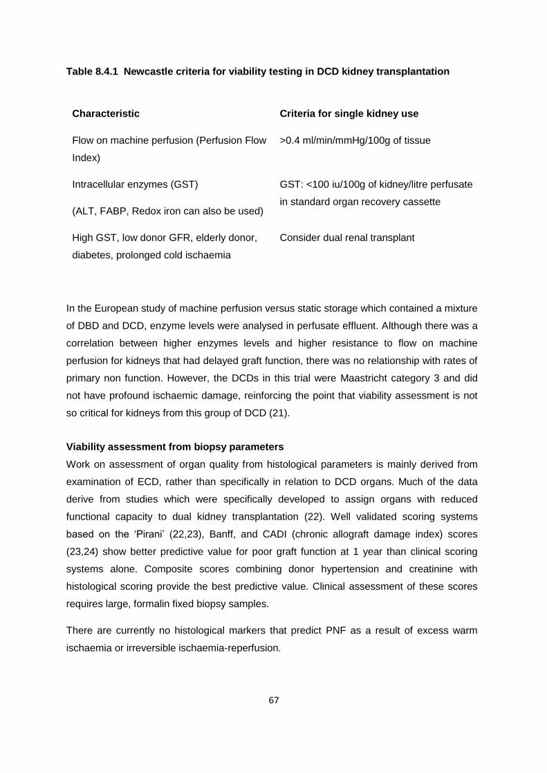
67
Table 8.4.1 Newcastle criteria for viability testing in DCD kidney transplantation
Characteristic Criteria for single kidney use
Flow on machine perfusion (Perfusion Flow
Index)
>0.4 ml/min/mmHg/100g of tissue
Intracellular enzymes (GST)
(ALT, FABP, Redox iron can also be used)
GST: <100 iu/100g of kidney/litre perfusate
in standard organ recovery cassette
High GST, low donor GFR, elderly donor,
diabetes, prolonged cold ischaemia
Consider dual renal transplant
In the European study of machine perfusion versus static storage which contained a mixture
of DBD and DCD, enzyme levels were analysed in perfusate effluent. Although there was a
correlation between higher enzymes levels and higher resistance to flow on machine
perfusion for kidneys that had delayed graft function, there was no relationship with rates of
primary non function. However, the DCDs in this trial were Maastricht category 3 and did
not have profound ischaemic damage, reinforcing the point that viability assessment is not
so critical for kidneys from this group of DCD (21).
Viability assessment from biopsy parameters
Work on assessment of organ quality from histological parameters is mainly derived from
examination of ECD, rather than specifically in relation to DCD organs. Much of the data
derive from studies which were specifically developed to assign organs with reduced
functional capacity to dual kidney transplantation (22). Well validated scoring systems
based on the ‘Pirani’ (22,23), Banff, and CADI (chronic allograft damage index) scores
(23,24) show better predictive value for poor graft function at 1 year than clinical scoring
systems alone. Composite scores combining donor hypertension and creatinine with
histological scoring provide the best predictive value. Clinical assessment of these scores
requires large, formalin fixed biopsy samples.
There are currently no histological markers that predict PNF as a result of excess warm
ischaemia or irreversible ischaemia-reperfusion.

68
Clinical donor risk scores
A range of increasingly complex scoring systems have been developed in an attempt to
predict outcomes in relation to pre-existing donor factors. These are not specific for use in
the context of DCD kidney donation. No scoring system, either alone or in combination with
pump parameters or histological scoring, has yet been shown to accurately define which
organs should be discarded due to an excessive risk of PNF or seriously impaired long-term
graft function (1, 25).
8.5 Recipient Selection
DCD graft outcome
In the UK, current data suggest that recipients of DCD kidneys have similar outcomes to
recipients of DBD kidneys (3). This is supported by UNOS registry data, which examined
44 035 deceased donor kidney transplant recipients, of which 1177 (3%) received a DCD
kidney, and found no difference in patient or graft survival at 5 years (26).
Factors that may influence outcome
NHSBT data show that increasing donor and recipient age and a cold ischaemic time of >12
hours are associated with worse outcome (3). UNOS data show that the incidence of DGF is
increased in DCD recipients, ranging from 41-51% compared with 24% in DBD recipients
(26,27). It is important to note that the most recent evidence suggests that ECD DCD donor
kidneys are no more likely to fail early than ECD DBD donor kidneys (1).
Extended criteria DCD organs
Given the progressive improvement in long term deceased kidney transplant outcomes,
with 5-year graft survival approaching 90% (28), it is important to avoid exposing young
patients to sensitisation from a poorly matched, poor quality organ which may fail within a
short period of time. Equally, the potential for serious morbidity or death resulting from
medical and/or other complications associated with DGF in older, more highly co-morbid
recipients raises the question how combined ECD DCD organs, with worse outcomes
associated with donor age and co-morbidity and increased risk of DGF due to DCD
retrieval, may best be utilised. This is largely an ethical issue, and is discussed further in
Chapter 5.

69
Recipient informed consent
Patient survival following receipt of a DCD kidney seems to be similar to that of a patient
receiving a DBD kidney. However, the increased risk of DGF and its consequences need to
be discussed with the recipient (see also Chapter 6).
8.6 Immunosuppression
Kidneys retrieved from DCD donors will inevitably have suffered warm ischaemic damage
and will be at increased risk of both PNF and DGF.
Widely favoured strategies to reduce the incidence of DGF and the risk of clinically silent
rejection occurring during periods of DGF include the use of induction therapies to reduce
the risk of rejection and the avoidance or delay in introduction of calcineurin inhibitors
(CNIs).
Use and choice of induction therapy
It is reasonable to assume that the benefits shown for induction therapy in non-DCD
transplantation can be extrapolated to DCD transplantation (29,30). Given the high rates of
DGF associated with the use of DCD organs, the reduced early rejection rate associated
with induction therapy is particularly desirable.
Retrospective cohort studies from Japan and Spain show reduced rates of DGF when
antibody induction therapy has been used in DCD transplantation (31,32). In the UK, a
prospective, randomised trial compared IL-2 receptor monoclonal antibody blockade
(Daclizumab) combined with delayed introduction of Tacrolimus when the creatinine had
fallen to <350 mol/L (or following biopsy-proven rejection) with initial Tacrolimus therapy
without antibody induction. This showed a significant reduction in DGF in the group treated
with Daclizumab and delayed Tacrolimus. However, this reduction only occurred in a sub-
group of machine-perfused organs and there was no significant difference in the primary end
point, which was graft function at three months (33). Larger studies in non-DCD populations
have failed to show any benefit, either in terms of DGF or other outcomes, from delayed CNI
introduction (34,35).
A common practice in North America is to use IL-2 receptor monoclonal antibody blockade
(with Basiliximab being the only agent currently available) in transplant recipients perceived
as being at low immunological risk. In high-risk recipients, such as the recipients of DCD
organs, the preferred regimen is to use lymphocyte depleting induction with polyclonal anti-

70
T cell agents such as thymoglobulin, or the monoclonal Alemtuzumab. There is an extensive
literature comparing different induction regimens used in this way but the number of grafts at
high risk due to DCD organ origin is small in comparison with other markers of risk such as
African-American race, high levels of sensitisation, long cold ischaemia time, and previous
graft loss. It is therefore difficult to extrapolate the reported superiority of anti-thymocyte
globulin over Basiliximab (36) or the equivalence of Alemtuzumab and thymoglobulin (37)
into the arena of DCD transplantation.
Choice of CNI
The majority of the recipients of DCD kidneys reported in the literature have received
Ciclosporin. There are few data comparing the use of Ciclosporin and Tacrolimus in this
context and the optimal immunosuppressive strategy remains to be established.
Other aspects of initial care
Systemic heparinisation is sometimes used, particularly if there is no immediate graft
function or the donor is Maastricht category 1, 2, 4 or 5, although there is little evidence to
support this.
In addition, if the donor is from one of the above categories, prophylactic antibiotic cover,
including anaerobic prophylaxis, is often administered for three days after transplantation,
although evidence for this practice is lacking.
It is not clear whether recipients of kidneys from DCD donors experience excessive
rejection, but rejection is certainly more difficult to diagnose in the presence of DGF. In the
presence of DGF, weekly graft biopsy is therefore recommended to exclude clinically silent
rejection or therapy-related toxicity (38). The histological appearances of the graft,
particularly of the blood vessels, are often atypical, with vascular changes that can mimic
‘vascular rejection’ being common.
A Health Technology Appraisal on Immunosuppressive Therapy Post Renal Transplantation
published in 2004 by The National Institute of Clinical Excellence (NICE) did not consider
recipients of DCD kidneys, and the recommendations should therefore not be applied to this
group (39).
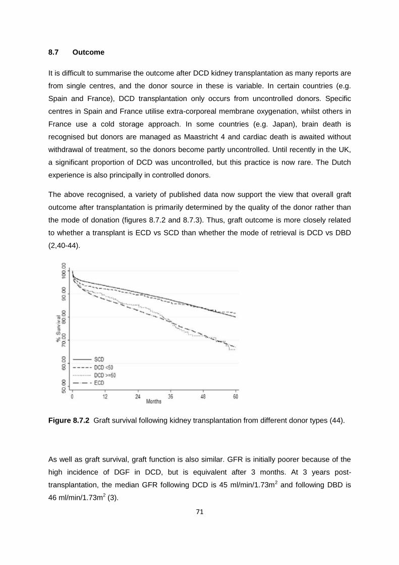
71
8.7 Outcome
It is difficult to summarise the outcome after DCD kidney transplantation as many reports are
from single centres, and the donor source in these is variable. In certain countries (e.g.
Spain and France), DCD transplantation only occurs from uncontrolled donors. Specific
centres in Spain and France utilise extra-corporeal membrane oxygenation, whilst others in
France use a cold storage approach. In some countries (e.g. Japan), brain death is
recognised but donors are managed as Maastricht 4 and cardiac death is awaited without
withdrawal of treatment, so the donors become partly uncontrolled. Until recently in the UK,
a significant proportion of DCD was uncontrolled, but this practice is now rare. The Dutch
experience is also principally in controlled donors.
The above recognised, a variety of published data now support the view that overall graft
outcome after transplantation is primarily determined by the quality of the donor rather than
the mode of donation (figures 8.7.2 and 8.7.3). Thus, graft outcome is more closely related
to whether a transplant is ECD vs SCD than whether the mode of retrieval is DCD vs DBD
(2,40-44).
Figure 8.7.2 Graft survival following kidney transplantation from different donor types (44).
As well as graft survival, graft function is also similar. GFR is initially poorer because of the
high incidence of DGF in DCD, but is equivalent after 3 months. At 3 years post-
transplantation, the median GFR following DCD is 45 ml/min/1.73m2 and following DBD is
46 ml/min/1.73m2 (3).

72
DCD kidneys are more prone to the detrimental effects of cold ischaemia than DBD kidneys,
as confirmed by a recent UK cohort study (1). The proposed national allocation scheme
should take this into account and ensure a policy that minimises CIT.
Figure 8.7.3 Graft survival following kidney transplantation from different donor types (3).
References
1. Summers DM, Johnson RJ, Hudson A, Collett D, Watson CJ, Bradley JA. Effect of
donor age and cold storage time on outcome in recipients of kidneys donated after
circulatory death in the UK: a cohort study. Lancet 2013; 381: 727–34.
2. Reid AW, Harper S, Jackson CH, et al. Expansion of the kidney donor pool by using
cardiac death donors with prolonged time to cardiorespiratory arrest. Am J Transplant
2011; 11: 995-1005.
3. Summers DM, Johnson RI, Fuggle SV, Collett D, Watson CJ, Bradley JA. Analysis of
factors that affect outcome after transplantation of kidneys donated after cardiac death
in the UK: a cohort study. Lancet 2010; 376: 1303-11.
4. Snoeijs MG, Buurman WA, Christiaans MH, et al. Histological assessment of
preimplantation biopsies may improve selection of kidneys from old donors after
cardiac death. Am J Transplant 2008; 8: 1844-51.
5. Wells AC, Rushworth L, Thiru S, et al. Donor kidney disease and transplant outcome
for kidneys donated after cardiac death. Br J Surg 2009; 96: 299-304.
Summers et al

73
6. Remuzzi G, Cravedi P, Perna A, et al. Long-term outcome of renal transplantation
from older donors. N Engl J Med 2006; 354: 343-52.
7. Nishikido M, Noguchi M, Koga S, et al. Kidney transplantation from non-heart-beating
donors: analysis of organ procurement and outcome. Transplant Proc 2004; 36: 1888-
90.
8. Teraoka S, Nomoto K, Kikuchi K, et al. Outcomes of kidney transplants from non-
heart-beating deceased donors as reported to the Japan Organ Transplant Network
from April 1995-December 2003: a multi-center report. Clin Transpl 2004; 91-102.
9. Patel AR, Eggener SE. Warm ischemia less than 30 minutes is not necessarily safe
during partial nephrectomy: every minute matters. Urol Oncol 2011; 29: 826-8.
10. Timsit MO, Tullius SG. Hypothermic kidney preservation: a remembrance of the past in
the future? Curr Opin Organ Transplant 2011; 16: 162-8.
11. Chatauret N, Thuillier R, Barrou B, Hauet T, Eugene M. Machine perfusion in clinical
trials: the preservation solution bias. Transpl Int 2011; 24: e81-2.
12. Treckmann J, Moers C, Smits JM, et al. Machine perfusion in clinical trials: "machine
vs. solution effects". Transpl Int 2012; 25: e69-70.
13. Moers C, Smits JM, Maathuis MH, et al. Machine perfusion or cold storage in
deceased-donor kidney transplantation. N Engl J Med 2009; 360: 7-19.
14. Jochmans I, Moers C, Smits JM, et al. Machine perfusion versus cold storage for the
preservation of kidneys donated after cardiac death: a multicenter, randomized,
controlled trial. Ann Surg 2010; 252: 756-64.
15. Watson CJ, Wells AC, Roberts RJ, Akoh JA, et al. Cold machine perfusion versus
static cold storage of kidneys donated after cardiac death: a UK multicentre
randomized controlled trial. Am J Transplant 2010; 10: 1991-9.
16. Moers C, Pirenne J, Paul A, Ploeg RJ. Machine perfusion or cold storage in deceased-
donor kidney transplantation. N Engl J Med 2012; 366: 770-1.
17. Hosgood SA, Nicholson ML. First in man renal transplantation after ex vivo
normothermic perfusion. Transplantation 2011; 92: 735-8.
18. Vogel T, Brockmann JG, Coussios C, Friend PJ. The role of normothermic
extracorporeal perfusion in minimizing ischemia reperfusion injury. Transplant Rev
2012; 26: 156-62.

74
19. Bon D, Chatauret N, Giraud S, Thuillier R, Favreau F, Hauet T. New strategies to
optimize kidney recovery and preservation in transplantation. Nat Rev Nephrol 2012;
8: 339-47.
20. Talbot D, D’Alessandro A. Organ donation and transplantation after cardiac death
(2009) ISBN 978-0-921733-5.
21. Moers C, Varnav OC, van Heurn E, et al. The value of machine perfusion perfusate
biomarkers for predicting kidney transplant outcome. Transplantation 2010; 90: 966-73.
22. Remuzzi G, Grinyò J, Ruggenenti P, et al. Early experience with dual kidney
transplantation in adults using expanded donor criteria. J Am Soc Nephrol 1999; 10:
2591-8.
23. Anglicheau D, Loupy A, Lefaucheur C, et al. A simple clinico-histopathological
composite scoring system is highly predictive of graft outcomes in marginal donors.
Am J Transplant 2008; 8: 2325-34.
24. Snoeijs MG, Buurman WA, Christiaans MH, et al. Histological assessment of
preimplantation biopsies may improve selection of kidneys from old donors after
cardiac death. Am J Transplant 2008; 8: 1844-51.
25. Jochmans I, Pirenne J. Graft quality assessment in kidney transplantation: not an
exact science yet! Curr Opin Org Transplant 2011; 16: 174-9.
26. Doshi MD, Hunsiker LG. Short and Long term outcomes with the use of kidneys and
livers donated after cardiac death. Am J Transplant 2007; 7: 122-9.
27. Gagandeep S, Matsuoka L, Mateo R, et al. Expanding the donor kidney pool: utility of
renal allografts procured in a setting of uncontrolled cardiac death. Am J Transplant
2006; 6: 1682-8.
28. NHS Blood and Transplant Annual Activity report. Accessed at
http://www.organdonation.nhs.uk/ukt/statistics/transplant_activity_report/
29. Webster AC, Ruster LP, McGee R, et al. Interleukin 2 receptor antagonists for kidney
transplant recipients. Cochrane Database Syst Rev 2010; 1: CD003897.
30. Gaber AO, Knight RJ, Patel S, Gaber LW. A review of the evidence for use of
thymoglobulin induction in renal transplantation. Transplant Proc 2010; 42: 1395-400.
31. Teraoka S, Nomoto K, Kikuchi K, et al. Outcomes of kidney transplants from non-
heart-beating deceased donors as reported to the Japan Organ Transplant Network
from April 1995-December 2003: a multi-center report. Clin Transpl 2004; 91-102.

75
32. Sánchez-Fructuoso AI, Marques M, Conesa J, et al. Use of different
immunosuppressive strategies in recipients of kidneys from non heart-beating donors.
Transpl Int 2005; 18: 596-603.
33. Wilson CH, Brook NR, Gok MA, Asher JF, Nicholson ML, Talbot D. Randomized
clinical trial of daclizumab induction and delayed introduction of tacrolimus for
recipients of non-heart-beating kidney transplants. Br J Surg 2005; 92: 681-7.
34. Andrés A, Marcén R, Valdés F, et al. A randomized trial of basiliximab with three
different patterns of cyclosporin A initiation in renal transplant from expanded criteria
donors and at high risk of delayed graft function. Clin Transplant 2009; 23: 23-32.
35. Andrés A, Budde K, Clavien P-A, et al. A randomized trial comparing renal function in
older kidney transplant patients following delayed versus immediate tacrolimus
administration. Transplantation 2009; 88: 1101-8.
36. Brennan DC, Daller JA, Lake KD, Cibrik D, Del Castillo D. Rabbit antithymocyte
globulin versus basiliximab in renal transplantation. N Engl J Med 2006; 355: 1967-77.
37. Hanaway MJ, Woodle ES, Mulgaonkar S, et al. Alemtuzumab induction in renal
transplantation. N Engl J Med 2011; 364: 1909-19.
38. Silva DM, Garcia JP, Ribeiro AR, et al. Utility of biopsy in kidney transplants with
delayed graft function and acute dysfunction. Transplant Proc 2007; 39: 376-7.
39. NICE Technology Appraisal. Renal transplantation - immunosuppressive regimens
(adults) (TA85). Accessed at www.nice.org.uk/guidance/ta85
40. Doshi MD, Hunsiker LG. Short and Long term outcomes with the use of kidneys and
livers donated after cardiac death. Am J Transplant 2007; 7: 122-9.
41. Barlow AD, Metcalfe MS, Johari Y, Elwell R, Veitch PS, Nicholson ML. Case matched
comparison of long-term results of non heart beating and heart beating donor renal
transplants. Br J Surg 2009; 96: 685-91.
42. Snoeijs MG, Winkens B, Heemskerk MB, et al. Kidney transplantation from donors
after cardiac death: a 25 year experience. Transplantation 2010; 90: 1106-12.
43. Sanni AO, Wilson CH, Wyrley-Birch H, et al. Non-heart-beating kidney transplantation:
6-year outcomes. Transplant Proc 2006; 38: 3396-7.
44. Locke JE, Segev DL, Warren DS, Dominik F, Simpkins CE, Montgomery RA.
Outcomes of kidneys from donors after cardiac death: implications for allocation and
preservation. Am J Transplant 2007; 7: 1797-807.

76
9 LIVER
Statements of Recommendation
Livers transplanted from Maastricht 3 DCD donors are a useful resource and
should be used where deemed safe. (B1)
Short and medium term outcome appears inferior to livers from DBD donors
with more PNF and ischaemic cholangiopathy, and a higher rate of re-
transplantation. (B1)
DCD and DBD subjects transplanted with MELD >30 or on organ-perfusion
support have similar graft survival. However, there is no survival benefit when
DCD livers are transplanted in patients with MELD ≤30 or in those not receiving
organ-perfusion support. (B2)
The incidence of biliary stricture is significantly lower when a low viscosity
solution is used to cold flush the aorta. (C2)
DCD liver use should be matched with the need of the recipients on the waiting
list, with allocation to those who will have the greatest transplant benefit. (B2)
The outcome of DCD liver transplantation is improved with short CIT, which is
best kept to under 8hrs. (B1)
A favourable outcome can be expected if an ‘ideal’ DCD liver is transplanted.
An ‘ideal’ DCD liver donor is <50 years old, has a functional WIT time <20 min,
a shorter CIT <10 hr, and <10% steatosis. (B1)
Using more restrictive DCD Donor criteria including BMI <29 kg/m2 and a
functional WIT <20 min (SAP <50 mmHg), equivalent 1 and 3-year patient and
graft survival rates can be achieved for both DCD and DBD liver transplants.
(B1)
Long FWIT is associated with an increased risk of ischaemic cholangiopathy
Potential recipients of DCD liver grafts should be informed of the potential risk
and be offered the choice to refuse such organs prior to transplant listing. (C2)
DCD grafts are best avoided in recipients of re-transplantation (B1)
DCD liver grafts should be ideally used in younger recipients with age <60
years. (B1)

77
Factors predictive of graft failure are: age ≥55 years, male sex, African–
American race, HCV positivity, metabolic liver disorder, transplant MELD ≥ 35,
hospitalisation at transplant, and the need for life support at transplant.
Recipient predictors of mortality are age ≥55 years, hospitalisation at
transplant, and re-transplantation. (B1)
A national audit should be performed to identify factors for non-utilisation of
extended criteria donors to establish the incidence of cholangiopathy under
ideal circumstances, identify more suitable risk factors, identify further
subgroups of DCDs that could potentially be utilised, allow for medical
interventions to be evaluated, and provide qualitative data regarding centre-
specific performance. (C1)
9.1 Introduction
The burden of chronic liver disease continues to grow worldwide and the UK is no
exception. Consequently, the indications for liver transplantation continue to expand with
increasing numbers of patients listed (52% increase over the last 4 years in the UK) but a
continuing shortage of donor livers (1). The liver transplant waiting list mortality in the UK is
currently 8-22%, comprising patients who die waiting for a transplant or are removed from
the list because they become too ill.
Besides the national campaigns to increase donor numbers, strategies for increasing the
liver donor pool include a UK wide split liver program and expansion of the use of DCD
organs. The long term outcome for liver transplantation from DCD donors has been
considered inferior to organs from DBD donors (2,3). However, recent reports suggest
comparable outcomes for DBD and DCD livers after transplantation (4,5). It is also
becoming apparent that the outcome of DCD livers depends on a number of donor and
recipient factors and technical factors in relation to the organ retrieval and implantation (6).
9.2 Donor Selection
There are now numerous single centre series (7-11) and large registry analyses (12-15)
which confirm that advanced donor age is associated with an increased risk of recipient
complications: specifically, ischaemic cholangiopathy, graft loss and mortality, with the donor
reference age being 40-45 years. Increasing donor weight/BMI is also associated with
78
increased risk of graft loss (15) and recipient mortality (6), especially in combination with
prolonged ischaemic times. Donor BMI <30 kg/m2 or an absolute donor weight <100 kg is
preferable.
Peri-extubation hemodynamics in the donor also seem to have an influence. Three single
institution studies (15-17) and one multi-centre study (18) provide evidence for peri-
extubation hypotension and/or hypoxia as risk factors for poor outcome, but the exact values
(BP and tissue oxygenation) at which irrecoverable liver/biliary injury occurs are still unclear.
An increased risk of ischaemic cholangiopathy is associated with a prolonged period of
FWIT. A mean arterial pressure of 50 mmHg for no more than 20 minutes is suggested,
although a higher risk of graft failure (composite end-point) is seen with a WIT of as low as
15 minutes (18).
With respect to primary WIT, both an early study from King’s College (17) and a more recent
study (the largest single centre report on DCD liver transplantation) (10) report increasing
duration of WIT being associated with recipient complications, specifically for ischaemic
cholangiopathy and graft failure. Primary functional WIT of less than 20 minutes (9,13,16,17)
and time to perfusion from donor asystole of less than 10 minutes are preferred (10).
Increasing cold ischaemia time (CIT) is also a significant factor for risk of biliary
complications including ischaemic cholangiopathy (6,9), graft failure (16), and recipient
mortality (14); the exact CIT cut-off is between 6-8 hours (8,9,16).
Absolute contraindications
In addition to the general absolute contraindications to organ donation defined by NHSBT
(i.e. invasive or haematological malignancy, untreated systemic infection, prion disease and
HIV disease), obvious absolute contraindications to the use of organs for DCD liver
transplantation are:
End-stage liver disease (CLD, cirrhosis and portal hypertension)
Acute liver failure (drug, viral)
Moderate and severe steatosis (>30% fat)
Acute liver injury that is not improving
The DCD donor can be categorised in two ways. Firstly, by the criteria used in DBD
selection, i.e. ‘good’ livers for transplantation:
Age <50 years
Normal liver function
79
<5 days on ICU
Fat content <30%
Absence of high dose pressor support
Absence of active infection
The second categorisation is restricted to DCDs. Within the DCD donor group there are
those that can be regarded as ‘ideal’ and those that are ‘marginal’ (ECD).
The ideal DCD donor profile includes:
Age <50 years
Weight <100 kg
ICU stay <5 days
WIT <20 minutes
Cold ischaemia time <8 hours, no steatosis (<15%)
The marginal (ECD) DCD donor profile includes:
Age >50 years
Weight >100 kg
ICU stay >5 days
WIT >20 minutes (up to 30 minutes)
CIT >8 hours (up to 12 hours)
Steatosis >15%

80
Table 9.2.1 Categorisation of DCD liver donors
Ideal DCD Marginal (ECD) DCD
Age (y) <50 >50
Weight (kg) <100 >100
ICU stay (days) < 5 >5
Warm ischaemia time (min) ≤ 20 20-30
Cold ischaemia time (min) ≤ 8 >8-12
Steatosis (%) ≤15 >15
Recommendation All potential liver donors fulfilling
these criteria should be used
These grafts should be
used selectively
9.3 Organ Preservation
Effective washout of the DCD liver microvasculature during retrieval is essential for optimal
preservation. If the blood remnants in the liver are not completely washed out of the
microcirculation, perfusion of the biliary tree and graft viability may be compromised. Low
viscosity preservation solutions (e.g. HTK, Marshall’s) are believed to do this more
effectively than high viscosity solutions (e.g. UW).
As non pressurised in situ aortic perfusion has been shown to be inadequate in delivering
physiological pressures in the hepatic artery, high pressure in situ 120 mmHg (Marshall
Solution) or 200 mmHg (UW solution) and ex situ perfusion 120 mmHg (UW Solution) has
been suggested to improve perfusion of the hepatic arterial tree, therefore more effectively
flushing the microcirculation of the bile ducts. The incidence of biliary strictures may be lower
when a low viscosity solution is used to cold flush the aorta in comparison with when a high-
viscosity solution is used. (These data are from a series of DBD patients and results can
only be extrapolated to the DCD situation) (19).
Type of flush fluid: A retrospective study analysing UNOS data has suggested that the use of
HTK to flush and store livers after retrieval from DCD donors is associated with a poorer

81
outcome compared to the use of UW solution (20). Therefore, using a low viscosity flush for
the arterial perfusion (such as Marshall’s solution) and a more viscous flush for portal
perfusion (such as UW) seems an attractive combination (17,19).
Operative technique: King’s College Hospital, London recommend decompressing the
inferior vena cava prior to starting the aortic flush to avoid detrimental congestion of the liver,
which is always present following circulatory collapse (17).
In DBD transplantation, pressurised aortic flush (19) and pressurised back-table flush (21)
decrease the risk of ischaemic and other biliary complications. However, this has not been
investigated in studies of DCD transplantation.
9.4 Organ Quality Assessment
There are currently no clearly defined measures of graft quality. Other than the suggested
clinical ECD criteria such as advanced age (>65 years), serum sodium >155 mmol/l,
prolonged ICU stay (>4 days with ventilatory support), donor serum creatinine >106 µmol/l,
BMI >30 kg/m2, positive serology, carcinoma outside the liver, high vasopressor support and
split-grafts, macrovesicular steatosis (>40%) is probably the best measure of poor quality
especially when combined with a prolonged FWIT and CIT >12 hours.
Steatosis, especially macrosteatosis, is an established risk factor for primary non function
and graft dysfunction. Hepatic steatosis is more common in donors of advanced age, as well
as in those with a history of obesity, dyslipidemia, metabolic disorders, or diabetes. Severely
fatty livers are more susceptible to warm and cold ischaemia reperfusion injury. In contrast to
macrovesicular steatosis, livers with predominantly microvesicular steatosis show less injury
and allograft survival rates are similar to those in nonsteatotic grafts. Extrapolating from the
DBD experience, hepatic allografts with moderate macrovesicular steatosis (30-60%) can be
used selectively in critical situations; mild macrovesicular steatosis (<30%) is relatively safe,
and severe steatosis (>60%) increases the risk of PNF. As macroscopic evaluation is
unreliable in the appraisal of the severity of steatosis, microscopic evaluation before
transplantation is recommended in cases where significant steatosis is suspected. The
percentage of steatosis should be determined by liver biopsy before transplantation. Current
practice is such that the majority of steatotic DCD grafts are declined.

82
9.5 Recipient Selection
As experience increases worldwide, more studies are attempting to address the factors that
can predict adverse outcomes after DCD liver transplantation. Although there is now a
clearer understanding of some of the donor factors, there is still a lack of high level
evidence relating to both donor and recipient factors that may affect outcome. In 2006,
Mateo et al (12) analysed the outcome of 367 DCD liver transplants performed in the US
between Jan 1996 and Dec 2003. Several recipient risk factors were identified as being
associated with DCD graft loss. These included recipients:
Age >60 years
Transplanted while on life support
With a long ICU stay prior to transplantation
Needing re-transplantation
Dialysis dependent prior to transplantation
With severe renal dysfunction (creatinine at transplant >177 µmol/l).
A more recent review of the Scientific Registry of Transplant Recipient (SRTR) database
identified 1567 recipients of DCD liver transplantation between Sept 2001 and April 2009
(14). In this analysis, recipient factors predicting graft failure included some common factors
such as:
Age (<18 or >55 years)
ICU admission prior to transplantation
The presence of life support
Other identified adverse recipient factors included:
Female sex
African American decent
Transplanted with metabolic disorders
MELD score >35
Positive Hepatitis C virus serology
Predictors of mortality included age >55 years, re-transplantation, and ICU stay at the time
of transplantation. Analysis of the same registry over a shorter period of time identified
recipient hepatocellular carcinoma (HCC) as a factor that significantly amplified mortality
risk (14). However, most patients with HCC on the transplant list tend to have a lower
MELD score and, with the exception of age, are consequently less likely to have the other

83
recognised adverse recipient risk factors. As a result, there is a tendency to use DCD grafts
in fit HCC patients and this may lead to bias. It is generally accepted that the use of ECD
DBD or DCD grafts in fit patients with HCC is a good use of this resource.
Organ allocation
The current allocation system for DBD liver grafts in the UK is a centre-based system with
grafts allocated according to need and transplant benefit. Following a consensus meeting in
March 2012, it was agreed that a national allocation system should instead be
implemented. The final decision about the best way to allocate DCD grafts is still awaited,
but most centres favour regional allocation in order to keep CIT as short as possible and to
ensure the best outcome.
In the USA, Mateo et al developed a classification of DCD liver grafts as low-risk or high risk,
depending on whether the FWIT was ≤30 min and CIT was ≤10 hr (12). They also grouped
recipients into low or high risk groups based on a recipient cumulative relative risk (RCRR
1-5.17, i.e. no risk to maximum risk) using age, medical condition at transplantation, regraft
status, dialysis received, and serum creatinine. When low risk recipients received low risk
DCD liver grafts, their graft survival rates at 1 and 3 years were comparable to those of the
DBD group. A further analysis of the same data has suggested similar results from similar
matching, but with an emphasis on donor age <45 years (13).
Based on these analyses, this group has suggested an algorithm for selecting potential
recipients for a DCD liver graft (13), but this needs external validation before implementation
in routine clinical practice.
9.6 Immunosuppression
Although there are no data to support an optimal immunosuppressive regimen for DCD liver
transplantation, consideration should be given to renal sparing regimens with delayed CNI
introduction. Induction immunosuppression and T cell depletion may also be considered, but
risk and benefit must be balanced in any choice of regimen.
With regards to HCV, intense immunosuppression often promotes HCV recurrence and the
need for treatment of acute cellular rejection (with pulse dosed corticosteroids) leads to
increased HCV RNA values, decreased graft survival and increased overall mortality (22,23).

84
The concept that a particular CNI may impact the natural history of HCV in liver transplant
patients is controversial (24). Although overall survival is similar regardless of the CNI use,
preliminary data suggest that ciclosporin-based immunosuppressive regimens, when used in
conjunction with interferon-alpha and ribavirin to treat HCV, may lead to a greater likelihood
of virological response to therapy, possibly through greater T cell regulatory inhibition (25).
Further confirmatory studies are required.
9.7 Outcome
In 2009, of the 646 deceased organ donors, 110 were DCD donors from whom livers were
recovered, and 76 of these livers were transplanted. This increased to 97 in 2010. However,
studies have shown that the graft survival of DCD livers is inferior to that of DBD donor livers
(7,12,26-28), with one study estimating a relative risk of graft failure of 1.85 times higher in
DCD recipients (27). The incidence of PNF using DCD livers has ranged from 0-5.5%, while
biliary complication rates have been reported to be higher compared with DBD allografts (13-
37% vs 1-20%) (5,7,15). The majority of studies on DCD outcomes have come from reviews
of national databases such as the UNOS database (12,27,28), the Scientific Registry of
Transplant Recipients (SRTR) database, and the UK transplant registry, or single-centre
studies that have reported on relatively small numbers of patients (5,7,15,17,29,30). There
are no widely accepted DCD donor parameters predictive of good outcomes that could guide
surgeons on whether or not to use or discard a DCD liver. In addition, it is unclear and
controversial as to which patients would benefit the most from the use of these allografts
(12,27).
Recently, Jay et al (14) compared 1113 DCD and 42 254 DBD recipients from the SRTR
database transplanted between 1996 and 2007. Patient survival was examined using
Kaplan-Meier analysis and Cox regression. DCD recipients had worse patient survival than
DBD recipients (p<0.001). One and three year survival was 82% and 71% for DCD
compared to 86% and 77% for DBD recipients. Moreover, DCD recipients required re-
transplantation more frequently (DCD 14.7% vs DBD 6.8%, p<0.001), and survival after re-
transplantation was markedly worse than survival after primary transplantation irrespective of
graft type. Amplification of mortality risk was observed when DCD was combined with a CIT
>12 hours (HR 1.81), shared organs (HR 1.69), recipient hepatocellular carcinoma
(HR 1.80), recipient age >60 years (HR 1.92), and recipient renal insufficiency (HR 1.82).
In addition, the Pittsburgh experience of DCD liver transplant outcome was recently reported
(9). 141 patients who underwent DCD liver transplantation between 1993 and 2007 were

85
compared to a matched cohort of 282 patients who received DBD livers. Patient survival
was similar, but 1, 5 and 10-year graft survival was significantly lower in DCD (69%, 56%,
44%) vs DBD (82%, 73%, 63%) recipients (p<0.0001). Primary non function and biliary
complications were more common in DCD patients, accounting for 67% of early graft failure.
Donor WIT >20 minutes, CIT >8 hours, and donor age >60 years were associated with
poorer DCD outcomes. There was a lack of survival benefit in DCD livers used in patients
with MELD ≤30 or those not on organ support, as graft survival was significantly lower
compared to DBD patients. However, DCD and DBD subjects transplanted with MELD >30
or on organ support had similar graft survival, suggesting a potentially greater benefit of
DCD livers in critically ill patients. The attrition in DCD grafts occurred early within the first
year after transplant and was due to a high incidence of PNF/DGF and biliary complications,
which also led to a higher re-transplantation rate in the DCD group. Of note, the survival
curves of both cohorts became parallel after the first year, suggesting that interventions to
decrease the occurrence of these complications will significantly improve the survival of
these grafts. PNF/DGF were the leading causes of early graft failure in the DCD group,
accounting for 50% of the cases, and by multivariate analyses were found to be associated
with the transplantation of male donor livers to female recipients and with recipients >60
years or with a BMI >30 kg/m2. CIT ≤8 hours was associated with a much lower incidence of
PNF, though this did not reach statistical significance because of low patient numbers.
By contrast, using restrictive criteria (Maastricht 3 DCD donors aged <55 years with a BMI
<29 kg/m2, donor WIT <30 min and hypotensive episodes <15 min), a multi-centre Dutch
study produced 1 and 3-year patient survival rates which were similar for DCD (85 and 80%)
and DBD (86·3 and 80·8%) transplants (p=0·763), and equivalent graft survival rates (74%
and 68% vs 80.4% and 74.5%; p=0.212) (4). Of concern, the 3-year cumulative percentage
of surviving grafts developing non-anastomotic biliary strictures was 31% after DCD and
9·7% after DBD transplantation (p<0·001). The re-transplantation rate was similar overall
(p=0.081), but that for biliary strictures was higher in the DCD group (p<0.001). Risk factors
for 1-year graft loss after DBD liver transplantation were transplant centre, recipient WIT and
donors with severe head trauma. After DCD liver transplantation, they were transplant
centre, donor WIT and CIT. As previously reported, use of DCD grafts was a risk factor for
non-anastomotic biliary strictures.
The preoperative identification of factors associated with poor outcomes in DCD liver
transplantation remains an important challenge. The period between extubation and
asystole may help predict graft function. Less liver damage may occur if the donor
progresses quickly to cardiac death, as opposed to maintaining a circulation in the presence
86
of significant hypoxia or hypotension. Consistent with this, restrictions on recovery are now
imposed by most UK centres based on donor time to death and the duration of hypoxia or
hypotension. This FWIT starts when the systolic blood pressure has a sustained (i.e. at least
2 min) fall below 50 mmHg (or the haemoglobin oxygen saturation falls below 70%) and
extends up to the onset of cold in situ perfusion. The duration of the FWIT is the important
determinant of outcome. The recent document ‘Donation After Circulatory Death’ published
by a steering group on behalf of the British Transplantation Society and Intensive Care
Society (31) suggested that the stand-down time from the onset of functional warm
ischaemia for DCD liver transplantation was 30 minutes (although 20 minutes is ideal), and
that age was an important factor.
Similarly, the 2006 Report of the National Conference on Donation after Cardiac Death in the
US recommended recovering livers for transplantation if the time from extubation to cold
perfusion was less than 30 minutes (32). These recommendations are based on animal
models which show that increasing this time from extubation to organ recovery decreases
survival (33,34).
A retrospective examination of the New England Organ Bank DCD database concentrated
on donor factors including vital signs after withdrawal of support (18). The outcome of DCD
liver transplantation was based on a composite endpoint chosen to represent a failed graft
which included any transplant-related death, primary non function, graft loss within 1 year, or
diffuse intra-hepatic biliary abnormalities (requiring re-transplantation). Data on 37
transplanted livers demonstrated that the donors tended to be young (average age 34.3 ±
13.2 years) and thin (average BMI 25.1 ± 5.8 kg/m2). The average time from extubation to
asystole was 17.6 ± 8.7 min and the average time from extubation to flush was 28.6 ± 8.8
min. Recipient MELD scores averaged 22.8 ± 7.5. Mean follow-up was 416 ± 328 days.
One-year patient and graft survival rates were 81.1% and 78.9% respectively. A total of 14
(37.8%) recipients reached the composite endpoint of death, graft loss within 1 year, or
diffuse intra-hepatic biliary abnormalities. Recipients who reached the primary endpoint
tended to have received livers from donors who were older, exhibited longer profound
hypotension in the post-extubation period, and had slightly longer CIT. To determine
clinically relevant parameters for predicting the primary endpoint, receiver operating
characteristic analysis on donor and recipient parameters were performed, including donor
age, CIT, time from extubation to asystole, time from extubation to flush, time with oxygen
saturation less than 50% to flush, and time from systolic BP <50 mmHg to flush. The best
predictive test was time from systolic BP <50 mmHg to flush (area under curve 0.652), and
the next best was donor age (area under curve 0.589).
87
A meta-analysis examined the risks of biliary complications, particularly ischaemic
cholangiopathy, after DCD compared with DBD liver transplantation (35). Eleven
retrospective cohort studies were reviewed, involving 489 DCD and 4455 DBD recipients.
DCD recipients had a 2.4-fold increased risk of biliary complications and a 10.8-fold
increased risk of ischaemic cholangiopathy. Ischaemic cholangiopathy was present in 16%
of DCD compared with 3% of DBD recipients. DCD recipients also experienced higher odds
of 1-year patient mortality (OR 1.6) and graft failure (OR 2.1). In addition, a single centre
experience of DCD compared to DBD liver transplantation showed following multivariate
analysis that CIT >8 hours (HR 2.46; p=0.05) and donor age >40 years (HR 2.90; p<0.01)
significantly increased the risk of ischaemic cholangiopathy (8).
A favourable outcome can be expected if an ‘ideal’ DCD liver is transplanted. An ‘ideal’ DCD
donor is <50 years old (8), has a shorter WIT than 30 min (14,15,18), has a CIT <10 hours
(32) and has <15% steatosis. Although Mateo et al (12) speculated that recipient MELD
scores in candidates who received DCD livers would be a predictive prognostic factor, most
other data have shown no relationship between the recipient MELD and prognosis and a
more robust algorithm needs to be developed to best match DCD donor livers with recipients
awaiting transplantation. However, the expected lifetime of a liver transplant candidate
offered a DCD liver should be compared with the expected lifetime of that candidate if they
were to turn down the DCD offer and continue to wait for a DBD liver. That is to say, even if
graft and/or patient survival is lower with a DCD liver, it may be better than dying on the
waiting list.
Considering the current shortage of donor organs, every liver transplant candidate should be
informed at the time of evaluation that the choice in the UK is frequently not between a
marginal (including DCD) liver and a standard liver, but between a marginal (DCD) liver or
no liver at all. Until an algorithm for the use of the DCD donor is determined,
recommendations by transplant teams and decisions by liver transplant candidates should
be predicated on full disclosure of the known risks and potential benefits of DCD liver
transplantation (5).
References
1. http://www.organdonation.nhs.uk/ukt/statistics/transplant_activity_report/archive_activi
ty_reports/pdf/ukt/activity_report_2009_10.pdf

88
2. Selck FW, Grossman EB, Ratner LE, Renz JF. Utilization, outcomes, and
retransplantation of liver allografts from donation after cardiac death: implications for
further expansion of the deceased-donor pool. Ann Surg. 2008; 248: 599-607.
3. Yamamoto S, Wilczek HE, Duraj FF, Groth CG, Ericzon BG. Liver transplantation with
grafts from controlled donors after cardiac death: a 20-year follow-up at a single
center. Am J Transplant 2010; 10: 602-11.
4. Dubbeld J, Hoekstra H, Farid W, et al. Similar liver transplantation survival with
selected cardiac death donors and brain death donors. Br J Surg 2010; 97: 744-53.
5. Fujita S, Mizuno S, Fujikawa T, et al. Liver transplantation from donation after cardiac
death: a single center experience. Transplantation 2007; 84: 46-9.
6. Mathur AK, Heimbach J, Steffick DE, Sonnenday CJ, Goodrich NP, Merion RM.
Donation after cardiac death liver transplantation: predictors of outcome. Am J
Transplant 2010; 10: 2512-9.
7. Foley DP, Fernandez LA, Leverson G, et al. Donation after cardiac death: the
University of Wisconsin experience with liver transplantation. Ann Surg 2005; 242:
724-31.
8. Foley DP, Fernandez LA, Leverson G, et al. Biliary complications after liver
transplantation from donation after cardiac death donors: an analysis of risk factors
and long-term outcomes from a single center. Ann Surg 2011; 253: 817-25. E
9. de Vera ME, Lopez-Solis R, Dvorchik I, et al. Liver transplantation using donation
after cardiac death donors: long-term follow-up from a single center. Am J Transplant
2009; 9: 773-81.
10. Taner CB, Bulatao IG, Willingham DL, et al. Events in procurement as risk factors for
ischemic cholangiopathy in liver transplantation using donation after cardiac death
donors. Liver Transpl 2012; 18: 100-11.
11. Skaro AI, Jay CL, Baker TB, et al. The impact of ischemic cholangiopathy in liver
transplantation using donors after cardiac death: the untold story. Surgery 2009; 146:
543-52; discussion 52-3.
12. Mateo R, Cho Y, Singh G, et al. Risk factors for graft survival after liver
transplantation from donation after cardiac death donors: an analysis of OPTN/UNOS
data. Am J Transplant 2006; 6: 791-6.

89
13. Lee KW, Simpkins CE, Montgomery RA, Locke JE, Segev DL, Maley WR. Factors
affecting graft survival after liver transplantation from donation after cardiac death
donors. Transplantation 2006; 82: 1683-8.
14. Jay C, Ladner D, Wang E, et al. A comprehensive risk assessment of mortality
following donation after cardiac death liver transplant - an analysis of the national
registry. J Hepatol 2011; 55: 808-13.
15. Chan EY, Olson LC, Kisthard JA, et al. Ischemic cholangiopathy following liver
transplantation from donation after cardiac death donors. Liver Transpl 2008; 14: 604-
10.
16. Hong JC, Yersiz H, Kositamongkol P, et al. Liver transplantation using organ donation
after cardiac death: a clinical predictive index for graft failure-free survival. Arch Surg
2011; 146: 1017-23.
17. Muiesan P, Girlanda R, Jassem W, et al. Single-center experience with liver
transplantation from controlled non-heartbeating donors: a viable source of grafts.
Ann Surg 2005; 242: 732-8.
18. Ho KJ, Owens CD, Johnson SR, et al. Donor postextubation hypotension and age
correlate with outcome after donation after cardiac death. Transplantation 2008; 85:
1588-94.
19. Pirenne J, Van Gelder F, Coosemans W, et al. Type of donor aortic preservation
solution and not cold ischemia time is a major determinant of biliary strictures after
liver transplantation. Liver Transpl 2001; 7: 540-5.
20. Stewart ZA, Cameron AM, Singer AL, Montgomery RA, Segev DL. Histidine-
Tryptophan-Ketoglutarate (HTK) is associated with reduced graft survival in deceased
donor livers, especially those donated after cardiac death. Am J Transplant 2009; 9:
286-93.
21. Moench C, Moench K, Lohse AW, Thies J, Otto G. Prevention of ischemic-type biliary
lesions by arterial back-table pressure perfusion. Liver Transpl 2003; 9: 285-9.
22. Charlton M, Seaberg E. Impact of immunosuppression and acute rejection on
recurrence of hepatitis C: Results of the National Institute of Diabetes and Digestive
and Kidney Diseases Liver Transplantation Database. Liver Transpl Surg 1999; 5(S1):
S107-14.

90
23. Berenguer M, Aguilera V, Prieto M, et al. Significant improvement in the outcome of
HCV-infected transplant recipients by avoiding rapid steroid tapering and potent
induction immunosuppression. J Hepatol 2006; 44: 717-22.
24. Villamil F, Levy G, Grazi GL, et al. Long-term outcomes in liver transplant patients
with hepatic C infection receiving tacrolimus or cyclosporine. Transplant Proc 2006;
38: 2964-7.
25. Miroux C, Morales O, Carpentier A, et al. Inhibitory effects of cyclosporine on human
regulatory T cells in vitro. Transplant Proc 2009; 41: 3371-4
26. Available from: http://www.unos.org
27. Merion RM, Pelletier SJ, Goodrich N, Englesbe MJ, Delmonico FL. Donation after
cardiac death as a strategy to increase deceased donor liver availability. Ann Surg
2006; 244: 555-62.
28. Abt PL, Desai NM, Crawford MD, et al. Survival following liver transplantation from
non-heartbeating donors. Ann Surg 2004; 239: 87-92.
29. Fukumori T, Kato T, Levi D, et al. Use of older controlled non-heartbeating donors for
liver transplantation. Transplantation 2003; 75: 1171-4.
30. Reich DJ, Munoz SJ, Rothstein KD, et al. Controlled nonheartbeating donor liver
transplantation. Transplantation 2000; 70: 1159-66.
31. Donation After Circulatory Death Steering Group, on behalf of the British
Transplantation Society and Intensive Care Society. Accessed at
http://www.bts.org.uk/transplantation/standards-and-guidelines/
32. Bernat JL, D’Alessandro AM, Port FK, et al. Report of a national conference on
donation after cardiac death. Am J Transplant 2006; 6: 281-91.
33. Monbaliu D, van Pelt J, De Vos R, et al. Primary graft nonfunction and Kupffer cell
activation after liver transplantation from nonheart-beating donors in pigs. Liver
Transpl 2007; 13: 239-47.
34. Garcia-Valdecasas JC, Tabet J, Valero R, et al. Evaluation of ischemic injury during
liver procurement from non-heart-beating donors. Eur Surg Res 1999; 31: 447-56.
35. Jay CL, Lyuksemburg V, Ladner DP, et al. Ischemic cholangiopathy after controlled
donation after cardiac death liver transplantation: a meta-analysis. Ann Surg 2011;
253: 259-64.
91
10 PANCREAS
Statements of Recommendation
Although DCD organs can be used for isolated pancreas transplants in
pancreas transplant alone or pancreas after kidney, available evidence and
current practice are increasingly in favour of their use for simultaneous
pancreas and kidney (SPK) transplantation. (C2)
Pancreas transplants from DCD donors are at increased risk of reperfusion
pancreatitis and thrombosis and this may be exacerbated by prolonged cold
ischaemia time and higher donor body mass index. The contribution of donor
age to the incidence of reperfusion pancreatitis/thrombosis in DCD organs is
poorly studied but may constitute a further risk factor. Ideal donors should be
<60 years old and have BMI <30 kg/m2. (C2)
The pancreas team should stand down after a functional warm ischaemia time
(systolic BP <50 mmHg and oxygen saturation of 70%) of 60 minutes. (C2)
A primary focus of any DCD pancreas transplant should be to achieve the
minimum cold ischaemic time. (B2)
There is little evidence regarding recipient risk factors but it is logical to
assume that higher recipient BMI, age, cardiovascular morbidity, and technical
surgical factors may contribute to poorer outcome. (C2)
Reported outcomes for DCD donor pancreas transplantation are broadly
similar to those from DBD donors, although considerably greater donor
selection is likely to have taken place. (C2)
Graft loss from thrombosis is twice as common in DCD as DBD pancreas
transplants. Particular attention should be paid to measures to prevent
thrombosis in recipients of DCD organs. A DCD organ should not be
transplanted into a recipient with a history of thrombo-embolic disease unless
this is monitored and treated. (C2)
Selected transplant centres will have built up a volume of expertise with DCD
extended criteria and these guidelines should not restrict innovative but safe
practice of pancreas transplantation. (Not graded)
92
10.1 Donor Selection
The selection of DCD donors for pancreas transplantation is more restrictive than that for
DBD donors. A recent review of the UK experience of 134 DCD pancreas transplants
compared to 875 contemporaneous DBD grafts reported that DCD donors tended to be
younger (median age 28 vs 37 years, p<0.001), had a lower BMI (23 vs 24 kg/m2, p=0.04),
and were less commonly due to cerebrovascular cause of death (33 vs 60%, p<0.0001) (1).
The median cold ischaemic time was similar in both DCD and DBD transplants (750 vs.
752 min), although achieving such cold ischaemia may be more challenging in DCD
transplantation due to the shorter lead time available to complete recipient selection, travel
to the recipient centre, and cross-match before organ retrieval.
Numbers transplanted do not allow a rigorous analysis of factors affecting donor selection,
but the following broad comments reflect current experience and practice in the UK.
Age
Hitherto unpublished analysis of the UK transplant registry data suggest that donor age is a
significant factor contributing to transplant outcome for DBD pancreata in the UK. Numbers
do not allow extensive analysis of data for DCD transplants, but the effect is likely to be the
same. Current criteria are to consider all potential donors up to the age of 60 years.
Body mass index
A low donor BMI (<28 kg/m2) is preferred, but potential donors with higher BMI should still be
referred and organs should be retrieved when the donor BMI is ≤30 kg/m2. Pancreata from
donors with a BMI >30 kg/m2 should be referred for consideration of islet transplantation.
Waiting time
A functional warm ischaemic time, from the point at which the systolic pressure is ≤50 mmHg
for 30 minutes, is a reasonable indication to abandon pancreas retrieval. Otherwise, a donor
in whom the blood pressure is stable and physiological may still yield a transplantable
pancreas some time after treatment withdrawal. In general, the retrieval team should be
prepared to retrieve the pancreas for up to 3 hours following the withdrawal of support. The
decision to stand down sooner should be made on the basis of the blood pressure profile
and other donor criteria. Donors with a withdrawal period over 60 minutes have been used
successfully (1,2).

93
Inotropes
Although high doses of inotropes are widely agreed to be detrimental, there is no good
evidence on which to base national criteria. Inotrope levels should not, therefore, be used to
exclude the referral of DCDs for pancreas retrieval.
Amylase and glucose levels
There is no good evidence that the level of either amylase or glucose has prognostic
significance and these should not be used to exclude either the referral or the transplantation
of DCD pancreata.
Steatosis and fibrosis
These factors are largely subjective and difficult to quantify, and the precise significance of
differing degrees of steatosis or fibrosis are uncertain. For these reasons, retrieval should
proceed unless the changes are gross. It is important that the transplanting surgeon should
be able to speak to the retrieving surgeon to discuss the quality of the donor organs.
Caution
Suggested donor criteria for pancreas transplantation are shown in table 10.2.1. However,
this is an evolving field. It is important to note that transplanting centres in the UK will have
built up a volume of expertise with extended DCD criteria, and these criteria should not
restrict innovative but safe practice of pancreas transplantation.
Table 10.2.1 Donor criteria for pancreas transplantation
Ideal DCD Marginal (ECD) DCD
Age (year) <45 45-60
BMI (kg/m2) <28 28-30
WIT (min) ≤ 30 >30
CIT (hr) ≤ 9 >9
Steatosis None Mild-moderate
Recommendation All potential pancreas donors
fulfilling these criteria should
be used
These grafts should be used
selectively

94
10.2 Organ Preservation
Warm ischaemia time
The endocrine function of the pancreas appears to tolerate warm ischaemia relatively well,
but the complication of reperfusion pancreatitis has serious implications in terms of both
morbidity and graft survival and this is probably (in part) related to the duration of warm
ischaemia. A WIT less than 30 minutes is preferred (ideal), but organs with WIT of more than
30 minutes should be considered.
In situ perfusion and preservation
In situ perfusion for pancreas donors should be via the aorta only (with additional perfusion
of the liver via the portal vein being instituted by opening the portal vein without impeding
venous drainage from the pancreas). UW solution is the preferred preservation solution both
for the initial flush and preservation.
Thrombolysis
The evidence for the benefit of an initial streptokinase flush comes from DCD kidney
retrieval. There is no equivalent evidence in DCD pancreas transplantation. A national trial of
the use of streptokinase in DCD donation is under consideration.
Cold ischaemia time
As with other organs, there is good evidence that cold ischaemia is detrimental to the
outcome of pancreas transplantation in proportion to its duration. UK data suggest that a CIT
of >12 hours in DBD pancreas transplantation is associated with a significantly increased
risk of graft loss; it is likely that a shorter CIT (such as 9 hours) will be more appropriate
when transplanting a DCD pancreas.
10.3 Organ Quality Assessment There is no ‘standard’ of quality assessment. Assessment is largely based on a visual
inspection and clinical experience. In general, a quality assessment can be made by looking
at the quality of perfusion of the gland and duodenum, as well as the amount of fat
infiltration, and by feeling the texture of the gland for fibrosis. These observations should be
considered alongside the donor characteristics and the haemodynamics and duration of the
withdrawal period.

95
10.4 Recipient Selection
There is relatively little reported experience of DCD pancreas transplantation to guide
recipient selection (see also section 10.6). While it is possible that the recipients of DCD
pancreas transplants are selected more rigorously than those receiving DBD transplants,
analysis of national databases does not show significant differences in recipient
characteristics between the two groups. Therefore, the indications for DCD pancreas
transplantation are no different to those for pancreas transplantation in general, and are
summarised in the National Protocol for Assessment of Kidney and Pancreas Transplant
Patients at http://www.organdonation.nhs.uk/about_transplants/organ_allocation/pancreas/.
However the UK data do suggest a poorer outcome when the DCD pancreas is transplanted
alone rather than with a kidney (1).
Organ allocation
As with other organs, there is good evidence that cold ischaemia is detrimental to the
outcome of pancreas transplantation in proportion to its duration. The implication is that, as
at present, DCD organs should be transplanted locally rather than transported for use in
sensitised patients. Current allocation arrangements in the UK are summarised on the
NHSBT website at http://www.organdonation.nhs.uk/about_transplants/organ_allocation/
pancreas/. The current scheme aims to minimise the distance the pancreas travels to a
recipient centre.
UK data suggest that the outcome of SPK transplantation using DCD organs is better than
that of isolated pancreas transplantation and this is therefore the preferred way of using
these organs (1).
Recent proposals for changing the allocation of DCD kidneys will enable a DCD kidney to be
preferentially offered with a DCD pancreas.
10.5 Immunosuppression
There are no data that indicate an optimal immunosuppressive regimen for a DCD pancreas.
The immunosuppressive requirements of pancreas transplantation appear to be greater than
those of kidney transplantation, but otherwise consideration of a kidney-friendly protocol
seems appropriate, with an induction agent such as Daclizumab or Alemtuzumab followed
by mycophenolate and an initial low dose tacrolimus regimen

96
10.6 Outcome
Pancreatic transplantation from DCD donors comprises only a small percentage of the total
number of pancreatic transplants, although there have been an increasing number
performed in recent years, especially in the UK (3). Worldwide, there have been fewer than
300 DCD pancreas transplants in the last 25 years.
Most of the published outcome data come from the USA and the UK. These suggest that
outcomes from DCD transplantation are broadly comparable to those of DBD pancreata
transplanted together with a kidney (SPK). The results of pancreas alone transplantation are
worse than those of combined SPK transplantation, with DCD pancreas alone
transplantation being non-significantly worse than that of DBD transplantation (1).
The US experience of 57 transplants (47 SPK, 10 solitary pancreas) from controlled
(Maastricht 3) DCD donors between 1993 and 2003 was collated from the UNOS registry in
2006 (4). Over this period, there were 4038 DBD pancreas transplants. Patient, kidney and
pancreas graft survival from DBD and DCD donors were equivalent at 1, 3 and 5 years after
transplantation (Table 10.6.1). Early graft loss due to vascular thrombosis was higher with
DCD donors (12.8 vs 6.1%), although 37 of the 57 DCD transplants were performed at one
centre, where no vascular thromboses were experienced.
A separate publication from Wisconsin summarised their experience of 37 transplants also
performed between 1993 and 2003 (5); these patients probably formed the bulk of the US
experience summarised in the 2006 paper.
In one US centre, four DCD donors were treated using an extracorporeal perfusion circuit
with heparinisation and cannulation of the femoral vessels prior to treatment withdrawal.
These donors were perfused with oxygenated blood and cooled to 22°C. The four pancreata
were transplanted as part of a combined pancreas and kidney transplant, with immediate
function of both pancreas and kidney in each case and all grafts surviving over 2 years at the
time of reporting (6).
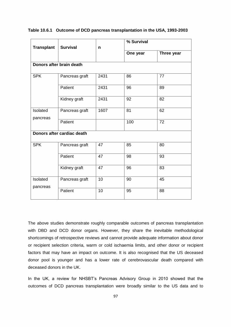
97
Table 10.6.1 Outcome of DCD pancreas transplantation in the USA, 1993-2003
Transplant Survival n
% Survival
One year Three year
Donors after brain death
SPK Pancreas graft 2431 86 77
Patient 2431 96 89
Kidney graft 2431 92 82
Isolated
pancreas
Pancreas graft 1607 81 62
Patient 100 72
Donors after cardiac death
SPK Pancreas graft 47 85 80
Patient 47 98 93
Kidney graft 47 96 83
Isolated
pancreas
Pancreas graft 10 90 45
Patient 10 95 88
The above studies demonstrate roughly comparable outcomes of pancreas transplantation
with DBD and DCD donor organs. However, they share the inevitable methodological
shortcomings of retrospective reviews and cannot provide adequate information about donor
or recipient selection criteria, warm or cold ischaemia limits, and other donor or recipient
factors that may have an impact on outcome. It is also recognised that the US deceased
donor pool is younger and has a lower rate of cerebrovascular death compared with
deceased donors in the UK.
In the UK, a review for NHSBT’s Pancreas Advisory Group in 2010 showed that the
outcomes of DCD pancreas transplantation were broadly similar to the US data and to

98
transplants from DBD donors, although the numbers were small (Table 10.6.2) (7). Of note,
1 year pancreas survival was substantially less for isolated pancreas transplantation in
comparison to SPK transplantation (76 vs 86%), although small numbers meant that this
difference was not statistically significant. This was almost exclusively due to a higher rate of
graft thrombosis in one centre following DCD donor isolated pancreas transplantation (12 vs
2% from the largest centre). Given this, particular attention should be paid to measures to
prevent thrombosis in recipients of DCD organs. A DCD organ should not be transplanted
into a recipient with a history of thrombo-embolic disease unless the coagulopathy is
corrected and closely monitored.
Table 10.6.2 Pancreas transplant outcomes at one year in the UK (6)
Transplant Organ n % survival 95% confidence
intervals
Donors after brain death 1/4/07 to 31/3/09
SPK Pancreas graft 294 88 83-91
Patient 296 96 93-98
Kidney graft 302 93 90-95
Isolated
pancreas
Pancreas graft 55 76 62-85
Patient 56 94 83-98
Donors after circulatory death 1/4/05 to 31/3/09
SPK Pancreas graft 36 86 69-94
Patient 36 94 79-99
Kidney graft 38 95 80-99
Isolated
pancreas
Pancreas graft 33 76 57-87
Patient 33 97 79-99

99
References
1. Muthusamy AS, Mumford L, Hudson A, Fuggle SV, Friend PJ. Pancreas
transplantation from donors after circulatory death from the United Kingdom. Am J
Transplant 2012; 12: 2150-6.
2. Qureshi MS, Callaghan CJ, Bradley JA, Watson CJE, Pettigrew GJ. Outcomes of
simultaneous pancreas and kidney transplantation from brain-dead donors and
controlled circulatory death donors. Br J Surg 2012; 99: 831-8.
3. Dominguez-Gil B, Haase-Kromwijk B, Van Leiden H, et al on behalf of the European
Committee (Partial Agreement) on Organ Transplantation. Council of Europe (CD-P-
TO). Current situation of donation after circulatory death in European countries.
Transplant Int 2011; 24: 676-86.
4. Salvalaggio PR,Fernandez LA, Kaufman DB. Outcomes of pancreas transplantation in
the United States using cardiac-death donors. Am J Transplant 2006; 6: 1059-65.
5. Fernandez LA, Di Carlo A, Odorico JS, et al. Simultaneous pancreas-kidney
transplantation from donation after cardiac death: successful long-term outcomes. Ann
Surg 2005; 242: 716-23.
6. Farney AC, Singh RP, Hines MH, et al. Experience in renal and extrarenal
transplantation with donation after cardiac death donors with selective use of
extracorporeal support. J Am Coll Surg 2008; 206: 1028-37.
7. Personal Communication. Alex Hudson, Principal Statistician, NHSBT.

100
11 PANCREATIC ISLETS
Statements of Recommendation
Selection criteria for recipients of islets from DCD donors should be the same
as for DBD donors. (B2)
Organs from DCD donors for islet isolation and transplantation should be
allocated through the National Pancreas Allocation Scheme. (B2)
The long term outcome of islet transplantation from DCD donors has been
satisfactory in the UK but the cohort is small. Further audit and research are
required. (C2)
Satisfactory functional islet preparations can be routinely obtained from DCD
donors. (C2)
11.1 Donor Selection
There is very limited clinical experience of islet transplantation from DCD donation and
limited information as to what constitutes the ideal donor for islet transplants procured from a
DCD pancreas.
Given the above, ideal donor criteria have to be extrapolated from the ideal DBD donor as
recommended by the National Clinical Guideline islet transplant program. Young donors
yield fewer islets after islet isolation because of the effect of collagenase on the pancreatic
extracellular matrix (1,2). The smaller the donor weight, the smaller the donor pancreas and
thus fewer islets are isolated for transplantation (3-9). Obese non-diabetic donors are
excellent for islet isolation because of a higher beta cell mass. Human islets are very
intolerant to prolonged periods of both warm and cold ischaemia and this is crucial to
successful islet transplantation (10).
The following summarises the current criteria for donor selection in the UK:
Optimal (ideal)
Age 18-45 yr
Weight 50-100 kg
BMI 21-35 kg/m2
Cold ischaemia <8 hr

101
Warm ischaemia <5 min
Marginal (ECD)
Age 16-18 or 45-65 yr
Weight 40-50 or >100 kg
BMI < 21 or >35 kg/m2
Cold ischaemia 8-16 hr
FWIT 5-30 min
Previous extensive abdominal surgery
Absolute contra-indications
Age >65 yr
BMI >40 kg/m2
Cold ischaemia > 16 hr
FWIT >30 min (systolic BP <50 mmHg, SpO2 <70%)
Diabetes mellitus
Evidence of pancreatic disease (e.g. chronic pancreatitis)
Pancreatic duct transection/traumatic capsular damage
Positive for HCV, HIV, HBV
Variant CJD
Untreated systemic infection
Evidence of intravenous drug use
Malignancy, myeloma, lymphoma, leukaemia
Invasive cancer in the last 3 years, excluding non-melanoma skin cancer and primary brain
tumour
11.2 Organ Preservation
Human islets are very intolerant to prolonged periods of both warm and cold ischaemia. It is
important that pancreas retrieval for islet transplantation is performed to the same high
standard as for whole pancreas transplantation, cooling the donor organs as rapidly as
possible during the retrieval process.

102
11.3 Organ Quality Assessment
The same quality criteria apply as described in section 10.3.
11.4 Recipient Selection
Selection of recipients of islets from DCD donors should be as for those from DBD donors:
Insulin sensitive patients with Type I diabetes and normal renal function who
experience recurrent severe hypoglycaemia despite optimised specialist
management.
Insulin sensitive patients with a renal allograft who are unable to maintain HbA1c
<7.0% (53 mmol/mmol) despite optimised specialist management.
There is no evidence that particular subsets of these patients benefit from DCD organs, and
those waiting for prolonged periods for second infusions are automatically given priority
through the National Pancreas Allocation Scheme.
Allocation
Organs for islet isolation and transplantation should be allocated through the National
Pancreas Allocation Scheme. Current allocation arrangements in the UK may be found on
the NHSBT website http://www.organdonation.nhs.uk/about_transplants/organ_allocation/
pancreas/
11.5 Immunosuppression
There are no data to indicate an optimal immunosuppressive regimen for DCD islet
transplantation. The current practice for the small number performed in the UK includes an
induction agent such as Daclizumab or Alemtuzumab, followed by mycophenolate and an
initial low dose tacrolimus regimen
11.6 Outcome
The first islet transplant from a DCD donor was described in 2003 (11) and since then the
majority of the clinical outcome data originate from Japan (5,8,12).

103
DCD islet preparations can be obtained that are comparable to preparations from DBD
donors and the early clinical outcomes (restoration of hypoglycaemic unawareness) are
similar. Although insulin independence from a single islet infusion from a DCD donor has
been described, the Japanese data only describe 3/64 patients achieving insulin
independence, which is significantly lower than would be expected from DBD islet
preparations.
There are no good data on long term outcomes of islet transplants from DCD donors and
only a small number have been carried out in the UK, but data from the King’s group have
demonstrated that satisfactory functional islet preparations can be routinely obtained from
DCD donors (13).
Factors that appear to predict good clinical outcomes are FWIT <25 minutes, short CIT, and
donor age <55 years.
References
1. Balamurugan An, Chang Y, Bertera S, et al. Suitability of human juvenile pancreatic
islets for clinical use. Diabetologia 2006; 49: 1845-54.
2. Sung-Hee I, Matsumoto I, Sawad T, et al. Effect of donor age on the function of
isolated human islets. Diabetes 2006; 55: 1361-8.
3. Lakey JRT, Warnock GL, Rajotte RV, et al. Variables in organ donors that affect the
recovery of human islets of Langerhans. Transplantation 1996; 61: 1047-53.
4. Kin T, Murdoch TB, Shapiro AM, Lakey JR. Estimation of pancreas weight from donor
variables. Cell Transplant 2006; 15: 181-5.
5. Liu X, Matsumoto S, Okitsu T, et al. Analysis of donor and isolation variables from
NHBD using Kyoto islet isolation method. Cell Transplant 2008; 17: 649-56.
6. Matsumoto I, Sawada T, Nakano M, et al. Improvement in islet yield from obese
donors for human islet transplants. Transplantation 2004; 78: 880-5.
7. Ridgway D, Manas D, Shaw J, White SA. Preservation of the donor pancreas for
whole pancreas and islet transplantation. Clin Transplant 2010; 24: 1-19.
104
8. Saito T, Gotoh M, Satomi S, et al. Islet transplantation using donors after cardiac
death: report of the Japan Islet Transplantation Registry. Transplantation 2010; 90:
740-7.
9. Zhao M, Muiesan P, Amiel SA, et al. Human islets derived from donors after cardiac
death are fully biofunctional. Am J Transplant 2007; 7: 2318-25.
10. Lakey JR, Kneteman NM, Rajotte RV, et al. Effect of core pancreas temperature
during cadaveric procurement on human islet isolation and functional viability.
Transplantation 2002; 73: 1106-10.
11. Markmann JF, Deng S, Desai NM, et al. The use of non-heart-beating donors for
isolated pancreatic islet transplantation. Transplantation 2003; 75: 1423-9.
12. Kenmochi T, Maruyama M, Saigo K, et al. Successful islet transplantation from the
pancreata of non heart beating donors. Transplant Proc 2008; 40: 2568-70.
13. Zhao M, Muiesan P, Amiel SA, et al. Human islets derived from donors after cardiac
death are fully biofunctional. Am J Transplant 2007; 7: 2318-25.

105
12 LUNG
Statements of Recommendation
The donor selection criteria for lung DCD should be the same as for DBD. (B2)
All patients on the lung transplant waiting list have the potential to receive DCD
lungs. (C1)
DCD lungs should not be regarded as extended or marginal. Transplant quality and
outcome is at least similar to DBD organs. (B1)
Perform antegrade and retrograde flush perfusion at the time of lung retrieval. (B2)
Pre-transplant ex vivo lung perfusion (EVLP) is advised in case of uncertain graft
performance to safely extend donor and procedural criteria (long warm ischaemia,
bad flush, clots). (B1)
Acceptance criteria on EVLP may include measures of pulmonary compliance,
vascular resistance, and gas exchange. (C2)
12.1 Donor Selection
All DCD organ donors age <65 years who do not have any of the contraindications listed
below are suitable to donate lungs:
Chest trauma with extensive bilateral lung contusions
Convincing radiological evidence of bilateral pneumonic consolidation
Pre-existing structural lung changes (e.g. emphysema or multiple large bullae)
Previous complex intra-pleural thoracic surgery or dense adhesions prohibiting safe
procurement
Systemic arterial PO2 <30 kPa on 100% FiO2 and 5 cmH2O PEEP, or equivalent
FiO2:PaO2 within 12 hours
Bronchoscopy (if available) showing inflammation/soiling of the airway, and recurrent
secretions in the distal airway after adequate toilet
Sustained peak airway pressure >30 cmH2O

106
All UK centres are currently participating in DEVELOP-UK, a trial of the efficacy of ex-vivo
lung perfusion in marginal lungs, or lungs (such as after DCD donation) where an objective
assessment cannot be made. The entry criteria of lungs into the study have therefore
become adopted as the national standard.
The decision with regard to ex-vivo lung perfusion (EVLP) is taken by the implanting centre,
not by the offering SN-OD or the retrieving centre. If EVLP is not being considered, the
following are also required:
The proposed transplant satisfies the criteria as for standard DBD donor lungs (if
information available)
DCD donor from Maastricht Category 2, 3, or 4
Systemic arterial PO2 >40 kPa on 100% FiO2 and 8 cmH2O PEEP, or equivalent
FiO2:PaO2 within previous 12 hours
Functional warm ischaemic time (FWIT) <30 minutes (FWIT starts when donor systolic
BP <50 mmHg and/or O2 saturation <70%)
Withdrawal of life support (WLS) time <120 minutes
The following are the criteria by which a DCD lung can be entered into the EVLP arm of the
study:
Any one or more of the following:
FWIT >30 minutes and <60 minutes for DCD donors
Chest X-ray findings prohibitive to standard transplantation
Systemic arterial PO2 <35-40 kPa and/or selective PV gas <30 kPa on 100% FiO2 and
8 cmH2O PEEP
History of aspiration with bronchoscopic inflammation/soiling of the airway, or recurrent
but not prohibitive secretions in the distal airway after adequate bronchial toilet
Difficult to recruit atelectasis
Sustained peak airway pressure >30 cmH2O
Unsatisfactory deflation test on disconnecting endotracheal tube
Unsatisfactory palpation of the lungs identifying undetermined masses, nodules or
gross oedema
Deterioration or cardiac arrest in the donor prior to retrieval such that uncertainty over
assessment remains
Unsatisfactory inspection of the lung after administration of the preservation flush and
107
procurement
Logistical reasons that will extend donor lung ischaemic time >10-12 hr
Information on Referral
‘Organ Donation after Circulatory Death: a Consensus Statement from the British
Transplantation Society and Intensive Care Society’ (1) specified that “Donor assessment
and recipient identification rely in part on a series of blood tests. It is both acceptable and
lawful for samples of blood to be taken from the donor to allow these various tests to be
carried out (including FBC, U&E, LFT, blood gases, virology screening, HLA-typing, and
blood group) and this should be done as soon as possible to minimise delays. It is vital that
all blood samples are correctly and fully labelled, particularly if they are to be sent for
analysis in laboratories other than those of the donor hospital”.
For lung donation, the following additional donor information should be available at the time
of offer:
Age
Sex
Height
Blood group
HLA Tissue Type*
Serological result for HIV, HBV, HCV*
*or the time that this information will be available
The following information should be provided to determine the likelihood of death occurring:
Primary diagnosis and past medical history
The use and dose of inotropes
Presence of a gag reflex
Presence of a cough reflex
Respiratory rate when disconnected from the ventilator
The fraction of inspired oxygen (FiO2)
Arterial oxygen saturation and pH
Ventilation mode
Planned time of withdrawal of treatment
Planned mode of withdrawal of treatment - i.e. extubation, reduction of inotropes

108
The following should be available, but with varying detail:
Arterial blood gases, ideally on 100% and PEEP 5 cmH2O. Not all ICUs will report
this, but the ABG and FiO2 of the last blood gas, within less than 12 hours of retrieval,
should be available.
Chest X-ray within 24 hours of retrieval. The chest X-ray report should be available
(on the basis of which some of the contraindications may be identified, i.e. extensive
bilateral lower lobe consolidation).
Bronchoscopy. The consensus document states “bronchoscopy to assess the
potential for lung donation may be appropriate, if it does not cause the patient
distress”. This needs specific discussion with the patient’s family. Bronchoscopy
should be requested as a routine, although there is no obligation for the ICU to carry
it out. There is no need for gram staining of tracheal or central airway secretions.
12.2 Organ Preservation
The lungs are antegradely flushed with supplemented (3.6% THAM 3.3 ml, 0.6 ml CaCl +
2.5 ml prostacyclin /litre) Perfadex®, the first litre at room temperature, the rest at 4°C. A
minimum volume of 60 ml/kg is given. The lungs are gently ventilated to maximise
distribution of the perfusate.
After the antegrade dose, 200 ml will be given down each pulmonary vein as a final
retrograde flush. This can be given in situ or on the back table.
For lungs destined for EVLP, an adequate portion of main pulmonary artery (PA), left atrial
cuff and particularly at least 4 cm of trachea will be taken by the retrieval surgeon. A piece of
aorta will be required to extend a deficient main PA (divided in close proximity to the
bifurcation) to allow for successful cannulation and bilateral perfusion
12.3 Organ Quality Assessment
Testing the graft function of warm perfused organs prior to transplantation is considered the
‘gold standard’. However, warm perfusion of most donated organs is difficult to perform and
usually impractical. Warm viability testing of donor lungs using ex situ lung perfusion is the
exception. The test is performed by ventilating the lungs and perfusing them with Steen
solution +/- added red blood cells. The ability of the ventilated lung to oxygenate the

109
perfusate is assessed together with lung compliance, airway resistance and tidal volume via
the ventilator.
12.4 Recipient Selection
There are now internationally used acceptance criteria for listing lung transplant patients (2).
All recipients on the lung transplant waiting list have the potential to receive DCD lungs. Most
centres in the UK have included ‘DCD donor’ in their list of categories for which specific
consent is obtained, but do not regard it as implying any additional risk.
Allocation
Time is of the essence, and organs may be lost because of delays in allocation. The
consensus document (1) states: “At present most organs are offered to transplant centres
sequentially rather than simultaneously. This can result in considerable delays that place an
intolerable and unnecessary burden on referring units and donor families. Simultaneous
offering to all relevant centres would reduce these delays significantly, and must be
addressed by NHSBT as a matter of urgency”.
The simplest routine is to use the current zones (on the basis that all teams can retrieve and
assess DCD lungs). The zonal centre has primacy of use, but a provisional offer is made by
NHSBT Duty Office to all the other 4 centres at 5 minute intervals, using the regular
offering sequence.
If the zonal centre turns down the organ, the current agreement is that, as long as there is no
contraindication, the decision to proceed falls on the accepting centre
12.5 Immunosuppression
There is currently no evidence to suggest that there should be any difference in the
immunosuppression protocol for DCD vs DBD lung transplantation.

110
12.6 Outcome
Since the first published results in the modern era (3), there have been institutional reports of
lung transplantation after retrieval from Category 3 donors from a number of centres around
the world. These include series from Melbourne (4), Toronto (5), St Louis (6), and Leuven
(7). Early US data was summarised by Mason (8). In general, outcomes were at least as
good as contemporaneous transplants from DBD donors, although there may be an element
of publication bias. Comparisons made include 30 day and one year mortality, and in some
of the papers, the incidence of primary graft dysfunction, acute rejection and early
obliterative bronchiolitis. In no series was there a higher morbidity or mortality (9).
The use of EVLP after DCD lung donation has been recently reported in both abstracts and
in one landmark paper (10), again with excellent results. A further update has confirmed
excellent outcomes with this treatment (11).
UK data have recently been collected on the first 100 lung transplants from DCD donors,
dating from 2003. These represented 7% of the lung transplants done in this era, rising to
14% of transplants performed in the past three years. The outcomes from DBD and DCD
transplantation were identical over this era, with a one-year survival of 79% (Figure 12.6.1).

111
Category 2 DCD Lung Donation
One of the earliest DCD reports was the case report of Steen describing EVLP for the
evaluation of lungs from a Category 2 donor, and the subsequent successful transplant (12).
Subsequently, the early Spanish experience with Category 2 donors was reported.
Outcomes were perceptibly less good than for DBD lungs, and especially for DCD lungs
from Category 3 donors, although the evaluation technique may have been suboptimal.
More recently, the Madrid group have been using EVLP for Category 2 donors and in a
series of six, report 100% patient survival with no primary graft dysfunction.
DONOR TYPE AS ON DATABASE 1 2
% p
atie
nt s
urvi
val
0
10
20
30
40
50
60
70
80
90
100
Years post-transplant
0 2 4 6 8 10
DBD
DCD
Figure 12.6.1 Patient survival following adult lung only
transplants, January 2002 to October 2012, by donor type
p=0.98

112
In conclusion, and in contrast with some other organs, the results of transplantation with
lungs from Category 3 donors are excellent. The published data are quite scanty, however,
and there are no randomised trials. EVLP will further expand this activity, and is probably
important or even essential for Category 1 and 2 donors.
References 1. Donation after Circulatory Death: a Consensus Statement from the British
Transplantation Society and Intensive Care Society. Available at www.ics.ac.
uk/intensive_care_professional/standards_and_guidelines/dcd
2. Orens JB, Estenne M, Arcasoy S, et al. International guidelines for the selection of
lung transplant candidates: 2006 update. A consensus report from the Pulmonary
Scientific Council of the International Society for Heart and Lung Transplantation. J
Heart Lung Transplant 2006; 25: 745-55.
3. Snell GI, Levvey BJ, Oto T, et al. Early lung transplantation success utilizing controlled
donation after cardiac death donors. Am J Transplantation 2008; 8: 1282-9.
4. Cypel M, Sato M, Yildirim E, et al. Initial experience with lung donation after
cardiocirculatory death in Canada. J Heart Lung Transplant 2009; 28: 753-8.
5. Puri V, Scavuzzo M, Guthrie T, et al. Lung transplantation and donation after cardiac
death: a single center experience. Ann Thoracic Surgery 2009; 88: 1609-14;
discussion 1614-5.
6. De Vleeschauwer SI, Wauters S, Dupont LJ, et al. Medium-term outcome after lung
transplantation is comparable between brain-dead and cardiac-dead donors. J Heart
Lung Transplantation 2011; 30: 975-81.
7. Mason DP, Murthy SC, Gonzalez-Stawinski GV, et al. Early experience with lung
transplantation using donors after cardiac death. J Heart Lung Transplant 2008; 27:
561-3.
8. Wigfield CH, Love RB. Donation after cardiac death lung transplantation outcomes.
Curr Opin Organ Transplantation 2011; 16: 462-8.
9. Cypel M, Yeung JC, Keshavjee S. Novel approaches to expanding the lung donor
pool: donation after cardiac death and ex vivo conditioning. Clin Chest Medicine 2011;
32: 233-44.
10. Cypel M, Yeung JC, Liu M, et al., Normothermic ex vivo lung perfusion in clinical lung
transplantation. N Engl J Med 2011; 364: 1431-40.
11. Cypel M, Yeung JC, Machuca T, et al. Experience with the first 50 ex vivo lung
perfusions in clinical transplantation. J Thorac Cardiovasc Surg 2012; 144: 1200-6.

113
12. Steen S, Sjöberg T, Pierre L, Liao Q, Eriksson L, Algotsson L. Transplantation of lungs
from a non-heart-beating donor. Lancet 2001; 357: 825-9.

114
13 HEART
Statement of Recommendation
In the UK, heart transplantation from DCD donors is currently NOT standard of
care. Because of a number of ethical issues, the use of DCD hearts is not
currently recommended. Once these are resolved this guideline will be
updated. (Not graded)
Transplantation of the heart from a DCD donor is established in the US (1) but to date has
not been performed in the UK. The principle of DCD heart transplantation is the same as any
other transplant – the heart is removed, its blood supply restored (via the coronary arteries)
and resumption of function is anticipated. The following relates to the US experience.
13.1 Donor Selection
The donor selection criteria for DCD cardiac transplantation are as for any other cardiac
donor.
13.2 Organ Retrieval
It is believed that the DCD heart requires prompt resumption of a blood supply to avoid
irrecoverable myocardial loss. With this in mind two approaches have been proposed in the
adult donor:
1. To perfuse within the donor by Extra Corporeal Membrane Oxygenation (ECMO)
support, with exclusion of the cerebral circulation by cross clamping the aortic arch
vessels. ECMO is constructed by aortic and right atrial cannulation after 30 000 u
heparin has been injected directly into the right atrium and massaged around the
circulation, but only after the aortic arch vessel clamp has been placed. The DCD
donor heart is prevented from beating by hyperkalaemia (serum potassium level
around 10 mmol/l) or the placement of a fibrillator within the pericardial cavity.
2. To take the DCD heart and immediately place it onto an ex situ normothermic
perfusion device (normothermic retrograde aortic perfusion with 25% blood-bank

115
blood, 10 ml 20% mannitol and the remaining volume made up of Gelofusin®). The
device offers left atrial perfusion for the beating heart, so permitting assessment of
left ventricular function
13.3 Organ Preservation
Prompt re-perfusion of the heart should be undertaken with warm blood-based perfusate
(see above) for 1 hour followed by isolation and perfusion with cold St Thomas’ solution. The
heart should be removed in standard fashion and transported in 2 litres of cold (4 °C) normal
saline.
This is an evolving field, and it is possible that different approaches to organ preservation
and perfusion will be adopted in clinical trials.
13.4 Organ Quality Assessment
No accepted parameters have been established. These will be agreed by participating units
when ethical approval has been obtained and research protocols agreed.
13.5 Recipient Selection
The approach should either be through:
Acceptance that the DCD heart is a good donor heart and so is offered to the most
deserving, previously consented and best matched recipient on the waiting list, or
Allocation to a group of carefully consented patients with drug-resistant and severe
heart failure who are not eligible for heart transplantation because of minor co-
morbidity, but who would be prepared to accept a DCD heart as an alternative to not
receiving an organ at all.
These approaches would both require a secondary DCD heart recipient waiting list.
Allocation
Time is of the essence in transplanting these organs. In practice, DCD organs will not be
moved between centres for the foreseeable future. If DCD cardiac transplantation becomes
established in the UK, a future issue to consider will be the national access to such organs.

116
13.6 Immunosuppression
There is no reason to assume that the DCD heart will require a different immunosuppressive
programme than that established for DBD donor heart transplantation:
Induction therapy: RATG
Maintenance therapy: CNI, prednisolone and mycophenolate
13.7 Outcome
The audit outcomes of DCD heart transplantation will be those of current DBD donor heart
transplant programme:
Ventilation time
Support (Pharmaceutical: beta agonists, alpha agonist, phosphodiesterase inhibitor
infusion; Mechanical: IABP, ECMO and VAD)
Duration of stay on ICU
Duration of stay in hospital
Ventricular function pre discharge and at 1 year (echocardiography)
30 day mortality (peri-operative)
90 day mortality
1 year mortality
Freedom from acute rejection (1 month, 3 monthly right ventricular biopsy)
Freedom from acute infection (bacterial, protozoal, viral, and particularly CMV)
Freedom from chronic rejection (coronary angiography at 12 months and yearly
intervals)
Reference
1. Boucek MM, Mashburn C, Dunn SM, et al. Pediatric heart transplantation after
declaration of cardiocirculatory death. N Eng J Med 2008; 359: 709-14

117
14 PAEDIATRIC DCD TRANSPLANTATION
Although DCD paediatric donors may constitute a small percentage of the entire paediatric
donor pool, DCD donation should be considered for any child, and their family, who are
facing end-of-life issues. It is essential that transplant and donor teams work with colleagues
who are comfortable with this type of donation in caring and working for with those families
who request donation (1).
There is great potential for DCD donation to increase organ availability for children. For
example, Koogler et al (2) showed that with routine requesting within the paediatric age
group, the potential for DCD donors to increase organ donation in their institution would be
42%.
Plans are currently being finalised for a UK paediatric DCD kidney allocation system,
planned to start in April 2014.
Renal and liver grafts from paediatric DCD donors have graft function and transplant
recipient survival rates comparable with organs recovered from SCD donors (3). A recent
review of the limited UK data on DCD donors in paediatric renal transplantation has
suggested that better outcomes are obtained from younger donors (up to 40 years of age)
and shorter functional warm ischaemia times (up to 60 minutes).
Lungs from DCD donors are being recovered and transplanted with good success, and four
hearts from paediatric DCD donors have been transplanted with good success (personal
communication, NHSBT).
Families of children are more willing to agree to donation than families of adult patients (1,4).
Parents and guardians want to be asked about organ donation, including DCD, and parents
make no distinction between cardiac and neurologic death (5,6).
In the UK currently, NHSBT figures show that the consent rate for potential DCD donors
aged 18-24 years is 58%, but the paediatric age group (0-17 years) still has the lowest
consent/authorisation rate for both potential DBD and DCD donors (59% and 37%,
respectively).

118
References
1. Meert KL, Thurston CS, Samaik AP. End-of-life decision making and satisfaction with
care: parental perspectives. Pediatr Crit Care Med 2000; 1: 179-85.
2. Koogler T, Costarino AT Jr. The potential benefits of the pediatric nonheartbeating
organ donor. Pediatrics 1998; 101: 1049-52.
3. Morris JA, Wilcox TR, Frist WH. Pediatric organ donation: the paradox of organ
shortage despite the remarkable willingness of families to donate. Pediatrics 1992; 89:
411-5.
4. Myers G, Kaplan K, Koogler T. Parents’/guardians’ views of organ donation after brain
death. Crit Care Med 2005; 32: A105.
5. Walker JA, McGrath PJ, MacDonald NE, et al. Parental attitudes toward pediatric
organ donation: a survey. CMAJ 1990; 142: 1383-7.
6. Kaplan K, Myers G, Koogler T. Parents’/guardians’ view of organ donation after
cardiac death. Crit Care Med 2005; 32: A105.