transglutaminase gene products - european patent office ep1317548 b1
TRANSCRIPT

Note: Within nine months of the publication of the mention of the grant of the European patent in the European PatentBulletin, any person may give notice to the European Patent Office of opposition to that patent, in accordance with theImplementing Regulations. Notice of opposition shall not be deemed to have been filed until the opposition fee has beenpaid. (Art. 99(1) European Patent Convention).
Printed by Jouve, 75001 PARIS (FR)
(19)E
P1
317
548
B1
TEPZZ_¥_7548B_T(11) EP 1 317 548 B1
(12) EUROPEAN PATENT SPECIFICATION
(45) Date of publication and mentionof the grant of the patent:18.11.2009 Bulletin 2009/47
(21) Application number: 01967485.2
(22) Date of filing: 14.09.2001
(51) Int Cl.:C12N 15/54 (2006.01) C12N 9/10 (2006.01)
C12Q 1/48 (2006.01) C07K 16/18 (2006.01)
G01N 33/53 (2006.01)
(86) International application number:PCT/GB2001/004120
(87) International publication number:WO 2002/022830 (21.03.2002 Gazette 2002/12)
(54) TRANSGLUTAMINASE GENE PRODUCTS
TRANSGLUTAMINASE GENPRODUKTE
PRODUITS GENIQUES DE TRANSGLUTAMINASE
(84) Designated Contracting States:AT BE CH CY DE DK ES FI FR GB GR IE IT LI LUMC NL PT SE TR
(30) Priority: 15.09.2000 GB 002276816.05.2001 GB 0111995
(43) Date of publication of application:11.06.2003 Bulletin 2003/24
(73) Proprietor: Zedira GmbH64293 Darmstadt (DE)
(72) Inventors:• AESCHLIMANN, Daniel, Peter
Cardiff CF10 3US (GB)• GRENARD, Pascale, Marie
Cardiff CF10 3US (GB)
(74) Representative: Arth, Hans-Lothar et alABK Patent AttorneysJasminweg 914052 Berlin (DE)
(56) References cited:WO-A-99/10507 US-A- 5 726 051
• AESCHLIEMANN ET AL: "ISOLATION OF A cDNAENCODING A NOVEL MEMBER OF THETRANSGLUTAMINASE GENE FAMILY FROMHUMAN KERATINOCYTES" JOURNAL OFBIOLOGICAL CHEMISTRY, AMERICAN SOCIETYOF BIOLOGICAL CHEMISTS, BALTIMORE, MD,US, vol. 273, no. 6, 6 February 1998 (1998-02-06),pages 3452-3460, XP002089593 ISSN: 0021-9258cited in the application

EP 1 317 548 B1
2
5
10
15
20
25
30
35
40
45
50
55
Description
[0001] The present invention relates to the identification of novel transglutaminase enzymes TGz and TGY.[0002] Transglutaminases are a family of structurally and functionally related enzymes that catalyze the post-trans-lational modification of proteins via a Ca2+ dependent transferase reaction between the γ-carboxamide group of a peptide-bound glutamine residue and various primary amines. Most commonly, γ-glutamyl-ε-lysine cross links are formed in orbetween proteins by reaction with the ε-amino group of lysine residues. Analysis of the three-dimensional structure ofthe a-subunit of factor XIII showed that transglutaminases contains a central core domain containing enzymatic activity,and a N-terminal p-sandwich domain and two C-terminal β-barrel domains, which are thought to be involved in theregulation of enzyme activity and specificity.[0003] Seven different transglutaminase genes have been characterised in higher vertebrates on the basis of theirprimary structure (Aeschlimann, D, and Paulsson, M (1994) Thromb. Haemostasis 71: 402-415 Aeschlimann, D. et al:(1998) J. Biol Chem 273, 3542-3460). Transglutamimases can be found throughout the body, but each transglutaminaseis characterised by its own typical tissue distribution, although each may be present in a number of different tissue typesoften in combination with other transglutaminases. Transglutaminase gene products have specific functions in the crosslinking of particular proteins or tissue structures. For review see Aeschlimann and Paulsson (1994) (supra) and Ae-schlimann, D. and Thomazy V. (2000) Connective Tissue Res.41, 1-27. For example, factor XIIIa stabilises the fibrinclot in haemostasis, whereas prostate transglutaminase (TGP) is involved in semen coagulation. Other transglutaminaseshave adopted additional functions such as the tissue transglutaminase (TGC), which is involved in GTP-binding inreceptor signalling, and band 4.2 protein which functions as a structural component of the cytoskeleton. Four trans-glutaminases have been shown to be expressed during the different stages of epidermal growth and differentiation.Three of these, keratinocyte transglutaminase (TGK), epidermal transglutaminase (TGE) and TGX, are associated withkeratinocyte terminal differentiation and the cross-linking of structural proteins to form the cornified envelope. The fourthenzyme TGC, is expressed in skin primarily in the basal cell layer, and plays a role in the stabilisation of the dermo-epidermal junction. The importance of proper cross-linking of the cornified envelope is exemplified by the pathologyseen in patients suffering from a severe form of the skin disease referred to as congenital ichthyosis, which has beenlinked to mutations in the gene encoding TGK.[0004] All transglutaminase enzymes appear to be encoded by a family of closely related genes. Alignment of thesegenes demonstrates that all members of the transglutaminase family exhibit a similar gene organisation, with remarkableconservation of intron distribution. Furthermore, phylogenetic analysis indicates that an early gene duplication eventsubsequently gave rise to two different transglutaminase lineages; one comprising TGC, TGE, and band 4.2 protein; theother, factor XIIIa, TGK and possibly also TGP (Aeschlimann and Paulsson (1994) (supra)). The genes encoding TGKand factor XIIIa have been mapped to human chromosome 14q11.2 and chromosome 6p24-25 respectively, whereasTGC and TGE have been mapped to chromosome 20q11, and TGP has been mapped to chromosome 3p21-22.[0005] Comparison of the structure of the individual transglutaminase genes shows that they may be divided into twosubclasses, wherein the genes encoding TGC, TGE, TGP and band 4.2 protein comprise 13 exons and 12 introns, andthe genes encoding factor XIIIa and TGK contain two extra exons. Exon IX of the former group is separated into twoexons (X and XI) in TGK and factor XIIIa, and the amino-terminal extensions of TGK and factor XIIIa comprise an additionalexon. However, except for the acquisition of an additional intro and the recruitment of an exon by the genes encodingfactor XIIIa and TGK, the gene structure is remarkably conserved among all members of the transglutaminase genefamily. Not only is the position of intron splice points highly conserved, but also the intron splice types. This similarity ingene structure and homology of the primary structure of the transglutaminases providers further support for the propositionthat the different transglutaminase genes are derived from a common ancestral gene.[0006] US 5,726,051 discloses a homologue of tissue transglutaminase (TGC) that is designated as TGase-H. Howeverthis homologue does not correspond to the transglutaminase sequences newly disclosed by the inventors in this appli-cation. It constitutes a splice variant derived from the TGM2 gene.[0007] The inventors have previously isolated a cDNA encoding a novel member of the transglutaminase gene familyTGX, from human foreskin keratinocytes (Aeschlimann et al (1998) J. Biol. Chem., 273, 3452-3460). Two related tran-scripts with an apparent size of 2.2 and 2.8 kb were obtained. The deduced amino acid sequence for the full-lengthgene product encodes a protein with 720 amino acids and a molecular mass of 81kDa, A sequence comparison of TGXto the other members of the transglutaminase gene family revealed that the domain structure and the residues requiredfor enzymatic activity and Ca2+ binding are conserved and show an overall sequence identity of about 35%, with thehighest similarity being found within the enzyme’s catalytic domain.[0008] The inventors subsequently determined that TGX is the product of a ~35kb gene located on chromosome 15,comprising 13 exons and 12 introns. The intron splice sites were found to conform to the consensus for splice junctionsin eukaryotes. The transcription initiation site is localised to a point 159 nucleotides upstream of the initiator methionineand the likely polyadenylation site is localised ~600 nucleotides downstream of the stop codon. The two mRNA isoformsare the result of alternative splicing of exon III and give rise to 2 protein variants of TGX which comprise catalytic activity.

EP 1 317 548 B1
3
5
10
15
20
25
30
35
40
45
50
55
TGX is expressed predominately in epithelial cells, and most prominently during foetal development, in epidermis andin the female reproductive system.[0009] The inventors have now localised the TGM5 gene to chromosome 15q15 by fluorescent in situ hybridization(Fig. 5A and B). Band 4.2 protein has previously been mapped to this chromosomal region (Sung L. A. et al (1992)Blood 79: 2763-2770; Najfeld V. et al (1992) Am. J. Hum. Genet 50: 71-75) and has subsequently been assigned toposition 15q15.2 by expression mapping of the LGMD2A locus on chromosome 15 (Chiannikulchai N. et al (1995) Hum.Mol. Genet 4: 717-725). A short sequence encompassing the left arm of one of the YAC (yeast artificial chromosome)clones (926G10; CEPH-Généthon) used for expression mapping matched with the sequence of intron 12 of the TGM5gene placing the genes encoding TGX and band 4.2 protein in close proximity on chromosome 15 (Fig. 5C). PCR withspecific primers for 5’ (exon I) or 3’ (exon XIII) sequences of band 4.2 protein as well as southern blot analysis revealedthat the BAC (bacterial artificial chromosome) clones containing the TGM5 gene also contained the EPB42 gene andthat the 2 genes are arranged in tandem.[0010] Further analysis by the inventors has recently led to the identification of two novel transglutaminase genesTGM7 and TGM6 which encode the proteins TGZ and TGY respectively. Alternative mRNA sequences of the TGM7gene are given in Fig. 6A and Fig. 6B. The TGM7 derived mRNA (Fig. 6A and Fig. 6B) comprises an open reading frameof 2130 nucleotides and a polyadenylation signal (AATAAA) 158 nucleotides downstream of the termination codon(TGA). The deduced protein for TGZ consists of 710 amino acids. The deduced protein for TGz from Fig. 6A has amolecular mass of 79, 908 Da and an isoelectric point of 6.7. The deduced protein for TGZ from Fig. 6B has a molecularweight of 80,065 and an isoelectric point of 6.6.[0011] The TGM6 full length transcript (Fig. 10A) comprises an open reading frame of 2109 nucleotides. The deducedprotein for the long form of TGY consists of 708 amino acids and has a calculated molecular mass of 79, 466 Da andan isoelectric point of 6.9. The transcript for the short form of TGY (Fig. 10B) comprises an open reading frame of 1878nucleotides and the deduced protein consists of 626 amino acids with a molecular mass of 70, 617 Da and an isoelectricpoint of 7.6.[0012] To analyse the relationship between the different transglutaminase genes, the inventors calculated their aminoacid similarity based upon sequence alignments, and calculated their evolutionary distances using different algorithms.All the algorithms used predicted a close relationship between TGX, TGZ, TGY, TGE, band 4.2 protein and TGC, andfactor XIIIa and TGK, respectively. The grouping of TGX, TGZ, TGY, TGE, TGC, and band 4.2 protein in one subclassand factor XIIIa and TGK in another is supported by the results of this analysis and by the gene structure and genomicorganisation of the different transglutaminase genes (Fig.9).[0013] The inventors have therefore determined the structure of the human TGM5 gene, and its flanking sequences,and have mapped the gene to the 15q15 region of chromosome 15. Further, the inventors have determined that thehuman TGM5 gene comprises 13 exons separated by 12 introns spanning roughly 35kb, and that the structure of theTGM5 gene is identical to that of EPB42 (band 4.2 protein), TGM2 (TGC) and TGM3 (TGE) genes. Southern blot analysishas also shown that TGM5 is a single copy gene in the haploid genome. The inventors developed a method for detectionand identification of transglutaminase gene products based on RT-PCR with degenerate primers and using this methodhave discovered the gene product of the TGM5 gene in keratinocytes (Aeschlimann et al (1998) J. Biol Chem. 273,3452-3460). Using this method, the inventors have identified another new transglutaminase gene product in humanforeskin keratinocytes and in prostate carcinoma tissue which has been designated TGz or transglutaminase type VII.A fall-length cDNA for this gene product was obtained by anchored PCR. Long range genomic PCR was used comprisingdifferent combinations of primers designed from the flanking sequences of the TGM5 - EPB42 gene sequence and theTGz cDNA sequence to explore whether the gene encoding TGz (TGM7) was present in close proximity to the other twotransglutaminase genes. This placed the TGM7 gene approximately 9kb upstream of the TGM5 gene and demonstratedthat the genes are arranged in tandem fashion (Fig. 5C). The inventors have therefore determined that the transglutam-inase genes, TGM5 (TGX), TGM7 (TGZ) and EPB42 (band 4.2 protein) are positioned side by side within approximately100 kb on chromosome 15. It has also been found that the mouse homologues of these genes are similarly arrangedon mouse chromosome 2. Finally, the inventors have identified and determined the nucleotide and amino acid Sequencesas well as tissue distribution for the novel transglutaminase gene products TGz and TOY.[0014] According to a first aspect of the invention there is provided a nucleotide sequence comprising at least a portionof the nucleotide sequence of Fig. 10A or Fig. 10B; a nucleotide sequence which hybridise to the nucleotide sequenceof Fig. 10A or Fig. 10B; a nucleotide sequence which is degenerate to the nucleotide sequence of Fig. 10A or Fig. 10B;all of which nucleotide sequences encode a polypeptide having transglutaminase activity.[0015] Preferably the nucleotide sequence consists of the nucleotide sequence of Fig. 10A or Fig. 10B.[0016] The first aspect of the present invention also provides a nucleotide sequence which hybridises under stringentconditions to the nucleotide sequences of Fig. 10A or Fig. 10B and which encodes a polypeptide having transglutaminaseactivity. Preferably the nucleotide sequence has at least 80%, more preferably 90% sequence homology to the nucleotidesequence shown in Fig. 10A or Fig. 10B. Homology is preferably measured using the BLAST program.[0017] The invention further provides a method of expressing a polypeptide comprising inserting a nucleotide sequence

EP 1 317 548 B1
4
5
10
15
20
25
30
35
40
45
50
55
according to the first aspect of the present invention into a suitable host and expressing that nucleotide sequence inorder to express a polypeptide having transglutaminase activity.[0018] The invention also provides a vector comprising a nucleotide sequence according to the first aspect of thepresent invention.[0019] According to another aspect of the invention there is provided a polypeptide having an amino acid sequencecomprising at least a portion of the amino acid sequence of Fig. 10A or Fig. 10B, wherein the polypeptide has trans-glutaminase activity.[0020] The invention also provides a polypeptide sequence which is at least 90% identical to the amino acid sequenceof Fig. 10A or Fig. 10B and which has transglutaminase activity. The amino acid sequence of the polypeptide havingtransglutaminase activity may differ from the amino acid sequence given in Fig. 10A or Fig. 10B by having the addition,deletion or substitution of some of the amino acid residues. Preferably the polypeptide of the present invention onlydiffers by about 1 to 20, more preferably 1 to 10 amino acid residues from the amino acid sequence given in Fig. 10Aor Fig. 10B.[0021] The invention also provides a composition comprising the polypeptide of the present invention for use intransamidation reactions on peptides and polypeptides.[0022] According to a further aspect of the invention, there is provided a polypeptide comprising exons II through toexon IV or exons X through to exon XII of the sequence shown in Fig. 10A or Fig. 10B. The positions of the exons inthe sequence shown in Fig. 10A or Fig. 10B can be determined from Fig. 8 where intron splice sites are marked witharrow heads.[0023] According to another aspect of the invention there is provided a composition comprising the polypeptide ac-cording to the present invention for use in the cross-linking of proteins.[0024] According to a further aspect of the invention there is provided a diagnostic method comprising detectingexpression of the polypeptide according to the present invention in a subject or in cells derived from a subject.[0025] The invention also provides an antibody directed against the polypeptide according to the present invention.The antibody may be any antibody molecule capable of specifically binding the polypeptide including polyclonal ormonoclonal antibodies or antigen binding fragments such as Fv, Fab, F(ab’)2 fragments and single chain Fv fragments.[0026] In accordance with a further aspect of the invention there is provided a method of diagnosis of autoimmunedisease comprising taking a sample from a subject and testing that sample for the presence of a transglutaminaseencoded by the nucleotide sequence of Fig. 10A or Fig 10B or portions thereof. Preferably the transglutaminase isdetected by using an antibody having affinity for the transglutaminase.[0027] The autoimmune disease to be diagnosed can be selected from Addison’s disease, Al haemolytic anaemia,Al thrombocytopenic purpura, Al thyroid diseases, atrophic gastritis - pernicious anaemia, Crohn’s disease, colitis ulce-rosa, Goodpasture syndrome, IgA nephropathy or IGA glomerulonephritis, myasthenia gravis, partial lipodystrophy,polymyositis, primary biliary cirrhosis, primary sclerosing cholangitis, progressive systemic sclerosis, recurrent pericar-ditis, relapsing polychondritis, rheumatoid arthritis, rheumatism, sarcoidosis, Sjögren’s syndrome, SLE, splenic atrophy,type 1 (insulin-dependent) diabetes mellitus, diabetis mellitus, Wegener granulomatosis, ulcerative colitis, vasculitis(both systemic and cutaneous), vitiligo.[0028] The invention also provides a competitive protein binding assay for the differential diagnosis of autoimmunediseases comprising the detection of antibodies against the transglutaminase encoded by the nucleotide sequence ofFig. 10A or Fig 10B, or portions thereof.[0029] Preferably the protein binding assay comprises using exogenous transglutaminase TGz or TGy, or both, as acompetitive antigen.[0030] The assay can be a competitive immunoassay selected from RIA, EIA/ELISA, LiA and FiA.[0031] The invention will now be described with reference to the accompanying Figures 1 to 11, in which:
Fig. 1 is a representation of the genomic organisation of the human TGM5 gene. The human TGM5 gene is repre-sented with the exons numbered I to XIII indicated by solid boxes separated by the introns 1 to 12. The sizes of theintrons and exons are given in bp (base pairs). The 5’- and 3’- untranslated regions in exon I and XIII, respectively,are represented by hatched boxes with functional elements defining the transcript indicated. Additional sequenceelements found in the TGM5 gene are indicated as follows: Alu, Alu 7SL derived retroposon; STS, sequence taggedsite. Below the genomic map, a representation of the sequences present in the individual BAC clones is depicted.
Fig. 2 is a representation of the structure of the 5’ untranslated region of the human TGM5 gene and mapping ofthe transcriptional start site. A. Primer extension analysis of poly (A+) RNA isolated from primary human keratinocyteprior to (lane 1) or after (lane 2) culture in suspension for 12h. Extension products were separated on denaturingpolyacrylamide gel alongside a Sanger dideoxynucleotide sequencing reaction of the appropriate genomic DNAfragment primed with the same oligonucleotide. The transcriptional start site is indicated by the arrow. B. Nucleotidesequence of the proximal 5’ region of the TGM5 gene, 5’ ends of mRNA from primary keratinocytes mapped by

EP 1 317 548 B1
5
5
10
15
20
25
30
35
40
45
50
55
RACE are indicated by arrowhead. The major transcription start site identified by primer extension is highlightedwith an asterisk (labeled +1). Consensus sequences for putative regulatory elements are underlined.
Fig. 3 is a representation of the structure of the 3’ untranslated region of the human TGM5 gene. 3’-flanking sequenceis shown with sequences homologous to known consensus sequences for 3’ professing of transcripts (AATAAA,CAYTG and YGTGTTYY) underlined. The termination points of cDNA’s isolated from human keratinocytes (Ae-schlimann et al (1998) J. Biol Chem 273, 3453-3460) by 3’ RACE are indicated by arrowhead. A pair of invertedlong repeat sequences is highlighted in italics.
Fig. 4 is a southern blot analysis of human genomic DNA hybridised to genomic TGX probes. Human genomic DNAwas digested with BamHI, EcoRI, and HindIII restriction enzymes and hybridised with short 32P-labelled DNA frag-ments corresponding to intron 2 and flanking sequences of exon II and III (left panel) and exon X (right panel),respectively. The migration positions of the HindIII DNA size markers is indicated on the left.
Fig. 5 shows the chromosomal localisation of the human TGM5 gene by fluorescence in situ hybridisation. A.representative picture of fluorescine-labelled genomic DNA of BAC-228(P20) (fluorescence, arrows) hybridised tometaphase spreads of human chromosome stained with propidium iodide. B. An ideogram of banded chromosome15 showing the localisation of the fluorescent signal on 13 chromosomes. C. Is a schematic map of the respectivelocus showing the organisation of the genes encoding TGx (TGM5), band 4.2 protein (EPB4.2) and TGz (TGM7)as well as other genes [mitochondrial ATPase subunit D pseudogene; D-type cyclin-interacting protein 1 (DIP1);EST Genbank AA457639, AA457640)], L1 repetitive element and genetic markers.
Fig. 6 shows A. the nucleotide sequence and deduced amino acid sequence for human TGZ. B. an alternativenucleotide sequence and deduced amino acid sequence for human TGZ. The initiation and termination codons aswell as polyadenylation signal (AATAAA) are underlined.
Fig.7 is a representation of the different tissue expression patterns for TGx, band 4.2 protein TGY and TGZ in differentfetal and mature human tissues. Human tissue Northern dot blot normalised for average expression of 9 differenthousekeeping genes probed with a fragment corresponding to the C-terminal β-barrel domains of TGX (A), TGZ(B), (C) TGY and band 4.2 (D). A diagram showing the type of poly (A)+ RNA dotted onto the membrane is shownin panel E.
Fig. 8 is a comparison of the structure of the different human transglutaminase genes. A. is an alignment of the ninecharacterised human gene products (TGX, TGY, TGZ (shown in Fig. 6A), TGC, TGE, band 4.2, factor XIII a-subunit,TGK, TGP,) is shown, with dashes indicating gaps inserted for optimal sequence alignment and underlined residuesrepresenting amino acids conserved in at least five gene products. The sequences are arranged to reflect thetransglutaminase domain structure, based on the crystal structure of factor XIII a-subunit. N-terminal propeptidedomain (d1), β-sandwich domain (d2), catalytic core domain (d3) and β-barrel domains 1 (d4) and 2 (d5) (from topto bottom). Known intron splice sites are marked by t. B. is an alignment of the nine characterised human geneproducts (TGX, TGY, TGZ (shown in Fig. 6B), TGC, TGE, band 4.2, factor XIII a-subunit, TGK, TGP,) is shown, withdashes indicating gaps inserted for optimal sequence alignment and underlined residues representing amino acidsconserved in at least five gene products. The sequences are arranged to reflect the transglutaminase domainstructure, based on the crystal structure of factor XIII a-subunit. N-terminal propeptide domain (d1), β-sandwichdomain (d2), catalytic core domain (d3) and β-barrel domains 1 (d4) and 2 (d5) (from top to bottom). Known intronsplice sites are marked by arrowheads.
Fig. 9 is a phylogenetic tree of the transglutaminase gene family and genomic organisation of the genes in man andin mouse. Sequences were aligned to maximise homology as shown in Fig. 8 except including sequences fromdifferent species as available: h, human; m, mouse; r, rat. Note, the mouse sequence for TGx3 is at present incompleteand no information is available for the N-terminal domain. In panel A5, a hypothetical pedigree for the gene familyis given that is consistent with the data on the sequence relationship of the individual gene products (A) as well aswith the data on the gene structure and genomic organisation (B). Phylogenetic trees based on the amino acidsequence homology of the gene products have been constructed using the NJ method (Saitou and Nei, 1987 Mol.Biol. Evol. 4, 406-425) of the PHYLIP software package for (A1) the N-terminal β-sandwich domain (A2) the catalyticcore domain, (A3) the C-terminal β-barrel domains, and (A4) the entire gene products (C). The table given in C.Shows the similarity of TGX to the other transglutaminase gene products. The domain structure is based on the x-ray crystallographic structure of the factor XIII a-subunit dimer and inferred on the other gene products based uponthe sequence alignment shown in Fig. 8. The numbers reflect % sequence identity.

EP 1 317 548 B1
6
5
10
15
20
25
30
35
40
45
50
55
Fig. 10. shows the nucleotide sequence and deduced amino acid sequence of TGY. A. Shows the nucleotide anddeduced amino acid sequence for the long form of TGY. B. Shows the nucleotide sequence and deduced aminoacid sequence for the short form of TGY.
Fig. 11 is a schematic representation of the organisation of the identified transglutaminase gene clusters in thehuman genome.
Isolation and Determination of the Structure of the Human TGM5 Gene.
[0032] A unique insertion sequence of about 30 amino acids between the catalytic core domain and β-barrel domain1 found in TGz was used as a template to design specific primers for the screening of a human genomic library. Thecharacterisation of several genes of the transglutaminase gene family showed that the positions of the introns has beenhighly conserved and a comparison of the TGX sequence to the sequences of the other transglutaminases indicatedthat this unique sequence is present within an exon, exon X (see Fig. 8, aa 460-503) in TGX. A PCR reaction from humangenomic DNA using oligonucleotides P1 and P2 that match sequences at either end of this unique segment yielded aDNA fragment of expected size which was confirmed to be the correct product by sequencing (results not shown).Screening of a human genomic DNA BAC library by PER using these oglionucleotides revealed two positive clones,BAC-33(p5) and BAC-228(P20) that were subsequently shown by Southern blotting with different cDNA probes to containsequences spanning at least exon II to exon X of the TGM5 gene (results not shown).[0033] Restriction analysis further indicated that each of the BAC clones contained substantially more than 50kb ofhuman genomic DNA.[0034] The similarity in the gene structure of the different transglutaminase genes prompted us to approach thecharacterisation of introns by PCR amplification using oligonucleotide primers corresponding to the flanking exon se-quences at the presumptive exon/intron boundaries. All intron/exon boundaries were sequenced from the PCR productsobtained in at least two independent PCR reactions, where applicable from both BAC clones, to exclude mutationsintroduced by Taq DNA polymerase, and the results compared. When sequences of PCR products comprising adjacentintrons had no overlap, the intervening sequence (exon sequence) was determined by direct sequencing from isolatedBAC plasmid DNA to confirm the absence of additional introns. Similarly, the 3’-untranslated region was obtained bystep-wise extension of the known sequence using direct sequencing of BAC plasmid DNA. Both BAC clones terminatedshort of exon I and all attempts at isolating clones spanning exon I by screening of BAC and P 1 libraries failed. Exon Iand intron 1 sequences were finally derived by nested PCR from human genomic DNA using conditions optimised forlong range genomic PCR.[0035] We established that the TGM5 gene comprises approximately 35kb of genomic DNA and contains 13 exonsand 12 introns (Fig. 1). All intron/exon splice sites conform to the known GT/AG donor/acceptor site rule and essentiallyto the consensus sequence proposed by Mount S.M (1982) Nucleic Acids Res. 10, 459-472. (Table I). A sequencehomologous to the branch point consensus CTGAC (Keller E. B and Noon. W. A (1984) Proc. Natl. Acad. Sci. USA 81:7417-7420) was found 24 to 44 nucleotides upstream of the 3’ splice site in introns 1, 3-6, and 9-12. The size of theintrons varied considerably, ranging from 106bp to more than 6kb (Fig. 1, Table Is. The sequence obtained from thetwo different BAC clones matched with the exception of a deletion spanning the sequence from intron 6 to intron 8 inBAC-33(P5) (Fig. 1).[0036] Further, we also resequenced the entire coding sequence of TGX and found 3 point mutations as comparedto the previously reported cDNA sequence (Aeschlimann. et al (1998) J. Biol Chem 273 3452-3460) One of the nucleotideexchanges is silent, the other two result in an amino acid exchange (Table III). The first two mutations were found inboth BAC clones, the third was only present in BAC-228(P20) due to the deletion in the other BAC clone. These differencesmay result from sequence polymorphism in the human gene pool as there was no ambiguity of the cDNA-derivedsequence in this position determined from multiple independently amplified PCR products. However, the fact that aserine and alanine residue are changed into a proline and glycine residue that constitute the conserved amino acid inthese positions in the transglutaminase protein family (see Fig. 8, aa 67 and aa 352 in TGX) suggested that these mayhave been PCR related mutations in the DNA sequence. To clarify this issue, we have prepared cDNA from humanforeskin keratinocytes from different individuaLs, amplified full-length cDNA with high fidelity DNA polymerase, andsequenced the respective portions of the cloned cDNAs. The data confirmed that allelic variants exist with differencesin these positions (Table III).[0037] The isolation and sequencing of cDNAs encoding TGX and Northern blotting with TGX DNA probes revealedexpression of at least two differentially spliced mRNA transcripts for TGX in human keratinocytes. Solving the genestructure confirmed the short form of TGX to be the result of alternative splicing of exon III as predicted. A third isolatedcDNA that differed also at the exon III/exon IV splice junction turned out to be the result of incomplete or absent splicingout of intron 3 as the sequence upstream of exon IV in the cDNA matched with the 3’ sequence of intron 3. Exon 3encodes part of the N-terminal β-barrel domain of TGX and the absence of the sequence encoded by exon 3 is expected

EP 1 317 548 B1
7
5
10
15
20
25
30
35
40
45
50
55
to result in major structural changes in at least this domain of the protein. Nevertheless, expression of TGx in 293 cellsusing the full-leagth cDNA resulted in synthesis of two polypeptides with a molecular weight consistent with the predictedproducts from the alternatively spliced transcripts (results not shown).[0038] Initially, 5’ RACE was used to determine the 5’ end of TGX cDNAs. Transcripts starting 77,96 and 157 nucleotidesupstream of the initiator ATG were isolated in addition to the previously described shorter transcript (Fig. 2B arrowheads).All of these transcripts were recovered repeatedly in independent experiments. Finally, primer extension experimentslocated the major transcription initiation site used in keratinocytes 157 nucleotides upstream of the translation start codon(Fig. 2). The proximal promoter region was analysed for potential binding sites of transcription factors using MatInspector(Genomatix, Munich Germany) and GCG (Genetics Computer Group, Inc., Madison, Wisconsin) software packages.No classical TATA-box sequence was found but a number of other potential transcription factor binding sites could beidentified (Fig. 2B), suggesting that TGx promoter is a TATA-less promoter. Interaction of CBP (may bind to CAAT-box),nuclear factor I (NF1) and upstream stimulatory factor (USF) to form a core proximal promoter has been demonstratedin a number of TATA-less genes. c-Myb is found in TATA-less proximal promoters of genes involved in hematopoiesisand often interacts with Ets-factors, and these sites maybe operative in the expression of TGX in hematopoetic cells,e.g HEL cells AP1, Ets and SP1 elements are typically found in keratinocyte-specific genes and may be involved intranscriptional regulation in keratinocytes. Several AP1 sites are present within 2.5kb of the upstream sequence andcould interact with the proximal AP1 factor for activation. SP1 sites are properly positioned upstream of the start pointsof the shorter transcripts raising the possibility that these could also be functional, though to a lesser degree.[0039] The last exon, exon XIIII, contained a consensus polyadenylation signal AATAAA-600bp downstream of thetermination codon (Fig. 3). This is in good agreement with the size of the mRNA (2.8kb) encoding full-length TGXexpressed in human keratinocytes as detected by Northern blotting considering the length of the coding sequence(2160bp.). A CAYTG signal that binds to U4 snRNA which is identical for 4 out of 5 nucleotides is present in tandem in3 copies 7 nucleotides downstream of the polyadenylation signal. A close match (YCTGTTYY) of another consensussequence YGTGTTYY that is found in many eukaryotic transcripts and provides a signal for efficient 3’ processing ispresent 46 nucleotides downstream of the polyadenylation signal. However, we have previously reported that all cDNAsisolated by RT-PCR with an oligo(dT) oligonucleotide from human keratinocytes ended within 9 to 34 nucleotides down-stream of the pentanucleotide ATAAA at position 2169. It has been shown that this pentanucleoditde functions as apolyadenylation signal and these shorter transcripts are selectively enhanced by PCR amplification because of thesmaller size of the PCR product.
Chromosomal Localisation of the TGM5 Gene.
[0040] To address the genomic organisation and identify the chromosomal localisation of the TGM5 gene or genesin the human genome, we performed Southern blot analysis of human genomic DNA cut with BamHI, EcoRI and HindIIIrestriction enzymes using probes derived from intron 2 as well as from the sequence encoded by exon X that is uniqueto TGX (Fig.4). Bands of 4.5, 6.0,10.5, 4.3, 9.3 and 2.6kb were revealed with the respective probes. The simple patternof restriction fragments hybridising with the probes indicated that the haploid human genome contains only one TGM5gene.[0041] The TGM5 gene was subsequently localised to chromosome 15 by fluorescent in situ hybridisation of humanmetaphase chromosome spreads using genomic DNA derived from either BAC clone as a probe (Fig. 5A). A comparisonof the probe signal to the DAPI banding pattern localised the TGM5 gene to the 15q15 region. The localisation wassubsequently refined by determining the distance of the fluorescent signal to the centromere as well as to either end ofthe chromosome on 13 copies of chromosome 15 and expressing it as a fractional distance of the total length of thechromosome. These measurements placed the TGM5 gene close to the centre of the 15q15 region, i.e. to the 15q15.2locus (Fig. 5B).
TGM5 is part of a cluster of transglutaminase genes.
[0042] The EPB42 gene has previously been assigned to locus 15q15.2 on chromosome 15. This raised the possibilitythat the EPB42 gene may be arranged in tandem with the TGM5 gene. Indeed, PCR with specific primers for sequencesderived from the 5’ and 3’ of the EPB42 gene yielded products of appropriate size from both, BAC-33(P5) and BAC-228(P20) (results not shown) and sequencing confirmed the identity of the PCR products. Southern blotting of BAC plasmidDNA with cDNA probes comprising the 5’ or 3’ end of the EPB42 gene and subsequent comparison of the pattern oflabelled restriction fragments with that of the TGM5 gene allowed us to map this locus in more detail. The EPB42 geneand TGM5 are arranged in the same orientation being spaced apart by ~11kb (Fig. 5C) approximately 30% of whichwas sequenced to characterise the 3’ and 5’ flanking UTR of the TGM5 and EPB42 gene, respectively.[0043] To analyse the relationship between the most closely homologous genes of the transglutaminase gene family(TGM7, TGM5 and EPB42 on human chromosome 15q15.2 and TGM2 and TGM3 on chromosome 20q11/12), we

EP 1 317 548 B1
8
5
10
15
20
25
30
35
40
45
50
55
mapped the respective mouse genes using radiation hybrid mapping. All genes mapped to the distal part of mousechromosome 2. The genes for tgm7, tgm5, and epb42 showed a best fit location for the segment defined by D2Mit104proximal and D2Mit 396 distal, with a highest LOD of 14.5, 16.6 and 15.5 to the anchor marker D2Mit395 (66.9cM) andan LOD of >20 to D2Ertd616e (69.0cM). This is in good agreement with the assigned locus 67.5cM distal from thecentromere (White, R.A et al., 1992 Nat. Genet. 2, 80-83). This tgm3 gene showed a best fit location for the segmentdefined by D2Mit447 proximal and D2Mit258 distal, with a highest LOD of 14.8 and 122 to D2Mit258 (78.0cM) andD2Mit338 (73.9cM), respectively The tgm2 gene showed a best fit location for the segment defined by D2Mit139 proximal(86.0cM) and D2Mit225 distal (91.0cM), with a highest LOD of 17.0 to the anchor marker. D2Mit287, consistent with it’sassigned locus 89.0cM from the centromere (Nanda, N., et al., 1999. Arch. Biochem. Biophys. 366, 151-156).[0044] We developed a method for detection and identification of transglutaminase gene products based on RT-PCRwith degenerate primers and using this method discovered the gene product of the TGM5 gene in keratinocytes. Usingthis same method, we have identified another new transglutaminase gene product in human foreskin keratinocytes andin prostate carcinoma tissue which we designated TGz or transglutaminase type VII A full-length cDNA for this geneproduct was obtained by anchored PCR (see below). We used long range genomic PCR with different combinations ofprimers designed film the flanking sequences of the TGM5 - EPB42 gene segment and the TGz cDNA sequence toexplore whether the gene encoding TGz, TGM7, was present in close proximity to the other two transglutaminase genes.This placed the TGM7 gene approximately 9kb upstream of the TGM5 gene and demonstrated that the genes arearranged in tandem fashion (Fig. 5C). The 5’ UTR of the TGM5 gene was sequenced (Fig. 2). The genes encoding TGC,TGE, which are more closely related to the TGM5, TGM7, and EPB42 genes than the other transglutaminase genesbased on amino acid sequence comparison and similarity in gene structure (Fig. 9) have been mapped to humanchromosome 20qll (Gentile V. et al (1994) Genomics 20, 295-297; Kim I. G. et al (1994) J. Invest. Dermatol 103, 137-142).The syntenic regions of the 15q15 and 20q11 locus in the mouse are present in a short segment, 2F1-G, of chromosome2, which puts all five transglutaminase genes in proximity (Fig. 9B). The mouse homologue of band 4.2 protein has beenmapped to this region of mouse chromosome 2 (White R. A. et al (1992) Nat. Genet 2, 80-83). We have isolated a BACclone containing the gene encoding the mouse homologue of TGx and shown that the tgm5 gene is located next to theepb42 gene in tandem fashion, similar to the organisation in the human genome. Furthermore, this clone was shownto contain the gene encoding the mouse homologue of TGz. Genomic sequences derived from this BAC clone and alsocDNA sequences derived from cDNA prepared from mouse uterus showed that the mouse and human gene productsare 85% identical on the nucleotide level. To analyse the relationship between the transglutaminase genes in moredetail, we calculated the amino acid similarity (Fig. 9C) based on the sequence alignment shown in Fig. 8 and calculatedevolutionary distances using different algorithms (Fig. 9A). All algorithms predicted a close relationship between TGXand TGE, and band 4.2 protein and TGC, raising the possibility that a single transglutaminase gene initially locallyduplicated to generate a cluster of 3 genes, followed by a duplication of a larger segment of the chromosomal region,gave rise to the organisation of the genes in mouse. In humans these chromosomal regions were apparently redistributedto two different chromosomes. This hypothesis led us to spectulate on the existence of an additional gene on humanchromosome 20q11. Careful analysis of the chromosomal sequences of this locus derived by the human genome projectrevealed the presence of a candidate gene, TGM6, located approximately 45kb downstream of the TGM3 gene consistentwith our hypothesis (Fig. 11). To confirm that this is in fact a functional gene and not a pseudogene, we screened alarge number of cell lines for expression of a respective gene product by PCR. A corresponding gene product, TGY, ortransglutaminase type VI could be identified in a small cell lung carcinoma cell line and a full-length cDNA was subse-quently derived by anchored PCR.
Determination of cDNA and amino acid sequences of TGM6 (TGY) and TGM7 (TGZ) gene products
[0045] A full-length cDNA sequence for TGz was obtained by anchored PCR using oligo(dT)-Not I primed cDNAprepared from human foreskin keratinocytes, prostate carcinoma tissue and human carcinoma cell line PC3, essentiallyfollowing the strategy previously described (Aeschlimann et al (1998) J. Biol Chem 273: 3245-3460). The oligo(dT)-NotI primer was used as the anchoring primer to obtain the 3’ end of the cDNA. 5’ RACE was used to determine the 5’ endof the cDNA. The obtained sequence information (Fig. 6) contained an open reading frame 2130 nucleotides and apolyadenylation signal (AATAAA) 158 nucleotides downstream of the termination codon (TGA). The deduced proteinconsists of 710 amino acids. The cDNA and amino acid sequence in Fig. 6A was first determined and the deducedprotein has a calculated molecular mass of 79,908 Da and an isoelectric point of 6.7. The cDNA and amino acid sequencein Fig. 6B was then determined. This sequence differs by a few nucleotides and amino acids from the sequence givenin Fig. 6A. The protein deduced from the sequence given in Fig. 6B has a calculated molecular mass of 80,065 and anisoelectric point of 6.6. A number of aberrantly spliced gene products were isolated which lacked part of exon IX (5’end)or-retained the whole or part of intron 11. These products are unlikely to be of physiological significance but may pointout that splicing of certain introns in this gene is a difficult and inefficient process.[0046] A full-length cDNA sequence for TGY was obtained by PCR using oligo(dT) primed cDNA prepared from the

EP 1 317 548 B1
9
5
10
15
20
25
30
35
40
45
50
55
lung small cell carcinoma cell line H69, and using sequence specific primers based on the presumptive transcribedgenomic sequence. 5’ RACE was used to determine the 5’ end of the cDNA. The obtained sequence information forthe long form of TGY (Fig. 10A) contained an open reading frame of 2109 nucleotides. The deduced protein for the longform of TGY consists of 708 amino acids and has a calculated molecular mass of 79,466 Da and an isoelectric point of6.9. A shorter transcript was also isolated which apparently resulted from alternative splicing of the sequence encodedby exon XII. The absence of exon XII results in a frame shift and thereby in premature termination within exon XIII. Theobtained sequence information for the short form of TGY (Fig. 10B) contained an open reading frame of 1878 nucleotides.The deduced protein for the short form of TGY consists of 626 amino acids and has a calculated molecular mass of70,671 Da and an isoelectric point of 7.6. The sequence alterations due to the splicing result in a short protein whichterminates just after the first C-terminal β-barrel domain. The β-barrel domains have been implicated in the regulationof enzyme-substrate interaction, and the lack of the second C-terminal β-barrel domain (see Fig. 8, d5) is likely to be ofbiological significance.[0047] The catalytic mechanism of transglutaminases has been solved based on biochemical data available for severaltransglutaminases and the X-ray crystallographic structure of the factor XIII a-subunit dimer. The reaction center isformed by the core domain and involves hydrogen bonding of the active site Cys to a His and Asp residue to form acatalytic triad reminiscent of the Cys-His-Asa triad found in the papain family of cysteine proteases. The residuescomprising the catalytic triad are conserved in TGY (Cys276, His335, Asp358) and TGZ (Cys227, His336, Asp359) (Fig.8) and the core domain shows a high level of conservation as indicated by a sequence identity of about 50% betweenthese gene products and the other transglutaminases (Fig. 9). A Tyr residue in barrel 1 domain of the a subunit of factorXIII is hydrogen bonded to the active site Cys residue and it has been suggested that the glutamine substrate attacksfrom the direction of this bond to initiate the reaction based on analogy to the cysteine proteases. In TGY, the Tyr residueis conserved (Tyr 540) while in TGZ the Tyr residue has been replaced by His538 similar to TGX (Fig. 8). This is expectedto be a conservative change which is supported by our data demonstrating that recombinant TGX from 293 cells hastransglutaminase activity. Crystallization experiments with factor XIIIa further indicated that 4 residues are involved inbinding of Ca2+-ion, including the main chain carbonyl of Ala457 and the side chain carboxyl groups of Asp438, Glu485,and Glu490. All three acidic residues are conserved in TGY and in TGZ (Fig. 8). None of the residues critical to enzymefunction are affected by the alternative splicing of TGY. Based on the preservation of critical residues for enzyme functionand domain folding and the extensive overall similarity of the TGY and TGZ isoforms to the other members of thetransglutaminase protein family, it can be predicted that the characterized cDNAs are encoding active transglutaminases.
Tissue Expression Patterns for TGX, TGY and TGZ.
[0048] We have previously shown that TGX is expressed in a number of different cell types (Aeschlimann et al (1998)J. Biol. Chem. 273, 3452-3460). To obtain a more complete picture on the expression of TGX and the novel geneproducts, we performed a dot blot Northern blot analysis of more than 50 adult and fetal human tissues. Band 4.2 proteinwas expressed at high level in bone morrow and fetal spleen and liver, consistent with its role in hematopoietic cells,and virtually undetectable in all other tissues. In contrast, TGX, TGY and TGZ showed widespread expression at lowlevel, with highest levels of TGX, TGY and TGZ mRNA present in the female reproductive system, in the central nervoussystem, and in testis, respectively (Fig. 7).[0049] RT-PCR analysis on human cell lines and tissues shows that TGZ is expressed in osteosarcoma cells (MG-63), dermal fibroblasts (TJ6F, HCA2), erythroleukemia cells (HEL), in primary keratinocytes, mammary epithelium car-cinoma cells (MCF7), HELA cells, skin, brain, heart, kidney, lung, pancreas, placenta, skeletal muscle, fetal liver, prostateand in prostate carcinoma tissue. A similar analysis for TGY revealed expression only in a lung small cell carcinoma cellline (H69) and extremely low levels of expression in tissues.[0050] In conclusion, TGZ is expressed widely in cells and tissues and expression levels are not apparently affectedby cellular differentiation, (i.e keratinocyte differentiation or fibroblast senescence). TGY expression, on the other hand,was very restricted and expression was only found in H69 cell line. This cell line has characteristics of neuronal cellssuch as then expression of neuron-specific enolase and brain isozyme of creatine kinase which together with widespreadexpression in tissues of the nervous system suggests that TGY expression may be specific to neuronal cells. Trans-glutaminase action has been implicated in the formation of aberrant protein complexes in the central nervous systemleading to nerve cell degeneration, e.g in Alzheimers and Huntington’s disease. Based on its expression pattern, Toyis a logical candidate to bring about the underlying transglutaminase-related pathological changes.
Reagents
[0051] Oligonucleotides were from Oligos. Etc. Inc. (Wilsonville, OR) or life technologies and restriction enzymes fromPromega Corp. (Madison, WI).

EP 1 317 548 B1
10
5
10
15
20
25
30
35
40
45
50
55
Genomic Library Screening
[0052] A human BAC library established in a F-factor-based vector, pBeloBAC 11, and maintained in E. coli DH10Bwas screened by PCR (Genome Systems, Inc., St. Louis, MO). A 147bp DNA fragment unique to TGX was amplifiedfrom 100ng of genomic DNA in 100ml of 10mM Tris/HCl, pH 8.3, 50mM KCl containing 2mM MuCl2, 0.2mM dNTPs using2.5 units of Tag DNA polymerase (Fisher Scientific Corp. Pittsburgh, PA) and 50pmol of upstream primer P1,5’- CCA-CATGTTGCAGAAGCTGAAGGCTAGAAGC and downstream primer P2, 5’-CCACATGTCCACATCACTGGGTC-GAAGGGAAGG. PCR cycles were 45 sec at 94˚C (denaturation), 2 min at 60˚C (annealing), and 3 min at 72˚C (elon-gation) for a total of 37 cycles, with the first cycle containing an extended denaturation period (6 min) during which thepolymerase was added (hot start), and the last cycle contained an extended elongation period (10 min). Two positiveclones were identified, BAC-33(P5) and BAC-228(P20) (Genome Systems), and their identity verified by Southernblotting. Plasmid DNA was prepared using a standard alkaline lysis protocol. 2mg plasmid DNA was restricted withBamHI, EcoRI, and SpeI and probed with a 32P-labelled-500bp NcoI/BspHI and ~600bp BspHI/NdeI cDNA fragment ofTGX, respectively, as described below.
Amplification of TGM5 Intron Sequences
[0053] PCRs were carried out with 2.5 units of Taq DNA polymerase (Fisher Scientific) and 100-200ng of plasmidDNA from BAC clones in 100ml of 10mM Tris/HCl, pH 8.3 50mM KC1 containing 2mM MgCl2, 0.2mM dNTPs and 50pmolof the desired oligonucleotide primers. The PCR cycles were 45 sec at 94˚C (denaturation), 1 min at 60˚C (annealing),and 5 min at 72˚C (elongation). A total of 32 cycles were carried out, with the first cycle comprising an extendeddenaturation period (6 min) during which the polymerase was added (hot start) and the last cycle comprised an extendedelongation period (10 min). The following oligonucleotides were used as upstream and downstream primers, respectively,in the individual reactions:
intron 2, 5’-GGACCACCTGCTTGTTCGCCGGGG,5’-AGGGGCTGGGGCTGTGATGGCGTG;intron 3,5’-ACCTCTTGAAAATCCACATCGACTCCT, 5’-CAGTTCTTGCTGCCTTGGTAGATGAAGCC;intron 4,5’-GACAGTGAACCCCAGAGGCAGGAG, 5’-TCTGTGGCTGGGTCAGTCTGGAAGTGCA (P3);intron 5,5’-GCCTGCACTTCCAGACTGACCCAGCCACA, 5’-TCCAGTTTCCATTGAGCACCCCA;intron 6,5’-TGCTGGGTCTTTGCTGCCGTCATGTGC, 5’-TCCTTCTTCTTATTCCCCAAAATCCTGCC;intron 7, 5’-TAGATGAGTATTATGACAACACAGGCAGG, 5’-GCGTCCAGCACCTGCCAGCCTCC;intron 8,5’-TGAGTGCTGGATGGCCCGGAAGG, 5’-CCCGCTCGTCACTCTGGATGCTC;intron 9, 5’-TTCACCAGGACACGAGTTCTGTTGGCA, P2 (see above);intron 10, P1 (see above), 5’-TCAGGACTGCTTTCTCTTCACCC;intron 11, 5’-ACCCCTGCAAAATCTCCTATTCCC, 5’-AATATCACCTGTATGGAGAGTGGCTGG;intron 12, 5’-TTGAGGACTGTGTGCTGACTGTGGM 5’AATGATGCTTGTTGGGG.
[0054] PCR’s were carried out with 1.25 units of Pfu Turbo DNA polymerase (Stratagene) and 260ng genomic DNAin a total of 100ml of supplied reaction buffer supplemented with 0.2mM dNTPs, 2ml DMSO and 50 pmol primers. ThePCR cycles were 45 sec at 94˚C (denaturstion), 1 min at 68˚C (annealing), and 2 min at 72˚C (elongation). A total of 37cycles were made, with the first cycle containing an extended denaturation period (6 min) during which the polymerasewas added (hot start), and the last cycle containing an extended elongation period (10 min).
Rapid Amplification of 5’-mRNA End
[0055] A modified RACE protocol was used to determine the transcription start site and obtain additional sequenceinformation of exon L Double stranded cDNA was prepared from poly(A+) RNA of cultured normal human keratinocytes(Aeschlimann et al (1998) J. Biol Chem. 273, 3452-3460) with the Copy Kit (Invitrogen, San Diego, CA). The cDNA waspurified from nucleotides using the GlassMax DNA Isolation Kit (Life Technologies, Inc.) and tailed in the presence of200mM dCTP with 10 units of terminal deoxynucleotidyl transferase (Promega) for 30 min at 37˚C to anchor the PCRat the 5’-end. The PCR reaction was anchored by performing a total of 5 cycles of one-sided PCR at a lower annealingtemperature (37˚C) with the abridged anchor primer (Life Technologies, Inc.) only Following transfer of 25% of thisreaction at 94˚C to a new tube containing abridged anchor primer and TGX-specific primer P3 (see above), the firstround of amplification was carried out for a total of 37 cycles under the conditions described above except for annealingwhich was carried out at 55˚C. Nested PCR was done with the universal amplification primer (Life Technologies, Inc.)and TGX-specific primer P4, 5’-TGAAGTACAGGGTGAGGTTGAAGG, as described above (annealing at 60˚C) using1.0 ml from the first round PCR

EP 1 317 548 B1
11
5
10
15
20
25
30
35
40
45
50
55
Primer Extension Analysis
[0056] Oligonucleotide P5 5’-CATGGTAGCTGCCTCCGGTTCCTG containing a 5’-infrared label (IRD 800) was pur-chased from MWG Biotech (Ebesberg, Germany). Primer P5 (5.3pmol) was hybridised to 1 ug of poly (A+)RNA fromprimary keratinocytes (Aeschlimann et al (1998) J. Biol. Chem. 273, 3452-3460) and reverse transcription performedwith 200 units of Superscript II RNAse H reverse transcriptase (Life Technologies) in a total of 20ml for 90 min at 42˚Caccording to the manufacturer’s instructions. Enzyme was heat inactivated and primer extension products extractedwith phenol chloroform, precipitated with ethanol, and then analysed on a 4.5% denaturing polyacrylamide gel adjacentto dideoxynucleotide chain termination sequencing reactions (Thermo Sequenase Cycle Sequencing Kit; Amersham)derived from a double-stranded genomic DNA fragment using the same primer.
DNA Preparation and Sequencing
[0057] Plasmid DNA from BAC clones was further purified for direct sequencing by digestion with 200mg/ml of RNaseA (Sigma, St. Louis, MO) for 1h at 37˚C and by subsequent micro-dialysis using Spectra/Por 2 membranes (SpectrumMedical Industries, Inc. Laguana Hills, CA). PCR produts were gel purified using the QIA quick Gel Extraction Kit (Qiagen,Inc. Chatsworth, CA) for sequencing. Cycle sequencing was performed by the dideoxy chain termination method usingthe Cyclist Exo-Pfu DNA Sequencing Kit (Stratagene, LaJolla, CA) and pre-cast 6% polyacrylamide gels with the Cast-Away Sequencing System (Stratagene) or using the dRhodamine Terminator Cycle Sequencing Ready Reaction Kit(PE Biosystems) and an ABI 310 automated sequencer.
Southern Blotting
[0058] 18mg human genomic DNA was digested with BamHI, EcoRI, and HindIII restriction enzymes, separated in a0.8% agarose gel and transferred to a Zeta-probe membrane (Bio-Rad, Labs. Hercules, CA). The gel was calibratedusing the Lambda DNA/HindIII markers (Promega). 32P-labelled probes were prepared by random prime labelling usingthe Multiprime DNA Labelling System (Amersham, Int. Amersham, UK) and PCR products corresponding to intron 2,intron 12, and exon X (see above) as DNA templates. Probes were hybridised to the blot overnight at 65˚C in 500 mMNaH2PO4, pH 7.5, containing 1mM EDTA and 7% SDS. The membrane was washed at 65˚C to a final stringency of40mM NaH2PO4, pH 7.5, 1mM EDTA, and 1% SDS, and the result developed by exposure of the membrane to BioMaxMR film (Eastman Kodak, Rochester, NY).
Chromosomal Localisation
[0059] Human peripheral blood lymphocytes were used to prepare metaphase chromosome spreads (Bebbington C.R. and Hentschel, C. C. G. (1987) in DNA cloning (Volume III) 184-188, IRL Press, Oxford UK). Cells were cultured inPB-Max Karyotyping medium (Gibco, BRL, Gaithersburg, MD) for 72h, and synchronised by culture in the presence of10-7M amethophterin (Fluka) for another 24h. Cells were released from the mitotic block by extensive washing andsubsequent culture in the above medium containing 10-5M thymidine for 5h. Cells were subsequently arrested in met-aphase by addition of colcemid to a final concentration of 0.1 mg/ml (Gibco BRL). Harvester cells were incubated in0.075M KCl for 25 min at 37˚C, fixed in methanol/acetic acid (3:1) solution, and chromosome spreads prepared bydropping the cells onto the glass slides. After air drying, chromosomes were treated with 100mg/ml of RNase A in 2xSSC for 1h at 37˚C, denatured in 70% (v/v) formamide in 2x SSC for 3 min at 75˚C, and dehydrated in a graded ethanolseries. DNA probes were prepared by random prime labelling of plasmid DNA of BAC-33(P5) and BAC-228(P20) withfluorescine-conjugated dUTP using the Prime-It Fluor Fluorescence Labelling Kit (Stratagene). Probes were denaturedat 75˚C for 10 min in hybridisation buffer consisting of 50% formamide (v/v) and 10% dextran sulphate (w/v) in 4x SSCand prehybridised at 42˚C for 20 min to 0.2mg/ml human competitor DNA (Stratagene) to block repetitive DNA sequences.Probes were subsequently hybridised to the chromosome spreads at 37˚C overnight, followed by washing to a finalstringency of 0.1x SSC at 60˚C. Spreads were mounted in phosphate-buffered glycerol containing 200 ng/ml propidiumiodide to counterstain chromosomes. Slides were examined by epifluorescence microscopy using a 100x objective andimages captured with a DC-330 CCD camera (DAGE-MTI, Inc. Michigan City, IN) using a LG-3 frame grabber board(Scion Corp. Frederick, MD) in a Macintosh 8500 workstation and a modified version of the NIH image 1.6 software(Scion Corp.). Images representing fluorescine-labelling and propidium iodide staining of the same field were superim-posed using Adobe Photoshop 3.0 (Adobe Systems, Inc. Mountain View, CA) to map the gene to a chromosomal region.
Cloning of Novel Transglutaminase Gene Products by Anchored PCR
[0060] For cloning of TGY, poly(A)+RNA was prepared from about 106 H69 cells (American Type Culture Collection,

EP 1 317 548 B1
12
5
10
15
20
25
30
35
40
45
50
55
Rockville, MD) by oligo(dT)-cellulose column chromatography using the Micro-Fast Track Kit (Invitrogen, San Diego,CA) and recovered in 20ml 10mM Tris/HCI, pH 7.5. The poly(A)+RNA (5.0ml) was reverse transcribes into DNA in atotal volume of 20ml using the cDNA Cycle Kit (Invitrogen) with 1.0ml oligo(dT) primer (0.2mg/ml). Overlapping fragmentsof TGY were amplified by PCR using oligonucleotides 5’-ATCAGAGTCACCAAGGTGGAC, 5’-AGAAACACATCGTC-CTCTGCACACC (P6), 5’-CAGGCTTTCCTCTCACCGCAAACAC, 5’-CGTACTTGACTGGCTTGTACCTGCC, 5’-TC-TACGTCACCAGGGTCATCAGTGC, 5’-GCCTGTTCACCGCCTTGCTGT, 5’-CATCACTGACCTCTACAAGTATCC, 5’-ACGGCGTGGGATTCATGCAGG, 5’-CATCCTCTATACCCGCAAGCC, and 5’-AGGTTGAGGCAGGATTAACTGAG-GCCTC. PCRs were carried out with 1.25 units of AmpliTaq Gold DNA polymerase (PE Biosystems) and 2.0ml cDNAin a total of 50ml of supplied reaction buffer supplemented with 2mM MgCI2, 0.2mM dNTPs and 25 pmol of the appropriategene-specific primers. A total of 40 PCR cycles were made, with an elevated annealing temperature of 65˚C for theinitial 5 cycles and an annealing temperature of 60˚C for the remaining cycles. The 5’-end of the cDNA was isolated by5’-RACE as described above with the exception of using the gene-specific oligonucleotides P6, 5’-GATGTCT-GGAACACAGCTTTGG, and 5’-TCACAGTCCAGGGCTCTGCTCAG. The PCR-products were either directly sequencedor when desired, cloned by taking advantage of the 3’ A-overhangs generated by Taq DNA polymerase using the OriginalTA-Cloning Kit (Invitrogen).[0061] For cloning of TGZ, we used a series of degenerate and gene-specific oligonucleotides to isolate overlappingDNA fragments, essentially following our previously described strategy. TGZ-specific oligonucleotide primers were 5’-CAACCTTGCGGCTTGAGTCTGTCG, 5’-CAGCAGCTCTGACGGCTTGGGTC (P7), 5’-ATCACCTTTGTGGCTGA-GACCG, 5’-CAAGGGTTAAAAAGTAGGATGAAAGTTC, 5’-CACAGTGTGACTTACCCGCTG, 5’-CATACAC-CACGTCGTTCCGCTG, 5’-CTTAAAGAACCCGGCCAAAGACTG, 5’-CGATGGTCAAGTTCCTATCCAXGTTG, 5’-TGTTGTTTCCAATTTCCGTTCCGC, 5’-TCTGGCACCCTCTGGATACGCAG, 5’-CTTAGGGATCAGCCAGCGCAGC,5’-GCGGATGAACCTGGACTTTGG, 5’-GGGTGACATGGACTCTCAGCG, 5’-TGGGCAAGGCGCTGAGAGTCCATG,5’-GCTGGAGGGCGGGTCTCAGGGAGC, and 5’-AGGACAGAGGTGGAGCCAAGACGACATAGCC. Preparation ofcDNA from human foreskin keratinocytes and prostate carcinoma tissue has been described previously. The PCRs wereperformed under the conditions described above or for PCR with degenerate primers as described previously. NestedPCRs were done by replacing the cDNA with 1.0ml from the first PCR reaction. The 5’-end of the cDNA was isolated by5’-RACE as described above with the exception of using the gene-specific oligonucleotides P7, 5’-TGAAGCTCAGCCG-GAGGTAGAAG, and 5’-GACAGACTCAAGCCGCAAGGTTG.
Northern Hybridization
[0062] A human RNA Master Blot containing poly(A)+ mRNA of 50 different tissues was obtained from ClonetechLaboratories, Inc (Palo Alto, CA). 32P-labeled probes were prepared by random prime labelling of DNA fragments of thedifferent transglutaminase gene products using the Multiprime DNA Labelling System (Amersham, Int, Amersham, UK).DNA fragments of 500-700bp compromising the 3’-end of TGX, TGZ, and band 4.2 protein, were generated by restrictionwith Pst I and Acc I, Nco I and Not I (exon XII and XIII), and Xho I, respectively. The cDNA encoding human band 4.2protein (Korsgren et al. 1990 Biochemistry 87,613-617) was kindly provided by Dr Carl M. Cohen, Boston, MA. A ∼220bp 32P-labeled fragment of TGY was generated by PCR using oligonucleotides 5’-CAGCCTCAGTCACCGCCATC-CGC and 5’-GATACTTGTAGAGGTCAGTGATG. Hybridization was performed under the conditions recommended bythe manufacturer. The labeled membrane was exposed to BioMax MR film (Eastman Kodak) and films developed after15 to 24h for first exposure and 3 to 5 days for second exposure.
Amplification of TGY and TGZ from different tissues
[0063] cDNA from various cell lines and human tissue was prepared as previously described. A panel of cDNAs fromhuman tissue (Multiple Tissue cDNA Panel I) were also obtained from Clonetech Laboratories. A 365 or 287bp fragmentof TGZ was amplified by PCR using oligonucleotides 5’-TGGGCAAGGCGCTGAGAGTCCATG and 5’-CTCTGGAG-GGCGGGTCTCAGGGAGC or 5’-AGGACAGAGGTGGAGCCAAGACGACATAGCC, respectively, with an annealingtemperature of 60˚C. A 218 or 170bp fragment of TGY was amplified by PCR using oligonucleotides 5’-CAGCCTCAGT-CACCGCCATCCGC and 5’-GATACTTGTAGAGGTCAGTGATG or 5’-GTGAAGGACTGTGCGCTGATG and 5’-CG-GGAAGTGAGGGCTTACAAG, respectively, and identical conditions as above.
Mapping of Transglutaminase Genes in Mouse Genome
[0064] The 100 radiation hybrid (RH) clones of the T31 mouse/hamster RH panel (McCarthy et al., (1997), GenomeRes., 7, 1153-1161) (Research Genetics, Huntsville, AL) were screened by PCR. A 139bp fragment of the tgm5 genewas amplified with primers5’-TGAGGACTGTGTGCTGACCTTG (f) and 5’-TCCTGTGTCTGGCCTAGGG (r), a 149bpfragment of the epb42 gene with primers 5’-CAGGAGGAGTAAGGGGAATTGG (f) and 5’-TGCAGGCTACTGGAATC-

EP 1 317 548 B1
13
5
10
15
20
25
30
35
40
45
50
55
CACG (r), a 400bp fragment of tgm7 with primers 5’-GGGAGTGGCCTCATCAATGG (f) and 5’-CCTTGACCTCACT-GCTGCTGA (r), a -600bp fragment of tgm3 with primers 5’-TCGGTGGCAGCCTCAAGATTG (f) and 5’-AGACATCAAT-GGGCAGGCAGTGG (r), and 655bp and 232bp fragments of tgm2 with primers 5’-TTGGGGAGCTGGAGAGCAAC (f)and 5’-ATCCAGGACTCCACCCAGCA (r) and primers 5’-(GCGGCCGCTAGT)CCACATTGCAGGGCTCCTGACT (f)and 5’-GCTAGCCTGTGCTCACCATGAGG (r), respectively. PCRs were carried out in a GeneAmp 9600 thermalcyclerwith 0.035 units/ml AmpliTaq Gold polymerase in standard reaction buffer containing 2mM MgCl2, 0.2mM dNTPs, 0.4mMof each primer and 2.5 ng/ml genomic DNA in a total reaction volume of 25ml. PCR conditions were: polymerase activationfor 10min at 95˚C, annealing at 60˚C for 45sec, extension at 72˚C for 1min and denaturation at 94˚C for 30sec for 35cycles with a final extension of 3.5min at 72˚C. PCR reactions were analyzed by agarose gel electrophoresis using 1%or 1.5% gels. The hybrid cell panel was analyzed at least twice in each case to exclude PCR related errors. The datawas submitted to the Jackson Laboratory Radiation Hybrid Database for analysis and mapped relative to known genomicmarkers (http://www.jax.org/resources/documents/cmdata.rhmap).

EP 1 317 548 B1
14
5
10
15
20
25
30
35
40
45
50
55
Tab
leI.
Sp
lice
do
no
ran
dac
cep
tor
seq
uen
ces
inth
eh
um
anT
GM
5g
ene.
Res
idue
sco
nsis
tent
with
the
splic
esi
teco
nsen
sus
sequ
ence
(MA
G/G
TR
AG
and
YA
G/G
)ar
e un
derli
ned.
Intr
onnu
mbe
rD
onor
seq
uenc
eA
ccep
tor
sequ
ence
1 2 3 4 5 6 7 8 9 10 11 12

EP 1 317 548 B1
15
5
10
15
20
25
30
35
40
45
50
55
Claims
1. A nucleotide sequence which has at least 80% homology to the nucleotide sequence of Fig. 10A or Fig. 10B andwhich nucleotide sequences encode a polypeptide having transglutaminase activity.
2. A nucleotide sequence according to claim 1 consisting of the nucleotide sequence of Fig. 10A or Fig. 10B.
3. A nucleotide sequence which hybridises under stringent conditions to the nucleotide sequence of Fig. 10A or Fig.10B and which encodes a polypeptide having transglutaminase activity.
4. A method of expressing a polypeptide comprising inserting a nucleotide sequence according to any one of claims1 to 3 into a suitable host cell and expressing that nucleotide sequence in order to express a polypeptide havingtransglutaminase activity.
5. A vector comprising a nucleotide sequence according to any one of claims 1 to 3.
6. A polypeptide having an amino acid sequence which is at least 90% identical to the amino acid sequences of Fig.10A or Fig. 10B and which has transglutaminase activity.
7. A polypeptide according to claim 6 wherein the amino acid sequence differs from that given in Fig. 10A or Fig. 10Bby about 1 to 20 amino acid additions, deletions or substitutions.
8. A polypeptide according to any one of claims 6 to 7 comprising exons II through to exon IV of the sequence shownin Fig. 10A or Fig. 10B.
Table II. Intron sizes and splice types in the human TGM5 gene. Sizes of introns are estimated to be within abouta 100bp unless indicated to be sequenced entirely.
Intron number Splice type Size Method
1 1 6,300bp PCR2 1 102bp Sequencing3 1 3,300bp PCR4 0 2,900bp PCR5 0 600bp PCR
6 1 ∼11,800bpPCR and Restriction
Analysis7 2 1,600bp PCR8 1 106bp Sequencing9 1 2,900bp PCR10 1 545bp Sequencing11 0 1100bp PCR12 2 209bp Sequencing
Table III. Apparent polymorphisms in the cDNA and genomic DNA sequences for TGX. The positions withnucleotide and amino acid variations are underlined.
Residue cDNA (a) Gene cDNA (b)
67 S TCA P CCA P CCA220 Y TAC Y TAT Y TAC352 A GCA G GGA A GCA
a. Aeschlimann et al., 1998b. additional sequence variant isolated in this work

EP 1 317 548 B1
16
5
10
15
20
25
30
35
40
45
50
55
9. A polypeptide according to any one of claims 6 to 7 comprising exon X through to exon XII of the sequence shownin Fig. 10A or Fig. 108.
10. A composition comprising a polypeptide according to any one of claims 6 to 9 suitable for use in cross-linking proteins.
11. A composition comprising a polypeptide according to any one of claims 6 to 9 suitable for use in a transamidationreaction on peptides and polypeptides.
12. A diagnostic method comprising detecting expression of a polypeptide according to any one of claims 6 to 9 in cellsderived from a subject.
13. An antibody directed against a polypeptide according to any one of claims 6 to 9.
14. A method of diagnosis of autoimmune disease comprising testing a sample isolated from a subject for the presenceof a transglutaminase encoded by the nucleotide sequences of Fig. 10A or Fig. 10B, or portions thereof.
15. A method according to claim 14 wherein the autoimmune disease to be diagnosed is selected from Addison’sdisease, Al haemolytic anaemia, Al thrombocytopenic purpura, Al thyroid diseases, atrophic gastritis - perniciousanaemia, Crohn’s disease, colitis ulcerosa, Goodpasture syndrome, IgA nephropathy or IGA glomerulonephritis,myasthenia gravis, partial lipodystrophy, polymyositis, primary biliary cirrhosis, primary sclerosing cholangitis, pro-gressive systemic sclerosis, recurrent pericarditis, relapsing polychondritis, rheumatoid arthritis, rheumatism, sar-coidosis, Sjögren’s syndrome, SLE, splenic atrophy, type I (insulin-dependent) diabetes mellitus, diabetis mellitus,Wegener granulomatosis, ulcerative colitis, vasculitis (both systemic and cutaneous), vitiligo.
16. A method of diagnosis of autoimmune diseases comprising the detection in a sample isolated from a subject ofantibodies against a transglutaminase encoded by the nucleotide sequences of Fig. 10A or Fig. 10B or portionsthereof.
17. A method according to claim 16 comprising non-endogenous transglutaminase TGY (Fig 10A or 10B) as a competitiveantigen.
18. A method according to claim 17 also comprising non-endogenous transglutaminase TGZ (Fig. 6A or 6B) as acompetitive antigen.
19. A method according to claim 16 to 18, wherein the method is a competitive immunoassay selected from RIA, EIA/ELISA, LiA and FiA.
Patentansprüche
1. Eine Nukleotidsequenz, die mindestens 80% Homologie zu der Nukleotidsequenz in Fig. 10A oder Fig. 10B aufweistund deren Nukleotidsequenzen für ein Polypeptid codieren, das Transglutaminase-Aktivität aufweist.
2. Eine Nukleotidsequenz gemäß Anspruch 1, die aus der Nukleotidsequenz aus Fig. 10A oder Fig. 10B besteht.
3. Eine Nukleotidsequenz, die unter stringenten Bedingungen an die Nukleotidsequenz aus Fig. 10A oder Fig. 10Bhybridisiert und die für ein Polypeptid, das Transglutaminase-Aktivität aufweist, codiert.
4. Ein Verfahren zur Expression eines Polypeptids umfassend eine Nukleotidsequenz gemäß einem der Ansprüche1 bis 3 in eine geeignete Wirtszelle einzubringen und diese Nukleotidsequenz zu exprimieren, um ein Polypeptidzu exprimieren, das Transglutaminase-Aktivität aufweist.
5. Ein Vektor, der eine Nukleotidsequenz gemäß einem der Ansprüche 1 bis 3 aufweist.
6. Ein Polypeptid, das eine Aminosäuresequenz aufweist, die zu mindestens 90% identisch mit der Aminosäurese-quenz aus Fig. 10A oder Fig. 10B ist und das Transglutaminase-Aktivität aufweist.
7. Ein Polypeptid gemäß Anspruch 6, wobei sich die Aminosäuresequenz von derjenigen, die in Fig. 10A oder Fig.

EP 1 317 548 B1
17
5
10
15
20
25
30
35
40
45
50
55
10B angegeben ist, in ungefähr 1 bis 20 zusätzlichen Aminosäuren, Aminosäuredeletionen oder -substitutionenunterscheidet.
8. Ein Polypeptid gemäß einem der Ansprüche 6 oder 7, das die Exons II durchgehend bis Exon IV der Sequenzumfasst, die in Fig. 10A oder Fig. 10B angegeben ist.
9. Ein Polypeptid gemäß einem der Ansprüche 6 oder 7, das Exon X durchgehend bis Exon XII der Sequenz umfasst,die in Fig. 10A oder Fig. 10B angegeben ist.
10. Eine Zusammensetzung, die ein Polypeptid gemäß einem der Ansprüche 6 bis 9 umfasst, die zur Verwendung beider Kreuzvernetzung von Proteinen geeignet ist.
11. Eine Zusammensetzung, die ein Polypeptid gemäß einem der Ansprüche 6 bis 9 umfasst, die zur Verwendung beieiner Transamidierungsreaktion an Peptiden und Polypeptiden geeignet ist.
12. Ein diagnostisches Verfahren, daß die Detektion der Expression eines Polypeptids gemäß einem der Ansprüche 6bis 9 in von einer Person gewonnenen Zellen umfasst.
13. Ein Antikörper, der gegen ein Polypeptid gemäß einem der Ansprüche 6 bis 9 gerichtet ist.
14. Ein Verfahren zur Diagnose einer Autoimmunerkrankung, das das Testen einer von einer Person gewonnenenProbe auf die Gegenwart einer Transglutaminase umfasst, die von den Nukleotidsequenzen aus Fig. 10A oder Fig.10B oder Abschnitten daraus kodiert wird.
15. Ein Verfahren gemäß Anspruch 14, wobei die Autoimmunerkrankung, die diagnostiziert werden soll, aus MorbusAddison, autoimmunvermittelter hämolytischer Anämie, autoimmunvermittelter thrombozytopenischer Purpura, au-toimmunvermittelter Schilddrüsenerkrankungen, atrophischer Gastritis - perniziöser Anämie, Morbus Crohn, Colitisulcerosa, Goodpasture-Syndrom, IgA-Nephropathie oder IgA-Glomerulonephritis, Myasthenia gravis, partieller Li-podystrophie, Polymyositis, primärer biliärer Zirrhose, primär sklerosierender Cholangitis, progressiver systemischerSklerose, rezidivierender Perikarditis, rezidivierender Polychondritis, rheumatoider Arthritis, Rheumatismus, Sar-koidose, Sjögren-Syndrom, SLE, Milzatrophie, Diabetes mellitus Typ 1 (Insulin-abhängig), Diabetes mellitus, Wege-ner-Granulomatose, Colitis ulcerosa, Vaskulitis (sowohl systemisch als kutan), Weißfleckenkrankheit ausgewähltwird.
16. Ein Verfahren zur Diagnose von Autoimmunerkrankungen, das die Detektion von Antikörpern gegen eine Trans-glutaminase, die von den Nukleotidsequenzen aus Fig. 10A oder Fig. 10B oder Abschnitten davon kodiert wird, ineiner von einer Person gewonnenen Probe umfasst.
17. Ein Verfahren gemäß Anspruch 16, das die nicht-endogene Transglutaminase TGγ (Fig. 10A oder Fig. 10B) alskompetitives Antigen umfasst.
18. Ein Verfahren gemäß Anspruch 17, das zudem die nicht-endogene Transglutaminase TGZ (Fig. 6A oder Fig. 6B)als kompetitives Antigen umfasst.
19. Ein Verfahren gemäß Anspruch 16 bis 18, wobei das Verfahren ein kompetitiver Immunoassay ist, der aus RIA,EIA/ELISA, LiA und FiA ausgewählt wird.
Revendications
1. Une séquence nucléotidique ayant au moins 80% d’homologie avec la séquence nucléotidique de fig. 10A ou fig.10B et où lesdites séquences nucléotidiques encodent pour un polypeptide ayant activité transglutaminase.
2. Une séquence nucléotidique selon la revendication 1 consistant en la séquence nucléotidique de fig. 10A ou fig. 10B.
3. Une séquence nucléotidique qui s’hybride avec la séquence nucléotidique de fig. 10A ou fig. 10B dans des conditionsstringentes et qui encode pour un polypeptide ayant activité transglutaminase.

EP 1 317 548 B1
18
5
10
15
20
25
30
35
40
45
50
55
4. Un procédé pour exprimer un polypeptide comprenant l’insertion d’une séquence nucléotidique selon l’une desrevendications 1 à 3 dans une cellule hôte appropriée et l’expression de cette séquence nucléotidique pour exprimerun polypeptide ayant activité transglutaminase.
5. Un vecteur comprenant une séquence nucléotidique selon l’une des revendications 1 à 3.
6. Un polypeptide ayant une séquence d’acides aminés qui est identique au moins à 90% avec la séquence d’acidesaminés de la fig. 10A ou de la fig. 10B et qui a activité transglutaminase.
7. Un polypeptide selon la revendication 6 dans lequel la séquence d’acides aminés diffère de celle donnée en fig.10A ou fig. 10B par à peu près 1 à 20 additions, délétions ou substitutions d’acides aminés.
8. Un polypeptide selon l’une des revendications 6 à 7 comprenant les exons II jusqu’à l’exon IV de la séquence donnéen fig. 10A ou fig. 10B.
9. Un polypeptide selon l’une des revendications 6 à 7 comprenant l’exon X jusqu’à l’exon XII de la séquence donnéen fig. 10A ou fig. 10B.
10. Une composition comprenant un polypeptide selon l’une des revendications 6 à 9 appropriée pour l’usage dans laliaison transversale de protéines.
11. Une composition comprenant un polypeptide selon l’une des revendications 6 à 9 appropriée pour une réaction detransamidation sur des peptides et polypeptides.
12. Un procédé diagnostique comprenant la détection de l’expression d’un polypeptide selon l’une des revendications6 à 9 dans des cellules dérivées d’une personne.
13. Un anticorps dirigé contre un polypeptide selon l’une des revendications 6 à 9.
14. Un procédé de diagnostic d’une maladie auto-immune comprenant l’analyse d’un prélèvement isolé d’une personnesur la présence d’une transglutaminase encodée par la séquence nucléotidique de fig. 10A ou fig. 10B ou sectionsde laquelle.
15. Un procédé selon la revendication 14 dans laquelle la maladie auto-immune à diagnostiquer est choisi de la maladied’Addison, anémie hémolytique auto-immune, purpura thrombocytopénique auto-immune, maladies de thyroïdeauto-immunes, atrophie gastrique - anémie pernicieuse, maladie de Crohn, colite ulcéreuse, syndrome de Good-pasture, néphropathie à IgA ou glomérulonéphrite à IgA, myasthénie grave, lipodystrophie partielle, polymyosite,cirrhose biliaire primitive, cholangite sclérosante primitive, sclérose systémique progressive, péricardite récurrente,polychondrite récidivante, polyarthrite rhumatoïde, rhumatisme, sarcoïdose, syndrome de Goujerot-Sjögren, LED,atrophie splénique, diabète de type 1 (insulino-dépendante), diabète sucré, granulomatose de Wegener, rectocolitehémorragique, vascularite (systémique et cutanée), vitiligo.
16. Un procédé de diagnostic de maladies auto-immunes comprenant la détection d’anticorps contre une transgluta-minase encodée par la séquence nucléotidique de fig. 10A ou fig. 10B ou sections de laquelle dans un prélèvementisolé d’une personne.
17. Un procédé selon la revendication 16 comprenant la transglutaminase non-endogène TGγ (fig. 10A ou fig. 10B)comme antigène compétitif.
18. Un procédé selon la revendication 17 comprenant en outre la transglutaminase non-endogène TGZ (fig. 6A ou fig.6B) comme antigène compétitif.
19. Un procédé selon les revendications 16 à 18 dans lequel le procédé est une analyse immunologique choisi de RIA,EIA/ELISA, LiA et FiA.

EP 1 317 548 B1
19

EP 1 317 548 B1
20

EP 1 317 548 B1
21
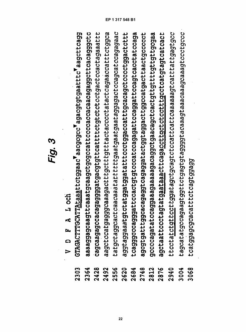
EP 1 317 548 B1
22

EP 1 317 548 B1
23

EP 1 317 548 B1
24
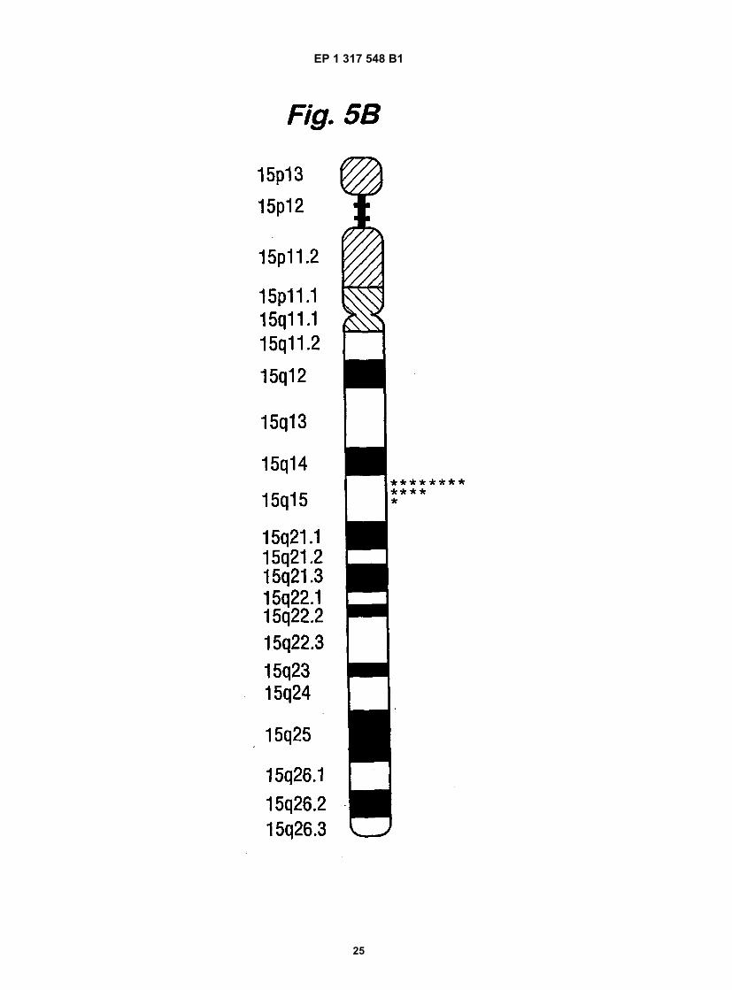
EP 1 317 548 B1
25
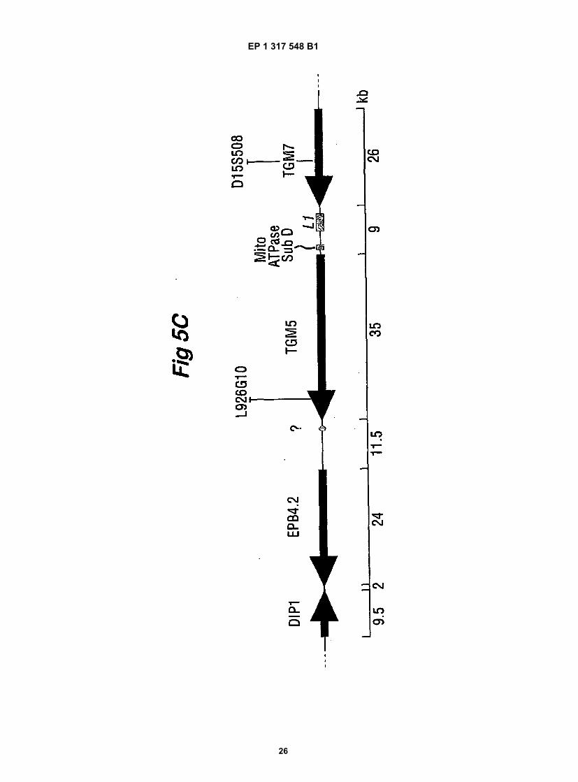
EP 1 317 548 B1
26

EP 1 317 548 B1
27

EP 1 317 548 B1
28

EP 1 317 548 B1
29

EP 1 317 548 B1
30

EP 1 317 548 B1
31

EP 1 317 548 B1
32

EP 1 317 548 B1
33

EP 1 317 548 B1
34

EP 1 317 548 B1
35

EP 1 317 548 B1
36

EP 1 317 548 B1
37

EP 1 317 548 B1
38

EP 1 317 548 B1
39

EP 1 317 548 B1
40

EP 1 317 548 B1
41

EP 1 317 548 B1
42

EP 1 317 548 B1
43

EP 1 317 548 B1
44

EP 1 317 548 B1
45

EP 1 317 548 B1
46

EP 1 317 548 B1
47

EP 1 317 548 B1
48

EP 1 317 548 B1
49

EP 1 317 548 B1
50
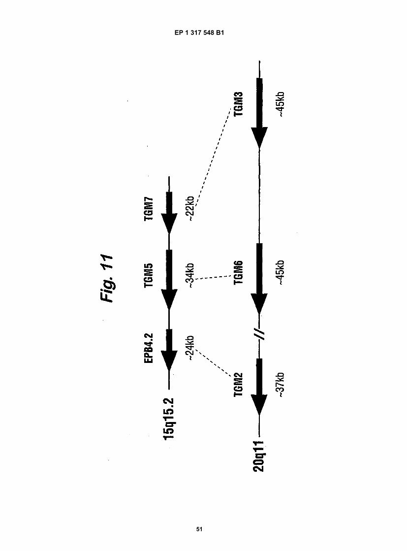
EP 1 317 548 B1
51

EP 1 317 548 B1
52
REFERENCES CITED IN THE DESCRIPTION
This list of references cited by the applicant is for the reader’s convenience only. It does not form part of the Europeanpatent document. Even though great care has been taken in compiling the references, errors or omissions cannot beexcluded and the EPO disclaims all liability in this regard.
Patent documents cited in the description
• US 5726051 A [0006]
Non-patent literature cited in the description
• Aeschlimann, D ; Paulsson, M. Thromb. Haemos-tasis, 1994, vol. 71, 402-415 [0003]
• Aeschlimann, D. et al. J. Biol Chem, 1998, vol. 273,3542-3460 [0003]
• Aeschlimann, D. ; Thomazy V. Connective TissueRes., 2000, vol. 41, 1-27 [0003]
• Aeschlimann et al. J. Biol. Chem., 1998, vol. 273,3452-3460 [0007] [0048] [0056]
• Sung L. A. et al. Blood, 1992, vol. 79, 2763-2770[0009]
• Najfeld V. et al. Am. J. Hum. Genet, 1992, vol. 50,71-75 [0009]
• Chiannikulchai N. et al. Hum. Mol. Genet, 1995,vol. 4, 717-725 [0009]
• Aeschlimann et al. J. Biol Chem., 1998, vol. 273,3452-3460 [0013] [0055]
• Aeschlimann et al. J. Biol Chem, 1998, vol. 273,3453-3460 [0031]
• Saitou ; Nei. Mol. Biol. Evol., 1987, vol. 4, 406-425[0031]
• Mount S.M. Nucleic Acids Res., 1982, vol. 10,459-472 [0035]
• Keller E. B ; Noon. W. A. Proc. Natl. Acad. Sci. USA,1984, vol. 81, 7417-7420 [0035]
• Aeschlimann et al. J. Biol Chem, 1998, vol. 273,3452-3460 [0036]
• White, R.A et al. Nat. Genet., 1992, vol. 2, 80-83[0043]
• Nanda, N. et al. Arch. Biochem. Biophys, 1999, vol.366, 151-156 [0043]
• Gentile V. et al. Genomics, 1994, vol. 20, 295-297[0044]
• Kim I. G. et al. J. Invest. Dermatol, 1994, vol. 103,137-142 [0044]
• White R. A. et al. Nat. Genet, 1992, vol. 2, 80-83[0044]
• Aeschlimann et al. J. Biol Chem, 1998, vol. 273,3245-3460 [0045]
• Bebbington C. R. ; Hentschel, C. C. G. DNA clon-ing. IRL Press, 1987, vol. III, 184-188 [0059]
• Korsgren et al. Biochemistry, 1990, vol. 87, 613-617[0062]
• McCarthy et al. Genome Res., 1997, vol. 7,1153-1161 [0064]