transformation of quiescent adult oligodendrocyte …ng2-creer does not target adult neural stem...
TRANSCRIPT

Transformation of quiescent adult oligodendrocyteprecursor cells into malignant glioma througha multistep reactivation processRui Pedro Galvaoa, Anita Kasinaa, Robert S. McNeillb, Jordan E. Harbinc, Oded Foremand, Roel G. W. Verhaake,Akiko Nishiyamaf, C. Ryan Millerb,g, and Hui Zonga,h,1
aInstitute of Molecular Biology, University of Oregon, Eugene, OR 97403; bDepartment of Pathology and Laboratory Medicine, University of North CarolinaSchool of Medicine, Chapel Hill, NC 27599; cDepartment of Biological Sciences, California Polytechnic State University, San Luis Obispo, CA 93401;dDepartment of Pathology, Genentech, South San Francisco, CA 94080; eDepartment of Genomic Medicine, Department of Bioinformatics and ComputationalBiology, University of Texas MD Anderson Cancer Center, Houston, TX 77030; fDepartment of Physiology and Neurobiology, University of Connecticut, Storrs,CT 06269; gLineberger Comprehensive Cancer Center, Neurosciences Center, and Department of Neurology, University of North Carolina School of Medicine,Chapel Hill, NC 27599; and hDepartment of Microbiology, Immunology, and Cancer Biology, University of Virginia, Charlottesville, VA 22908
Edited by Ben A. Barres, Stanford University School of Medicine, Stanford, CA, and approved August 28, 2014 (received for review August 1, 2014)
How malignant gliomas arise in a mature brain remains a mystery,hindering the development of preventive and therapeutic inter-ventions. We previously showed that oligodendrocyte precursorcells (OPCs) can be transformed into glioma when mutations areintroduced perinatally. However, adult OPCs rarely proliferate com-pared with their perinatal counterparts. Whether these relativelyquiescent cells have the potential to transform is unknown, which isa critical question considering the late onset of human glioma.Additionally, the premalignant events taking place between initialmutation and a fully developed tumor mass are particularly poorlyunderstood in glioma. Here we used a temporally controllable Cretransgene to delete p53 and NF1 specifically in adult OPCs and dem-onstrated that these cells consistently give rise to malignant gliomas.To investigate the transforming process of quiescent adult OPCs, wethen tracked these cells throughout the premalignant phase, whichrevealed a dynamic multistep transformation, starting with rapid buttransient hyperproliferative reactivation, followed by a long period ofdormancy, and then final malignant transformation. Using pharmaco-logical approaches, we discovered that mammalian target of rapamy-cin signaling is critical for both the initial OPC reactivation step andlate-stage tumor cell proliferation and thusmight be a potential targetfor both glioma prevention and treatment. In summary, our resultsfirmly establish the transformingpotential of adultOPCs and reveal anactionable multiphasic reactivation process that turns slowly dividingOPCs into malignant gliomas.
cancer | cellular quiescence | cellular reactivation | mTOR signaling
The quiescence and reactivation of progenitor cells play es-sential roles in tissue homeostasis, regeneration, and cancer
(1–5). As tissues mature, progenitor cells often enter a quiescentstate characterized by greatly reduced proliferation relative toembryonic levels, although they can be transiently reactivatedinto high levels of proliferation by physiologic stimuli or signalsfor tissue repair. Although most progenitors become increasinglyquiescent with aging (6–9), cancer incidence is highest in seniors,suggesting a critical role of cellular reactivation for tumorigenesis.Because not all cell types can be equally activated (10–13), iden-tifying the cell of origin for adult cancers and understanding themechanisms of their reactivation should provide critical insightsfor cancer prevention and treatment.Malignant gliomas are devastating, incurable brain tumors, and
particularly little is known about the early stages of their de-velopment. Recently, we showed that perinatal oligodendrocyteprecursor cells (OPCs) can give rise to glioma upon the loss of p53and neurofibromatosis 1 (NF1) (14), two of the most frequentlymutated tumor suppressor genes in human glioblastoma (GBM)(15, 16). However, the transforming potential of perinatal OPCscould result from a synergy between genetic lesions and the rapid
proliferation of OPCs at this age. Compared with their perinatalcounterparts, adult OPCs are relatively quiescent (9, 17–19). Al-though all adult OPCs retain the ability to proliferate, they do so ata very slow rate, dividing only once in 36 d at P60 (60-d-old mice)compared with every 4 d at P6 (9, 19). Furthermore, adult OPCsadopt a transcriptional program with much decreased expression ofcell cycle genes (20, 21), thus contributing to their infrequentproliferation. Reactivation of adult quiescent OPCs has beenpreviously characterized in response to neuronal activity and tobrain injury, both of which trigger proliferation followed by dif-ferentiation into myelinating oligodendrocytes (22–24), but neverin the context of oncogenic mutations. Because 87% of humanmalignant gliomas occur after 55 y of age (25), it is necessary toinvestigate whether adult OPCs can be transformed into tumors,and if so, how they become reactivated. Mechanisms regulatingprogenitor cell quiescence are beginning to emerge (1, 26),including the mammalian target of rapamycin (mTOR) path-way, which has known functions in normal OPC biology (27–30). If adult OPCs can form gliomas, such pathways might playimportant roles in the tumorigenic process.Here we focused on adult high-grade gliomas rather than
pediatric gliomas, which are molecularly and clinically distinct
Significance
How malignant gliomas arise in a mature brain remains a mys-tery, which hinders the development of effective treatments.Which cell types can escape their quiescent, adult state and howthey do so is unknown. Additionally, because gliomas are onlydetected at advanced stages, the full course of transformationremains uncharacterized. Here we report that adult oligoden-drocyte precursor cells, despite their relatively quiescent prop-erties, can be reactivated to a highly proliferative state by p53and NF1 mutations and give rise to malignant gliomas. Further-more, we describe the early phase of gliomagenesis for the firsttime, revealing a multistep process of reactivation, dormancy,and final transformation in which mammalian target of rapa-mycin signaling plays a critical role at both early and late steps.
Author contributions: R.P.G., R.S.M., C.R.M., and H.Z. designed research; R.P.G., A.K., R.S.M.,and J.E.H. performed research; A.N. contributed new reagents/analytic tools; R.P.G., A.K.,R.S.M., J.E.H., O.F., and R.G.W.V. analyzed data; and R.P.G. and H.Z. wrote the paper.
The authors declare no conflict of interest.
This article is a PNAS Direct Submission.
Data deposition: The microarray data reported in this paper have been deposited in theGene Expression Omnibus (GEO) database, www.ncbi.nlm.nih.gov/geo (accession nos.GSE61282 and GSE26676).1To whom correspondence should be addressed. Email: [email protected].
This article contains supporting information online at www.pnas.org/lookup/suppl/doi:10.1073/pnas.1414389111/-/DCSupplemental.
E4214–E4223 | PNAS | Published online September 22, 2014 www.pnas.org/cgi/doi/10.1073/pnas.1414389111
Dow
nloa
ded
by g
uest
on
May
27,
202
0

(31, 32). Therefore, we inactivated p53 and NF1 specifically inadult OPCs and found that these cells consistently give rise tomalignant gliomas. Interestingly, we found that gliomagenesis fromadult OPCs is not a simple linear process. Upon mutation, OPCsbecome immediately reactivated, reaching the proliferative rate ofperinatal OPCs. Rather than continue to proliferate, mutant OPCsreturn to a dormancy state, until eventually one or a few cells es-cape dormancy for a second time, and malignant transformationensues in specific brain regions. Knowledge of this process couldyield novel opportunities to halt the progression of gliomagenesis.To exploit that possibility, we tested the role of mTOR signalingand found that it is critical for both initial OPC reactivation andlater-stage tumor cell proliferation, making this pathway a goodcandidate for both glioma therapy and prevention.
ResultsEstablishing a Model to Examine the Transforming Potential of AdultOPCs. To determine whether adult OPCs can be transformed intogliomas, we established a model that contains (i) an NG2-CreERtransgene to excise floxed alleles specifically in OPCs in a tamoxi-fen (Tmx)-dependent fashion (33), (ii) floxed alleles of p53 andNF1 (p53KO/flox,NF1flox/flox), two of the most frequently mutated tu-mor suppressor genes in human glioma (15), and (iii) a Cre-inducible reporter gene, ROSA26-LSL-tdTomato (tdT), to identifyrecombined cells (Fig. 1A). We will refer to mice with homozygousconditional p53 and NF1 mutations as conditional KOs (CKOs)and to their controls (no p53 or NF1mutations) as WT. To induce
mutations specifically in adults, we administered Tmx at either 45or 180 d of age (young or aged adults).To ensure that our model targets mutations to OPCs in the
adult, we first confirmed the temporal and cell type specificity ofthe NG2-CreER transgene. To test the temporal specificity ofNG2-CreER, we analyzed the brains of adult mice not given Tmxand found very few tdT+ cells, whereas 5 d of Tmx yielded manylabeled cells (Fig. S1A), indicating that CreER activity is highlydependent on Tmx administration. Labeled cells were distributedevenly throughout the brain, with slightly fewer in the cerebellum(Fig. 1B), consistent with the known distribution of OPCs in theadult brain (34). These results suggest that our approach targetsOPCs without spatial bias. To test the cell type specificity of NG2-CreER, we characterized all tdT+ cells 1 d after a 5-d Tmxtreatment (5 dpi). The majority of tdT+ cells are platelet-derivedgrowth factor (PDGF) receptor α (PDGFRα)+ OPCs (72.1 ±1.4% SEM in olfactory bulb, n= 3mice; Fig. 1C). Other tdT+ cellsare mostly PDGFRβ+ pericytes (Fig. S1B), a nonneural cell typeknown to express NG2, as well as some CC1+ oligodendrocytes(Fig. S1C), which likely differentiated from labeled OPCs. NotdT+ astrocytes were detected (Fig. 1D). Although rare, tdT+
cortical pyramidal neurons were also found (Fig. S1D); their la-beling likely indicates a low level of NG2-CreER transgene expres-sion directly in mature neurons (33) because our experimentaltiming is too short and too late for cortical neurogenesis to occur.We conclude that our mouse model effectively targets mutations toadult OPCs without spatial bias.
Fig. 1. Glioma mouse model using inducible NG2-CreER to specifically mutate adult OPCs. (A) Beforetamoxifen (Tmx) administration, all cells are p53heterozygous (p53KO/flox), NF1 WT (NF1flox/flox), andcolorless because CreER is sequestered in the cyto-plasm. To induce mutations in floxed p53 and NF1alleles and expression of the tdTomato (tdT) re-porter transgene, Tmx was given for 5 d, starting atP45 or P180, causing CreER protein in NG2+ cells toenter the nucleus. Mice were then killed at differentdpi depending on the experimental purpose. (B)Sagittal sections of mouse WT whole brain given 5 dTmx at P45 and killed 1 d later, showing thatrecombination (tdT+ cells) is evenly distributedthroughout all brain regions. (C and D) Immuno-histochemical characterization of tdT+ cells showingthat recombination occurs in OPCs (PDGFRα+, Olig2+)(C) but not in astrocytes (Aldolase-C+, GFAP+) (D).(Scale bars, 40 μm.)
Galvao et al. PNAS | Published online September 22, 2014 | E4215
GEN
ETICS
PNASPL
US
Dow
nloa
ded
by g
uest
on
May
27,
202
0

NG2-CreER Does Not Target Adult Neural Stem Cells. Although ourdata above reinforced the reliability of our mouse model, to un-ambiguously determine the transformation potential of adult OPCs,it is particularly important to ensure that NG2-CreER does not labeladult neural stem cells (NSCs), which are known to give rise to gli-oma upon oncogenic mutations (35). To address this question, wefirst characterized tdT+ cells in the subventricular zone (SVZ), thelargest stem cell niche in the adult brain. The SVZ contains GFAP+
(glial fibrillary acidic protein) NSCs (type B cells), which generateAscl1+ transient amplifying cells (type C cells) that differentiate intoPSA-NCAM+ neuroblasts (36) (type A cells; Fig. 2A). We found notdT+ cells within the SVZ that could be identified as typeB (GFAP+),type C (Ascl1+/PDGFRα−), or type A (PSA-NCAM+/Olig2−)cells (Fig. 2 B–E), strongly suggesting that SVZ NSCs are notlabeled by NG2-CreER. In a complementary experiment, weanalyzed the olfactory bulb (OB), the final destination of SVZ
NSC-derived neuroblasts, for signs of adult-generated neuronsderived from tdT+ NSCs (Fig. S2). We administered BrdU(5-bromo-2′-deoxyuridine) to adult WT mice to label newly gen-erated cells (Fig. S2E) and stained for BrdU and NeuN (Neu-ronal Nuclei, neuron marker) at 51 dpi (Fig. S2 C and D), whenall NSC-derived neurons would have migrated to the OB andmatured (37, 38). If NG2-CreER mediates recombination inNSCs, we would expect to see a population of newly formed tdT+
OB neurons (tdT+/BrdU+/NeuN+). Although we found many newneurons (BrdU+/NeuN+), none of them were tdT+, suggesting thatNG2-CreER does not induce recombination in SVZ NSCs. Basedon both SVZ and OB analyses, we conclude that NG2-CreER doesnot induce recombination in SVZ NSCs.
Adult OPCs Generate Malignant Gliomas upon p53/NF1 Mutation.Having confirmed the reliability of our mouse model, we next
Fig. 2. NG2-CreER does not target adult SVZ neural stem cells. (A) Diagrams illustrating the cell types in the SVZ and oligodendrocyte lineages and the molecularmarkers used to identify them. (B–D) WTmice were given Tmx for 5 d at P45 and then killed 2 d (6 dpi; B), 3 d (7 dpi; C), or 7 d (11 dpi; D) later, allowing sufficienttime for each cell type to be labeled or generated (85). Brains were then immunostained to detect tdT+ cells expressing markers of type B (GFAP, B), type C (Ascl1,C), or type A cells (PSA-NCAM, D) in the SVZ. DAPI nuclear staining (omitted for simplicity) was used to define the boundaries of the cell-dense SVZ (dotted lines).PDGFRα, expressed by OPCs but not C cells, was used to distinguish these cell types (both can be Ascl1+). Olig2, expressed by OPCs but not A cells, was used todistinguish these cell types (both can be PSA-NCAM+). (E) Quantification results, showing that tdT+ cells in the SVZ are either in the oligodendrocyte lineage orlikely pericytes (PDGFRβ+), but not part of the SVZ stem cell lineage (n = 4 mice for type A cells, n = 3 mice for others). (Scale bars, 50 μm.)
E4216 | www.pnas.org/cgi/doi/10.1073/pnas.1414389111 Galvao et al.
Dow
nloa
ded
by g
uest
on
May
27,
202
0
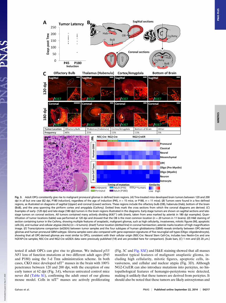
tested if adult OPCs can give rise to gliomas. We induced p53/NF1 loss of function mutations at two different adult ages (P45and P180) using the 5-d Tmx administration scheme. In bothcases, CKO mice developed tdT+ masses in the brain with 100%penetrance between 120 and 200 dpi, with the exception of oneearly tumor at 62 dpi (Fig. 3A), whereas untreated control micenever did (Table S1), confirming the adult onset of our gliomamouse model. Cells in tdT+ masses are actively proliferating
(Fig. 3C and Fig. S3E) and H&E staining showed that all massesmanifest typical features of malignant anaplastic glioma, in-cluding high cellularity, mitotic figures, apoptotic cells, in-vasiveness, and cellular and nuclear atypia (Fig. 3D). AlthoughNG2-CreER can also introduce mutations into pericytes, no his-topathological features of hemangio-pericytoma were detected,making it unlikely that these tumors are derived from pericytes. Itshould also be noted that these tumors are likely astrocytomas and
Fig. 3. Adult OPCs consistently give rise to malignant proneural gliomas in defined brain regions. (A) Tmx-treated mice developed brain tumors between 120 and 200dpi in all but one case (62 dpi, P180 induction), regardless of the age of induction (P45, n = 15 mice, or P180, n = 11 mice). (B) Tumors were found in a few definedregions, as illustrated on diagrams of sagittal (Upper) and coronal (Lower) sections. These regions include the olfactory bulb (OB), habenula (Hab), bottom of the brain(BoB), and the area spanning the piriform cortex and amygdala (Cx/Amy). Dotted lines mark the cross sections from which the coronal diagrams are derived. (C)Examples of early- (120 dpi) and late-stage (180 dpi) tumors in the brain regions illustrated in the diagrams. Early-stage tumors are shown on sagittal sections and late-stage tumors on coronal sections. All tumors contained many actively dividing (Ki67+) cells (Insets, taken from area marked by asterisk in 180 dpi examples). Quan-tification of tumor locations (table) was performed at 120 dpi and showed that the OB is the most common location (n = 20 tumors in 11 brains). (D) H&E staining ofsection containing tumor in the Cx/Amy, showing multiple features of anaplastic, malignant gliomas, such as high cellularity, invasiveness, mitotic figures (M), apoptoticcells (A), and nuclear and cellular atypias (Ak/Ac) (n = 6 tumors). (Inset) Tumor location (dotted line) in coronal hemisection; asterisk marks location of highmagnificationimage. (E) Transcriptome comparison (ssGSEA) between tumor samples and the four subtypes of human glioblastoma (GBM) reveals similarity between OPC-derivedgliomas and human proneural GBM subtype. Glioma samples were also compared with gene expression signatures of five neuroglial cell types (Oligo: oligodendrocyte),showing that all OPC-derived gliomas are most similar to OPCs, consistent with their cellular origin (NSC-Cre: Neural Stem Cell-Cre, includes two Nestin-Cre and onehGFAP-Cre samples; NSC-Cre and NG2-Cre ssGSEA data were previously published (14) and are provided here for comparison). [Scale bars, (C) 1 mm and (D) 20 μm.]
Galvao et al. PNAS | Published online September 22, 2014 | E4217
GEN
ETICS
PNASPL
US
Dow
nloa
ded
by g
uest
on
May
27,
202
0

not oligodendrogliomas based on the presence of angular, pleo-morphic, rather than rounded, isomorphic nuclei and the lack ofperinuclear halos or branched capillaries (Fig. 3D) (39, 40). Theengraftment of tumor cells into the brains of NOD/SCID miceconsistently gave rise to large, invasive secondary and tertiary tumorsafter 35–50 d (Table S1), confirming the malignancy of OPC-derived gliomas. Additionally, tumor cells cultured in clonalconditions exhibited self-renewing and multilineage differentia-tion potential (Fig. S3 A–D), suggesting the existence of tumor-propagating cells within adult OPC-derived gliomas. Therefore,we conclude that even relatively quiescent OPCs in a fully ma-ture brain can give rise to glioma.Interestingly, we noticed that tumors were not randomly dis-
tributed, despite equal distribution of mutant OPCs in all brain
regions (Fig. 1B). Because it is difficult to pinpoint the anatomicalorigin of late-stage tumors due to their large size and infiltrativenature, we collected a set of brains at 120 dpi, when tumors aresmall, to quantify tumor frequency at distinct locations. The mostcommon location was the OB (40%), followed by the habenula, asmall region in the dorsal thalamus (25%), piriform cortex/amyg-dala (15%), and the space between the hypothalamus and the baseof the skull (15%) (Fig. 3 B and C and Table S1). Altogether, weconclude that OPCs in the adult brain can exit their quiescent stateand give rise to malignant anaplastic gliomas upon p53/NF1 deletion.
Adult-Induced Gliomas Are Similar to Human Proneural Glioma andMaintain OPC Features. Recent studies have used genomic andtranscriptomic approaches to define glioma subtypes within
Fig. 4. Reactivation of mutant OPCsupon NG2-CreER–mediated mutagene-sis. (A and B) Representative images ofOB sagittal sections from 12 dpi WT (A)and CKO (B) mice immunostained forthe reporter gene (tdT), an OPC marker(PDGFRα), and a proliferation marker(BrdU). (C) Quantification of the fractionof proliferating tdT+ OPCs in the OB atvarious time points starting before Tmxwas administered (0 dpi), as well as12, 50, and 90 dpi, revealing a transientspike in mutant OPC proliferation at 12dpi (for 0 dpi, proliferation was quanti-fied among all PDGFRα+ cells; no tdT+
cells). Dashed line, proliferation level ofperinatal (P6) OPCs (n = 4). (D) Quanti-fication in the OB of the total number oftdT+ OPCs starting before Tmx was ad-ministered (0 dpi), as well as 12, 50, and90 dpi, showing that mutant OPCs ac-cumulate throughout the premalignantphase, even after the proliferative ratereturns to basal level (n = 4). (E) Locationof images shown in A and B and diagramof experimental procedure. (F and G)Representative images of OB sagittalsections from 90 dpi WT (F) and CKO (G)mice immunostained for an OPC marker(PDGFRα) and a mature oligodendrocytemarker (CC1) (white arrows, tdT+/PDGFRα+ cells; blue arrows, tdT+/CC1+
cells). (H) Quantification of the percent-age of tdT+/PDGFRα+ (OPCs), tdT+/CC1+
(oligodendrocytes), or tdT+/PDGFRα+/CC1+
cells in the OB of WT and CKO mice at90 dpi, suggesting mutant OPCs cannotdifferentiate effectively (n = 4 CKO and3 WT mice). (I) Location of images shownin F and G and diagram of experimentalprocedure. Error bars represent SEM andstatistical significance was determinedby Student t test (*P < 0.05; **P < 0.01;***P < 0.001). (Scale bars, 50 μm.)
E4218 | www.pnas.org/cgi/doi/10.1073/pnas.1414389111 Galvao et al.
Dow
nloa
ded
by g
uest
on
May
27,
202
0

pathologically indistinguishable tumor samples (41–46). Thesemolecular classifications provide valuable insight for prognosticpredictions and development of targeted therapies and emphasizethe importance of matching distinct mouse glioma models tohuman subtypes. Therefore, we used microarray data to comparethe transcriptional profile of 12 adult OPC-derived gliomas to thatof human GBM samples by single sample Gene Set EnrichmentAnalysis (ssGSEA), which uses predefined sets of genes to de-termine the relative similarity between samples (Fig. 3E). Thegliomas selected for analysis were large, terminal tumors thatsometimes spanned several brain regions but likely originated inthe OB (tumors 1, 3, 7, and 8), thalamus (tumor 10), piriformcortex/amygdala (tumors 2, 4, 5, and 9), and base of the skull (tumor6), as well as two secondary tumors grafted from the OB and cortex/amygdala into the striatum (tumors 11 and 12, respectively; TableS1). We found that, regardless of anatomical origin or age of in-duction, most tumors from our model resemble the proneuralsubtype of human GBM (46), although some could not be un-ambiguously identified as proneural or classical (tumors 5–8; Fig.3E). Consistent with their proneural signature, our adult-inducedgliomas were similar to OPCs, but not neurons or astrocytes (Fig.3E and Fig. S3 E–I) (46). Markers of pericytes and hemangio-pericytomas (47) were also absent from these gliomas, consistentwith their OPC origin (Fig. S3I). Interestingly, although manyGFAP+ cells were detected within the tumor masses, these werenot tdT+ (Fig. S3H), indicating that they are most likely local re-active astrocytes. These data concur with our previous findingswith neonatal OPC-derived tumors (14) (Fig. 3E), suggesting that
gliomas originated from p53/NF1-null OPCs generally belong tothe proneural subtype regardless of the timing of mutations.
Adult OPC Gliomagenesis Follows a Multistep Process of Reactivation,Dormancy, and Malignancy. Having confirmed the transformationcapability of adult OPCs, we next investigated how these rarelydividing progenitors become reactivated. To pinpoint the timingof OPC reactivation, we quantified both proliferative rate and totalnumber of OPCs at several time points during the premalignantphase (preceding a detectable tumor). We initially focused on theOB, where gliomas most commonly develop in our model (Fig. 4A–E). Before Tmx is given (0 dpi), WT and CKOOPCs have equallylow proliferative rates. Just 12 d after Tmx (12 dpi), the proliferativerate ofmutantOPCs increased to perinatal levels (54.9± 3.6%SEM;n = 3), but then quickly decreased to below WT levels at 50 dpi andremained low even at 90 dpi, just 30 d before gliomas begin to appear(Fig. 4C). As a consequence of reactivation, the number of tdT+
mutant OPCs rose sharply in the first 12 d after Tmx administra-tion. Surprisingly, the number of mutant OPCs continued to ex-pand long after cells returned to WT proliferation levels (Fig. 4D),likely due to defects in differentiation of mutant OPCs (see below).In contrast, the number of labeled WT OPCs remained constant(Fig. 4D). Interestingly, we observed similar proliferation and cellnumber dynamics not only in other tumor hot spots (habenula), butalso in regions where gliomas did not normally form (cingulate/motor cortex and corpus callosum; Fig. S4 A–F) except pro-liferation in the corpus callosum, which was identical in WT andCKOmice. Overall, these results suggest that additional events areneeded for malignant transformation.
Fig. 5. mTOR signaling is necessary for mutant OPC reactivation. (A and B) Immunostaining of the OB of CKO mice treated with vehicle or temsirolimus,showing that the levels of phospho-S6 (pS6, mTOR downstream target) are drastically reduced by the drug. (C) Western blot for pS6 and total S6 protein inthe OB, showing temsirolimus blocks phosphorylation of S6 by mTOR. Mice were treated for 10 (A and B) or 5 (C) d and then killed 4 h after the last dose. (D)Diagrams of experimental procedure and brain region analyzed (red arrow). (E) Quantification of the fraction of proliferating tdT+ OPCs at 12 dpi in the OBof CKO mice treated with vehicle (Veh, control) or temsirolimus (Tem). Tem significantly decreases mutant OPC proliferation to WT adult OPC proliferationlevels (dotted line) (Veh, n = 3; Tem, n = 4; **P < 0.01). (Scale bar, 100 μm.)
Galvao et al. PNAS | Published online September 22, 2014 | E4219
GEN
ETICS
PNASPL
US
Dow
nloa
ded
by g
uest
on
May
27,
202
0

In addition to proliferation, the differentiation of OPCs intomature oligodendrocytes also affects their homeostatic balance. Todeterminewhether p53/NF1mutations perturbOPCdifferentiation,we quantified the percentage of tdT+ cells expressing PDGFRα(OPCs) or CC1 (oligodendrocytes) in theOB ofWT andCKOmiceat 90 dpi (Fig. 4 F–I). Compared with WT mice, CKO mice hadsignificantly more tdT+/PDGFRα+ cells and significantly fewertdT+/CC1+ cells, indicating an impairment in OPC differentiation.As with proliferation, impaired differentiation was observed in allregions tested (Fig. S4G), including the corpus callosum, whichcould explain the increase in OPC numbers despite a lack ofhyperproliferation in that region. Therefore, compromised dif-ferentiation of mutant OPCs, together with the early spike inproliferation, likely contributes to their continuous expansionthroughout the premalignant phase, eventually leading to gli-oma. Importantly, OPCs mutated at P180 exhibited the samehyperproliferative (49% vs. 20% BrdU+, P < 0.01) and un-differentiated (88% vs. 72% PDGFRα+, P < 0.01) behaviors at12 dpi (n = 3), indicating that OPC reactivation competence isnot lost over time. In summary, we found that gliomagenesisfrom adult OPCs involves multiple steps of cellular behavioralternations, including robust but transient reactivation in allbrain regions immediately after mutagenesis, followed by slow butsteady mutant cell expansion during a long period of apparentdormancy, and ending with sudden malignant transformation.
mTOR Inhibition Blocks Mutant OPC Reactivation Step. To study themolecular nature of the reactivation step, we focused on themTOR signaling pathway, which has recently emerged as a keymechanism in the reactivation of quiescent neural, hematopoi-etic, and muscle stem cells (48–51) and is known to play a rolein OPC proliferation and differentiation (27–30, 52). Therefore,we decided to test its involvement in mutation-induced OPCreactivation by administering the mTOR inhibitor temsirolimusthroughout the 12 dpi period. The effectiveness of target inhibitionin the brain was confirmed by immunostaining and Western blot(Fig. 5 A–C), both of which showed that brains of mice treatedwith temsirolimus have drastically reduced levels of phosphory-lated S6 ribosomal protein (pS6), a downstream target of mTOR.
Quantification of proliferation showed that OPCs in CKO micetreated with temsirolimus no longer underwent the characteristic12 dpi spike, but instead remained at WT levels (Fig. 5E). TotalOPC numbers were also reduced by mTOR inhibition (Fig. S5).These results suggest that mTOR signaling is necessary for theearly reactivation of mutant OPCs.
mTOR Signaling Rises Dramatically in Malignant OPCs and Is Criticalfor Glioma Cell Proliferation. As our data above implicate mTORsignaling in the initial reactivation step of the premalignantphase, we next investigated whether this pathway is necessary atthe malignant phase as well. We first quantified the percentage ofpS6+ OPCs at premalignant and malignant stages. We found thatmost pS6+ cells in the adult OB are neurons (Fig. S6A), and that,unexpectedly, only 2% of OPCs are pS6+ in WT or CKO mice at12 dpi (Fig. 6B and Fig. S6 B and C), perhaps due to low levels ofmTOR signaling (Discussion). In stark contrast, the percentage ofpS6+ OPCs increased drastically to 20% in the tumor mass (Fig. 6 Aand B). We observed the same trend when staining for the phos-phorylated form of another mTOR downstream target, 4E-bindingprotein 1 (4E-BP1), which was rarely seen before malignancy butquite common in tumor cells (Fig. S6D). Therefore, mTOR sig-naling appears to be highly up-regulated in malignant OPCs.To test the role of mTOR signaling in OPC-derived glioma cells,
we cultured cells from one of our primary gliomas with temsir-olimus. Tumor cell growth was acutely sensitive to this mTORinhibitor at doses that inhibited S6 and 4E-BP1 phosphorylation(Fig. 6D and Fig. S6F). Cell cycle analysis by flow cytometry dem-onstrated that this effect was due to cell cycle arrest at the G1 stage(Fig. 6 E and F), consistent with the known roles of mTOR sig-naling in promoting cell cycle progression at the G1 to S transition(53). These results were confirmed with EdU (5-ethynyl-2′-deoxy-uridine) quantifications by immunostaining, as well as usinganother mTOR inhibitor (rapamycin, from which temsirolimusis derived) and another primary glioma cell line (Fig. S6G–I). Weconclude that OPC-derived glioma cells greatly increase cell in-trinsic mTOR signaling, which is important for their proliferation.
Fig. 6. mTOR signaling rises dramatically in malignantOPCs and is critical for glioma cell proliferation. (A) Immu-nostaining for phospho-S6 (pS6, mTOR target) in gliomatissue. (B) Quantification of the percentage of OPCs (tdT+/PDGFRα+) and OPC-derived glioma cells (tdT+ cells in tumormass, typically all PDGFRα+ as seen on Fig. S6D; adjacentsection of tumor in A and Fig. S3F) labeled with pS6, in-dicating higher levels of mTOR signaling in glioma cells (n = 3;**P < 0.01). (C) Diagrams of location of image shown in A andexperimental procedure. (D) Cellular viability assay of culturedglioma cells treated with temsirolimus, revealing a significantimpact of this mTOR inhibitor on glioma cell growth in vitro.(E) Quantification of the fraction of glioma cells at differentphases of the cell cycle (G0/G1, S, G2/M) and apoptosis (sub-G1) after exposure to increasing doses of temsirolimus,showing an increase in G0/G1 and decrease in G2/M thatsuggests G1 arrest. (F) DNA content distribution of untreatedand temsirolimus-treated tumor cells, illustrating the drug-induced cell cycle arrest at G1 (jagged black line, actual datacurve; smooth black line, fitted data curve; colored areas,cells presumed to be in different cell cycle stages, as per colorcode). (Scale bar, 100 μm.)
E4220 | www.pnas.org/cgi/doi/10.1073/pnas.1414389111 Galvao et al.
Dow
nloa
ded
by g
uest
on
May
27,
202
0

DiscussionA deep understanding of the timing and process of gliomagenesisshould greatly facilitate the development of effective therapiesand biomarkers for early detection. We developed a geneticmouse model with both cell type specificity and temporal controlto study gliomagenesis from adult OPCs for the first time. Usingthis model, we showed that quiescent adult OPCs can be reac-tivated upon mutagenesis and transform into malignant gliomasthat preferentially develop in defined locations. Time courseanalysis revealed a surprising, multistep behavior of mutant OPCreactivation, dormancy, and malignant transformation. Finally, weuncovered an important role for mTOR signaling at both early andlate stages of gliomagenesis.Although previous studies have suggested NSCs as the cell of
origin for glioma (35, 54, 55), our prior work showed that eventhough introducing mutations in NSCs can lead to glioma, ma-lignant transformation only occurs in OPCs, thus warranting adistinction between cell of mutation (NSCs) and cell of origin(OPCs) (14). It remains possible that additional cell types also actas a cell of origin, such as the OPC-like type C* cells described byothers (56), or that different glioma subtypes are derived fromdifferent cells of origin, such as low-grade ocular tract gliomas,which might not be OPC-derived (57). Nonetheless, overwhelmingevidence now supports OPCs as a cell of origin for glioma (14, 58–64). However, these studies either introduced oncogenes into OPCsearly in development, as in our previous work (14), or did not uselineage tracing tools (58, 59). Thus, it is possible that the glioma-genic potential of OPCs could be dependent on their active peri-natal state (65, 66) and that adult OPCs may not be transformablebecause they rarely divide (9, 17–21). Our current study convinc-ingly resolved this issue by introducing mutations specifically intoadult OPCs and demonstrating their capacity for transformation.Because most human malignant gliomas occur in elderly adults(25), our findings not only firmly establish OPCs as a cell of originfor glioma but also highlight the clinical relevance of this discovery.Because gliomas have no known morphologically detectable
preneoplastic lesions, it is difficult to study early events before finalmalignancy. Taking advantage of the lineage marker tdTomato inour mouse model, we followed the progression of gliomagenesisand found that it is not a linear process but rather marked bysuccessive steps of reactivation, dormancy, and malignant trans-formation. The reactivation step is likely caused by signaling per-turbations due to the acute loss of p53 and NF1, but mutant OPCsquickly return to quiescence/dormancy. This dormancy could bedue to cell intrinsic tumor suppressor mechanisms activated by NF1loss of function (67) or cell extrinsic mechanisms including com-petition for limited trophic support or cell contact-mediatedgrowth inhibition as the number of mutant OPCs increases (68).Interestingly, mutant OPCs continue to accumulate throughoutthis dormant period, likely due to an inability to differentiate intooligodendrocytes. This lack of differentiation distinguishesmutation-induced from physiological or injury-induced OPCreactivation, which is a controlled process ending with differen-tiation of the reactivated cells (22–24). Eventually, one or moremutant cells exit dormancy to undergo malignant transformation,triggering the formation of a tumor mass. The mechanism formalignant transformation remains unclear, but likely depends onadditional mutations that allowOPCs to overcome the barriers thatkept them dormant after the initial reactivation. Overall, the dis-covery of a stepwise process of gliomagenesis providesmultiple newpotential therapeutic approaches, such as early interventions toprevent OPC reactivation or to induce their differentiation, whichmight help prevent gliomagenesis by halting the progression towardmalignant transformation.The malignant transformation step could also be dependent
on niche properties of certain brain regions, as suggested by ourfinding that malignant gliomas develop preferentially in specific
locations. In fact, regional differences in the expression of sig-naling molecules have been suggested to account for the uniqueproperties of CNS progenitors in those areas as well as their re-sponse to oncogenic mutations (12, 13, 69, 70). Alternatively, OPCsthemselves could have heterogeneous characteristics depending onthe brain region (52, 71, 72), which could influence their trans-formation potential. Future studies investigating these possibilitiesmight provide critical insights into what drives gliomagenesis.The mTOR pathway has recently emerged as a key mechanism
in stem cell reactivation. In Drosophila, nutrient-stimulatedTOR signaling reactivates quiescent neural stem cells during theembryo-to-larva transition (51). In mammals, adult hematopoieticstem cells are kept quiescent via TSC1-mediated inhibition ofmTOR (48, 49), and quiescent adult muscle stem cells display anmTOR-dependent increase in proliferation and cell size after in-jury (50). In OPCs, mTOR signaling promotes proliferation anddifferentiation during development (27–30, 52), and its activity isblocked by p53 via stress-induced up-regulation of mTOR inhib-itors (73) and NF1 via Ras inhibition (74, 75). Therefore, mTORcould be part of the molecular mechanism behind mutation-induced adult OPC reactivation. Consistent with its role in de-velopment, we found that pharmacological inhibition of mTORsignaling with temsirolimus decreased WT adult OPC proliferation(22.6% vs. 5.8% BrdU+, P = 0.004) and differentiation (55.6% vs.65.6% PDGFRα+, P = 0.01). Importantly, temsirolimus treatmentalso blocked mutant OPC hyperproliferation at 12 dpi, indicatingthat this pathway is necessary for the initial reactivation step. In-terestingly, temsirolimus did not affect mutant OPC differentiation,likely because this process was already impaired by the loss of p53and NF1. Because the mTOR pathway is also active in a variety ofcancers (76), we investigated its role in malignant OPC proliferationand found that blocking mTOR strongly inhibits tumor cell growth.Overall, our results show thatmTOR signaling is involved in both theinitial reactivation of p53/NF1-mutant OPCs as well as the un-controlled proliferation of OPC-derived gliomas. These findingsshould provide the rationale and facilitate the regimen design foran effective use of the next generation of mTOR inhibitors cur-rently being developed (76). Specifically, treatment early in thecourse of disease could be beneficial and could prevent low- tohigh-grade glioma progression. Due to the complex mutationallandscape of malignant gliomas (15, 16), such treatments wouldlikely be most effective as part of a combinatorial strategy in whichmultiple deregulated pathways would be targeted.Given the clear effect of the mTOR inhibitor temsirolimus in
blockingOPC proliferation at 12 dpi, we were surprised to find thatvery few OPCs at this time point have detectable levels of pS6 orp4E-BP1, both downstream targets of mTOR and common indi-cators of pathway activity. This lack of pS6 or p4E-BP1 signalscould be due to mTOR signaling occurring below detection levelsin these cells, which could be necessary to avoid cellular senescenceand/or apoptosis, as reported for Kras and Myc oncogene sig-naling (77–81). Alternatively, mTOR signaling in premalignantOPCs could occur through untested downstream targets ortemsirolimus could exert its antiproliferative effect via mTOR-in-dependent mechanisms (82, 83). One intriguing possibility is thatmTOR signaling in pS6+ cells in the brain could have a paracrineeffect on OPCs, as occurs in nutrient-dependent reactivation ofquiescent Drosophila neural stem cells (51). Nevertheless, oncemutant OPCs become malignant, mTOR’s proliferative role is likelycell autonomous based on their robust labeling with pS6 and p4E-BP1 in vivo and their sensitivity to temsirolimus in vitro. Although itsexact mode of action warrants further investigation, temsirolimusprevents both mutant OPC reactivation and tumor cell pro-liferation, pinpointing mTOR signaling as an integral part of thegliomagenic process.In summary, our results have solidified the role of OPCs as a
cell of origin for glioma and shed light on the poorly understoodpremalignant phase of gliomagenesis. Our findings have also led
Galvao et al. PNAS | Published online September 22, 2014 | E4221
GEN
ETICS
PNASPL
US
Dow
nloa
ded
by g
uest
on
May
27,
202
0

to important questions. How do mutant OPCs become dormantafter the initial reactivation? What causes their eventual escapefrom the dormant state to form a tumor? What mechanisms areresponsible for tumor hot spots? The answers to these questionswill continue to advance our understanding of glioma and willhopefully bring us closer to effective treatments.
Materials and MethodsAdditional details are available in SI Materials and Methods.
In Vivo Drug Delivery. All mice were given daily tamoxifen doses of 200 mg/kgfor several consecutive days (2 d for perinatal mice, 5 d for adult mice, exceptmice used for dosage curve, Fig. S1A; and mice used for detection of new OBneurons, Fig. S2). BrdU was given by i.p. (adult mice) or s.c. (perinatal mice)injection at 50 mg/kg. Mice were given one injection per day for 7 d andkilled 24 h after the last injection. Temsirolimus (CCI-779; Selleckchem) wasinjected i.p. at 100 mg/kg (40 μL/g body weight) daily for 10 d. Mice werekilled 4 h after the last dose was delivered.
Tumor Cell Grafting. CKO mice suspected of having a primary brain tumorwere decapitated to dissect the tumor mass, which was then dissociated to asingle cell suspension; 105 cells were immediately grafted into the striatumof each NOD-SCID mouse.
Sphere-Forming Assay. Cultured tumor cells were plated onto nonadhering96-well plates and 60-mm dishes at clonal density (100 cells/μL, 104 cells/wellfor 96-well plates, 5 × 10⁵ cells/dish for 60-mm dishes) in medium supple-mented with EGF (20 ng/mL) and FGF2 (10 ng/mL). To differentiate, sphereswere placed on matrigel-coated plates in medium without EGF/FGF2 andwith 1% FBS for 6 d.
Microarray Analysis and Single Sample Gene Set Enrichment Analysis. 44KMouse Development OligoMicroarrays from Agilent Technologies were usedto analyze total RNA extracted from bulk tumor tissue that was hybridizedagainst a common reference sample of RNA pooled from four WT P17 mousebrain neocortices. Single sample GSEA was performed as described elsewhere(46). To compare tumor samples to the different mouse brain cell types, datafrom a previous publication (84) was downloaded and preprocessed as de-scribed previously (46) into sets of 250 variably expressed gene clusters. Theclusters used in the present study contained postnatal day 16 myelin oligo-dendrocytes (n = 4), postnatal day 6–7 OPCs (n = 2), postnatal day 7 neurons(n = 4), and postnatal day 17–30 astrocytes (n = 6). Gene sets for four pre-viously described GBM subtypes (46) were defined by selecting the top 250marker genes as provided.
Cell Growth Assay. OPC-derived glioma cells were treated with increasingconcentrations of temsirolimus (Selleckchem), rapamycin (Sigma-Aldrich), orDMSO (untreated). Growth of treated vs. untreated cells was determined at5 d after treatment using CellTiter 96 Aqueous One Solution Cell ProliferationAssay (MTS; Promega). Cell growthwas determined in quadruplicate per drugconcentration and data pooled from two independent experiments.
Cell Cycle Analysis. OPC-derived glioma cells were treated with increasingconcentrations of temsirolimus, harvested 48 h later, and stained with theGuava Cell Cycle Reagent (EMDMillipore), and DNA content was determinedusing a Guava EasyCyte Plus flow cytometer. Data were obtained in triplicateper drug concentration and data pooled from two independent experiments.For quantification of proliferation by EdU incorporation, tumor cells werecultured for 3 d in 100 nM temsirolimus and then given EdU for 8 h andimmediately fixed in 4% (wt/vol) formaldehyde.
Immunoblots. For in vivo experiments, OB tissue fromWTmice treated for 5 dwith vehicle or temsirolimus was collected 4 h after the last dose and im-mediately flash-frozen in liquid nitrogen until further processing. For in vitroexperiments, OPC-derived glioma cells treatedwith temsirolimus for 24 hwereharvested and lysed. For both types of experiments, protein concentrationwasquantified using the BCA assay (BioRad), and immunoblots were performed byresolving equal amounts of protein by gradient [8–16% (vol/vol)] SDS/PAGE(Bio-Rad) and transferred to PVDF membranes (EMD Millipore) forimmunodetection.
Quantifications and Statistics. Cryosections (16 μm thick; or 30-μm-thickvibratome sections for type A and C cell quantifications) were imaged witha confocal microscope. The significance of the quantification data was cal-culated by Student t test. All error bars represent SEM.
ACKNOWLEDGMENTS. We thank A. Henner for technical support andC. Liu, P. Gonzalez, M. Yao, M. Kohwi, D. Brautigan, Y. Zhu, K. Park,J. Kim, and S. Gutkind for critical comments on the manuscript. This workwas supported in part by National Institutes of Health Grant R01-CA136495 (to H.Z.), Medical Research Program of W. M. Keck Foundation(H.Z.), and the University of North Carolina Cancer Research Fund(C.R.M.). J.E.H. was supported by the University of Oregon Summer Pro-gram in Undergraduate Research. C.R.M. is a Damon Runyon-GenentechClinical Investigator supported in part by a Clinical Investigator award fromthe Damon Runyon Cancer Research Foundation (CI-45-09). R.S.M. isa Robert H. Wagner Scholar in the UNC Molecular and Cellular PathologyGraduate Program and a trainee in the UNC Graduate Training Programin Translational Medicine, supported in part by the Howard HughesMedical Institute. H.Z. is a Pew Scholar in Biomedical Sciences, sup-ported by the Pew Charitable Trusts.
1. Cheung TH, Rando TA (2013) Molecular regulation of stem cell quiescence. Nat RevMol Cell Biol 14(6):329–340.
2. Matsumoto A, Nakayama KI (2013) Role of key regulators of the cell cycle inmaintenance of hematopoietic stem cells. Biochim Biophys Acta 1830(2):2335–2344.
3. Montarras D, L’honoré A, Buckingham M (2013) Lying low but ready for action: Thequiescent muscle satellite cell. FEBS J 280(17):4036–4050.
4. Udagawa T (2008) Tumor dormancy of primary and secondary cancers. APMIS116(7-8):615–628.
5. Valcourt JR, et al. (2012) Staying alive: Metabolic adaptations to quiescence. Cell Cycle11(9):1680–1696.
6. Maslov AY, Barone TA, Plunkett RJ, Pruitt SC (2004) Neural stem cell detection,characterization, and age-related changes in the subventricular zone of mice.J Neurosci 24(7):1726–1733.
7. Molofsky AV, et al. (2006) Increasing p16INK4a expression decreases forebrain pro-genitors and neurogenesis during ageing. Nature 443(7110):448–452.
8. Pietras EM, Warr MR, Passegué E (2011) Cell cycle regulation in hematopoietic stemcells. J Cell Biol 195(5):709–720.
9. Psachoulia K, Jamen F, Young KM, Richardson WD (2009) Cell cycle dynamics of NG2cells in the postnatal and ageing brain. Neuron Glia Biol 5(3-4):57–67.
10. Chen W, et al. (2008) Malignant transformation initiated by Mll-AF9: Gene dosageand critical target cells. Cancer Cell 13(5):432–440.
11. Johnson RA, et al. (2010) Cross-species genomics matches driver mutations and cellcompartments to model ependymoma. Nature 466(7306):632–636.
12. Lee Y, Gianino SM, Gutmann DH (2012) Innate neural stem cell heterogeneitydetermines the patterning of glioma formation in children. Cancer Cell 22(1):131–138.
13. Lee Y, Yeh TH, Emnett RJ, White CR, Gutmann DH (2010) Neurofibromatosis-1 reg-ulates neuroglial progenitor proliferation and glial differentiation in a brain region-specific manner. Genes Dev 24(20):2317–2329.
14. Liu C, et al. (2011) Mosaic analysis with double markers reveals tumor cell of origin inglioma. Cell 146(2):209–221.
15. Cancer Genome Atlas Research Network (2008) Comprehensive genomic character-
ization defines human glioblastoma genes and core pathways. Nature 455(7216):
1061–1068.16. Parsons DW, et al. (2008) An integrated genomic analysis of human glioblastoma
multiforme. Science 321(5897):1807–1812.17. Shi J, Marinovich A, Barres BA (1998) Purification and characterization of adult oli-
godendrocyte precursor cells from the rat optic nerve. J Neurosci 18(12):4627–4636.18. Wolswijk G, Noble M (1989) Identification of an adult-specific glial progenitor cell.
Development 105(2):387–400.19. Young KM, et al. (2013) Oligodendrocyte dynamics in the healthy adult CNS: Evidence
for myelin remodeling. Neuron 77(5):873–885.20. Belachew S, et al. (2002) Cyclin-dependent kinase-2 controls oligodendrocyte pro-
genitor cell cycle progression and is downregulated in adult oligodendrocyte pro-
genitors. J Neurosci 22(19):8553–8562.21. Lin G, Mela A, Guilfoyle EM, Goldman JE (2009) Neonatal and adult O4(+) oligo-
dendrocyte lineage cells display different growth factor responses and different gene
expression patterns. J Neurosci Res 87(15):3390–3402.22. Gibson EM, et al. (2014) Neuronal activity promotes oligodendrogenesis and adaptive
myelination in the mammalian brain. Science 344(6183):1252304.23. Franklin RJ, Ffrench-Constant C (2008) Remyelination in the CNS: From biology to
therapy. Nat Rev Neurosci 9(11):839–855.24. McTigue DM, Tripathi RB (2008) The life, death, and replacement of oligodendrocytes
in the adult CNS. J Neurochem 107(1):1–19.25. Dolecek TA, Propp JM, Stroup NE, Kruchko C (2012) CBTRUS statistical report: Primary
brain and central nervous system tumors diagnosed in the United States in 2005-2009.
Neuro-oncol 14(Suppl 5):v1–v49.26. Li L, Bhatia R (2011) Stem cell quiescence. Clin Cancer Res 17(15):4936–4941.27. Baron W, Metz B, Bansal R, Hoekstra D, de Vries H (2000) PDGF and FGF-2 signaling in
oligodendrocyte progenitor cells: Regulation of proliferation and differentiation by
multiple intracellular signaling pathways. Mol Cell Neurosci 15(3):314–329.
E4222 | www.pnas.org/cgi/doi/10.1073/pnas.1414389111 Galvao et al.
Dow
nloa
ded
by g
uest
on
May
27,
202
0

28. Guardiola-Diaz HM, Ishii A, Bansal R (2012) Erk1/2 MAPK and mTOR signaling se-quentially regulates progression through distinct stages of oligodendrocyte differ-entiation. Glia 60(3):476–486.
29. Tyler WA, et al. (2009) Activation of the mammalian target of rapamycin (mTOR) isessential for oligodendrocyte differentiation. J Neurosci 29(19):6367–6378.
30. Zou J, et al. (2011) Rheb1 is required for mTORC1 and myelination in postnatal braindevelopment. Dev Cell 20(1):97–108.
31. Paugh BS, et al. (2010) Integrated molecular genetic profiling of pediatric high-gradegliomas reveals key differences with the adult disease. J Clin Oncol 28(18):3061–3068.
32. Sturm D, et al. (2014) Paediatric and adult glioblastoma: Multiform (epi)genomicculprits emerge. Nat Rev Cancer 14(2):92–107.
33. Zhu X, et al. (2011) Age-dependent fate and lineage restriction of single NG2 cells.Development 138(4):745–753.
34. Dawson MR, Polito A, Levine JM, Reynolds R (2003) NG2-expressing glial progenitorcells: an abundant and widespread population of cycling cells in the adult rat CNS.Mol Cell Neurosci 24(2):476–488.
35. Alcantara Llaguno S, et al. (2009) Malignant astrocytomas originate from neural stem/progenitor cells in a somatic tumor suppressor mouse model. Cancer Cell 15(1):45–56.
36. Ihrie RA, Alvarez-Buylla A (2011) Lake-front property: A unique germinal niche by thelateral ventricles of the adult brain. Neuron 70(4):674–686.
37. Petreanu L, Alvarez-Buylla A (2002) Maturation and death of adult-born olfactorybulb granule neurons: Role of olfaction. J Neurosci 22(14):6106–6113.
38. Winner B, Cooper-Kuhn CM, Aigner R, Winkler J, Kuhn HG (2002) Long-term survivaland cell death of newly generated neurons in the adult rat olfactory bulb. Eur JNeurosci 16(9):1681–1689.
39. Louis DN, Ohgaki H, Wiestler OD, Cavenee WK (2007) WHO Classification of Tumours ofthe Central Nervous System (International Agency for Research on Cancer, World HealthOrganization, Geneva), 4th Ed, p 309.
40. Sarkar C, Jain A, Suri V (2009) Current concepts in the pathology and genetics ofgliomas. Indian J Cancer 46(2):108–119.
41. Brennan C, et al. (2009) Glioblastoma subclasses can be defined by activity amongsignal transduction pathways and associated genomic alterations. PLoS ONE 4(11):e7752.
42. Cooper LA, et al. (2010) The proneural molecular signature is enriched in oligoden-drogliomas and predicts improved survival among diffuse gliomas. PLoS ONE 5(9):e12548.
43. Freije WA, et al. (2004) Gene expression profiling of gliomas strongly predicts survival.Cancer Res 64(18):6503–6510.
44. Nutt CL, et al. (2003) Gene expression-based classification of malignant gliomas cor-relates better with survival than histological classification. Cancer Res 63(7):1602–1607.
45. Phillips HS, et al. (2006) Molecular subclasses of high-grade glioma predict prognosis,delineate a pattern of disease progression, and resemble stages in neurogenesis.Cancer Cell 9(3):157–173.
46. Verhaak RG, et al.; Cancer Genome Atlas Research Network (2010) Integrated ge-nomic analysis identifies clinically relevant subtypes of glioblastoma characterized byabnormalities in PDGFRA, IDH1, EGFR, and NF1. Cancer Cell 17(1):98–110.
47. Armulik A, Genové G, Betsholtz C (2011) Pericytes: Developmental, physiological, andpathological perspectives, problems, and promises. Dev Cell 21(2):193–215.
48. Chen C, et al. (2008) TSC-mTOR maintains quiescence and function of hematopoieticstem cells by repressing mitochondrial biogenesis and reactive oxygen species. J ExpMed 205(10):2397–2408.
49. Gan B, et al. (2008) mTORC1-dependent and -independent regulation of stem cell re-newal, differentiation, and mobilization. Proc Natl Acad Sci USA 105(49):19384–19389.
50. Rodgers JT, et al. (2014) mTORC1 controls the adaptive transition of quiescent stemcells from G0 to G(Alert). Nature 510(7505):393–396.
51. Sousa-Nunes R, Yee LL, Gould AP (2011) Fat cells reactivate quiescent neuroblasts viaTOR and glial insulin relays in Drosophila. Nature 471(7339):508–512.
52. Hill RA, Patel KD, Medved J, Reiss AM, Nishiyama A (2013) NG2 cells in white matterbut not gray matter proliferate in response to PDGF. J Neurosci 33(36):14558–14566.
53. Fingar DC, et al. (2004) mTOR controls cell cycle progression through its cell growtheffectors S6K1 and 4E-BP1/eukaryotic translation initiation factor 4E. Mol Cell Biol24(1):200–216.
54. Zheng H, et al. (2008) p53 and Pten control neural and glioma stem/progenitor cellrenewal and differentiation. Nature 455(7216):1129–1133.
55. Zhu Y, et al. (2005) Early inactivation of p53 tumor suppressor gene cooperating withNF1 loss induces malignant astrocytoma. Cancer Cell 8(2):119–130.
56. Wang Y, et al. (2009) Expression of mutant p53 proteins implicates a lineage re-lationship between neural stem cells and malignant astrocytic glioma in a murinemodel. Cancer Cell 15(6):514–526.
57. Solga AC, Gianino SM, Gutmann DH (2014) NG2-cells are not the cell of origin formurine neurofibromatosis-1 (Nf1) optic glioma. Oncogene 33(3):289–299.
58. Assanah M, et al. (2006) Glial progenitors in adult white matter are driven to formmalignant gliomas by platelet-derived growth factor-expressing retroviruses.J Neurosci 26(25):6781–6790.
59. Lei L, et al. (2011) Glioblastoma models reveal the connection between adult glialprogenitors and the proneural phenotype. PLoS ONE 6(5):e20041.
60. Lindberg N, Kastemar M, Olofsson T, Smits A, Uhrbom L (2009) Oligodendrocyteprogenitor cells can act as cell of origin for experimental glioma. Oncogene 28(23):2266–2275.
61. Masui K, et al. (2010) Glial progenitors in the brainstem give rise to malignant gliomasby platelet-derived growth factor stimulation. Glia 58(9):1050–1065.
62. Persson AI, et al. (2010) Non-stem cell origin for oligodendroglioma. Cancer Cell 18(6):669–682.
63. Sugiarto S, et al. (2011) Asymmetry-defective oligodendrocyte progenitors are gliomaprecursors. Cancer Cell 20(3):328–340.
64. Weiss WA, et al. (2003) Genetic determinants of malignancy in a mouse model foroligodendroglioma. Cancer Res 63(7):1589–1595.
65. Lei L, Canoll P (2011) MADM gives new insights into gliomagenesis. J Mol Cell Biol3(5):273–275.
66. Sukhdeo K, Hambardzumyan D, Rich JN (2011) Glioma development: Where did it allgo wrong? Cell 146(2):187–188.
67. Courtois-Cox S, et al. (2006) A negative feedback signaling network underlies onco-gene-induced senescence. Cancer Cell 10(6):459–472.
68. Hughes EG, Kang SH, Fukaya M, Bergles DE (2013) Oligodendrocyte progenitorsbalance growth with self-repulsion to achieve homeostasis in the adult brain. NatNeurosci 16(6):668–676.
69. Ihrie RA, et al. (2011) Persistent sonic hedgehog signaling in adult brain determinesneural stem cell positional identity. Neuron 71(2):250–262.
70. Warrington NM, et al. (2010) Cyclic AMP suppression is sufficient to induce glioma-genesis in a mouse model of neurofibromatosis-1. Cancer Res 70(14):5717–5727.
71. Dimou L, Simon C, Kirchhoff F, Takebayashi H, Götz M (2008) Progeny of Olig2-expressing progenitors in the gray and white matter of the adult mouse cerebralcortex. J Neurosci 28(41):10434–10442.
72. Viganò F, Möbius W, Götz M, Dimou L (2013) Transplantation reveals regional differencesin oligodendrocyte differentiation in the adult brain. Nat Neurosci 16(10):1370–1372.
73. Feng Z (2010) p53 regulation of the IGF-1/AKT/mTOR pathways and the endosomalcompartment. Cold Spring Harb Perspect Biol 2(2):a001057.
74. Banerjee S, Crouse NR, Emnett RJ, Gianino SM, Gutmann DH (2011) Neurofibroma-tosis-1 regulates mTOR-mediated astrocyte growth and glioma formation in a TSC/Rheb-independent manner. Proc Natl Acad Sci USA 108(38):15996–16001.
75. Johannessen CM, et al. (2005) The NF1 tumor suppressor critically regulates TSC2 andmTOR. Proc Natl Acad Sci USA 102(24):8573–8578.
76. Zoncu R, Efeyan A, Sabatini DM (2011) mTOR: From growth signal integration tocancer, diabetes and ageing. Nat Rev Mol Cell Biol 12(1):21–35.
77. Feldser DM, et al. (2010) Stage-specific sensitivity to p53 restoration during lungcancer progression. Nature 468(7323):572–575.
78. Junttila MR, et al. (2010) Selective activation of p53-mediated tumour suppression inhigh-grade tumours. Nature 468(7323):567–571.
79. Murphy DJ, et al. (2008) Distinct thresholds govern Myc’s biological output in vivo.Cancer Cell 14(6):447–457.
80. Rad R, et al. (2013) A genetic progression model of Braf(V600E)-induced intestinaltumorigenesis reveals targets for therapeutic intervention. Cancer Cell 24(1):15–29.
81. Sarkisian CJ, et al. (2007) Dose-dependent oncogene-induced senescence in vivo andits evasion during mammary tumorigenesis. Nat Cell Biol 9(5):493–505.
82. Galat A (2013) Functional diversity and pharmacological profiles of the FKBPs andtheir complexes with small natural ligands. Cell Mol Life Sci 70(18):3243–3275.
83. Romano MF, et al. (2004) Rapamycin inhibits doxorubicin-induced NF-kappaB/Relnuclear activity and enhances the apoptosis of melanoma cells. Eur J Cancer 40(18):2829–2836.
84. Cahoy JD, et al. (2008) A transcriptome database for astrocytes, neurons, and oligo-dendrocytes: A new resource for understanding brain development and function.J Neurosci 28(1):264–278.
85. Doetsch F, García-Verdugo JM, Alvarez-Buylla A (1999) Regeneration of a germinallayer in the adult mammalian brain. Proc Natl Acad Sci USA 96(20):11619–11624.
Galvao et al. PNAS | Published online September 22, 2014 | E4223
GEN
ETICS
PNASPL
US
Dow
nloa
ded
by g
uest
on
May
27,
202
0