transcriptome analysis of five ovarian stages reveals
TRANSCRIPT
RESEARCH Open Access
Transcriptome analysis of five ovarianstages reveals gonad maturation in femaleMacrobrachium nipponenseYuning Zhang1, Sufei Jiang2, Hui Qiao2, Yiwei Xiong2, Hongtuo Fu1,2*, Wenyi Zhang2, Yongsheng Gong2,Shubo Jin2 and Yan Wu2
Abstract
Background: Macrobrachium nipponense is an economically important species of freshwater shrimp in China.Unlike other marine shrimps, the ovaries in adult female M. nipponense can mature rapidly and periodically duringthe reproductive period, but the resulting high stocking densities and environmental deterioration can negativelyimpact the harvest yield and economic benefits. To better understand ovary development in female M. nipponense,we performed systematic transcriptome sequencing of five different stages of ovarian maturation.
Results: We obtained 255,966 Gb of high quality transcriptome data from 15 samples. Of the 105,082 unigenes thatwere selected, 30,878 were successfully annotated. From these unigenes, we identified 17 differentially expressedgenes and identified three distinct gene expression patterns related to different biological processes. We found thatcathepins, legumains, and cystatin were enriched in the lysosome pathway, and they are related to vitellogeninhydrolysis. Additionally, we found that myosin heavy chain 67 participated in oocyte excretion.
Conclusions: We provide the first detailed transcriptome data relating to the ovarian maturation cycle in M.nipponense. Our results provide important reference information about the genomics, molecular biology,physiology, and population genetics of M. nipponense and other crustaceans. It is conducive to further solve theproblem of M. nipponense rapid ovarian maturation from the aspects of energy supply and cell division.
BackgroundMacrobrachium nipponense (Crustacea; Decapoda;Palaemonidae) is widely distributed in China, Japan,Korea, Vietnam, and Myanmar [1, 2]. There is a hugemarket demand for this species, with high prices and atight market, due to its taste and nutritional value. Dur-ing the reproductive period, the ovaries of adult femaleM. nipponense mature rapidly and periodically, resultingin the production of huge numbers of offspring in theculture ponds and subsequent high stocking densities
and environmental deterioration. This pattern of ovarianmaturation has not been reported in other shrimp spe-cies and appears to be unique to M. nipponense. It dif-fers markedly from the pathological sexual precocitydemonstrated by crustacean species such as Eriocheirsinensis and Macrobrachium rosenbergii [3–5]. This re-productive phenomenon in M. nipponense results in asignificant decline in the market specifications of fe-males, ultimately affecting the whole harvest yield andcausing economic losses. Thus, understanding the mo-lecular mechanisms that regulate ovarian maturation inthis species has dual significance in terms of productionpractices and scientific research.As the ovary of M. nipponense matures, its size and
colour undergo visible changes. From March to April
© The Author(s). 2021 Open Access This article is licensed under a Creative Commons Attribution 4.0 International License,which permits use, sharing, adaptation, distribution and reproduction in any medium or format, as long as you giveappropriate credit to the original author(s) and the source, provide a link to the Creative Commons licence, and indicate ifchanges were made. The images or other third party material in this article are included in the article's Creative Commonslicence, unless indicated otherwise in a credit line to the material. If material is not included in the article's Creative Commonslicence and your intended use is not permitted by statutory regulation or exceeds the permitted use, you will need to obtainpermission directly from the copyright holder. To view a copy of this licence, visit http://creativecommons.org/licenses/by/4.0/.The Creative Commons Public Domain Dedication waiver (http://creativecommons.org/publicdomain/zero/1.0/) applies to thedata made available in this article, unless otherwise stated in a credit line to the data.
* Correspondence: [email protected] Fisheries College, Nanjing Agricultural University, 214081 Wuxi, China2Key Laboratory of Freshwater Fisheries and Germplasm ResourcesUtilization, Freshwater Fisheries Research Center, Ministry of Agriculture,Chinese Academy of Fishery Sciences, 214081 Wuxi, China
Zhang et al. BMC Genomics (2021) 22:510 https://doi.org/10.1186/s12864-021-07737-5
every year, when water temperature increases, thewinter-stagnant ovaries begin to develop. Developmentproceeds as follows: undeveloped stage (stage I: transpar-ent, oocyte proliferation), developmental stage (stage II:khaki, original age of yolk), near maturity stage (stageIII: light green, secondary yolk), mature stage (stage IV:dark green, yolk termination), and recession stage (stageV: dark gray). The surface of the ovary in stage I is un-even, and it is transparent with small brown spots. Instage II, the ovaries are khaki and slightly larger in vol-ume, with a larger spot area, compared to stage I. Theovaries in stage III expand rapidly, increase significantlyin volume, are light green in colour, and have a smoothsurface. Ovaries in stage IV are the largest in size anddark green in colour, and distinct eggs can be seenthrough their walls. In stage V, ovaries in recession havebeen emptied of eggs, the size is close to that found atstage II, and the colour is opaque but darker than that ofthe developmental stage (。. S1).To date, researchers have cloned, expressed, and stud-
ied the functions of ovarian maturation-related genessuch as GIH, GnRH, VG, VGR, Cathepsin, and HSP90[6–10]. These genes also have been studied in species re-lated to M. nipponense, such as Macrobrachium rosen-bergii [11–13] and Penaeus vannamei [14, 15]. However,no key regulatory genes have been identified in M. nip-ponense, and the regulation of ovarian maturation re-mains unclear. No reference genome for M. nipponenseis available to date, and ovarian-related research cur-rently is based on transcriptome sequencing, which candynamically reflect the levels of gene transcription andprovide a molecular basis for biological research [16].Only two transcriptome libraries suitable for ovarian re-search in M. nipponense have been reported. Wu et al.[17] constructed an ovarian cDNA library and Jianget al. [18] compared normal and precocious sexually ma-ture ovaries by transcriptome analysis of M. nipponense.However, limitations in detection technology meant thatthe amount of effective information available in the li-braries was small. Moreover, Jiang et al. [18] distin-guished between and compared sexually precocious andnormal mature ovarian transcription groups; their re-sults reflected differences between the ovarian-transcription groups under specific conditions but didnot reveal the whole process of ovarian maturation.Thus, information about ovarian maturation is lackingfor M. nipponense.Transcriptomic characterizations of gonadal matur-
ation in fish species such as Coilia nasus [19] and Oreo-chromis niloticus [20] and other organisms (fish, turtles,decapods) have been carried out [21–24]. These studiesdemonstrate the feasibility of obtaining transcriptionalprofiles of ovaries at different stages of maturation toidentify the mechanisms that underlie M. nipponense
gonadal maturation. In this study, we first obtained tran-scriptome data integrated from five stages of gonad mat-uration in female M. nipponense, then we usedcomparative transcriptomics to identify differentiallyexpressed genes (DEGs) and annotated them using thegene ontology (GO) and Kyoto Encyclopedia of Genesand Genomes (KEGG) databases [25]. Cluster analysis il-lustrated different expression trends of DEGs involved invarious biological processes, and the 17 DEGs identifiedrevealed the genes and pathways involved in the internalregulation of ovarian development. These data are avaluable resource for elucidating the molecular mecha-nisms that underlie ovarian maturation in M.nipponense.
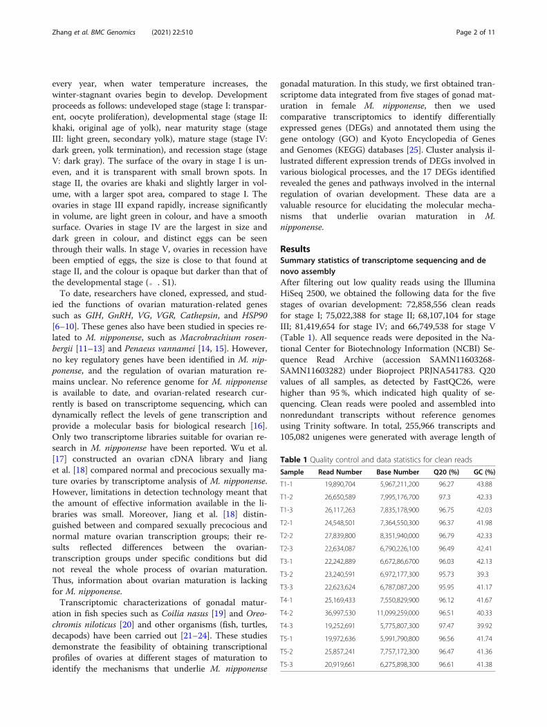
ResultsSummary statistics of transcriptome sequencing and denovo assemblyAfter filtering out low quality reads using the IlluminaHiSeq 2500, we obtained the following data for the fivestages of ovarian development: 72,858,556 clean readsfor stage I; 75,022,388 for stage II; 68,107,104 for stageIII; 81,419,654 for stage IV; and 66,749,538 for stage V(Table 1). All sequence reads were deposited in the Na-tional Center for Biotechnology Information (NCBI) Se-quence Read Archive (accession SAMN11603268-SAMN11603282) under Bioproject PRJNA541783. Q20values of all samples, as detected by FastQC26, werehigher than 95 %, which indicated high quality of se-quencing. Clean reads were pooled and assembled intononredundant transcripts without reference genomesusing Trinity software. In total, 255,966 transcripts and105,082 unigenes were generated with average length of
Table 1 Quality control and data statistics for clean reads
Sample Read Number Base Number Q20 (%) GC (%)
T1-1 19,890,704 5,967,211,200 96.27 43.88
T1-2 26,650,589 7,995,176,700 97.3 42.33
T1-3 26,117,263 7,835,178,900 96.75 42.03
T2-1 24,548,501 7,364,550,300 96.37 41.98
T2-2 27,839,800 8,351,940,000 96.79 42.33
T2-3 22,634,087 6,790,226,100 96.49 42.41
T3-1 22,242,889 6,672,86,6700 96.03 42.13
T3-2 23,240,591 6,972,177,300 95.73 39.3
T3-3 22,623,624 6,787,087,200 95.95 41.17
T4-1 25,169,433 7,550,829,900 96.12 41.67
T4-2 36,997,530 11,099,259,000 96.51 40.33
T4-3 19,252,691 5,775,807,300 97.47 39.92
T5-1 19,972,636 5,991,790,800 96.56 41.74
T5-2 25,857,241 7,757,172,300 96.47 41.36
T5-3 20,919,661 6,275,898,300 96.61 41.38
Zhang et al. BMC Genomics (2021) 22:510 Page 2 of 11
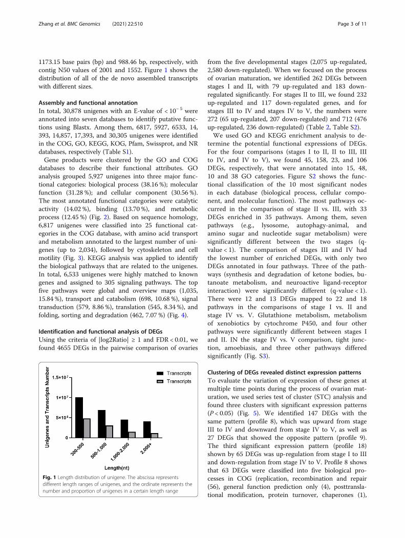
1173.15 base pairs (bp) and 988.46 bp, respectively, withcontig N50 values of 2001 and 1552. Figure 1 shows thedistribution of all of the de novo assembled transcriptswith different sizes.
Assembly and functional annotationIn total, 30,878 unigenes with an E-value of < 10− 5 wereannotated into seven databases to identify putative func-tions using Blastx. Among them, 6817, 5927, 6533, 14,393, 14,857, 17,393, and 30,305 unigenes were identifiedin the COG, GO, KEGG, KOG, Pfam, Swissprot, and NRdatabases, respectively (Table S1).Gene products were clustered by the GO and COG
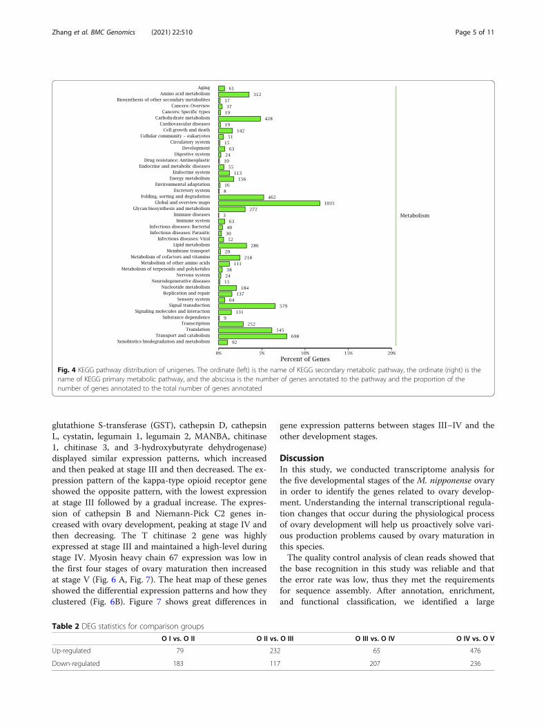
databases to describe their functional attributes. GOanalysis grouped 5,927 unigenes into three major func-tional categories: biological process (38.16 %); molecularfunction (31.28 %); and cellular component (30.56 %).The most annotated functional categories were catalyticactivity (14.02 %), binding (13.70 %), and metabolicprocess (12.45 %) (Fig. 2). Based on sequence homology,6,817 unigenes were classified into 25 functional cat-egories in the COG database, with amino acid transportand metabolism annotated to the largest number of uni-genes (up to 2,034), followed by cytoskeleton and cellmotility (Fig. 3). KEGG analysis was applied to identifythe biological pathways that are related to the unigenes.In total, 6,533 unigenes were highly matched to knowngenes and assigned to 305 signaling pathways. The topfive pathways were global and overview maps (1,035,15.84 %), transport and catabolism (698, 10.68 %), signaltransduction (579, 8.86 %), translation (545, 8.34 %), andfolding, sorting and degradation (462, 7.07 %) (Fig. 4).
Identification and functional analysis of DEGsUsing the criteria of |log2Ratio| ≥ 1 and FDR < 0.01, wefound 4655 DEGs in the pairwise comparison of ovaries
from the five developmental stages (2,075 up-regulated,2,580 down-regulated). When we focused on the processof ovarian maturation, we identified 262 DEGs betweenstages I and II, with 79 up-regulated and 183 down-regulated significantly. For stages II to III, we found 232up-regulated and 117 down-regulated genes, and forstages III to IV and stages IV to V, the numbers were272 (65 up-regulated, 207 down-regulated) and 712 (476up-regulated, 236 down-regulated) (Table 2, Table S2).We used GO and KEGG enrichment analysis to de-
termine the potential functional expressions of DEGs.For the four comparisons (stages I to II, II to III, IIIto IV, and IV to V), we found 45, 158, 23, and 106DEGs, respectively, that were annotated into 15, 48,10 and 38 GO categories. Figure S2 shows the func-tional classification of the 10 most significant nodesin each database (biological process, cellular compo-nent, and molecular function). The most pathways oc-curred in the comparison of stage II vs. III, with 33DEGs enriched in 35 pathways. Among them, sevenpathways (e.g., lysosome, autophagy-animal, andamino sugar and nucleotide sugar metabolism) weresignificantly different between the two stages (q-value < 1). The comparison of stages III and IV hadthe lowest number of enriched DEGs, with only twoDEGs annotated in four pathways. Three of the path-ways (synthesis and degradation of ketone bodies, bu-tanoate metabolism, and neuroactive ligand-receptorinteraction) were significantly different (q-value < 1).There were 12 and 13 DEGs mapped to 22 and 18pathways in the comparisons of stage I vs. II andstage IV vs. V. Glutathione metabolism, metabolismof xenobiotics by cytochrome P450, and four otherpathways were significantly different between stages Iand II. IN the stage IV vs. V comparison, tight junc-tion, amoebiasis, and three other pathways differedsignificantly (Fig. S3).
Clustering of DEGs revealed distinct expression patternsTo evaluate the variation of expression of these genes atmultiple time points during the process of ovarian mat-uration, we used series test of cluster (STC) analysis andfound three clusters with significant expression patterns(P < 0.05) (Fig. 5). We identified 147 DEGs with thesame pattern (profile 8), which was upward from stageIII to IV and downward from stage IV to V, as well as27 DEGs that showed the opposite pattern (profile 9).The third significant expression pattern (profile 18)shown by 65 DEGs was up-regulation from stage I to IIIand down-regulation from stage IV to V. Profile 8 showsthat 63 DEGs were classified into five biological pro-cesses in COG (replication, recombination and repair(56), general function prediction only (4), posttransla-tional modification, protein turnover, chaperones (1),
Fig. 1 Length distribution of unigene. The abscissa representsdifferent length ranges of unigenes, and the ordinate represents thenumber and proportion of unigenes in a certain length range
Zhang et al. BMC Genomics (2021) 22:510 Page 3 of 11

cell wall/membrane/envelope biogenesis (1), and cellmotility (1)) (Table S3). Only two DEGs were annotatedto general function prediction only and replication, re-combination and repair, respectively, in profile 9 (TableS4). In profile 18, 20 % of the DEGs (13) were enrichedto six biological processes, and most were in the generalfunction prediction only category (Table S5).
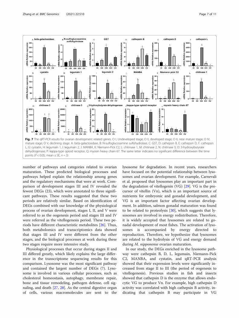
Screening and expression of genes related to ovarymaturationBased on the analysis of DEGs, we screened 17 genes re-lated to ovarian maturation by combining the q-value ofthe DEG enrichment pathways in the comparison groupsI vs. II, II vs. III, III vs. IV, and IV vs. V. Twelve genes(beta-galactosidase, N-sulfoglucosamine sulfohydrolase,
Fig. 2 GO classification of unigenes; The abscissa is the second level term under the three categories of GO. The ordinate represents the numberof genes annotated to the term and the percentage of all genes
Fig. 3 COG classification statistics of unigenes. The abscissa is the classification content of COG, and the ordinate is the number of genes
Zhang et al. BMC Genomics (2021) 22:510 Page 4 of 11
glutathione S-transferase (GST), cathepsin D, cathepsinL, cystatin, legumain 1, legumain 2, MANBA, chitinase1, chitinase 3, and 3-hydroxybutyrate dehydrogenase)displayed similar expression patterns, which increasedand then peaked at stage III and then decreased. The ex-pression pattern of the kappa-type opioid receptor geneshowed the opposite pattern, with the lowest expressionat stage III followed by a gradual increase. The expres-sion of cathepsin B and Niemann-Pick C2 genes in-creased with ovary development, peaking at stage IV andthen decreasing. The T chitinase 2 gene was highlyexpressed at stage III and maintained a high-level duringstage IV. Myosin heavy chain 67 expression was low inthe first four stages of ovary maturation then increasedat stage V (Fig. 6 A, Fig. 7). The heat map of these genesshowed the differential expression patterns and how theyclustered (Fig. 6B). Figure 7 shows great differences in
gene expression patterns between stages III–IV and theother development stages.
DiscussionIn this study, we conducted transcriptome analysis forthe five developmental stages of the M. nipponense ovaryin order to identify the genes related to ovary develop-ment. Understanding the internal transcriptional regula-tion changes that occur during the physiological processof ovary development will help us proactively solve vari-ous production problems caused by ovary maturation inthis species.The quality control analysis of clean reads showed that
the base recognition in this study was reliable and thatthe error rate was low, thus they met the requirementsfor sequence assembly. After annotation, enrichment,and functional classification, we identified a large
Fig. 4 KEGG pathway distribution of unigenes. The ordinate (left) is the name of KEGG secondary metabolic pathway, the ordinate (right) is thename of KEGG primary metabolic pathway, and the abscissa is the number of genes annotated to the pathway and the proportion of thenumber of genes annotated to the total number of genes annotated
Table 2 DEG statistics for comparison groups
O I vs. O II O II vs. O III O III vs. O IV O IV vs. O V
Up-regulated 79 232 65 476
Down-regulated 183 117 207 236
Zhang et al. BMC Genomics (2021) 22:510 Page 5 of 11
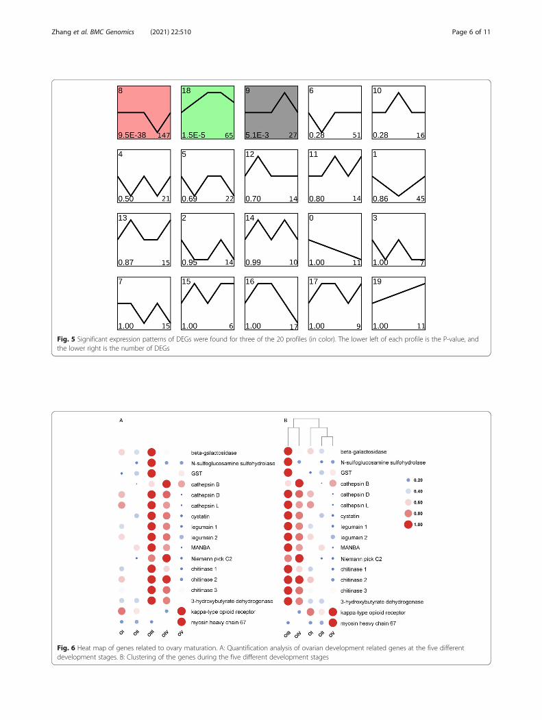
Fig. 5 Significant expression patterns of DEGs were found for three of the 20 profiles (in color). The lower left of each profile is the P-value, andthe lower right is the number of DEGs
Fig. 6 Heat map of genes related to ovary maturation. A: Quantification analysis of ovarian development related genes at the five differentdevelopment stages. B: Clustering of the genes during the five different development stages
Zhang et al. BMC Genomics (2021) 22:510 Page 6 of 11
number of pathways and categories related to ovarianmaturation. These predicted biological processes andpathways helped explain the relationship among genesand the regulatory mechanisms that were at work. Com-parison of development stages III and IV revealed thefewest DEGs (23), which were annotated to three signifi-cant pathways. These results suggested that these twoperiods are relatively similar. Based on identification ofDEGs combined with our knowledge of the physiologicalprocess of ovarian development, stages I, II, and V werereferred to as the oogenesis period and stages III and IVwere referred as the vitellogenesis period. These two pe-riods have different characteristic metabolites [26]. Thus,both metabolomics and transcriptomics data showedthat stages III and IV were different from the otherstages, and the biological processes at work during thesetwo stages require more intensive study.Physiological processes that occur during stages II and
III differed greatly, which likely explains the large differ-ence in the transcriptome sequencing results for thiscomparison. Lysosome was the most significant pathwayand contained the largest number of DEGs (7). Lyso-some is involved in various cellular processes, such ascholesterol homeostasis, autophagy, membrane repair,bone and tissue remodeling, pathogen defense, cell sig-naling, and death [27, 28]. As the central digestive organof cells, various macromolecules are sent to the
lysosome for degradation. In recent years, researchershave focused on the potential relationship between lyso-somes and ovarian development. For example, Carnevaliet al. proposed that lysosomes play an important part inthe degradation of vitellogenin (VG) [29]. VG is the pre-cursor of vitellin (Vn), which is an important source ofnutrients for embryonic and gonadal development, andVG is an important factor affecting ovarian develop-ment. In addition, salmon gonadal maturation was foundto be related to proteolysis [30], which suggests that ly-sosomes are involved in energy redistribution. Therefore,it is widely accepted that lysosomes are related to go-nadal development of most fishes. The activation of lyso-somes is accompanied by energy directed toreproduction. Therefore, we hypothesize that lysosomesare related to the hydrolysis of VG and energy demandduring M. nipponense ovarian maturation.In our study, the DEGs enriched in the lysosome path-
way were cathepsin B, D, L, legumain, Niemann-PickC2, MANBA, and cystatin, and qRT-PCR analysisshowed that their expression levels were significantly in-creased from stage II to III (the period of oogenesis tovitellogenesis). Previous studies in fish and insectsshowed that cathepsin D is the enzyme that allows endo-cytic VG to produce Vn. For example, high cathepsin Dactivity was correlated with high cathepsin B activity, in-dicating that cathepsin B may participate in VG
Fig. 7 The qRT-PCR results for ovarian development related genes. O I, Undeveloped stage; O II, developed stage; O II, near mature stage; O IV,mature stage; O V, declining stage. A: beta-galactosidase, B: N-sulfoglucosamine sulfohydrolase, C: GST, D: cathepsin B, E: cathepsin D, F: cathepsinL, G: cystatin, H: legumain 1, I: legumain 2, J: MANBA, K: Niemann-Pick C2, L: chitinase 1, M: chitinase 2, N: chitinase 3, O: 3-hydroxybutyratedehydrogenase, P: kappa-type opioid receptor, Q: myosin heavy chain 67. The same letter indicates no significant difference between the timepoints (P < 0.05; mean ± SE, n = 3)
Zhang et al. BMC Genomics (2021) 22:510 Page 7 of 11
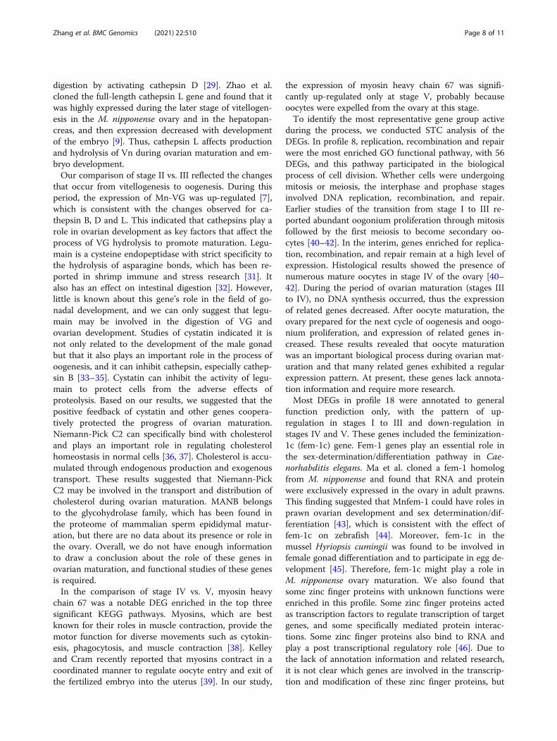
digestion by activating cathepsin D [29]. Zhao et al.cloned the full-length cathepsin L gene and found that itwas highly expressed during the later stage of vitellogen-esis in the M. nipponense ovary and in the hepatopan-creas, and then expression decreased with developmentof the embryo [9]. Thus, cathepsin L affects productionand hydrolysis of Vn during ovarian maturation and em-bryo development.Our comparison of stage II vs. III reflected the changes
that occur from vitellogenesis to oogenesis. During thisperiod, the expression of Mn-VG was up-regulated [7],which is consistent with the changes observed for ca-thepsin B, D and L. This indicated that cathepsins play arole in ovarian development as key factors that affect theprocess of VG hydrolysis to promote maturation. Legu-main is a cysteine endopeptidase with strict specificity tothe hydrolysis of asparagine bonds, which has been re-ported in shrimp immune and stress research [31]. Italso has an effect on intestinal digestion [32]. However,little is known about this gene’s role in the field of go-nadal development, and we can only suggest that legu-main may be involved in the digestion of VG andovarian development. Studies of cystatin indicated it isnot only related to the development of the male gonadbut that it also plays an important role in the process ofoogenesis, and it can inhibit cathepsin, especially cathep-sin B [33–35]. Cystatin can inhibit the activity of legu-main to protect cells from the adverse effects ofproteolysis. Based on our results, we suggested that thepositive feedback of cystatin and other genes coopera-tively protected the progress of ovarian maturation.Niemann-Pick C2 can specifically bind with cholesteroland plays an important role in regulating cholesterolhomeostasis in normal cells [36, 37]. Cholesterol is accu-mulated through endogenous production and exogenoustransport. These results suggested that Niemann-PickC2 may be involved in the transport and distribution ofcholesterol during ovarian maturation. MANB belongsto the glycohydrolase family, which has been found inthe proteome of mammalian sperm epididymal matur-ation, but there are no data about its presence or role inthe ovary. Overall, we do not have enough informationto draw a conclusion about the role of these genes inovarian maturation, and functional studies of these genesis required.In the comparison of stage IV vs. V, myosin heavy
chain 67 was a notable DEG enriched in the top threesignificant KEGG pathways. Myosins, which are bestknown for their roles in muscle contraction, provide themotor function for diverse movements such as cytokin-esis, phagocytosis, and muscle contraction [38]. Kelleyand Cram recently reported that myosins contract in acoordinated manner to regulate oocyte entry and exit ofthe fertilized embryo into the uterus [39]. In our study,
the expression of myosin heavy chain 67 was signifi-cantly up-regulated only at stage V, probably becauseoocytes were expelled from the ovary at this stage.To identify the most representative gene group active
during the process, we conducted STC analysis of theDEGs. In profile 8, replication, recombination and repairwere the most enriched GO functional pathway, with 56DEGs, and this pathway participated in the biologicalprocess of cell division. Whether cells were undergoingmitosis or meiosis, the interphase and prophase stagesinvolved DNA replication, recombination, and repair.Earlier studies of the transition from stage I to III re-ported abundant oogonium proliferation through mitosisfollowed by the first meiosis to become secondary oo-cytes [40–42]. In the interim, genes enriched for replica-tion, recombination, and repair remain at a high level ofexpression. Histological results showed the presence ofnumerous mature oocytes in stage IV of the ovary [40–42]. During the period of ovarian maturation (stages IIIto IV), no DNA synthesis occurred, thus the expressionof related genes decreased. After oocyte maturation, theovary prepared for the next cycle of oogenesis and oogo-nium proliferation, and expression of related genes in-creased. These results revealed that oocyte maturationwas an important biological process during ovarian mat-uration and that many related genes exhibited a regularexpression pattern. At present, these genes lack annota-tion information and require more research.Most DEGs in profile 18 were annotated to general
function prediction only, with the pattern of up-regulation in stages I to III and down-regulation instages IV and V. These genes included the feminization-1c (fem-1c) gene. Fem-1 genes play an essential role inthe sex-determination/differentiation pathway in Cae-norhabditis elegans. Ma et al. cloned a fem-1 homologfrom M. nipponense and found that RNA and proteinwere exclusively expressed in the ovary in adult prawns.This finding suggested that Mnfem-1 could have roles inprawn ovarian development and sex determination/dif-ferentiation [43], which is consistent with the effect offem-1c on zebrafish [44]. Moreover, fem-1c in themussel Hyriopsis cumingii was found to be involved infemale gonad differentiation and to participate in egg de-velopment [45]. Therefore, fem-1c might play a role inM. nipponense ovary maturation. We also found thatsome zinc finger proteins with unknown functions wereenriched in this profile. Some zinc finger proteins actedas transcription factors to regulate transcription of targetgenes, and some specifically mediated protein interac-tions. Some zinc finger proteins also bind to RNA andplay a post transcriptional regulatory role [46]. Due tothe lack of annotation information and related research,it is not clear which genes are involved in the transcrip-tion and modification of these zinc finger proteins, but
Zhang et al. BMC Genomics (2021) 22:510 Page 8 of 11
we speculate that these genes are closely related to ovar-ian maturation.
ConclusionsResults of this study provided comprehensive data aboutthe ovarian maturation transcriptome in M. nipponense.We obtained 30,878 unigenes, of which 4,655 werefound to be differentially expressed in comparisons ofthe five development stages of the ovary. We identified17 DEGs that may be related to ovary maturation in sig-nificant pathways identified in the comparisons. Amongthem, cathepins, legumains, and cystatin were enrichedin the lysosome pathway, and they are known to protectthe progress of VG hydrolysis. Additionally, myosinheavy chain 67 participated in oocyte excretion. Our re-sults highlighted differences between stages III-IV andthe other stages of ovary development. The reason ofthe difference may due to the biological processes hap-pened in each period. Our data provided new informa-tion about the regulation of ovarian maturation in M.nipponense and may be helpful for solving the problemof rapid M. nipponense development in the aquacultureindustry. However, the relationship between other regu-latory genes and ovarian maturation requires furtherstudy.
MethodsTissue preparation and RNA isolationWe obtained healthy female M. nipponense (2.75 ±1.45 g) from the Freshwater Fisheries Research Center ofthe China Academy of Fishery Sciences (Wuxi, China)(120°13’44′′E, 31°28′22′′N). Shrimp were cultured at100 individuals/m2, and the feeding conditions followedSun et al. [47]. According to previous studies [48], fivedifferent stages of ovarian maturation were distinguishedby colour (see Fig. S1) and immediately stored in liquidnitrogen at -190 °C. Tissues were collected from five in-dividuals at each stage and homogenized with TRIzol re-agent (Autolab Tech, Beijing, China) to extract totalRNA. The RNA concentration was detected using aQubit RNA Kit with Qubit 2.0 Fluorometer (Life Tech-nologies, Carlsbad, CA, USA), and its purity was de-tected using a Nanodrop 2000 spectrophotometer(Thermo Scientific, Waltham, MA, USA). The integrityof the RNA was assessed using an RNA Nano 6000 de-tection kit (2100 Bioanalyzer System; Agilent Technolo-gies, Santa Clara, CA, USA).
Library construction and sequencingA sequencing library was prepared with 3 µg of RNAfrom each sample using a NEBNext Ultra RNA LibraryPrep Kit (Illumina, San Diego, CA, USA) according tothe manufacturer’s instructions. The RNA was purifiedand broken into small random fragments using poly-T
oligo attached magnetic beads (Life Technologies, Carls-bad, CA, USA). Double-stranded DNA was synthesizedusing a TruSeq™ Stranded mRNA Prep Kit (Illumina).The DNA fragments in the library with a length of 150–200 bp were screened and purified using an Ampure XPsystem (Beckman Coulter, Beverly, MA, USA). The puri-fied double-stranded cDNA (size selection and connec-tion) was incubated with 3 ml of USER enzymes (NEB,Ipswich, MA, USA) at 37 °C for 15 min and then cul-tured at 95 °C for 5 min. Polymerase chain reaction(PCR) was performed with Phusion High-Fidelity DNApolymerase, universal PCR primers, and index (X) Pri-mer, and the products were then purified using theAmpure XP system. The composite samples were pairedand sequenced using a HiSeq™ 25,000 for 2 × 100 bp ac-cording to the manufacturer’s instructions. The PE read-ing of each lane was about 150 m (n = 3).
Assembly and dataset annotationAn Illumina HiSeq high-throughput sequencing plat-form based on sequencing by synthesis technology canproduce a large quantity of high-quality raw data.FastQC tools were used to truncate adapter and primersequences and remove reads with N > 10 % (where N in-dicates inability to determine base information) andthose with quality (Q) < 5 for > 50 % of reads. Trinity(http://trinityrnaseq.sourceforge.net/) was used to as-semble reads from scratch according to parametric tran-scription group. The minimum contig length was 300and K-mer was 27.We annotated the final set of unigenes comprehen-
sively using BLAST software (http://blast.ncbi.nlm.nih.gov/Blast.cgi) to compare unigene sequences with theNR (ftp://ftp.ncbi.nih.gov/blast/db/), Swiss-Prot (http://www.uniprot.org/), GO (http://www.geneontology.org/),COG (http://www.ncbi.nlm.nih.gov/COG/), KOG(http://www.ncbi.nlm.nih.gov/COG/), and KEGG (http://www.genome.jp/kegg/) databases (E-value ≤ 10− 5). Afterpredicting the amino acid sequences of the unigenes, wecompared them with the Pfam database (http://pfam.xfam.org/) using HMMER software (http://hmmer.janelia.org/).
DEG analysis and quantitative analysis of ovarianmaturation related gene expression at different ovariandevelopmental stagesWe used DESeq2 to analyze the differential expressionbetween the sample groups, and 10 pairwise comparativeDEG sets of ovaries in the five stages were obtained. TheBenjamini-Hochberg correction method was used tocorrect the significance of the p-value of the original testhypothesis to obtain the false discovery rate (FDR) [49],and fold-change was used to present the expression ratiobetween the comparison groups. |log2(fold change)| ≥ 1
Zhang et al. BMC Genomics (2021) 22:510 Page 9 of 11
and FDR < 0.05 were used as screening criteria to defineDEGs. The GO, COG, and KEGG annotation methodswere similar to those mentioned above in terms of uni-gene annotation, and DEG pathway enrichment analysisidentified significantly enriched pathways based on a q-value < 0.05.We validated the DEGs by qRT-PCR to evaluate the
sequencing and data analysis. Total RNA was extractedfrom ovaries (~ 100 mg) with 1 mL TRIzol reagent(TaKaRa, Japan), and first-strand cDNA was synthesizedusing a Reverse Transcriptase M-MLV Kit (TaKaRa).The qRT-PCR was performed using a Bio-Rad iCycleriQ5 real-time PCR system (Hercules, CA, USA), witheukaryotic translation initiation factor 5 A as the refer-ence gene [50]. The primers used are shown in Table S6.The reaction was amplified with 35 cycles at 94 °C for30 s, 50 °C for 30 s, and 72 °C for 1 min, followed by10 min incubation at 72 °C as a final extension step [51].Four replicates were run for each sample, and three con-trols were used for each reaction: nuclease-free water;primer-free water; and template-free water. The fluores-cence curve and data were recorded automatically by thesystem. The dissociation curves of the amplified prod-ucts were analyzed at the end of each PCR. The mRNAexpression levels were determined using the 2−ΔΔCT
method [52].
Supplementary InformationThe online version contains supplementary material available at https://doi.org/10.1186/s12864-021-07737-5.
Additional file 1:
Additional file 2:
Additional file 3:
Additional file 4:
Additional file 2:
Additional file 3:
Additional file 4:
AcknowledgementsNot applicable.
Authors' contributionsY.Z. conducted the experiment, performed data analyses and drafted themanuscript. H.Q., S.J. and Y.X. conceived the project and helped designedthe experiments. Y.Z. edited the manuscript. Data were analyzed by W.Z. andS.J. Language in the manuscript was improved by H.F., Y.G. and Y.W. Allauthors discussed the results and commented on the manuscript.
FundingThis research was supported by the National Key R&D Program of China(2018YFD0900201), the Central Public-interest Scientific Institution Basal Re-search Fund CAFS (2020TD36, 2019JBFM04), the Jiangsu Agricultural IndustryTechnology System, the China Agriculture Research System-48 (CARS-48),and the New Cultivar Breeding Major Project of Jiangsu Province(PZCZ201745).
Availability of data and materialsThe dataset supporting the conclusions of this article is available in the NCBISequence Read Archive (accession SAMN11603268-SAMN11603282) underBioproject PRJNA541783.
Declarations
Ethics approval and consent to participateWe obtained permission from the Tai Lake Fishery Management Council andthe Committee of the Freshwater Fisheries Research Center to conduct theexperimental programs. Tissues were collected from live animals in order toprevent the degradation of RNA for RNA-Seq and qPCR-PCR analysis. We se-dated the prawns with MS222 prior to removing the tissue samples. The ani-mals were sacrificed after tissue collection.
Consent for publicationNot applicable
Competing interestsThe authors declare that they have no competing interests.
Received: 3 February 2021 Accepted: 21 May 2021
References1. Cai Y, Ng P K L. The freshwater palaemonid prawns (Crustacea: Decapoda:
Caridea) of Myanmar[J]. Hydrobiologia, 2002, 487(1): 59–83.2. De Grave S, Ghane A. The establishment of the oriental river prawn,
Macrobrachium nipponense (De Haan W. 1833–1850. Crustacea, pp. i-xvii, i-xxxi, ix-xvi, 1-243, pls[J]. AJ, LQ, 1823: 1–55.
3. Subramoniam T. Sexual biology and reproduction in crustaceans[M].Academic Press, 2016.
4. Li X, Li Z, Liu J, et al. Advances in precocity research of the Chinese mittencrab Eriocheir sinensis[J]. Aquaculture International, 2011, 19(2): 251–267.
5. Jasmani S, Ohira T, Jayasankar V, et al. Localization of vitellogenin mRNAexpression and vitellogenin uptake during ovarian maturation in the giantfreshwater prawn Macrobrachium rosenbergii[J]. Journal of ExperimentalZoology Part A: Comparative Experimental Biology, 2004, 301(4): 334–343.
6. Qiao H, Xiong Y, Zhang W, et al. Characterization, expression, and functionanalysis of gonad-inhibiting hormone in Oriental River prawn,Macrobrachium nipponense and its induced expression by temperature[J].Comparative Biochemistry and Physiology Part A: Molecular & IntegrativePhysiology, 2015, 185: 1–8.
7. Bai H, Qiao H, Li F, et al. Molecular characterization and developmentalexpression of vitellogenin in the oriental river prawn Macrobrachiumnipponense and the effects of RNA interference and eyestalk ablation onovarian maturation[J]. Gene, 2015, 562(1):22–31.
8. Bai H, Qiao H, Li F, et al. Molecular and functional characterization of thevitellogenin receptor in oriental river prawn, Macrobrachium nipponense[J].Comp Biochem Physiol A Mol Integr Physiol, 2016, 194:45–55.
9. Zhao W, Chen L, Zhang F, et al. Molecular characterization of cathepsin LcDNA and its expression during oogenesis and embryogenesis in theoriental river prawn Macrobrachium nipponense (Palaemonidae)[J]. GenetMol Res, 2013, 12(4): 5215–5225.
10. Zhao W, Chen L, Qin J, et al. MnHSP90 cDNA characterization and itsexpression during the ovary development in oriental river prawn,Macrobrachium nipponense[J]. Molecular Biology Reports, 2011, 38(2): 1399–1406.
11. Okuno A, Yang W J, Jayasankar V, et al. Deduced primary structure ofvitellogenin in the giant freshwater prawn, Macrobrachium rosenbergii, andyolk processing during ovarian maturation[J]. Journal of ExperimentalZoology, 2002, 292(5): 417–429.
12. Arockiaraj J, Gnanam A J, Muthukrishnan D, et al. Macrobrachium rosenbergiicathepsin L: molecular characterization and gene expression in response toviral and bacterial infections[J]. Microbiological Research, 2013, 168(9): 569–579.
13. Roth Z, Khalaila I. Identification and characterization of the vitellogeninreceptor in Macrobrachium rosenbergii and its expression duringvitellogenesis[J]. Molecular Reproduction and Development, 2012, 79(7):478–487.
Zhang et al. BMC Genomics (2021) 22:510 Page 10 of 11
14. Le Boulay C, Van Wormhoudt A, Sellos D. Cloning and expression ofcathepsin l-like proteinases in the hepatopancreas of the shrimp Penaeusvannamei during the intermolt cycle[J]. Journal of Comparative PhysiologyB, 1996, 166(5): 310–318.
15. Peng J, Wei P, Zhang B, et al. Gonadal transcriptomic analysis anddifferentially expressed genes in the testis and ovary of the Pacificwhite shrimp (Litopenaeus vannamei)[J]. BMC Genomics, 2015, 16(1):1006.
16. Wang Z, Gerstein M, Snyder M. RNA-Seq: a revolutionary tool fortranscriptomics[J]. Nature Reviews Genetics, 2009, 10(1): 57.
17. Wu P, Qi D, Chen L, et al. Gene discovery from an ovary cDNA library oforiental river prawn Macrobrachium nipponense by ESTs annotation[J].Comparative Biochemistry & Physiology Part D Genomics & Proteomics,2009, 4(2):0–120.
18. Jiang H, Li X, Sun Y, et al. Insights into sexual precocity of female orientalriver prawn Macrobrachium nipponense through transcriptome analysis[J].Plos One, 2016, 11(6): e0157173.
19. Xu G, Du F, Li Y, et al. Integrated application of transcriptomics andmetabolomics yields insights into population-asynchronous ovarydevelopment in Coilia nasus[J]. Scientific Reports, 2016, 6: 31835.
20. Tao W, Yuan J, Zhou L, et al. Characterization of gonadal transcriptomesfrom Nile tilapia (Oreochromis niloticus) reveals differentially expressedgenes[J]. PloS One, 2013, 8(5): e63604.
21. Ravi P, Jiang J, Liew W C, et al. Small-scale transcriptomics revealsdifferences among gonadal stages in Asian seabass (Lates calcarifer)[J].Reproductive Biology and Endocrinology, 2014, 12(1): 5.
22. Lee S L J, Horsfield J A, Black M A, et al. Histological and transcriptomiceffects of 17α-methyltestosterone on zebrafish gonad development[J]. BMCGenomics, 2017, 18(1): 557.
23. Radhakrishnan S, Literman R, Neuwald J, et al. Transcriptomic responses toenvironmental temperature by turtles with temperature-dependent andgenotypic sex determination assessed by RNAseq inform the geneticarchitecture of embryonic gonadal development[J]. PloS One, 2017, 12(3):e0172044.
24. Chandler J C, Aizen J, Fitzgibbon Q P, et al. Applying the power oftranscriptomics: understanding male sexual development in decapodCrustacea[J]. Integrative and Comparative Biology, 2016, 56(6): 1144–1156.
25. Kanehisa M, Goto S. KEGG: kyoto encyclopedia of genes and genomes[J].Nucleic acids research, 2000, 28(1): 27–30.
26. Zhang Y, Fu Y, Jiang S, et al. Comparative metabolomics analysis of ovariandevelopmental stages in Macrobrachium nipponense[J]. ComparativeBiochemistry and Physiology Part D: Genomics and Proteomics, 2020, 34:100648.
27. Seymour E A. The effects of powdered carp pituitary on ovariandevelopment, ovarian ascorbic acid and ovulation in Carassius carassius L.exposed to various photoperiod and temperature regimes[J]. Journal of FishBiology, 1981, 19(6): 675–682.
28. Settembre C, Ballabio A. Lysosome: regulator of lipid degradationpathways[J]. Trends in Cell Biology, 2014, 24(12): 743–750.
29. Carnevali O, Cionna C, Tosti L, et al. Role of cathepsins in ovarian folliclegrowth and maturation[J]. General and Comparative Endocrinology, 2006,146(3): 195–203.
30. Mommsen T P. Salmon spawning migration and muscle proteinmetabolism: the August Krogh principle at work[J]. ComparativeBiochemistry and Physiology Part B: Biochemistry and Molecular Biology,2004, 139(3): 383–400.
31. Ren Q, Zhang X W, Sun Y D, et al. Two cysteine proteinases respond tobacterial and WSSV challenge in Chinese white shrimp Fenneropenaeuschinensis[J]. Fish & Shellfish Immunology, 2010, 29(4):0–556.
32. Sojka D, Hajdušek O, Dvořák J, et al. IrAE–An asparaginyl endopeptidase(legumain) in the gut of the hard tick Ixodes ricinus[J]. International Journalfor Parasitology, 2007, 37(7): 713–724.
33. Ahn S J, Bak H J, Park J H, et al. Olive flounder (Paralichthys olivaceus)cystatin B: cloning, tissue distribution, expression and inhibitory profile ofpiscine cystatin B[J]. Comparative Biochemistry and Physiology Part B:Biochemistry and Molecular Biology, 2013, 165(3): 211–218.
34. Frygelius J, Oscarson M, Nordqvist K, et al. The reproductive tissue specificcystatin subgroup of genes: expression during gonadal development inwildtype and testatin knockout animals[J]. Sexual Development, 2007, 1(6):363–372.
35. Hashmi S, Zhang J, Oksov Y, et al. The Caenorhabditis elegans CPI-2acystatin-like inhibitor has an essential regulatory role during oogenesis andfertilization[J]. Journal of Biological Chemistry, 2006, 281(38): 28415–28429.
36. Ko D C, Binkley J, Sidow A, et al. The integrity of a cholesterol-bindingpocket in Niemann-Pick C2 protein is necessary to control lysosomecholesterol levels[J]. Proceedings of the National Academy of Sciences,2003, 100(5): 2518–2525.
37. Storch J, Xu Z. Niemann–Pick C2 (NPC2) and intracellular cholesteroltrafficking[J]. Biochimica et Biophysica Acta (BBA)-Molecular and Cell Biologyof Lipids, 2009, 1791(7): 671–678.
38. Weiss A, Leinwand L A. The mammalian myosin heavy chain gene family[J].Annual Review of Cell and Developmental Biology, 1996, 12(1): 417–439.
39. Kelley C A, Cram E J. Regulation of actin dynamics in the C. elegans SomaticGonad[J]. Journal of Developmental Biology, 2019, 7(1): 6.
40. Du Y, Qiu G. Histological and histochemical observation on ovarydevelopment in oriental river prawn Macrobrachium nipponense[J].Guangdong Agricultural Sciences, 2014, 41(13):119–123 + 2.
41. Sharifian S, Kamrani E, Safaie M. Oogenesis and ovarian development in thefreshwater Crab Sodhiana iranica (Decapoda: Gecarcinuaidae) from thesouth of Iran[J]. Tissue and Cell, 2015, 47(2): 213–220.
42. Islam M S, Kodama K, Kurokora H. Ovarian development of the mud crabScylla paramamosain in a tropical mangrove swamps, Thailand[J]. Journal ofScientific Research, 2010, 2(2): 380–389.
43. Ma K Y, Liu Z Q, Lin J Y, et al. Molecular characterization of a novel ovary-specific gene fem-1 homolog from the oriental river prawn, Macrobrachiumnipponense[J]. Gene, 2016, 575(2): 244–252.
44. Shi J T, Li Z, Gui J F, et al. The cloning and expression analysis of zebrafishfem-1c, a member of fem-1 family[J]. Acta Hydrobiologica Sinica, 2015,39(3): 459–467.
45. Wang Y, Wu C, Guo P, et al. Molecular characterization and expression ofthe feminization-1c (fem-1c) in the freshwater mussel (Hyriopsis cumingii)[J].Aquaculture and Fisheries, 2018, 3(1): 6–13.
46. Li X, Zhang J. Structure and function of zinc finger proteins in Eucaryon[J].Chinese Journal of Biochemistry and Molecular Biology, 2009,25(03):206–211.
47. Sun S, Fu H, Gu Z, et al. Effects of stocking density on the individual growthand differentiation of the oriental river prawn Macrobrachium nipponense(de Haan, 1849) (Caridea: Palaemonidae)[J]. Journal of Crustacean Biology,2016, 36(6): 769–775.
48. Qiao H, Fu H, Xiong Y, et al. Molecular insights into reproduction regulationof female Oriental River prawns M. nipponense through comparativetranscriptomic analysis[J]. Scientific Reports, 2017, 7(1): 12161.
49. Thissen D, Steinberg L, Kuang D. Quick and easy implementation of theBenjamini-Hochberg procedure for controlling the false positive rate inmultiple comparisons[J]. Journal of Educational and Behavioral Statistics,2002, 27(1): 77–83.
50. Hu Y, Fu H, Qiao H, et al. Validation and evaluation of reference genes forquantitative real-time PCR in Macrobrachium Nipponense[J]. InternationalJournal of Molecular Sciences, 2018, 19(8): 2258.
51. Li F, Qiao H, Fu H, et al. Identification and characterization of opsin geneand its role in ovarian maturation in the oriental river prawn Macrobrachiumnipponense[J]. Comparative Biochemistry and Physiology Part B:Biochemistry and Molecular Biology, 2018, 218: 1–12.
52. Livak, K.J., Schmittgen, T.D., 2001. Analysis of relative gene expression datausing real-time quantitative PCR and the 2 – ΔΔCT method. Methods 25,402–408.
Publisher’s NoteSpringer Nature remains neutral with regard to jurisdictional claims inpublished maps and institutional affiliations.
Zhang et al. BMC Genomics (2021) 22:510 Page 11 of 11