transcriptional response to cardiac injury in the zebrafish
TRANSCRIPT
Rodius et al. BMC Genomics 2014, 15:852http://www.biomedcentral.com/1471-2164/15/852
RESEARCH ARTICLE Open Access
Transcriptional response to cardiac injury in thezebrafish: systematic identification of genes withhighly concordant activity across in vivo modelsSophie Rodius1, Petr V Nazarov2, Isabel A Nepomuceno-Chamorro3, Céline Jeanty1, Juan Manuel González-Rosa4,Mark Ibberson5, Ricardo M Benites da Costa6, Ioannis Xenarios5,6,7, Nadia Mercader8 and Francisco Azuaje1*
Abstract
Background: Zebrafish is a clinically-relevant model of heart regeneration. Unlike mammals, it has a remarkableheart repair capacity after injury, and promises novel translational applications. Amputation and cryoinjury models arekey research tools for understanding injury response and regeneration in vivo. An understanding of the transcriptionalresponses following injury is needed to identify key players of heart tissue repair, as well as potential targets forboosting this property in humans.
Results: We investigated amputation and cryoinjury in vivo models of heart damage in the zebrafish through unbiased,integrative analyses of independent molecular datasets. To detect genes with potential biological roles, we derivedcomputational prediction models with microarray data from heart amputation experiments. We focused on atop-ranked set of genes highly activated in the early post-injury stage, whose activity was further verified inindependent microarray datasets. Next, we performed independent validations of expression responses withqPCR in a cryoinjury model. Across in vivo models, the top candidates showed highly concordant responses at 1 and3 days post-injury, which highlights the predictive power of our analysis strategies and the possible biological relevanceof these genes. Top candidates are significantly involved in cell fate specification and differentiation, and include heartfailure markers such as periostin, as well as potential new targets for heart regeneration. For example, ptgis and ca2were overexpressed, while usp2a, a regulator of the p53 pathway, was down-regulated in our in vivo models.Interestingly, a high activity of ptgis and ca2 has been previously observed in failing hearts from rats and humans.
Conclusions: We identified genes with potential critical roles in the response to cardiac damage in the zebrafish. Theirtranscriptional activities are reproducible in different in vivo models of cardiac injury.
Keywords: Myocardial infarction, Zebrafish, Ventricular amputation, Ventricular cryoinjury, Heart regeneration,Transcriptional responses, Transcriptional association networks
BackgroundThe zebrafish (Danio rerio) has the capacity to regenerateits heart after undergoing severe injury [1,2]. This capabil-ity has been shown in adult zebrafish using differentin vivo models of cardiac damage, and involves the gener-ation of new cardiomyocytes from existing ones locatednear the injury site [3,4]. Thus, the zebrafish represents acompelling model to study heart injury and regenerationwith potential clinical impact. In the long-term, this will
* Correspondence: [email protected] Neuro-Oncology Laboratory, CRP-Santé, Luxembourg, LuxembourgFull list of author information is available at the end of the article
© 2014 Rodius et al.; licensee BioMed CentralCommons Attribution License (http://creativecreproduction in any medium, provided the orDedication waiver (http://creativecommons.orunless otherwise stated.
be crucial to address a major world-wide public healthproblem: heart attacks (myocardial infarction) followedby heart failure [5]. The latter is a consequence of theincapacity of the human heart to replace the lost cardi-omyocytes by newly formed myocardium, insteadforming an irreversible fibrotic scar [6,7].Identification of the cellular and molecular mechanisms
of zebrafish heart regeneration might thus allow to findnew therapeutic approaches for the treatment of myo-cardial infarction in humans. Despite obvious differencesbetween teleosts and mammals, the sequencing of thezebrafish genome revealed that about 70% of human genes
Ltd. This is an Open Access article distributed under the terms of the Creativeommons.org/licenses/by/4.0), which permits unrestricted use, distribution, andiginal work is properly credited. The Creative Commons Public Domaing/publicdomain/zero/1.0/) applies to the data made available in this article,

Rodius et al. BMC Genomics 2014, 15:852 Page 2 of 15http://www.biomedcentral.com/1471-2164/15/852
have at least one zebrafish orthologue [8]. A large amountof genetic screens have already been performed in zeb-rafish in order to identify genes involved in cardiacdevelopment and regeneration. Random mutagenesis,creation of transgenic strains using inducible geneticapproaches or targeted gene inactivation using morpholi-nos have led to generation of different models of humancardiac disorders [9-16], whose number will undoubtedlybe expanded by new genome editing techniques, such asCrisp/Cas9 and TALEN. Moreover, the external develop-ment of the zebrafish embryo and its transparency allowsdirect microscopic observation of cardiovascular structureswithout invasive instrumentation. Coupled to its ability tosurvive for several days without functional cardiovascularsystem, catching oxygen by passive diffusion [17], theseattributes make zebrafish particularly suitable to studythe phenotype of cardiovascular diseases. Besides, responseto drugs is well conserved between fish and mammals [18],making zebrafish a widely used model for toxicological ana-lysis and to study the possible cardiac effects of chemicalcompounds.Different techniques have been set up to study the re-
markable cardiac regeneration ability of adult zebrafish.Coronary artery ligation, mimicking ischemic injury thatleads to myocardial infarction in human, is commonlyused as model of myocardial infarction in mice. However,the size of the ventricle (around 1 mm3) renders this kindof injury almost impossible to achieve in zebrafish. Conse-quently, different alternative models have been used tostudy myocardial infarction in adult zebrafish: ventricularamputation, cryoinjury and cell-type specific ablation[19,20]. The oldest and most widely used technique isventricular amputation, which consists of removing about20% of the apex of the ventricle by surgical resection. Fol-lowing surgery, the apex is sealed by a clot of erythrocytes,further replaced by fibrin. Two months post-injury, theclot is not replaced by scar tissue but by cardiac muscleformed by cardiomyocytes proliferation, restoring thecontractile properties of the heart [21]. More recently, thecryocauterization model, which is already used in mouse[22], has been applied to zebrafish. In this technique, acryoprobe or dry ice is used to freeze 15 to 25% of theventricle [23-27]. Following cryocauterization, blood accu-mulates in the infarct area and massive cell death can beobserved at the injury site. A large fibrotic scar is formedand further replaced by cardiac tissue following cardio-myocytes proliferation. Total recovery is longer than forventricular amputation (up to 130 days [23]), which maybe due to the extra time needed to remove the necrotictissue and heal the scar. Finally, a genetic cell ablationmodel based on the Cre-lox strategy was set up to specif-ically kill cardiomyocytes in an inducible manner through-out the heart [28]. This technique allows destruction of60% of cardiomyocytes and promotes signs of cardiac
failure. Upon cell death, inflammatory cells are recruited,endocardial and epicardial cells are activated and cardio-myocytes de-differentiation and proliferation are enhanced,leading to muscle regeneration by 1 month after injury.Among these cardiac regeneration models, cryoinjury isperhaps the most relevant for translational biomedicalresearch. It resembles what is observed in the humanheart following myocardial infarction, including the factthat the injury affects all cardiac cell types and promotesmassive cell death and the formation of a fibrotic scar.The analysis of transcriptional responses following injury
is a powerful approach to identifying candidate drivers ofheart repair and regeneration, which may represent poten-tial targets for triggering or boosting cardiomyocyte renewalin mice and humans [29,30]. Mainly using amputation andgenetic ablation models, several studies have characterizeddifferential expression responses at different times afterinjury based on microarrays or in situ hybridizationtechniques [31]. These techniques in combination withgeneration of transgenic zebrafish strains and proteinconcentration analysis have demonstrated the regulatoryroles of biological pathways implicated in cell proliferationand differentiation, such as retinoic acid [32], platelet-derived growth factor [31,33], sonic hedgehog, Insulin-likegrowth factor, Transforming growth factor beta [34,35],Jak1/Stat3 [30] and Notch signalling [36].Despite the insights derived from previous research,
there is still a need to: a. apply biologically unbiasedapproaches to dissecting transcriptional responsesafter injury, and b. investigate the validity of relevantfindings across different in vivo models. The former in-volves the characterization of gene expression patternsbeyond the identification of genes differentially expressed.The latter is important not only to reconcile differentin vivo models that are promising for translational re-search, but also to identify genes with post-injury tran-scriptional behaviours that are highly robust regardless ofinjury model. Previous investigations conducted by ourteam and elsewhere have demonstrated that the applicationof network-based approaches to gene expression analysiscan: 1. enhance systems-level understanding of key bio-logical states; and 2. identify candidate markers of cardiacdamage, which are not only accurate for sample classifica-tion but also reproducible in independent datasets andmodel organisms.Here, our main objective is to identify genes exhibiting
important transcriptional perturbations after heart injuryin the zebrafish, which are both biologically meaningfuland reproducible in independent in vivo models.To address this question, we applied different computa-
tional analysis strategies with an emphasis on network-based approaches, which identify significant gene-gene as-sociations from the expression data. Our key premise wasthat genes with prominent patterns of “transcriptional

Rodius et al. BMC Genomics 2014, 15:852 Page 3 of 15http://www.biomedcentral.com/1471-2164/15/852
connectivity” in such networks represent genes withpotential critical roles in the response to injury. Here,we show that across in vivo models, a set of top candidatesshowed highly concordant responses at 1 and 3 days post-injury (dpi). This set of computationally predicted candi-dates are significantly associated with processes relevantto cell fate specification and differentiation. They includegenes with suspected functional roles in heart regener-ation, as well as novel genes that may exercise influentialroles in the early stages of injury response.
ResultsOverview of discovery frameworkFigure 1 summarises our analytical and modelling work-flow. First, we analysed a published dataset that consistsof gene expression measurements of sham-operated andinjured hearts at 1, 3, 5 and 7 days post-amputation [29].From now on, we will refer to this dataset as the modelderivation dataset. We detected genes significantly differ-entially expressed across all samples, and selected themfor further analysis. We then applied three published com-putational approaches to generating and analysing geneco-expression networks: Clustering with OverlappingNeighborhood Expansion (ClusterONE) [37], inference
Figure 1 Overview of analytical and modelling workflow.
of gene regression networks with model trees (RegNet)[38], and the Weighted Gene Co-Expression Network(WGCNA) algorithm [39]. This allowed us to identify:networks clusters, highly connected genes, and genesthat can be used as estimators of the expression valuesof other genes in the dataset (Methods). We independ-ently performed these analyses, focused on (statistically)top-ranked results, and selected a list of candidate genesfor further computational and experimental analysis. Inthe validation phase, we implemented the cryoinjurymodel at our laboratory and measured the expression ofthe candidate genes by RT-qPCR (Methods). From nowon, we will refer to this dataset as the model validationdataset. In the network models reported here, nodes andedges represent genes and between-gene expression corre-lations respectively.
Data pre-processing and differential expression analysisProcessed Affymetrix GeneChip Zebrafish microarray datawere downloaded from ArrayExpress (E-GEOD-17993)resulting of 15617 features with 10773 unique gene symbol[29]. Model derivation dataset consists of 15 microarrays: 3replicates over 5 time points (control, 1, 3, 5 and 7 dayafter operation). Pre-filtering of features decreased the size

Rodius et al. BMC Genomics 2014, 15:852 Page 4 of 15http://www.biomedcentral.com/1471-2164/15/852
of dataset to 7182 features corresponding to 5840 geneswith unique gene symbol. Limma analysis resulted in 3403unique annotated genes variable across the time pointswith FDR < 0.05, 88 genes at FDR ≤ 0.0001. The numberof differentially expressed genes (FDR < 0.05) after apply-ing 2-condition contrasts varied in time: 1907, 2349, 1914,and 330 annotated genes for control vs. 1, 3, 5 and 7 daysrespectively. Thus the highest dysregulation of the tran-scriptome was observed at the 3rd day after injury, whileother important events required for heart regenerationoccur in the days following injury.A second dataset comes from independent work by Kizil
et al. [40] (E-MEXP-1239). Here, 11 arrays were used: 4controls replicates, 1 replicate for each of 6 hours, 12 hoursand 1 day, 2 replicates for 3 and 5 days. We were ableto identify only 34 differentially expressed genes withFDR < 0.05 in this dataset.
Identification of potentially relevant genes with network-based approachesAfter computing between-gene correlations among top-differentially expressed genes in the derivation dataset(88 differentially expressed genes at FDR ≤ 0.0001), wegenerated a gene co-expression network. This networkconsisted of 3828 edges representing gene co-expressionrelationships estimated with the Pearson correlationcoefficient, r. To reduce the number of potential spuriousassociations and facilitate visualizations, we filtered outedges with │r│ < 0.95. To verify the potential statisticalrelevance of these associations, we estimated theirmaximal information coefficients (MICs) and con-firmed the high co-expression relationships of thesegenes (MIC > 0.95). The resulting network included 86nodes and 346 edges. A dynamic visualization of thisnetwork at different time points and their underlyingdata are available in Additional file 1. The ClusterONEalgorithm identified two highly statistically significantnetwork modules (Figure 2A): one consisting of 18nodes and 79 edges (P = 1.4E-7), and another consistingof 21 nodes and 107 edges (P = 3.3E-7). The first modulecontained postnb (periostin b). Periostin is a knownfibroblast marker, which is found in the scar tissue inboth mouse and zebrafish [41-46]. While its role inmyocardial proliferation remains to be fully elucidated,interestingly, its expression precedes heart regenerationin the zebrafish [46], which is consistent with our find-ings in two in vivo injury models. The second modulealso includes potentially interesting genes, such as usp2a(Ubiquitin Specific Peptidase 2) and mapk4 (Mitogen-Activated Protein Kinase 4), which are implicated in celldeath and proliferation signalling pathways [47,48].Independently, we applied the RegNet algorithm on the
reduced dataset (88 genes at FDR ≤ 0.0001) to extract tran-scriptional association networks (Methods). This modelling
technique specifies mathematical relationships between thegenes based on linear regression models. Such associationsare graphically represented with directed edges to indicatewhich genes can be used as reliable estimators of theexpression values of other genes in the dataset(Figure 2B). RegNet identified a statistically significantnetwork (alpha = 0.05, which is set to control the FDRusing Benjamin-Yekutieli procedure, and theta = 30%,which is set as a relative error threshold) containing 8nodes and 7 edges (Figure 2B), which overlaps with themodules identified previously (Figure 2A). In this network,ptgis (Prostaglandin I2 synthase) is shown as the centralnode, and its expression data are correlated (positivelyand negatively) with the other genes in the network. Thisallows a qualitative labelling of each network edge withthe direction of the observed gene-gene associations. Forexample, ptgis is negatively correlated with acta1a (uppergene in Figure 2B). This means that the expression valuesof these genes have opposite responses in the dataset.Therefore, we can use the sign of these associations totag the direction of the relationship between ptgis andthe other genes: UP (positive association) and DOWN(negative association). These results highlight ptgis as acandidate relevant gene based on its potential to estimatethe expression response of other genes in the dataset.To further assess the potential predictive relevance and
robustness of these findings, we applied the WGCNAalgorithm to a larger version of the dataset: 3403 differ-entially expressed genes at FDR ≤ 0.05, and with geneassociations calculated with the Spearman correlationcoefficient. As in the results from ClusterONE andRegNet, WGCNA found a strong association betweenptgis and acta1a within the same network module consist-ing of 1013 genes (Figure 2C, high resolution version inAdditional file 2). For ptgis, WGCNA reports a very highmodule membership (MM) score of 0.95 (P = 2.3E-08),which also highlights the prominent centrality of ptgiswithin this highly interconnected module. In this moduleacta1a shows a MM= −0.9 (P = 4.9E-06) that not onlyindicates its negative correlation with ptgis, but also itsmultiple strong connections with the other members ofthis module. We repeated this analysis on a network gen-erated with Pearson correlations, and consistent associa-tions between ptgis (MM= 0.9) and acta1a (MM= −0.87)were detected within the same, though smaller, module(616 genes). These results gave us further evidence of thepotential relevance of the association between ptgis andacta1a.As we are interested in identifying genes with robust
and reproducible transcriptional responses, we comple-mented our findings with an analysis of concordant geneexpression patterns in relation to an independent dataset[40]. As in our model derivation dataset, Kizil et al.’s data-set was obtained from microarray measurements from a

Figure 2 Computational models generated from public data. Different approaches highlight genes and associations with potential influentialroles in response to injury. A. Top network modules identified with the ClusterONE algorithm [37]. Network nodes shown as rectangles: geneswith exclusive membership in the modules shown, diamonds: genes with multiple module membership. Edges represent co-expression associations.B. Top candidate regulatory circuit inferred with the RegNet algorithm [38]. Edges represent gene co-expression association, with arrows defining thedirection of the association between ptgis and the other genes according to linear regression models. The latter are shown for each gene-geneassociation. C. Snapshot of network module identified with the WGCNA algorithm, which contains genes involved in A and B, including ptgis(higher resolution image in Additional file 2). D. List of genes with significant concordant expression values between our model derivation dataset [29]and a dataset obtained from an independent study based on amputation model [40]. Between-dataset correlation values are shown.
Rodius et al. BMC Genomics 2014, 15:852 Page 5 of 15http://www.biomedcentral.com/1471-2164/15/852
heart amputation model in zebrafish. Unlike our modelderivation dataset, Kizil et al.’s dataset exhibited a reducedset of significantly differentially expressed genes (34 genesdifferentially expressed at FDR < 0.05) across controland post-injury samples. We searched for highly con-cordant expression response between these two datasets(Pearson correlation > 0.8) at control and days 1, 3 and5 post-injury. Figure 2D displays the resulting list ofgenes with strong between-dataset expression correla-tions. Although none of these genes were highlighted aspotentially relevant by our network-based approaches,they deserved further investigation based on their highlyrobust behaviour in independent studies. Moreover, wenote that mmp13a (matrix metalloproteinase 13a) andLOC100332348 (an uncharacterized protein coding gene)were found together with ptgis and acta1a in the sameWGCNA module.
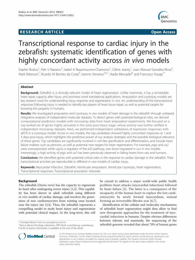
Selection of candidate genes for independentexperimental validationNext, we selected a list of genes for independent experi-mental validation at our laboratory. Among the genes thatwere identified above as potentially relevant, we decidedto focus our attention on 15 genes that met the followingcriteria: a. genes that were highlighted by at least one ofthe techniques reported above, b. genes that have not beenfunctionally characterized in the zebrafish or associatedwith its heart regeneration capacity, c. genes with un-ambiguous and accurate matching qPCR primers forthe zebrafish genome. Additionally, we included postnbbecause of its known activity in the early stages ofheart regeneration in zebrafish. Figure 3A shows thelist of selected candidate genes, and a summary of theirexpression responses in the model derivation dataset.In this visualisation we also plot the probe-specific
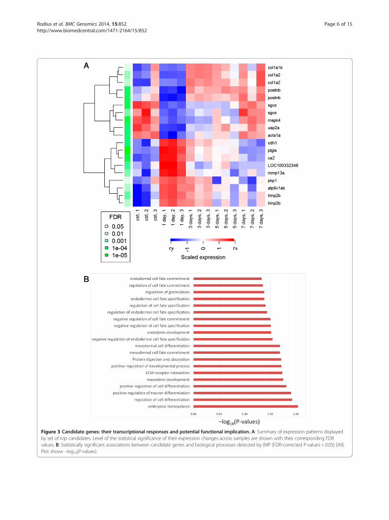
Figure 3 Candidate genes: their transcriptional responses and potential functional implication. A: Summary of expression patterns displayedby set of top candidates. Level of the statistical significance of their expression changes across samples are shown with their corresponding FDRvalues. B: Statistically significant associations between candidate genes and biological processes detected by IMP (FDR-corrected P-values < 0.05) [49].Plot shows –log10(P-values).
Rodius et al. BMC Genomics 2014, 15:852 Page 6 of 15http://www.biomedcentral.com/1471-2164/15/852

Rodius et al. BMC Genomics 2014, 15:852 Page 7 of 15http://www.biomedcentral.com/1471-2164/15/852
expression levels for those genes with multiple micro-array probes in the model derivation dataset. Suchcases show high expression consistency within thedataset.After selecting the candidate genes, we detected statisti-
cally significant associations between this gene set and cel-lular processes with the IMP (Integrative Multi-speciesPrediction) system [49]. IMP identified 20 significantassociations (FDR-corrected P-values < 0.05) betweenour candidate genes and diverse cellular processes rele-vant or related to cell proliferation, differentiation andcell-cell interactions (Figure 3B). Some are specificallyrelated to mesoderm and endoderm development. Inter-estingly, while cardiomyocytes derive from the mesoderm,factors secreted by the endoderm are necessary to inducedifferentiation of the mesoderm in heart tissue during em-bryonic development [50-54]. We might speculate thatthe crosstalk between endodermal and mesodermal cellsthat allows to regulate cardiogenesis during vertebrateembryonic development may be re-activated followingmyocardial infarction in adult to promote cardiac regener-ation. These computational predictions, made by IMPbased on the analysis of diverse independent moleculardatasets, further demonstrate the potential relevance ofour candidate genes.Also we assessed the transcriptional concordance of
our candidate genes in relation to the dataset by Lienet al.’s [30]. We detected a high positive correlation intheir responses (log2FC) at 3 dpi (Pearson correlation,r = 0.955). Moreover, there is a positive linear correlationat day 7 (r = 0.636). These analyses provide further evi-dence of the robust responses of our candidate genesacross time and in vivo injury models. It is also worth not-ing that the high concordance of our candidate genes at 3dpi was detected despite the fact that Lien et al.’s datasetshows a much smaller set of significantly differentiallyexpressed genes than our derivation dataset at 3dpi (326vs. 1123 genes, FDR < 0.01).
Independent validation with a cryoinjury modelIn order to independently validate, in vivo, the resultsobtained from the analysis of public microarray data, weinduced myocardial infarction in zebrafish using thecryoinjury technique. Experiments were performed onWIK wild-type fish aged 10–11 months. For sham surgery,we opened the body and the pericardial sac, withouttouching the ventricle, as previously described [29]. Ani-mals were sacrificed and hearts were recovered 1 or 3 dayspost-surgery. Three independent experiments comparinggene expression in sham vs. cryoinjured animals were per-formed. In each experiment, the ventricle was separatedfrom the bulbus and the atrium following dissection, and3 to 4 ventricles were pooled per sample in order to per-form the qPCR validation. To visualize the cryoinjured
area, we performed immunofluorescence stainings of theventricle of sham and cryoinjured animals. Hearts ofsham-operated animals expressed tropomyosin in thewhole ventricle and in the atrium, while tropomyosinstaining appeared very weak in the injured area oneday post-injury (Additional file 3). Furthermore, whileno apoptotic cells were visible in sham-operatedhearts, many TUNEL-positive cells were detected inthe lesioned area at day-1 post-injury (Additional file 3).These results confirmed massive cell death of cardiomyo-cytes in the injured area at day-1 following cryoinjury.We measured the expression of the candidate genes
with qPCR 1 and 3 days after cryoinjury and from shamoperated animals (3 injured vs. 3 sham samples for eachtime point). We assessed whether qPCR expression re-sponses were consistent with those observed in the ampu-tation model. We found a high concordance between theexpression responses at day-1 from the model derivationand validation datasets (Figure 4). More specifically,strong positive linear correlations were detected betweentheir (log2-transformed) expression fold-changes at day-1(Pearson’s correlation, r = 0.77, P = 0.0008). Our validationexperiments at day-3 also showed that the transcriptionalresponse of our candidate genes is highly concordant be-tween independent datasets and in vivo models. As before,we observed a high positive correlation between the deriv-ation and validation datasets (r = 0.84, Figure 5).Next, we tested the capacity of the RegNet-derived net-
work (Figure 2B) to reproduce its predicted associationsin our cryoinjury model. From this network, the followingcandidate genes were included in the independent qPCRdataset: ptgis, ca2, usp2a, sgce and acta1. Thus, wetested whether the following expression associationswere preserved:
A. ptgis and ca2 (positive association)B. ptgis and usp2a (negative association)C. ptgis and sgce (negative association)D. ptgis and acta1 (negative association)
Figures 6 and 7 illustrate the results from this independ-ent test, and indicate a good reproducibility of predictedassociations at day-1 and −3. This network model cor-rectly described the qualitative gene-gene (fold-change)relationships observed in the validation dataset. The onlyexception to the latter was the response of sgce, whichshowed a Log2FC = 0.003, instead of the negative valuepredicted. An alternative visualization of the changes in(log2) expression of these genes is available in Additionalfile 4. This is further evidence of the predictive potentialof this network model, and of the putative relevance ofptgis as a transcriptional control component in the earlyresponse to heart injury in both amputation and cryoin-jury models.
Figure 4 Candidate genes show significant concordance of expression patterns across independent in vivomodels at day-1 post-injury.Log2-transformed fold-changes (FC) observed at day-1 (in relation to controls). Independent observations show strong positive linear relationships(Pearson correlation coefficient: 0.77, coefficient of determination: 0.59).
Rodius et al. BMC Genomics 2014, 15:852 Page 8 of 15http://www.biomedcentral.com/1471-2164/15/852
DiscussionThe main contributions of our research are: a. based onthe combination of different computational approachesand information resources, we have identified genes withpotential functional roles in the early response to heartinjury in the zebrafish; b. we have demonstrated thepotential predictive value of network-based approachesto systematically dissect complex transcriptional phe-nomena; and c. we have shown that the identified geneexpression responses and their relationships are repro-ducible in two in vivo models of heart injury: amputa-tion and cryoinjury.A central objective of our study was to show transcrip-
tional response concordance between two in vivo modelsof cardiac injury: starting with the traditional one (resec-tion) and then validating the findings on a more recent,clinically-motivated one (cryoinjury). In particular, weaimed to demonstrate that the integrative bioinformaticsanalysis of public datasets could result in the identifica-tion of novel, biologically interesting candidate genes,whose responses to injury are sufficiently robust or re-producible across datasets and in vivo models.The top-15 candidate genes identified in our analysis
are mostly involved in cell fate specification and
differentiation. Among them, periostin, which regulatescardiac homeostasis, is a well-known marker of heartfailure [55]. Interestingly, among the 8 genes highlightedwith the RegNet algorithm, some of them were previ-ously associated with cardiac hypertrophy while othersare still of unknown function in the heart. The centralnode, ptgis, codes for prostaglandin I2 (prostacyclin)synthase, prostacyclin acting as a vasodilator and inhibit-ing platelet aggregation. Ptgis’s up-regulation was re-cently shown to be associated with cardiac hypertrophyand heart failure in rat [56]. Furthermore, recent re-search has established connections between prostaglan-dins and regeneration of the brain in zebrafish [57] andthe heart in mice [58].Likewise, ca2 mRNA and protein expression was also
found to increase during the progression of ventricularhypertrophy in human [59], suggesting that ca2 could bea prognostic marker of heart failure. Acta1, encodingskeletal muscle alpha actin, is one of the major compo-nents of the contractile apparatus. While the down-regulation of acta1a in our experiments might only re-flect a decrease in the number of cardiomyocytes uponinjury, it could also represent a bona fide downregula-tion of the gene. Indeed, mutations in the acta1 gene in

Figure 6 Predicted relationships between ptgis and its candidate regulated genes are reproduced in the cryoinjury model at day-1post-injury. Plots show the Log2FC observed in independent qPCR data validation at day-1 (in relation to sham-1d), which are qualitativelyconsistent with the associations predicted by the computational model inferred from public data (Figure 2B). Error bars: Standard errors based onexpression variability of 3 biological replicates for experimental and control conditions.
Figure 5 Candidate genes show significant concordance of expression patterns across independent in vivomodels at day-3 post-injury.Log2-transformed fold-changes (FC) observed at day-3 (in relation to controls). Independent observations show strong positive linear relationships(Pearson correlation coefficient: 0.84, coefficient of determination: 0.71).
Rodius et al. BMC Genomics 2014, 15:852 Page 9 of 15http://www.biomedcentral.com/1471-2164/15/852
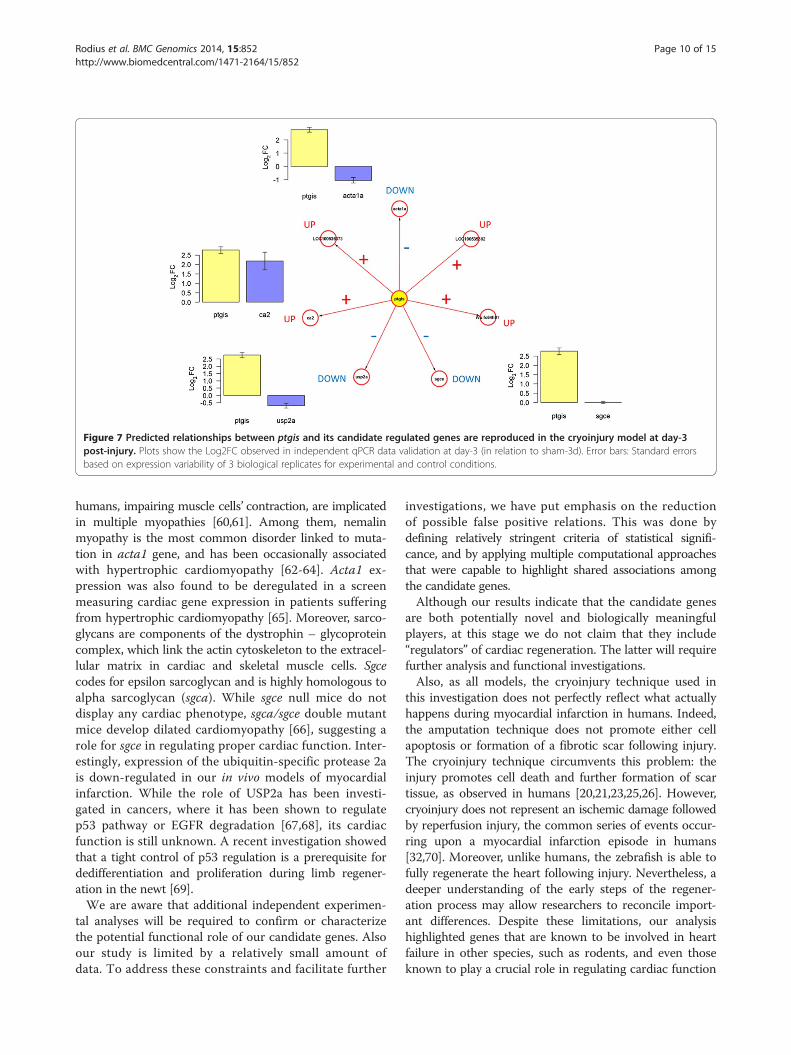
Figure 7 Predicted relationships between ptgis and its candidate regulated genes are reproduced in the cryoinjury model at day-3post-injury. Plots show the Log2FC observed in independent qPCR data validation at day-3 (in relation to sham-3d). Error bars: Standard errorsbased on expression variability of 3 biological replicates for experimental and control conditions.
Rodius et al. BMC Genomics 2014, 15:852 Page 10 of 15http://www.biomedcentral.com/1471-2164/15/852
humans, impairing muscle cells’ contraction, are implicatedin multiple myopathies [60,61]. Among them, nemalinmyopathy is the most common disorder linked to muta-tion in acta1 gene, and has been occasionally associatedwith hypertrophic cardiomyopathy [62-64]. Acta1 ex-pression was also found to be deregulated in a screenmeasuring cardiac gene expression in patients sufferingfrom hypertrophic cardiomyopathy [65]. Moreover, sarco-glycans are components of the dystrophin – glycoproteincomplex, which link the actin cytoskeleton to the extracel-lular matrix in cardiac and skeletal muscle cells. Sgcecodes for epsilon sarcoglycan and is highly homologous toalpha sarcoglycan (sgca). While sgce null mice do notdisplay any cardiac phenotype, sgca/sgce double mutantmice develop dilated cardiomyopathy [66], suggesting arole for sgce in regulating proper cardiac function. Inter-estingly, expression of the ubiquitin-specific protease 2ais down-regulated in our in vivo models of myocardialinfarction. While the role of USP2a has been investi-gated in cancers, where it has been shown to regulatep53 pathway or EGFR degradation [67,68], its cardiacfunction is still unknown. A recent investigation showedthat a tight control of p53 regulation is a prerequisite fordedifferentiation and proliferation during limb regener-ation in the newt [69].We are aware that additional independent experimen-
tal analyses will be required to confirm or characterizethe potential functional role of our candidate genes. Alsoour study is limited by a relatively small amount ofdata. To address these constraints and facilitate further
investigations, we have put emphasis on the reductionof possible false positive relations. This was done bydefining relatively stringent criteria of statistical signifi-cance, and by applying multiple computational approachesthat were capable to highlight shared associations amongthe candidate genes.Although our results indicate that the candidate genes
are both potentially novel and biologically meaningfulplayers, at this stage we do not claim that they include“regulators” of cardiac regeneration. The latter will requirefurther analysis and functional investigations.Also, as all models, the cryoinjury technique used in
this investigation does not perfectly reflect what actuallyhappens during myocardial infarction in humans. Indeed,the amputation technique does not promote either cellapoptosis or formation of a fibrotic scar following injury.The cryoinjury technique circumvents this problem: theinjury promotes cell death and further formation of scartissue, as observed in humans [20,21,23,25,26]. However,cryoinjury does not represent an ischemic damage followedby reperfusion injury, the common series of events occur-ring upon a myocardial infarction episode in humans[32,70]. Moreover, unlike humans, the zebrafish is able tofully regenerate the heart following injury. Nevertheless, adeeper understanding of the early steps of the regener-ation process may allow researchers to reconcile import-ant differences. Despite these limitations, our analysishighlighted genes that are known to be involved in heartfailure in other species, such as rodents, and even thoseknown to play a crucial role in regulating cardiac function

Rodius et al. BMC Genomics 2014, 15:852 Page 11 of 15http://www.biomedcentral.com/1471-2164/15/852
in humans, reinforcing thus the potential of our candidategenes as key regulators of heart regeneration.
ConclusionsBecause of its potential for translational research, we willcontinue investigating heart injury responses in thezebrafish using the cryoinjury model. We will expandthe scope and depth of our investigations, includingadditional biological replicates at post-injury times aswell as different control samples reflecting pre-injuryor non-damaged states. As larger, high-quality datasetsbecome available at our laboratory and elsewhere, wewill be able to further assess the predictive potential ofour network-based models.In conclusion, we identified genes with potential critical
roles in the response to cardiac damage in the zebrafish.Their transcriptional activities are reproducible in dif-ferent in vivo models of cardiac injury. These findingsmotivate additional research to confirm and function-ally characterize our set of candidate genes. These insightsand the application of network-based computationalmodels open new opportunities for cardiovascular transla-tional research.
MethodsAnimal procedure and cryoinjury modelAll procedures were approved by the national authoritiesresponsible for animal experiments in Luxembourg. Ex-periments were performed on wild-type adult zebrafishaged 10–11 months from the WIK strain (ZIRC, Eugene,OR, USA). Animals were maintained under standard la-boratory conditions in the Zebtec Stand Alone system(Tecniplast) at a density of 3 fish/L, at 28°C with day/nightlight cycles of 14 h light/10 h dark.Cryoinjury and hearts dissection were performed as
previously described [24]. Briefly, fish were anesthetizedin 0.04% tricaine (Sigma Aldrich, Bornem, BE) andplaced ventral side up in a damp foam. A small incisionwas performed through the body wall and the pericar-dium, the pericardial sac was opened and the ventricleexposed by gently compressing the abdomen. The sur-face of the ventricle was frozen by applying a cryoprobepreviously cooled in liquid nitrogen, until thawing wasobserved. Following surgery, fish were placed in a tankof fresh water, reanimated by gently pipetting wateronto the gills and further housed under standard condi-tions. Sham operations consisted of opening the bodywall and the pericardial sac, without touching the ex-posed ventricle. Animals were sacrificed 1 or 3 days aftersurgery by immersion in 0.16% tricaine (Sigma Aldrich,Bornem, BE). Hearts were dissected in PBS containingheparin and KCl and further used for histological stainingor RNA extraction.
ImmunohistochemistryHearts were fixed overnight in PBS containing 4% para-formaldehyde, dehydrated and embedded in paraffinwax. Seven μm sections were cut on a Leica Microtome,mounted on superfrost slides and dried for 1 h at 45°C.Samples were deparaffinized in xylol, rehydrated in etha-nol and washed in distilled water. Heat-induced epitoperetrieval was performed in citrate buffer pH 6. Sectionswere blocked for 1 h in 5% BSA – 0.5% tween 20. Primaryanti-tropomyosin antibody was CH1 (DSHB) used at a di-lution of 1/20. Secondary antibody was Alexa Fluor®568goat-anti mouse IgG (1/300; Molecular Probes). TUNELstaining was performed using the in situ Cell DeathDetection Kit, Fluorescein (Roche), according to themanufacturer’s instructions. Nuclei were stained withDAPI. Pictures were acquired using a confocal fluores-cence microscope (Zeiss Laser Scanning MicroscopeLSM 510).
Independent validation: RNA extraction, integrity andreverse transcriptionThree to four heart ventricles were pooled per sample inTRIzol (Invitrogen, Carlsbad, CA) and stored at −80°Cuntil extraction. Homogenisation of samples was per-formed with a Polytron® (Bohemia,USA). Aqueousphase was isolated with Phase lock gel-Heavy (5 Prime,Gaithersburg, MD), total RNA was precipitated with 100%isopropanol and purified using RNeasy Micro kit com-bined with an on-column DNase treatment followingthe manufacturer’s instructions (Qiagen, Valencia, CA).RNA quantity was assessed with a Nanodrop (ThermoScientific, Wilmington, USA) and quality was evaluatedwith the Agilent 2100 Bioanalyzer (Agilent Technologies,Palo Alto, CA). RNAs used in the present study were ofgood quality and un-degraded (Ratio A260/A280 ≈ 2 andRIN ≥ 8).500 ng of RNA were reverse transcribed into cDNA
using the SuperScript III (Invitrogen, Carlsbad, CA) re-verse transcriptase with the following protocol: RNAswere mixed with random primers, oligo (dT)12-18 anddNTPs in a total volume of 13 μl. Samples were heatedto 65°C for 5 min and incubated on ice for at least1 min. Then the 5X RT buffer, DTT, RNaseOUT andSuperScript III were added to a total volume of 20 μl.RT was allowed at 50°C for 60 min and was followedby enzyme inactivation at 70°C for 15 min. Final con-centrations were: 100 ng of oligo(dT)12-18, 50 ng ofrandom primers, 0.5 mM dNTPs, 50 mM Tris–HCl,75 mM KCl, 3 mM MgCl2, 5 mM DTT, 40U of RNase-OUT and 200U of SuperScript III. To remove RNAcomplementary to the cDNA, 2U of E. coli RNaseHwere added and samples were incubated at 37°C for20 min. In each RT-PCR a no template control (noRNA in RT) was performed.

Rodius et al. BMC Genomics 2014, 15:852 Page 12 of 15http://www.biomedcentral.com/1471-2164/15/852
Independent validation: qPCR experimentscDNAs obtained from RT of RNA were diluted 10-foldand 4 μL were mixed with SYBR®Green Master Mix(Biorad, Nazareth, Belgium) to a final volume of 20 μLcontaining 300nM of each primer. Amplification wascarried out in the CFX96 thermal cycler (BioRad) underthe following conditions: heating for 3 min at 95°C,40 cycles of denaturation for 30 s at 95°C, followed by anannealing/extension for 1 min. After each run a meltingcurve analysis was performed, ramping from 55°C to 95°Cin 20 min. A negative control without cDNA templatewas run in every assay and measures were performed induplicates.Primers were designed with the Beacon Designer Pro
8.10 software (Premier Biosoft, Palo Alto, USA), flankingan intron. Specificity was assessed using the NCBIBLAST tool [71], melting curves were performed ineach assay and gene-specific amplification was confirmedby a single band in 4% E-Gel® (Life technologies). Dataanalysis normalization was carried out against three refer-ence genes; ef1a, rpl13a, tuba1 and expression levels werecalculated using the CFX manager 3.0 software (Biorad) viathe delta-delta Cq method, taking into account the calcu-lated amplification efficiency for each primers pair. SeeAdditional file 5 for MIQE checklist [72] and details ofqPCR experiments.
Data pre-processing and differential expression analysisAs we were intended to use already pre-processed micro-array data, no normalization or probe summarization wasperformed in addition to those used in [29,40]. In order toincrease the power of statistical testing for detecting dif-ferential expressed genes, we performed pre-filtering offeatures removing those, which never show expressionover 6 in log2 scale. The cleaned dataset was then ana-lysed by limma package of R/Bioconductor [73] as it wasdescribed before [74,75]. Finally, a list of features signifi-cantly regulated at each time point was obtained using thesame “limma” model and a set of contrasts, comparingtime point samples versus controls. To control for falsediscovery rates (FDR), we used the Benjamini–Hochberg’sadjust of P-values. To report changes in gene expressionwe used standard heatmaps of R/Bioconductor.
Gene association network modelsWe generated and analysed gene co-expression networkswith different computational methodologies: Clusteringwith Overlapping Neighborhood Expansion (ClusterONE)[37], inference of gene regression networks with modeltrees (RegNet) [38], and the Weighted Gene Co-ExpressionNetwork (WGCNA) algorithm [39]. The capacity of thesetechniques to produce biologically informative resultshave previously been reported in different biomedicalresearch applications. These techniques allowed us to
identify: networks clusters, highly connected genes, andgenes that can be used as estimators of the expressionvalues of other genes in the dataset.The Clustering with Overlapping Neighborhood Expan-
sion (ClusterONE) algorithm identifies highly cohesivenetwork clusters (modules) through a 3-step process [37].First, starting from a seed node, ClusterONE implementsa greedy search of candidate groups showing high cohe-siveness. This procedure is started from different seednodes and leads to the formation of multiple, and possiblyoverlapping, clusters. Next, ClusterONE merges clusterswith significant overlaps according to a pre-definedthreshold. In the final step, candidate modules that donot meet user-defined scores (minimum number of nodesand module density) are removed from the list of candi-date modules. Here we used ClusterONE’s plugin (v1.0)for Cytoscape (v2.8.3) [76] with a minimum number ofnodes of 5, “Auto” minimum density, unweighted clus-tering, node penalty = 2, haircut threshold = 0; overlapthreshold = 0.8.RegNet [38] identifies transcriptional association net-
works in three steps. In the first step, an unsupervisedlearning algorithm analyses each gene as a target by takinginto account the remaining genes as inputs to a mathem-atical model that estimates the expression value of thatgene. The mathematical model consists of several linearmodels spread over separate areas of the search space, i.e.optimal partitions of gene expression samples. Each linearmodel represents localized similarities between specificgroups of genes. Moreover, the algorithm builds linearmodels under all samples (global similarity) if the optimalsubspace is defined by the complete set of gene expressionsamples. We have used M5’ model tree algorithm [77] tobuild the mathematical model for each target gene. In thesecond step, RegNet extracts, as hypothetical evidences ofgene-gene association, the linear dependency that existsbetween the target gene and every gene involved in thelinear models of the target gene. This step only includesthose mathematical models that have a relative error lessthan a threshold value. The third step involves building agraph model of gene co-expression network by assessingthe significance of the set of hypothetical evidences usingthe Benjamin-Yekutieli procedure for the control of falsediscovery rate. In our analysis RegNet was applied with athreshold value theta = 30%, i.e. the model trees with rela-tive error greater than theta were removed, and statisticalsignificance at 0.05.Module identification was also performed using
Weighted Correlation Network Analysis (WGCNA)[39]. With WGCNA, weighted correlations are calculatedbetween genes based on the gene’s expression in the dif-ferent samples; a weighted correlation is a correlationraised to a certain power (called the soft thresholdingpower). Weighted correlations are used as they are more

Rodius et al. BMC Genomics 2014, 15:852 Page 13 of 15http://www.biomedcentral.com/1471-2164/15/852
stringent for detecting true correlations, as they will favorhigher correlations over lower correlations (which canoften occur due to chance). In WGCNA, the weightedcorrelations are used to create a specific type of network(a topological overlap network). This network is based onnetwork topology rather than direct correlations betweengenes: genes are strongly linked together in this network ifthey share many correlated neighbours with each other.This topological overlap network is then analysed toidentify network modules of genes that are stronglylinked together. Identifying network modules from thegene expression in this way focuses on groups of genesthat are predicted to be functionally related based onlyon the gene expression data. WGCNA was performedon genes that were significantly differentially expressedbetween at least two conditions across all samples(FDR < 0.05). The network was constructed by calcu-lating an adjacency matrix using a soft-thresholdingpower of 10 and Spearman correlation using pairwisecomplete observations. A topological overlap matrix(TOM) was then calculated from the adjacency matrix,converted to distances, and clustered by hierarchicalclustering using average linkage clustering. Moduleswere identified with dynamic tree cut with minimummodule size = 20, using the hybrid method. Moduleeigengenes were calculated and similar modules weremerged together using a module eigengene distance of0.15 as the threshold.
Additional files
Additional file 1: Dynamic visualization of co-expression network atdifferent times (PDF) and underlying data (Excel file). In the networkvisualizations: nodes are color-coded to reflect Log2FC values (in relationto control), with scales indicated for each time-specific state.
Additional file 2: Detailed, higher resolution view of Figure 2C.
Additional file 3: Immunohistochemistry on sagittal sections ofsham-operated and cryoinjured hearts recovered one daypost-surgery. A – D: Staining is performed with an antibody againsttropomyosin (red). Note the reduced staining of tropomyosin in theinjured area of the ventricle compared to sham. E –H: TUNEL assay onhearts recovered one day post-surgery detects a high amount ofapoptotic cells (green) in the cryoinjured area while no staining isvisible in uninjured ventricles. Nuclei are stained with DAPI (blue).A/C: costaining DAPI/tropomyosin; B/D: staining of tropomyosinalone. E/G: costaining DAPI/TUNEL; F/H; TUNEL staining alone.Scale bar is 100 μm. A: atrium; B: bulbus arteriosus; V: ventricle;IA: injured area. Sham: sham-operated heart recovered oneday post-surgery; 1dpi: cryoinjured heart recovered atday-1 post-injury.
Additional file 4: Changes in (log2) expression of candidate genesin independent qPCR data validation with corresponding standarderrors (calculated based on expression variability of 3 biologicalreplicates for experimental and control conditions). (A) 1 day afterinjury vs sham-1d and (B) 3 days after injury vs sham-3d. Central node inFigures 6 and 7, ptgis, is marked yellow.
Additional file 5: MIQE checklist, including details of qPCRexperiments. Sheet 1: MIQE, Sheet 2: A-Primers B- Normalization dataanalysis information.
Competing interestsAll authors declare that they have no competing interests.
Authors’ contributionsSR performed in vivo work and immunohistochemistry and made significantcontributions to the preparation of the manuscript. FA conceived theinvestigation, performed computational analyses and led the writing of themanuscript. PN, IN, MI, and IX contributed computational analyses and inputsto manuscript writing. CJ performed qPCR validation, its data analysis andinputs to manuscript. JMGR, RMBC and NM contributed cryoinjury modelexpertise and validation experiments. All authors read and approved the finalmanuscript.
AcknowledgementsAt CRP-Santé: We thank Simone Niclou, Laurent Vallar and Arnaud Muller fortheir support. Celine Hoffmann for providing assistance with confocalmicroscopy. Jose Esposito and Raymond Rodius for their technical assistance.
FundingThis research was supported by the INTER program of Luxembourg’sNational Research Fund (FNR) and the Swiss National Research Foundation(SNF), INFUSED project (www.infused-project.eu). NM acknowledges supportfrom Spanish Ministry of Economy and Competivity (BFU2011-25297 andTerCel projects) and the Comunidad de Madrid (P2010/BMD-2321).
Author details1NorLux Neuro-Oncology Laboratory, CRP-Santé, Luxembourg, Luxembourg.2Genomics Research Unit, CRP-Santé, Luxembourg, Luxembourg.3Departamento Lenguajes y Sistemas Informáticos, Universidad de Sevilla,Seville, Spain. 4Cardiovascular Research Center, Massachusetts GeneralHospital and Harvard Medical School, Boston, USA. 5Vital-IT Systems Biology/Medicine Department, SIB Swiss Institute of Bioinformatics, Lausanne,Switzerland. 6Center for Integrative Genomics, University of Lausanne,Lausanne, Switzerland. 7Department of Biochemistry, University of Geneva,Geneva, Switzerland. 8Department of Cardiovascular Development andRepair, Centro Nacional de Investigaciones Cardiovasculares Carlos III, CNIC,Madrid, Spain.
Received: 15 September 2014 Accepted: 25 September 2014Published: 3 October 2014
References1. Bakkers J: Zebrafish as a model to study cardiac development and
human cardiac disease. Cardiovasc Res 2011, 91(2):279–288.2. Gemberling M, Bailey TJ, Hyde DR, Poss KD: The zebrafish as a model for
complex tissue regeneration. Trends Genet 2013, 29(11):611–620.3. Kikuchi K, Holdway JE, Werdich AA, Anderson RM, Fang Y, Egnaczyk GF,
Evans T, Macrae CA, Stainier DY, Poss KD: Primary contribution to zebrafishheart regeneration by gata4(+) cardiomyocytes. Nature 2010,464(7288):601–605.
4. Jopling C, Sleep E, Raya M, Marti M, Raya A, Izpisua Belmonte JC: Zebrafishheart regeneration occurs by cardiomyocyte dedifferentiation andproliferation. Nature 2010, 464(7288):606–609.
5. Go AS, Mozaffarian D, Roger VL, Benjamin EJ, Berry JD, Blaha MJ, Dai S, FordES, Fox CS, Franco S, Fullerton HJ, Gillespie C, Hailpern SM, Heit JA, HowardVJ, Huffman MD, Judd SE, Kissela BM, Kittner SJ, Lackland DT, Lichtman JH,Lisabeth LD, Mackey RH, Magid DJ, Marcus GM, Marelli A, Matchar DB,McGuire DK, Mohler ER, Moy CS, et al: Heart disease and strokestatistics–2014 update: a report from the American Heart Association.Circulation 2014, 129(3):e28–e292.
6. Laflamme MA, Murry CE: Heart regeneration. Nature 2011, 473(7347):326–335.7. Rosenzweig A: Medicine. Cardiac regeneration. Science 2012,
338(6114):1549–1550.8. Howe K, Clark MD, Torroja CF, Torrance J, Berthelot C, Muffato M, Collins JE,
Humphray S, McLaren K, Matthews L, McLaren S, Sealy I, Caccamo M, Churcher C,Scott C, Barrett JC, Koch R, Rauch GJ, White S, Chow W, Kilian B, Quintais LT,Guerra-Assuncao JA, Zhou Y, Gu Y, Yen J, Vogel JH, Eyre T, Redmond S, BanerjeeR, et al: The zebrafish reference genome sequence and its relationship to thehuman genome. Nature 2013, 496(7446):498–503.

Rodius et al. BMC Genomics 2014, 15:852 Page 14 of 15http://www.biomedcentral.com/1471-2164/15/852
9. Lawson ND, Wolfe SA: Forward and reverse genetic approaches for theanalysis of vertebrate development in the zebrafish. Dev Cell 2011,21(1):48–64.
10. Skromne I, Prince VE: Current perspectives in zebrafish reverse genetics:moving forward. Dev Dyn 2008, 237(4):861–882.
11. Chen JN, Haffter P, Odenthal J, Vogelsang E, Brand M, van Eeden FJ,Furutani-Seiki M, Granato M, Hammerschmidt M, Heisenberg CP, Jiang YJ,Kane DA, Kelsh RN, Mullins MC, Nusslein-Volhard C: Mutations affecting thecardiovascular system and other internal organs in zebrafish.Development 1996, 123:293–302.
12. Stainier DY, Fouquet B, Chen JN, Warren KS, Weinstein BM, Meiler SE,Mohideen MA, Neuhauss SC, Solnica-Krezel L, Schier AF, Zwartkruis F,Stemple DL, Malicki J, Driever W, Fishman MC: Mutations affecting theformation and function of the cardiovascular system in the zebrafishembryo. Development 1996, 123:285–292.
13. Huang CJ, Jou TS, Ho YL, Lee WH, Jeng YT, Hsieh FJ, Tsai HJ: Conditionalexpression of a myocardium-specific transgene in zebrafish transgeniclines. Dev Dyn 2005, 233(4):1294–1303.
14. Davison JM, Akitake CM, Goll MG, Rhee JM, Gosse N, Baier H, Halpern ME,Leach SD, Parsons MJ: Transactivation from Gal4-VP16 transgenicinsertions for tissue-specific cell labeling and ablation in zebrafish.Dev Biol 2007, 304(2):811–824.
15. Knopf F, Schnabel K, Haase C, Pfeifer K, Anastassiadis K, Weidinger G: Duallyinducible TetON systems for tissue-specific conditional gene expressionin zebrafish. Proc Natl Acad Sci U S A 2010, 107(46):19933–19938.
16. Vogel B, Meder B, Just S, Laufer C, Berger I, Weber S, Katus HA, Rottbauer W:In-vivo characterization of human dilated cardiomyopathy genes inzebrafish. Biochem Biophys Res Commun 2009, 390(3):516–522.
17. Pelster B, Burggren WW: Disruption of hemoglobin oxygen transport doesnot impact oxygen-dependent physiological processes in developingembryos of zebra fish (Danio rerio). Circ Res 1996, 79(2):358–362.
18. Zon LI, Peterson RT: In vivo drug discovery in the zebrafish. Nat Rev DrugDiscov 2005, 4(1):35–44.
19. Kikuchi K, Poss KD: Cardiac regenerative capacity and mechanisms. AnnuRev Cell Dev Biol 2012, 28:719–741.
20. Lien CL, Harrison MR, Tuan TL, Starnes VA: Heart repair and regeneration:recent insights from zebrafish studies. Wound Repair Regen 2012,20(5):638–646.
21. Poss KD, Wilson LG, Keating MT: Heart regeneration in zebrafish. Science2002, 298(5601):2188–2190.
22. van den Bos EJ, Mees BM, de Waard MC, de Crom R, Duncker DJ: A novelmodel of cryoinjury-induced myocardial infarction in the mouse: acomparison with coronary artery ligation. Am J Physiol Heart Circ Physiol2005, 289(3):H1291–H1300.
23. Gonzalez-Rosa JM, Martin V, Peralta M, Torres M, Mercader N: Extensive scarformation and regression during heart regeneration after cryoinjury inzebrafish. Development 2011, 138(9):1663–1674.
24. Gonzalez-Rosa JM, Mercader N: Cryoinjury as a myocardial infarctionmodel for the study of cardiac regeneration in the zebrafish. Nat Protoc2012, 7(4):782–788.
25. Schnabel K, Wu CC, Kurth T, Weidinger G: Regeneration of cryoinjuryinduced necrotic heart lesions in zebrafish is associated with epicardialactivation and cardiomyocyte proliferation. PLoS One 2011, 6(4):e18503.
26. Chablais F, Veit J, Rainer G, Jazwinska A: The zebrafish heart regeneratesafter cryoinjury-induced myocardial infarction. BMC Dev Biol 2011, 11:21.
27. Chablais F, Jazwinska A: Induction of myocardial infarction in adultzebrafish using cryoinjury. J Vis Exp 2012, (62):3666. doi:10.3791/3666.
28. Wang J, Panakova D, Kikuchi K, Holdway JE, Gemberling M, Burris JS, SinghSP, Dickson AL, Lin YF, Sabeh MK, Werdich AA, Yelon D, Macrae CA, PossKD: The regenerative capacity of zebrafish reverses cardiac failurecaused by genetic cardiomyocyte depletion. Development 2011,138(16):3421–3430.
29. Sleep E, Boue S, Jopling C, Raya M, Raya A, Izpisua Belmonte JC:Transcriptomics approach to investigate zebrafish heart regeneration.J Cardiovasc Med (Hagerstown) 2010, 11(5):369–380.
30. Fang Y, Gupta V, Karra R, Holdway JE, Kikuchi K, Poss KD: Translationalprofiling of cardiomyocytes identifies an early Jak1/Stat3 injury responserequired for zebrafish heart regeneration. Proc Natl Acad Sci U S A 2013,110(33):13416–13421.
31. Lien CL, Schebesta M, Makino S, Weber GJ, Keating MT: Gene expressionanalysis of zebrafish heart regeneration. PLoS Biol 2006, 4(8):e260.
32. Kikuchi K, Holdway JE, Major RJ, Blum N, Dahn RD, Begemann G, Poss KD:Retinoic acid production by endocardium and epicardium is an injuryresponse essential for zebrafish heart regeneration. Dev Cell 2011,20(3):397–404.
33. Kim J, Wu Q, Zhang Y, Wiens KM, Huang Y, Rubin N, Shimada H, Handin RI,Chao MY, Tuan TL, Starnes VA, Lien CL: PDGF signaling is required forepicardial function and blood vessel formation in regenerating zebrafishhearts. Proc Natl Acad Sci U S A 2010, 107(40):17206–17210.
34. Choi WY, Gemberling M, Wang J, Holdway JE, Shen MC, Karlstrom RO, PossKD: In vivo monitoring of cardiomyocyte proliferation to identifychemical modifiers of heart regeneration. Development 2013,140(3):660–666.
35. Chablais F, Jazwinska A: The regenerative capacity of the zebrafishheart is dependent on TGFbeta signaling. Development 2012,139(11):1921–1930.
36. Zhao L, Borikova AL, Ben-Yair R, Guner-Ataman B, MacRae CA, Lee RT, BurnsCG, Burns CE: Notch signaling regulates cardiomyocyte proliferationduring zebrafish heart regeneration. Proc Natl Acad Sci U S A 2014,111(4):1403–1408.
37. Nepusz T, Yu H, Paccanaro A: Detecting overlapping protein complexes inprotein-protein interaction networks. Nat Methods 2012, 9(5):471–472.
38. Nepomuceno-Chamorro IA, Aguilar-Ruiz JS, Riquelme JC: Inferring generegression networks with model trees. BMC Bioinformatics 2010, 11:517.
39. Langfelder P, Horvath S: WGCNA: an R package for weighted correlationnetwork analysis. BMC Bioinformatics 2008, 9:559.
40. Kizil C, Otto GW, Geisler R, Nusslein-Volhard C, Antos CL: Simple controlscell proliferation and gene transcription during zebrafish caudal finregeneration. Dev Biol 2009, 325(2):329–340.
41. Conway SJ, Molkentin JD: Periostin as a heterofunctional regulator ofcardiac development and disease. Curr Genomics 2008, 9(8):548–555.
42. Snider P, Standley KN, Wang J, Azhar M, Doetschman T, Conway SJ: Originof cardiac fibroblasts and the role of periostin. Circ Res 2009,105(10):934–947.
43. Oka T, Xu J, Kaiser RA, Melendez J, Hambleton M, Sargent MA, Lorts A,Brunskill EW, Dorn GW 2nd, Conway SJ, Aronow BJ, Robbins J, Molkentin JD:Genetic manipulation of periostin expression reveals a role in cardiachypertrophy and ventricular remodeling. Circ Res 2007, 101(3):313–321.
44. Shimazaki M, Nakamura K, Kii I, Kashima T, Amizuka N, Li M, Saito M, FukudaK, Nishiyama T, Kitajima S, Saga Y, Fukayama M, Sata M, Kudo A: Periostin isessential for cardiac healing after acute myocardial infarction. J Exp Med2008, 205(2):295–303.
45. Kuhn B, del Monte F, Hajjar RJ, Chang YS, Lebeche D, Arab S, Keating MT:Periostin induces proliferation of differentiated cardiomyocytes andpromotes cardiac repair. Nat Med 2007, 13(8):962–969.
46. Gonzalez-Rosa JM, Peralta M, Mercader N: Pan-epicardial lineage tracingreveals that epicardium derived cells give rise to myofibroblasts andperivascular cells during zebrafish heart regeneration. Dev Biol 2012,370(2):173–186.
47. Seger R, Krebs EG: The MAPK signaling cascade. FASEB J 1995,9(9):726–735.
48. Stevenson LF, Sparks A, Allende-Vega N, Xirodimas DP, Lane DP, Saville MK:The deubiquitinating enzyme USP2a regulates the p53 pathway bytargeting Mdm2. EMBO J 2007, 26(4):976–986.
49. Wong AK, Park CY, Greene CS, Bongo LA, Guan Y, Troyanskaya OG: IMP: amulti-species functional genomics portal for integration, visualizationand prediction of protein functions and networks. Nucleic Acids Res 2012,40(Web Server issue):W484–W490.
50. Van Vliet P, Wu SM, Zaffran S, Puceat M: Early cardiac development: a viewfrom stem cells to embryos. Cardiovasc Res 2012, 96(3):352–362.
51. Takeuchi JK, Bruneau BG: Directed transdifferentiation of mousemesoderm to heart tissue by defined factors. Nature 2009,459(7247):708–711.
52. Alsan BH, Schultheiss TM: Regulation of avian cardiogenesis by Fgf8signaling. Development 2002, 129(8):1935–1943.
53. Holtzinger A, Rosenfeld GE, Evans T: Gata4 directs development ofcardiac-inducing endoderm from ES cells. Dev Biol 2010, 337(1):63–73.
54. Rouleau M, Medawar A, Hamon L, Shivtiel S, Wolchinsky Z, Zhou H, De RosaL, Candi E, de la Forest DS, Mikkola ML, van Bokhoven H, Missero C, MelinoG, Puceat M, Aberdam D: TAp63 is important for cardiac differentiation ofembryonic stem cells and heart development. Stem Cells 2011,29(11):1672–1683.

Rodius et al. BMC Genomics 2014, 15:852 Page 15 of 15http://www.biomedcentral.com/1471-2164/15/852
55. Frangogiannis NG: Matricellular proteins in cardiac adaptation anddisease. Physiol Rev 2012, 92(2):635–688.
56. Lu B, Yu H, Zwartbol M, Ruifrok WP, van Gilst WH, de Boer RA, Sillje HH:Identification of hypertrophy- and heart failure-associated genes bycombining in vitro and in vivo models. Physiol Genomics 2012,44(8):443–454.
57. Kyritsis N, Kizil C, Zocher S, Kroehne V, Kaslin J, Freudenreich D, Iltzsche A,Brand M: Acute inflammation initiates the regenerative response in theadult zebrafish brain. Science 2012, 338(6112):1353–1356.
58. Hsueh YC, Wu JM, Yu CK, Wu KK, Hsieh PC: Prostaglandin E(2) promotespost-infarction cardiomyocyte replenishment by endogenous stem cells.EMBO Mol Med 2014, 6(4):496–503.
59. Alvarez BV, Quon AL, Mullen J, Casey JR: Quantification of carbonicanhydrase gene expression in ventricle of hypertrophic and failinghuman heart. BMC Cardiovasc Disord 2013, 13:2.
60. Nowak KJ, Wattanasirichaigoon D, Goebel HH, Wilce M, Pelin K, Donner K,Jacob RL, Hubner C, Oexle K, Anderson JR, Verity CM, North KN, IannacconeST, Muller CR, Nurnberg P, Muntoni F, Sewry C, Hughes I, Sutphen R, LacsonAG, Swoboda KJ, Vigneron J, Wallgren-Pettersson C, Beggs AH, Laing NG:Mutations in the skeletal muscle alpha-actin gene in patients with actinmyopathy and nemaline myopathy. Nat Genet 1999, 23(2):208–212.
61. Feng JJ, Marston S: Genotype-phenotype correlations in ACTA1mutations that cause congenital myopathies. Neuromuscul Disord 2009,19(1):6–16.
62. D’Amico A, Graziano C, Pacileo G, Petrini S, Nowak KJ, Boldrini R, Jacques A,Feng JJ, Porfirio B, Sewry CA, Santorelli FM, Limongelli G, Bertini E, Laing N,Marston SB: Fatal hypertrophic cardiomyopathy and nemaline myopathyassociated with ACTA1 K336E mutation. Neuromuscul Disord 2006,16(9–10):548–552.
63. Kim SY, Park YE, Kim HS, Lee CH, Yang DH, Kim DS: Nemaline myopathyand non-fatal hypertrophic cardiomyopathy caused by a novel ACTA1E239K mutation. J Neurol Sci 2011, 307(1–2):171–173.
64. Gatayama R, Ueno K, Nakamura H, Yanagi S, Ueda H, Yamagishi H, Yasui S:Nemaline myopathy with dilated cardiomyopathy in childhood. Pediatrics2013, 131(6):e1986–e1990.
65. Lim DS, Roberts R, Marian AJ: Expression profiling of cardiac genes inhuman hypertrophic cardiomyopathy: insight into the pathogenesis ofphenotypes. J Am Coll Cardiol 2001, 38(4):1175–1180.
66. Lancioni A, Rotundo IL, Kobayashi YM, D’Orsi L, Aurino S, Nigro G, Piluso G,Acampora D, Cacciottolo M, Campbell KP, Nigro V: Combined deficiency ofalpha and epsilon sarcoglycan disrupts the cardiac dystrophin complex.Hum Mol Genet 2011, 20(23):4644–4654.
67. Priolo C, Tang D, Brahamandan M, Benassi B, Sicinska E, Ogino S, Farsetti A,Porrello A, Finn S, Zimmermann J, Febbo P, Loda M: The isopeptidaseUSP2a protects human prostate cancer from apoptosis. Cancer Res 2006,66(17):8625–8632.
68. Liu Z, Zanata SM, Kim J, Peterson MA, Di Vizio D, Chirieac LR, Pyne S,Agostini M, Freeman MR, Loda M: The ubiquitin-specific protease USP2aprevents endocytosis-mediated EGFR degradation. Oncogene 2013,32(13):1660–1669.
69. Yun MH, Gates PB, Brockes JP: Regulation of p53 is critical for vertebratelimb regeneration. Proc Natl Acad Sci U S A 2013, 110(43):17392–17397.
70. Lepilina A, Coon AN, Kikuchi K, Holdway JE, Roberts RW, Burns CG, Poss KD:A dynamic epicardial injury response supports progenitor cell activityduring zebrafish heart regeneration. Cell 2006, 127(3):607–619.
71. BLAST: Basic Local Alignment Search Tool. 2012. Available from: http://www.ncbi.nlm.nih.gov/BLAST/Blast.cgi.
72. Bustin SA, Benes V, Garson JA, Hellemans J, Huggett J, Kubista M, Mueller R,Nolan T, Pfaffl MW, Shipley GL, Vandesompele J, Wittwer CT: The MIQEguidelines: minimum information for publication of quantitativereal-time PCR experiments. Clin Chem 2009, 55(4):611–622.
73. Smyth GK: Linear models and empirical bayes methods for assessingdifferential expression in microarray experiments. Stat Appl Genet Mol Biol2004, 3:Article 3.
74. Nazarov PV, Reinsbach SE, Muller A, Nicot N, Philippidou D, Vallar L, Kreis S:Interplay of microRNAs, transcription factors and target genes: linkingdynamic expression changes to function. Nucleic Acids Res 2013,41(5):2817–2831.
75. Gillespie CS, Lei G, Boys RJ, Greenall A, Wilkinson DJ: Analysing time coursemicroarray data using Bioconductor: a case study using yeast2Affymetrix arrays. BMC Res Notes 2010, 3:81.
76. Shannon P, Markiel A, Ozier O, Baliga NS, Wang JT, Ramage D, Amin N,Schwikowski B, Ideker T: Cytoscape: a software environment forintegrated models of biomolecular interaction networks. Genome Res2003, 13(11):2498–2504.
77. Malerba D, Esposito F, Ceci M, Appice A: Top-down induction of modeltrees with regression and splitting nodes. IEEE Trans Pattern Anal MachIntell 2004, 26(5):612–625.
doi:10.1186/1471-2164-15-852Cite this article as: Rodius et al.: Transcriptional response to cardiacinjury in the zebrafish: systematic identification of genes with highlyconcordant activity across in vivo models. BMC Genomics 2014 15:852.
Submit your next manuscript to BioMed Centraland take full advantage of:
• Convenient online submission
• Thorough peer review
• No space constraints or color figure charges
• Immediate publication on acceptance
• Inclusion in PubMed, CAS, Scopus and Google Scholar
• Research which is freely available for redistribution
Submit your manuscript at www.biomedcentral.com/submit