transcranial magnetic stimulation - alex reichenbach · tms localizer using a „hunting...
TRANSCRIPT
1
Session 4 – Virtual Lesion Approach ITranscranial Magnetic Stimulation
Alexandra ReichenbachMPI for Biological CyberneticsTübingen, Germany
Today‘s Schedule
Virtual Lesion Approach : Study Design Rationale Measurements Temporal modes Spatial modes Coil placement Stimulation intensity Control conditions
A. Reichenbach / A. Thielscher
2
Virtual Lesion Approach: Overview
Virtual lesion (Walsh & Rushworth, 1999): Mimic a brain lesion temporarily in a circumscribed area without cortical re-organization.
Use TMS pulses to interfere temporarily with the neural processing in the stimulated area while the subject is performing a behavioral task.
Effects of TMS on reaction times or error rate indicate that the stimulated area is crucially involved in the task.
A. Reichenbach / A. Thielscher
With the virtual lesion approach, TMS is a tool for neuroscience which allows to disentangle the necessity of brain regions for a specific task compared to the mere involvement of it (causality vs. correlation). Furthermore, this methodology can be used to measure the chronometry of neural information processing.
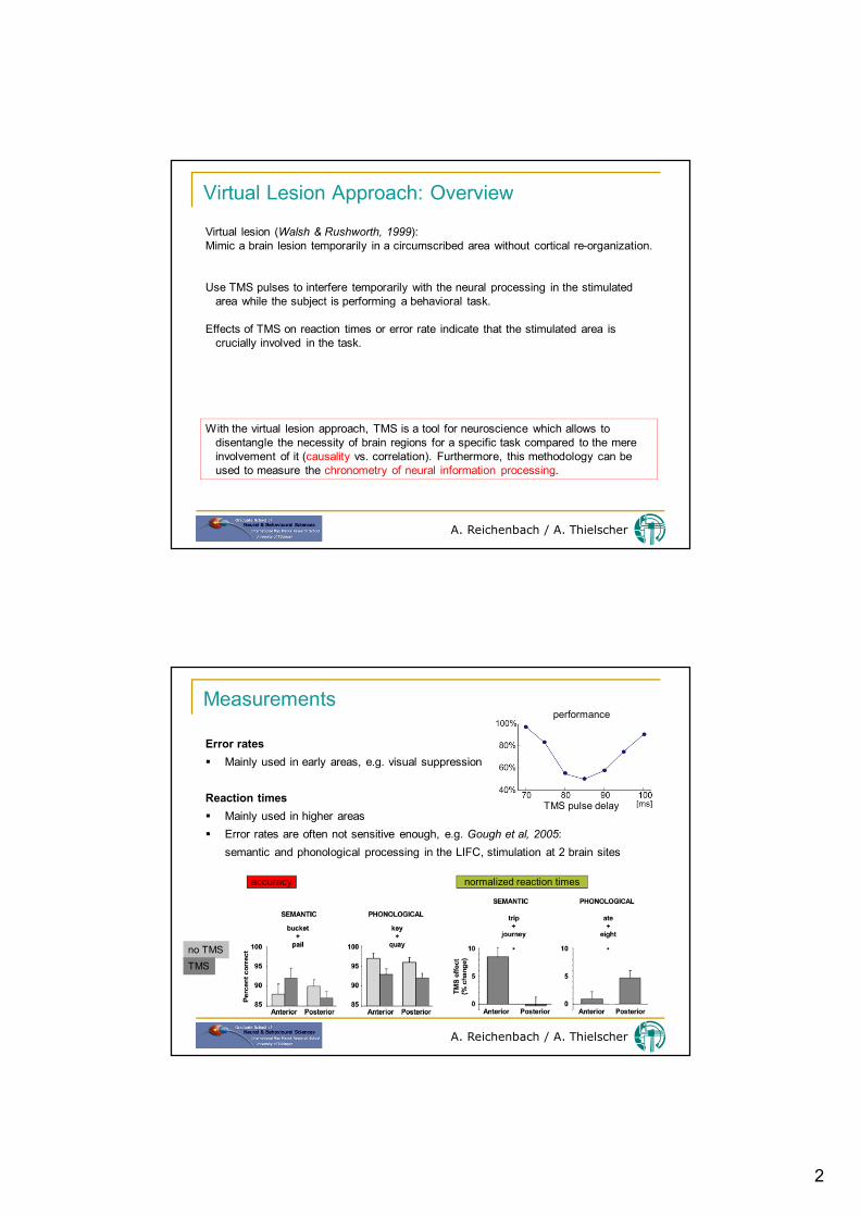
Measurements
A. Reichenbach / A. Thielscher
Error rates Mainly used in early areas, e.g. visual suppression
Reaction times Mainly used in higher areas Error rates are often not sensitive enough, e.g. Gough et al, 2005:
semantic and phonological processing in the LIFC, stimulation at 2 brain sites
no TMSTMS
normalized reaction timesaccuracy
performance
TMS pulse delay
3
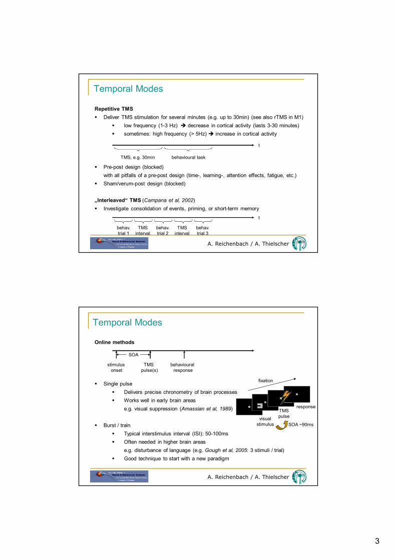
Temporal Modes
A. Reichenbach / A. Thielscher
Repetitive TMS Deliver TMS stimulation for several minutes (e.g. up to 30min) (see also rTMS in M1)
low frequency (1-3 Hz) decrease in cortical activity (lasts 3-30 minutes) sometimes: high frequency (> 5Hz) increase in cortical activity
Pre-post design (blocked)with all pitfalls of a pre-post design (time-, learning-, attention effects, fatigue, etc.)
Sham/verum-post design (blocked)
„Interleaved“ TMS (Campana et al, 2002) Investigate consolidation of events, priming, or short-term memory
t
TMS, e.g. 30min behavioural task
t
behav.trial 1
behav.trial 2
behav.trial 3
TMSinterval
TMSinterval
Temporal Modes
A. Reichenbach / A. Thielscher
Online methods
Single pulse Delivers precise chronometry of brain processes Works well in early brain areas
e.g. visual suppression (Amassian et al, 1989)
Burst / train Typical interstimulus interval (ISI): 50-100ms Often needed in higher brain areas
e.g. disturbance of language (e.g. Gough et al, 2005: 3 stimuli / trial) Good technique to start with a new paradigm
stimulus onset
TMS pulse(s)
behavioural response
SOA
fixation
visualstimulus
TMSpulse
response
SOA ~90ms
4
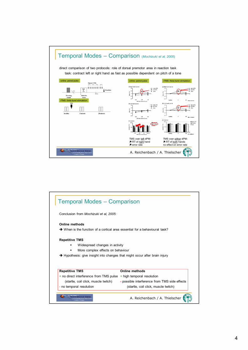
Temporal Modes – Comparison (Mochizuki et al, 2005)
A. Reichenbach / A. Thielscher
direct comparison of two protocols: role of dorsal premotor area in reaction tasktask: contract left or right hand as fast as possible dependent on pitch of a tone
online: paired pulse
rTMS: theta burst stimulation
online: paired pulse rTMS: theta burst stimulation
TMS over left dPM: RT of right hand error rate
TMS over either dPM: RT of both handsno effect on error rate
Temporal Modes – Comparison
A. Reichenbach / A. Thielscher
Conclusion from Mochizuki et al, 2005:
Online methods When is the function of a cortical area essential for a behavioural task?
Repetitive TMS Widespread changes in activity More complex effects on behaviour
Hypothesis: give insight into changes that might occur after brain injury
Repetitive TMS Online methods+ no direct interference from TMS pulse + high temporal resolution
(startle, coil click, muscle twitch) - possible interference from TMS side effects- no temporal resolution (startle, coil click, muscle twitch)

5
Spatial Modes
A. Reichenbach / A. Thielscher
Disrupt function at site of stimulation
Induce noise in a secondary site by
disinhibiting the site (a)
disrupting pre-processes to this site (b)
Investigate timing of interaction between two areas
+ -
a: inhibitory connection b: downstream area
Spatial Modes – Some Remarks
A. Reichenbach / A. Thielscher
Most commonly it is assumed that the function at site of stimulation is disrupted. This approach needs a hypothesis based on anatomy.
Think of possible effects from secondary sites when observing „strange“ effects.
Interleaved TMS/fMRI as tool to disentangle TMS effect on different brain sites.
Beware of rash conclusions when observing improvement of function!Alternative explanations can be Motor facilitation Intersensory facilitation (coil click, somatosensory stimulation)
6
Coil Placement
A. Reichenbach / A. Thielscher
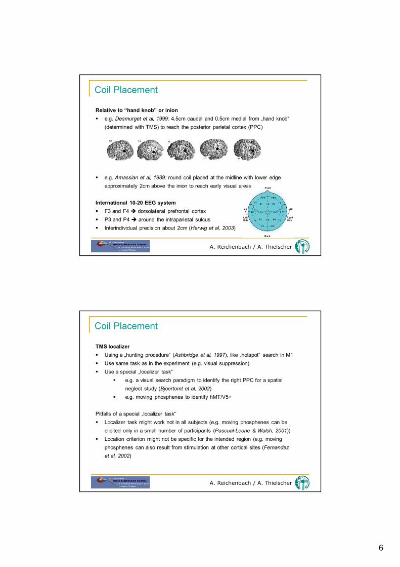
Relative to “hand knob” or inion e.g. Desmurget et al, 1999: 4.5cm caudal and 0.5cm medial from „hand knob“
(determined with TMS) to reach the posterior parietal cortex (PPC)
e.g. Amassian et al, 1989: round coil placed at the midline with lower edge approximately 2cm above the inion to reach early visual areas
International 10-20 EEG system F3 and F4 dorsolateral prefrontal cortex P3 and P4 around the intraparietal sulcus Interindividual precision about 2cm (Herwig et al, 2003)
Coil Placement
A. Reichenbach / A. Thielscher
TMS localizer Using a „hunting procedure“ (Ashbridge et al, 1997), like „hotspot“ search in M1 Use same task as in the experiment (e.g. visual suppression) Use a special „localizer task“
e.g. a visual search paradigm to identify the right PPC for a spatial neglect study (Bjoertomt et al, 2002)
e.g. moving phosphenes to identify hMT/V5+
Pitfalls of a special „localizer task“ Localizer task might work not in all subjects (e.g. moving phosphenes can be
elicited only in a small number of participants (Pascual-Leone & Walsh, 2001)) Location criterion might not be specific for the intended region (e.g. moving
phosphenes can also result from stimulation at other cortical sites (Fernandez et al, 2002)
7
Coil Placement
A. Reichenbach / A. Thielscher
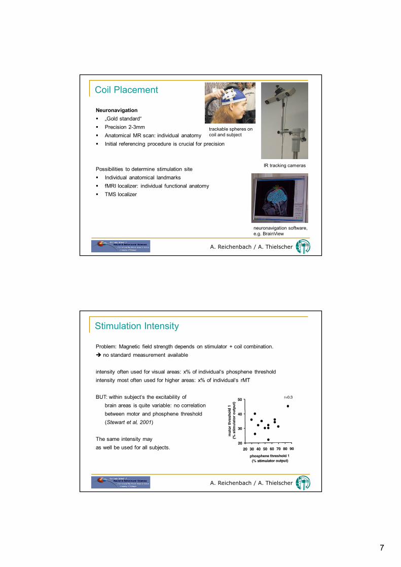
Neuronavigation „Gold standard“ Precision 2-3mm Anatomical MR scan: individual anatomy Initial referencing procedure is crucial for precision
Possibilities to determine stimulation site Individual anatomical landmarks fMRI localizer: individual functional anatomy TMS localizer
IR tracking cameras
neuronavigation software,e.g. BrainView
trackable spheres oncoil and subject
Stimulation Intensity
A. Reichenbach / A. Thielscher
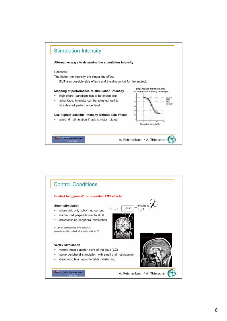
Problem: Magnetic field strength depends on stimulator + coil combination. no standard measurement available
intensity often used for visual areas: x% of individual‘s phosphene thresholdintensity most often used for higher areas: x% of individual‘s rMT
BUT: within subject‘s the excitability of brain areas is quite variable: no correlationbetween motor and phosphene threshold (Stewart et al, 2001)
The same intensity may as well be used for all subjects.
8
Stimulation Intensity
A. Reichenbach / A. Thielscher
Alternative ways to determine the stimulation intensity
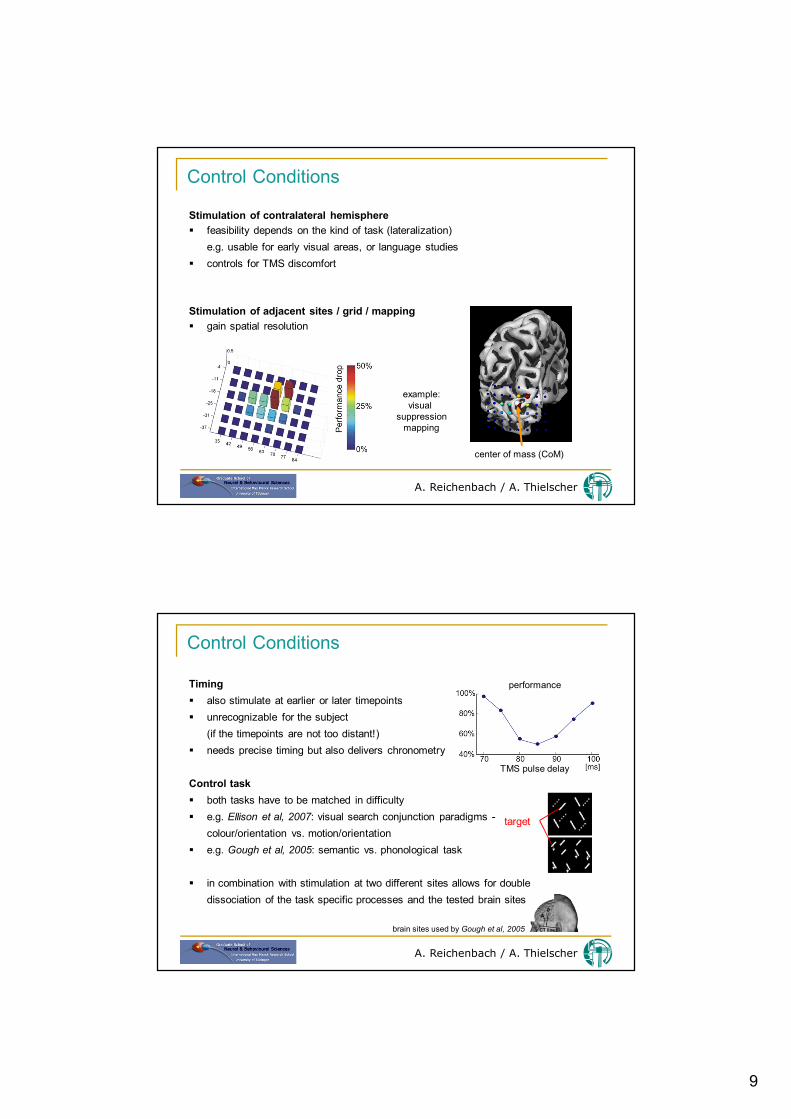
Rationale:The higher the intensity the bigger the effect
BUT also possible side effects and the discomfort for the subject.
Mapping of performance to stimulation intensity high efford, paradigm has to be known well advantage: intensity can be adjusted well to
fit a desired performance level
Use highest possible intensity without side effects avoid M1 stimulation if task is motor related
Stimulation Intensity [%]
Dependence of Performanceon Stimulation Intensity - Example
Control Conditions
A. Reichenbach / A. Thielscher
Control for „general“ or unwanted TMS effects!
Sham stimulation sham coil: only „click“, no current normal coil perpendicular to skull drawback: no peripheral stimulation
!!! use of control sites (see below) is sometimes also called „sham stimulation“ !!!
Vertex stimulation vertex: most superior point of the skull (CZ) some peripheral stimulation with small brain stimulation drawback: less uncomfortable / disturbing
„click“no current
9
Control Conditions
A. Reichenbach / A. Thielscher
Stimulation of contralateral hemisphere feasibility depends on the kind of task (lateralization)
e.g. usable for early visual areas, or language studies controls for TMS discomfort
Stimulation of adjacent sites / grid / mapping gain spatial resolution
center of mass (CoM)
example:visual
suppressionmapping
Control Conditions
A. Reichenbach / A. Thielscher
Timing also stimulate at earlier or later timepoints unrecognizable for the subject
(if the timepoints are not too distant!) needs precise timing but also delivers chronometry
Control task both tasks have to be matched in difficulty e.g. Ellison et al, 2007: visual search conjunction paradigms -
colour/orientation vs. motion/orientation e.g. Gough et al, 2005: semantic vs. phonological task
in combination with stimulation at two different sites allows for double dissociation of the task specific processes and the tested brain sites
TMS pulse delay
performance
target
brain sites used by Gough et al, 2005