transarticular screw fixation for atlantoaxial instability - journal of
TRANSCRIPT
RESEARCH ARTICLE Open Access
Transarticular screw fixation for atlantoaxialinstability - modified Magerl’s technique in38 patientsRaj Bahadur1,2†, Tarun Goyal3*†, Saravdeep S Dhatt4, Sujit K Tripathy4
Abstract
Background: Symptomatic atlantoaxial instability needs stabilization of the atlantoaxial joint. Among the varioustechniques described in literature for the fixation of atlantoaxial joint, Magerl’s technique of transarticular screwfixation remains the gold standard. Traditionally this technique combines placement of transarticular screws andposterior wiring construct. The aim of this study is to evaluate clinical and radiological outcomes in subjects ofatlantoaxial instability who were operated using transarticular screws and iliac crest bone graft, without the use ofsublaminar wiring (a modification of Magerl’s technique).
Methods: We evaluated retrospectively 38 subjects with atlantoaxial instability who were operated at our instituteusing transarticular screw fixation. The subjects were followed up for pain, fusion rates, neurological status andradiographic outcomes. Final outcome was graded both subjectively and objectively, using the scoring systemgiven by Grob et al.
Results: Instability in 34 subjects was secondary to trauma, in 3 due to rheumatoid arthritis and 1 had tuberculosis.Neurological deficit was present in 17 subjects. Most common presenting symptom was neck pain, present in 35of the 38 subjects.Postoperatively residual neck and occipital pain was present in 8 subjects. Neurological deficit persisted in only 7subjects. Vertebral artery injury was seen in 3 subjects. None of these subjects had any sign of neurological deficitor vertebral insufficiency. Three cases had nonunion. At the latest follow up, subjectively, 24 subjects had goodresult, 6 had fair and 8 had bad result. On objective grading, 24 had good result, 11 had fair and 3 had bad result.The mean follow up duration was 41 months.
Conclusions: Transarticular screw fixation is an excellent technique for fusion of the atlantoaxial complex. Itprovides highest fusion rates, and is particularly important in subjects at risk for nonunion. Omitting the posteriorwiring construct that has been used along with the bone graft in the traditional Magerl’ s technique achievesequally good fusion rates and is an important modification, thereby avoiding the complications of sublaminar wirepassage.
BackgroundAtlantoaxial articulation is the most unique part of thespine. It is the most mobile segment of the spine, andlargely depends on the ligamentous supports and theintegrity of the odontoid for its stability. Fusion of theC1-C2 complex may be required in cases of atlantoaxial
instability. Its extreme mobility places heavy demand onthe atlantoaxial fixation construct for sufficient rigidityrequired for its fusion. The causes of C1-C2 instabilityare numerous and include trauma, congenital malforma-tions, inflammatory arthritis, malignancies, skeletal dys-plasias, rotatory subluxations and pharyngeal infections.Symptoms of instability of the atlanto axial complex arevaried, such as neck pain, transient paresis, headaches,ataxia and intermittent loss of consciousness.Clinically or radiographically significant atlantoaxial
subluxation is best treated by reduction and fusion of
* Correspondence: [email protected]† Contributed equally3Dept of Orthopaedics, All India Institute of Medical Sciences, New Delhi,IndiaFull list of author information is available at the end of the article
Bahadur et al. Journal of Orthopaedic Surgery and Research 2010, 5:87http://www.josr-online.com/content/5/1/87
© 2010 Bahadur et al; licensee BioMed Central Ltd. This is an Open Access article distributed under the terms of the Creative CommonsAttribution License (http://creativecommons.org/licenses/by/2.0), which permits unrestricted use, distribution, and reproduction inany medium, provided the original work is properly cited.

the C1-C2 joint. Posterior C1-C2 fusion using transarti-cular screw (TAS), introduced by Magerl et al in 1979[1] is the gold standard for atlantoaxial arthrodesis. Ithas the advantage of a more rigid fixation with higherrates of fusion, avoiding need for postoperative halo, noplacement of implant in the spinal canal, and possibilityof its use in anomalies of odontoid process or the pos-terior arch [2-7]. Magerl et al used two transarticularscrews along with bone graft and interspinous wiring forfusion. But the use of sublaminar wiring is fraught withseveral complications, such as damage to the dura andthe cord during insertion of the wires and late compres-sion of the cord by wire breakage or loosening [8-12].Further, it has been found that there may be no impor-tant contribution of the wires in holding the graft forfusion, and comparable fusion rates have been achievedin these studies [4,13,14].Thus we designed our study to evaluate the outcome
of cases of atlantoaxial instability treated with transarti-cular screw fixation. We did not include supplementalwiring as described by Magerl et al in our technique.Postoperatively, the subjects were evaluated clinicallyand radiographically for the improvement in clinicalscores, fusion rates of the arthrodesis and any associatedcomplications.
MethodsWe studied 38 subjects of atlantoaxial instability whounderwent posterior fusion using transarticular screws.All the cases were operated by the senior author (RB)from 1995 to 2008. Instability was defined on flexion-extension X-rays, using atlanto dens interval (ADI). ADIof greater than 5 mm was taken as definition of atlan-toaxial instability (figure 1).All subjects were assessed with plain anteroposterior,
open mouth view and lateral flexion extension radio-graphs. Lateral radiographs help to verify that theC1-C2 complex has been reduced adequately before thesurgery and to find the estimated length of the screwsto be used (figure 2).A Computed Tomography Scan with saggital, coronal
and 3 D reconstruction was done in all the cases to lookat the transverse foramen of C2, understand the fractureanatomy, C2 isthmus size, space available for the cordand integrity of the C1 lateral masses. Magnetic Reso-nance Imaging (MRI) was done only in subjects withneurological deficit, to study the lesion of the cord andthe degree of canal compromise, in order to plan poster-ior decompression in these cases (figure 3).Subjects who had pathology of the C1-C2 facets and
C1 lateral masses, such as comminuted fractures or thetumors destroying the C1 lateral masses that precludescrew placement were excluded from the study. Subjectswho were found to have anomalous course of the
vertebral artery on Computed Tomography Scan werealso excluded from the study. This was studied usingaxial and saggital cuts of the CT scan in the region oftransverse foramen of the C2 vertebra. High riding ver-tebral artery was identified as having a too medial and/
Figure 1 Lateral radiograph of a subject with atlantoaxialinstability secondary to odontoid fracture showing markedatlantoaxial displacement.
Figure 2 Post reduction film of the same subject using skeletaltraction in the ward. Further complete reduction was obtainedintraoperatively using skeletal traction with crutchfield tongs.
Bahadur et al. Journal of Orthopaedic Surgery and Research 2010, 5:87http://www.josr-online.com/content/5/1/87
Page 2 of 8

or a cranial course, recognized by the medial or cepha-lad location of transverse foramen. This reduces the dis-tance between the spinal canal and the medial wall ofthe transverse foramen, thereby placing the vertebralartery in the path of the screw. The screw trajectory wastaken as neutral to about 15 degrees medial from thestarting point at the inferomedial angle of C2-C3 facet.All traumatic cases were screened for other associated
spinal and extraspinal injuries, using clinical examina-tion and necessary investigations. Other preoperativevariables that were assessed included risk factors fornonunion, pathological abnormality responsible forC1-C2 instability, subject’s clinical status including painand presenting radiological findings. The neurologicalstatus was documented using Frankel’s Grades.We used two transarticular screws for fixation of
C1-C2 complex combined with bone grafting. The pla-cement of transarticular screws was similar in technicaldetails to the technique described by Magerl et al in1979 [1]. We used iliac crest bone graft measuringabout 3 × 2 cm harvested from the posterior iliac crest.The lamina of the C2 vertebra and C1 arch were decor-ticated before application of the bone graft with a highspeed burr. C1 C2 facet joints were also curetted toenhance fusion. Bone graft was placed between the pos-terior arch of C1 and the spinous process of the C2 ver-tebra. The graft press fits in the space once nibbled toappropriate shape. In subjects where posterior decom-pression was carried out and laminectomy of the C1was done (n = 10), this graft could not be placed in themidline. We used morselised bone graft placed alongthe bilateral facet joints in these cases.Postoperatively, all subjects were kept in a Philadel-
phia collar for 6 weeks. The subjects were followed up
for pain, fusion rates, neurological status and radio-graphic outcomes. Initial follow up was at 3 months,then at 6 months and 1 year. Subsequent follow up wasdone annually. Fusion was defined radiologically as evi-dence of continuity of trabecular bone formationbetween C1 and C2 across the graft, without lucency orresorption of the graft or hardware failure. Position ofthe screws was assessed by transoral, anteroposteriorand lateral radiographs. A screw was considered wellpositioned when both the lateral and anteroposteriorprojections showed both screws lying entirely within thebone and crossing the joint space in the anteroposteriorview. Stability was accepted if there was no change inatlantodens interval during flexion and extension stu-dies. Range of neck motion in rotation was also notedin the follow up.Final outcome was graded both subjectively and objec-
tively, using the scoring system given by Grob et al [6].Subjectively, the results were graded as good (no seriouspain, no restriction of activity); fair (periods of pain,working capacity reduced); or bad (permanent severepain and disability). The objective rating was good (nopain, solid fusion); fair (moderate pain, solid fusion); orbad (nonunion with severe pain) [15].
ResultsA total of 38 subjects were studied. Of them 29 weremales (76%) and 9 were females. The mean age at thetime of surgery was 35 years (range 9 to 63 years).Trauma was the most common cause of atlantoaxialinstability, seen in 34 (89.5%) subjects. Most commonmode of trauma was road traffic accident, in 29 of these34 subjects. The distribution of subjects by etiology isgiven in table 1. All subjects with traumatic atlantoaxialinstability had fracture of the odontoid process. Type IID’ Olanzo fracture was seen in 30 of these subjects. In 4subjects it was type III fracture. Indications for arthrod-esis in these subjects with odontoid fracture were estab-lished nonunion or age more than 60 years. There werefive cases of non union of odontoid fracture secondaryto failed anterior screw fixation for the fracture of theodontoid. They were operated after mean of 7 monthsafter injury. In 8 subjects the initial injury to the uppercervical spine was missed at their initial referral center.These subjects presented late with neck pain and stiff-ness at 3-6 months from injury. In 21 subjects, fracture
Figure 3 MRI showing cord compression due to anteriortranslation of the axis over atlas in a subject with atlantoaxialinstability.
Table 1 Etiology of atlantoaxial instability
Etiology Frequency Percentage
trauma 34 89.5%
rheumatoid arthritis 3 8%
tuberculosis 1 2.5%
Total 38 100%
Bahadur et al. Journal of Orthopaedic Surgery and Research 2010, 5:87http://www.josr-online.com/content/5/1/87
Page 3 of 8
odontoid was managed conservatively at their initialreferral centre with immobilization or traction. Therewere three cases with rheumatoid arthritis. Mean ADIin these cases was 10.5 mm. All these subjects had neu-rological deficit.Most common presenting symptom was neck pain,
present in 35 of the 38 subjects (92%) in our series.Neurological deficit was present in 17 subjects (44.7%).Of these 15 subjects had quadriparesis and 2 subjectshad monoplegia. Out of the 34 traumatic cases 14 hadneurological deficit. All 3 subjects with rheumatoidarthritis had neurological deficit. Worsening of neurolo-gical deficit over time was seen in 3 subjects. Two ofthese subjects had rheumatoid arthritis, and the thirdhad history of road side accident.The most common risk factor for nonunion in our
subjects was smoking, seen in 8 subjects (21%). Theother factors included-rheumatoid arthritis, in 3 sub-jects; steroid intake in 3 subjects and diabetes mellitusin 3 subjects.Postoperative radiographs showed adequate reduction
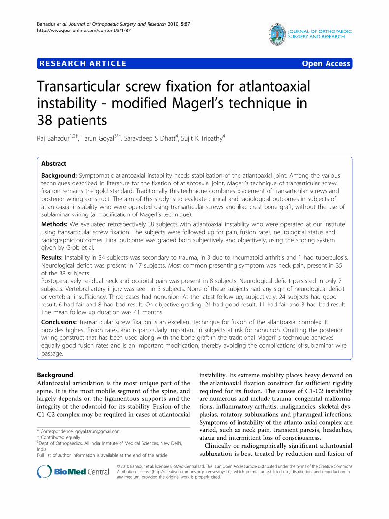
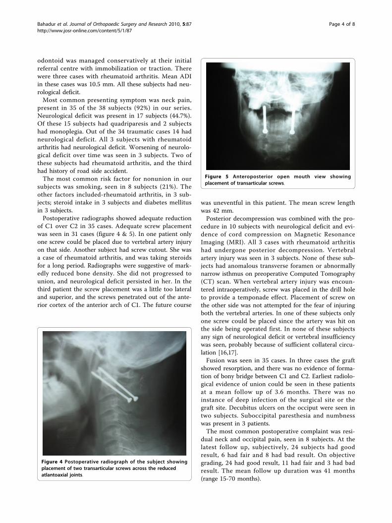
of C1 over C2 in 35 cases. Adequate screw placementwas seen in 31 cases (figure 4 & 5). In one patient onlyone screw could be placed due to vertebral artery injuryon that side. Another subject had screw cutout. She wasa case of rheumatoid arthritis, and was taking steroidsfor a long period. Radiographs were suggestive of mark-edly reduced bone density. She did not progressed tounion, and neurological deficit persisted in her. In thethird patient the screw placement was a little too lateraland superior, and the screws penetrated out of the ante-rior cortex of the anterior arch of C1. The future course
was uneventful in this patient. The mean screw lengthwas 42 mm.Posterior decompression was combined with the pro-
cedure in 10 subjects with neurological deficit and evi-dence of cord compression on Magnetic ResonanceImaging (MRI). All 3 cases with rheumatoid arthritishad undergone posterior decompression. Vertebralartery injury was seen in 3 subjects. None of these sub-jects had anomalous transverse foramen or abnormallynarrow isthmus on preoperative Computed Tomography(CT) scan. When vertebral artery injury was encoun-tered intraoperatively, screw was placed in the drill holeto provide a temponade effect. Placement of screw onthe other side was not attempted for the fear of injuringboth the vertebral arteries. In one of these subjects onlyone screw could be placed since the artery was hit onthe side being operated first. In none of these subjectsany sign of neurological deficit or vertebral insufficiencywas seen, probably because of sufficient collateral circu-lation [16,17].Fusion was seen in 35 cases. In three cases the graft
showed resorption, and there was no evidence of forma-tion of bony bridge between C1 and C2. Earliest radiolo-gical evidence of union could be seen in these patientsat a mean follow up of 3.6 months. There was noinstance of deep infection of the surgical site or thegraft site. Decubitus ulcers on the occiput were seen intwo subjects. Suboccipital paresthesia and numbnesswas present in 3 patients.The most common postoperative complaint was resi-
dual neck and occipital pain, seen in 8 subjects. At thelatest follow up, subjectively, 24 subjects had goodresult, 6 had fair and 8 had bad result. On objectivegrading, 24 had good result, 11 had fair and 3 had badresult. The mean follow up duration was 41 months(range 15-70 months).
Figure 4 Postoperative radiograph of the subject showingplacement of two transarticular screws across the reducedatlantoaxial joints.
Figure 5 Anteroposterior open mouth view showingplacement of transarticular screws.
Bahadur et al. Journal of Orthopaedic Surgery and Research 2010, 5:87http://www.josr-online.com/content/5/1/87
Page 4 of 8

At admission 17 subjects had neurological deficit. Ofthese 14 were Frankel’s grade C and 3 were Frankel’sgrade D. At discharge 10 subjects had completely recov-ered, with neurological deficit persisting in only 7 sub-jects. All these 7 subjects belonged to Frankel’s grade C.The mean range of neck motion was 40 degrees of lat-
eral rotation on left side and 35 degrees of rotation onright side. Since atlantoaxial fusion virtually eliminatesthe motion at C1-C2 joint, the residual rotation reflectsthe subaxial component of the neck motion. This rangeof motion was maintained on follow up. The meanrange of lateral rotation in cases of rheumatoid arthritiswas 25 degrees on each side. This is consistent with theview that rheumatoid spine has restricted range of atlan-toaxial and subaxial motion. The range of flexion andextension was maintained after surgery. The mean flex-ion and extension arc was 150 degrees. The mean rangeof lateral bending was 45 degrees on each side.
DiscussionThe aim of treatment of atlantoaxial instability is toachieve a solid fusion between C1 and C2, virtuallyeliminating any motion between them. This is expectedto relieve the neck pain and avoid the risk of furtherneurological deficit. The posterior wiring techniquespopularized by Gallie et al [18] and Brooks and Jenkins[19] had been the most common means of stabilizationin the past. In recent years, a variety of other techniqueshave been used, such as, interlaminar clamps, polyaxialscrew and rod fixation, transarticular screw fixation andC1 lateral mass screws with C2 pars screw fixation. Pos-terior C1-C2 transarticular screw fixation has becomethe gold standard for atlantoaxial fusion. It has lead toconsiderable improvement in the fusion rates upto morethan 95% [1-6] over C1-C2 wiring procedures, whosefailure rates range from 10% to 25% [20,21]. Taggard etal [7] conducted a case control study to compare thefusion rates using transarticular screws and posteriorwiring techniques. After a mean follow up of 31 monthsthey found that successful fusion was achieved in 13 of14 subjects treated with the TAS technique as comparedto 5 out of 13 subjects who underwent a posterior wir-ing technique. They observed that subjects with a radio-graphically solid fusion were 21 more times likely tohave undergone TAS than posterior wiring technique (p= 0.004). The position of the transarticular screws is clo-ser to the centre of axis of rotation and lateral bending,which provides better control of movements than othertechniques which rely on peripheral fixation.The biomechanics of surgical stabilization of the C1-C2
articulation can be divided into three different types.One-point fixation stabilizes the motion segment onlyposteriorly (e.g. Gallie wiring, Halifax clamps etc). Two-point fixation construct includes transarticular screws
through the laterally placed facet joints. Three-point fixa-tion consists of the combination of the two previousprinciples, thus stabilizing the C1-C2 motion segmentboth laterally and posteriorly. In biomechanical testingthree point fixation has been found to be superior toboth two-point and one-point fixations [22-26]. Thus thetension band construct provides two advantages-first, itenhances the stability of the TAS fixation; and second,the structural bone graft is stabilized by the wire. Butsublaminar wire passage carries the potential risk of neu-rological complications [9-11], especially in cases wherethe canal has already been compromised. Further thiswire-graft technique is technically demanding and timeconsuming [2,27]. Some reports have shown that metalwires or cables may bow anteriorly because of “springphenomenon” even without any breakage, leading toencroachment upon the spinal cord [13,28].It is controversial in literature whether posterior wir-
ing construct provide any additional contributiontowards fusion. Matsumoto et al reported 18 cases ofloosening of posterior wiring construct in 52 cases with95% fusion rate [14]. In Ito’s series, all cases had loosen-ing, but with 100% fusion rate. Thus, wire or cable loos-ening did not lead to nonunion or pseudarthrosis, but itmight endanger the spinal cord. From these observa-tions, Ito et al came to the conclusion that adding wireconstruct is not required [13]. Avoiding the placementof posterior wires may be especially important in situa-tions where inflammatory disease with soft tissue swel-ling and pannus has resulted in compromise of thespinal canal, or in the case of C1-C2 subluxation whichis not completely reducible [8]. Significant degenerativechanges or osteoporosis of the posterior elements of C1and C2 also preclude the use of posterior wiring techni-ques. Wang et al [4] achieved solid fusion in all their 57subjects, using only morselized autograft and transarti-cular screw, without any posterior wiring construct. Wedid not use the morselized graft but a strut of iliac crestgraft well fitted in the space between the C1 lamina andC2 spinous process. Thus, although from the biomecha-nical viewpoint, bilateral TAS fixation may not be asstable as the 3-point fixations, fusion rates have notbeen altered. There is only slight micromotion left inflexion-extension after fixation. We supposed that thismicromotion would not affect fusion. In our series,there is no loss of the reduction and the fusion rate is92%. This is in unison with the fusion rates achieved byother authors who used combination of Transarticularscrews and posterior wiring [1-7]. Randomized or a casecontrol study will be a better study design to study thiseffect. But correspondence of our results with those ofstudies using Magerl’s fixation suggests that this techni-que is a sound alternative thus simplifying the Magerl’stechnique.
Bahadur et al. Journal of Orthopaedic Surgery and Research 2010, 5:87http://www.josr-online.com/content/5/1/87
Page 5 of 8

Though single screw placement is expected to lead tononunion, there is no convincing data in this regard. Inour study single screw was placed in 1 subject. Solidunion was achieved in this subject at follow up. Songet al [23] concluded that unilateral C1-C2 transarticularscrew fixation with interspinous bone graft wiring is anexcellent alternative in the treatment of atlantoaxialinstability when bilateral screw fixation is contraindi-cated. They reported a solid fusion using this techniquein 18 of 19 subjects with atlantoaxial instability and uni-lateral anomalies. Grob et al [6] found that nonuniondid not follow incorrect placement of one screw, sobilateral fixation is not an indispensable condition for asatisfactory outcome.Posterior transarticular screw fixation has several
advantages over other fixation techniques. Contrary tothe traditional posterior fusion techniques, the integrityof the ring of C1 is not necessary for transarticular screwplacement. Thus this technique can be used even in casesof fracture or the absence of posterior arch of the atlas.This technique also provides approach for laminectomy,if needed for decompression of the cord. Further, there isno implant inside the spinal canal as in the wiring techni-ques and complications associated with wire looseningare avoided. A very important advantage is that it avoidsthe need for postoperative halo immobilization, whencompared to the posterior wiring techniques. This is animportant factor from the subjects’ point of view for theselection of the procedure. Achieving preoperative reduc-tion is imperative for safe atlantoaxial fusion. Displace-ment of C1 on C2 decreases the space available for thecord. This distorts the C1 C2 alignment, and the place-ment of transarticular screws is not completely safe. Thisalso increases the risk with sublaminar wire passage,because of increased chances of hitting the cord.Although some authors have used transarticular screwfixation for in situ fixation, the precise limit beyondwhich this technique is contraindicated is not defined.Thus in large fixed displacements of C1 on C2, occipito-cervical fusion with C1 decompression, or anteriordecompression and fusion are indicated [8].The disadvantages of this procedure include need for
an extensive skin incision and soft tissue dissection toexpose the entire dorsum of C2. This extensive posteriorexposure has been associated with a complication rateas high as 10%, including superficial infections and occi-pital nerve injury [8,29]. Screw placement requires anacute angle for proper screw trajectory, which may beimpeded by kyphotic deformities or by moving the neckanteriorly. Additionally, there is a steep learning curvefor this technique. Complications associated with thistechnique include the potential for vertebral arteryinjury, malposition of screws, pseudoarthrosis, implantfailure, dural tear, hypoglossal paresis, brain stem
infarction and death. Inconstant size and location of thetransverse foramen in the lateral mass of the axis placesthe vertebral artery at risk during drilling and screw pla-cement. Scans with saggital and coronal reconstructionshelp to assess the relationship of transverse foramen ofC2 and the C1-C2 facet joint to determine the correcttrajectory for the screw and avoid arterial injury [30,31].Radiographic and anatomical studies of the atlanto-axialcomplex suggest that upto 20% of the subjects haveatlanto axial anatomy that precludes safe bilateral screwplacement [32-34]. We had 3 cases of vertebral arteryinjury in our study (8%). Reported rates of vertebralartery injury using this technique vary from 0-10% indifferent series [17,8,29,32,35-39]. American Associationof Neurological Surgeons/Congress of Neurological Sur-geons (AANS/CNS) Section on disorders of SpinalNerves and Peripheral Nerves in their survey publishedby Wright and Lauryssen [35], estimated the risk of ver-tebral artery injury during C1-C2 transarticular screwfixation to be 2.2% per screw inserted. The risk of neu-rological deficit from vertebral artery injury was 0.2%per subject, and the mortality rate was 0.1%. Thus injuryto vertebral artery is well tolerated in the majority of thesubjects. Despite numerous reports of vertebral arteryinjuries, resultant neurological deficit is rare [8]. CoricD et al [40] reported a case of vertebral artery to epi-dural venous plexus fistula as a complication of poster-ior atlantoaxial facet screw fixation. Madawi et al [33]reported five cases of vertebral artery injury (8.2%) insubjects who underwent this operation. He also pointedout that incomplete reduction is a risk factor for inade-quate screw placement. Incidence of dural tears hasbeen reported to be 0.3%. suboccipital numbness is rela-tively common, seen in 16.8% patients in report byWright and Lauryssen [35]. In most of them however itresolved spontaneously with time.Despite excluding all the patients with dangerous
anatomy of the vertebral artery, we still had 3 patientsin whom vertebral artery injury was observed. Two ofthese patients were observed in the first half of thestudy period when the experience of the surgeon withthis technique was relatively recent. This is a highly sur-geon dependent technique and learning curve is high.Surgeon has to be familiar with the anatomy of thetransverse foramen in the upper cervical spine. Thisneeds experience with studying a large number of CTscans. Failure to meticulously identify the danger in thisregion may lead to catastrophy.The studies of RA subjects showed relatively lower
rates of bony union than did the studies with smallerpercentages of RA subjects [29,41-43]. Literature sug-gests that presence of rheumatoid arthritis entails therisk of posterior graft nonunion more than other disor-ders [6,41-43]. We achieved union in only of the
Bahadur et al. Journal of Orthopaedic Surgery and Research 2010, 5:87http://www.josr-online.com/content/5/1/87
Page 6 of 8

3 patients with RA. Due to the small sample size withonly 3 subjects with rheumatoid arthritis, no statisticallysignificant conclusion regarding effect of rheumatoiddisease on fixation and union can be reached. Ito T et alfound that in 5 of their 7 subjects with rheumatoidarthritis who had nonunion, C1-C2 complex was stabledue to fusion at the facet joints, as demonstrated byfunctional radiographs and computed tomography scans[13]. Thus atlantoaxial transarticular screws can bringthe facet fusion despite the posterior graft failure insuch cases.
ConclusionsThus, transarticular screw fixation is an effective techni-que for the fusion of the atlantoaxial complex. It pro-vides highest fusion rates, and is particularly importantin subjects at risk for nonunion. It has expanded theindications for atlantoaxial fusion and is an importantsalvage technique in subjects with previous failed proce-dures. Although its learning curve may be steep, it isassociated with few rates of complications in experthands.
AcknowledgementsAuthors have not received any funding for the study or during preparationof the manuscript.
Author details1Postgraduate Institute of Medical Education and Research, Chandigarh,India. 2Government Medical College and Hospital, Chandigarh, India. 3Deptof Orthopaedics, All India Institute of Medical Sciences, New Delhi, India.4Dept of Orthopaedics, Postgraduate Institute of Medical Education andResearch, Chandigarh, India.
Authors’ contributionsRB is the senior authors who carried out the surgical procedure, coordinatedthe planning of preoperative and postoperative protocols, and helped todraft the manuscript. TG had the instrumental role in the planning andexecution of perioperative and intraoperative design of the study andpreparation of the manuscript. SSD and ST helped in acquisition of data andin drafting of the manuscript. All authors read and approved the finalmanuscript.
Competing interestsThe authors declare that they have no competing interests.
Received: 26 November 2009 Accepted: 22 November 2010Published: 22 November 2010
References1. Magerl F, Seemann PS: Stable posterior fusion of the atlas and axis by
transarticular screw fixation. In Cervical spine I. Volume 1. Edited by: Kehr P,Weidner A. New York: Springer; 1987:322-327.
2. Stillerman CB, Wilson JA: Altanto-axial stabilization with posteriortransarticular screw fixation: technical description and report of 22cases. Neurosurgery 1993, 32:948-955.
3. Blauth M, Richter M, Lange U: Transarticular screw fixation C1/2 intraumatic atlantoaxial instabilities. Comparison between percutaneousand open procedures. Orthopade 1999, 28:651-661.
4. Wang C, Yan M, Zhou H, Wang S, Dang G: Atlantoaxial transarticularscrew fixation with morselized autograft and without additional internalfixation: technical description and report of 57 cases. Spine 2007,32(6):643-646.
5. Jeanneret B, Magerl F: Primary Posterior Fusions C1-2 in OdontoidFractures: Indications, Technique, and Results of Transarticular ScrewFixation. J Spinal Disord 1992, 5:464-475.
6. Grob D, Jeanneret B, Aebi M, Aebi M, Markwalder TM: Atlanto.-axial fusionwith transarticular screw fixation. J Bone Joint Surg Br 1991, 73(6):972-6.
7. Taggard DA, Kraut MA, Clark CR, Traynelis VC: Case-control studycomparing the efficacy of surgical techniques for C1-C2 arthrodesis. JSpinal Disord Tech 2004, 17(3):189-94.
8. Smith MD, Phillips WA, Hensinger RN: Complications of fusion to theupper cervical spine. Spine 1991, 16(7):702-5.
9. Coyne TJ, Fehlings MG, Wallace MC, Bernstein M, Tator CH: C1-C2 PosteriorCervical Fusion: Long Term Evaluation of Results and Efficacy. Neurosurg1995, 37:688-693.
10. Fraser AB, Sen C, Casden AM, Catalano PJ, Post KD: Cervical transduralintramedullary migration of a sublaminar wire: a complication of cervicalfixation. Spine 1994, 19:456-9.
11. Cervellati S, Bettini N, Bianco T, Parisini P: Neurological complications insegmental spinal instrumentation: analysis of 750 subjects. Eur Spine J1996, 5:161-6.
12. Blacklock JB: Fracture of a sublaminar stainless steel cable in the uppercervical spine with neurological injury. Case report. J Neurosurg 1994,81:932-3.
13. Ito T, Hayashi M, Takei H: Loosening of supplemental cable intransarticular screw fixation and bone grafting. J Orthop Surg 1998,6:71-4.
14. Matsumoto M, Chiba K, Nakamura M, Ogawa Y, Toyama Y, Ogawa J: Impactof interlaminar graft materials on the fusion status in atlantoaxialtransarticular screw fixation. J Neurosurg Spine 2005, 2(1):23-6.
15. McGuire RA Jr, Harkey HL: Modification of technique and results ofatlantoaxial transfacet stabilization. Orthopedics 1995, 18:1029-1032.
16. Taneichi H, Suda K, Kajino T, Kaneda K: Traumatically induced vertebralartery occlusion associated with cervical spine injuries: prospectivestudy using magnetic resonance angiography. Spine 2005, 30:1955-62.
17. Neo M, Fujibayashi S, Miyata M, Takemoto M, Nakamura T: Vertebral arteryinjury during cervical spine surgery: a survey of more than 5600operations. Spine 2008, 33(7):779-85.
18. Gallie WE: Fractures and Dislocation of the Cervical Spine. Am J Surg1939, 46:495-499.
19. Brooks AL, Jenkins EB: Atlanto-axial arthrodesis by wedge compressionmethod. J Bone Joint Surg Am 1978, 60:279-284.
20. Dickman CA, Sonntag VK: Posterior C1-C2 transarticular screw fixation foratlantoaxial arthrodesis. Neurosurgery 1998, 43(2):275-80.
21. Farey ID, Nadkarni S, Smith N: Modified Gallie technique versustransarticular screw fixation in C1-C2 fusion. Clin Orthop Relat Res 1999,359:126-135.
22. Melcher RP, Puttlitz CM, Kleinstueck FS, Lotz JC, Harms J, Bradford DS:Biomechanical testing of posterior atlantoaxial fixation techniques. Spine2002, 27(22):2435-40.
23. Mitchell TC, Sadasivan KK, Ogden AL, Mayeux RH, Mukherjee DP,Albright JA: Biomechanical study of atlantoaxial arthrodesis: transarticularscrew fixation versus modified Brooks posterior wiring. J Orthop Trauma1999, 13(7):483-9.
24. Naderi S, Crawford NR, Song GS, Sonntag VK, Dickman CA: Biomechanicalcomparison of C1-C2 posterior fixations. Cable, graft, and screwcombinations. Spine (Phila Pa 1976) 1998, 15(23):1946-55.
25. Montesano PX, Juach EC, Anderson PA, Benson DR, Hanson PB:Biomechanics of cervical spine internal fixation. Spine (Phila Pa 1976)1991, 16(3):S10-S16.
26. Grob D, Dvorak J, Panjabi MM, Hayek J: Dorsal atlantoaxial screw fixation.A stability test in vitro and in vivo. Orthopade 1991, 20(2):154-162.
27. Guiot B, Fessler RG: Complex atlantoaxial fractures. J Neurosurg 1999,91(Suppl 2):139-143.
28. Geremia GK, Kim KS, Cerullo L, Calenoff L: Complications of sublaminarwiring. Surg Neurol 1985, 23(6):629-35.
29. Gluf WM, Schmidt MH, Apfelbaum RI: Atlantoaxial transarticular screwfixation: a review of surgical indications, fusion rate, complications,and lessons learned in 191 adult subjects. J Neurosurg Spine 2005,2(2):155-63.
30. Dull ST, Toselli RM: Preoperative oblique axial computed tomographicimaging for C1-C2 transarticular screw fixation: technical note. JNeurosurg 1995, 37:150-1.
Bahadur et al. Journal of Orthopaedic Surgery and Research 2010, 5:87http://www.josr-online.com/content/5/1/87
Page 7 of 8

31. Nogueira-Barbosa MH, DeWno HLA: Multiplanar reconstructions of helicalcomputed tomography in planning of atlantoaxial transarticular fixation.Eur Spine J 2005, 14:493-500.
32. Yoshida M, Neo M, Fujibayashi S, Nakamura T: Comparison of theanatomical risk for vertebral artery injury associated with the C2-pediclescrew and atlantoaxial transarticular screw. Spine 2006, 31:E513-7.
33. Abou Madawi A, Solanki G, Casey AT, Crockard HA: Variation of the groovein the axis vertebra for the vertebral artery. Implications forinstrumentation. J Bone Joint Surg Br 1997, 79(5):820-823.
34. Miyata M, Neo M, Ito H, Yoshida M, Miyaki K, Fujibayashi S, Nakayama T,Nakamura T: Is Rheumatoid Arthritis a Risk Factor for a High-RidingVertebral Artery? Spine 2008, 33(18):2007-2011.
35. Wright NM, Lauryssen C: Vertebral artery injury in C1-2 transarticularscrew fixation: results of a survey of the AANS/CNS section on disordersof the spine and peripheral nerves. American Association ofNeurological Surgeons/Congress of Neurological Surgeons. J Neurosurg1998, 88(4):634-640.
36. Neo M, Matsushita M, Iwashita Y, Yasuda T, Sakamoto T, Nakamura T:Atlantoaxial transarticular screw fixation for a high-riding vertebralartery. Spine 2003, 28(7):666-70.
37. Haid RW Jr, Subach BR, McLaughlin MR, Rodts GE Jr, Wahlig JB Jr: C1-C2transarticular screw fixation for atlantoaxial instability: a 6-yearexperience. Neurosurgery 2001, 49(1):65-8.
38. Liang ML, Huang MC, Cheng H, Huang WC, Yen YS, Shao KN, Huang CI,Shih YH, Lee LS: Posterior transarticular screw fixation for chronicatlanto-axial instability. J Clin Neurosci 2004, 11(4):368-72.
39. Campanelli M, Kattner KA, Stroink A, Gupta K, West S: Posterior C1-C2transarticular screw fixation in the treatment of displaced type IIodontoid fractures in the geriatric population–review of seven cases.Surg Neurol 1999, 51(6):596-600.
40. Coric D, Branch CL Jr, Wilson JA, Robinson JC: Arteriovenous fistula as acomplication of C1-2 transarticular screw fixation. Case report andreview of the literature. J Neurosurg 1996, 85(2):340-3.
41. Casey AT, Madawi AA, Veres R, Crockard HA: Is the technique of posteriortransarticular screw fixation suitable for rheumatoid atlanto-axialsubluxation? Br J Neurosurg 1997, 11(6):508-19.
42. Kandziora F, Mittlmeier T, Kerschbaumer F: Stage-related surgery forcervical spine instability in rheumatoid arthritis. Eur Spine J 1999,8(5):371-81.
43. Shen FH, Samartzis D, Jenis LG, An HS: Rheumatoid arthritis: evaluationand surgical management of the cervical spine. Spine J 2004,4(6):689-700.
doi:10.1186/1749-799X-5-87Cite this article as: Bahadur et al.: Transarticular screw fixation foratlantoaxial instability - modified Magerl’s technique in 38 patients.Journal of Orthopaedic Surgery and Research 2010 5:87.
Submit your next manuscript to BioMed Centraland take full advantage of:
• Convenient online submission
• Thorough peer review
• No space constraints or color figure charges
• Immediate publication on acceptance
• Inclusion in PubMed, CAS, Scopus and Google Scholar
• Research which is freely available for redistribution
Submit your manuscript at www.biomedcentral.com/submit
Bahadur et al. Journal of Orthopaedic Surgery and Research 2010, 5:87http://www.josr-online.com/content/5/1/87
Page 8 of 8