tracer kinetics - journal of lipid research · abstract a noninvasive method for visualizing the...
TRANSCRIPT

tracer kinetics
Lovastatin enhances hepatic uptake of low density lipoprotein in humans
Kari Kervinen: Markku J. Savolainen: Juhani I. Heikkill,t and Y. Antero Keslniemi'**
Departments of Internal Medicine. and Clinical Chemistry,t and Biocenter of Oulu, University of Oulu, SF-90220 Oulu. Finland
Abstract A noninvasive method for visualizing the uptake of low density lipoprotein (LDL) was used to investigate the effect of hypolipidemic drugs on LDL uptake by the human liver in vivo. Fourteen hypercholesterolemic patients (six with familial hypercholesterolemia and eight with common hypercholesterole- mia) were studied. Autologous LDL particles were isolated and divided into two aliquots; one was labeled with 99mTC and the other with lS1I. and both preparations were reinjected simul- taneously. The labeled LDL was visualized 24 h later by scan- ning the thorax and abdomen with a gamma camera, and the livedheart ratio was calculated as an estimate of the hepatic up- take of LDL. The results of 99"TC-labeled LDL scintigraphy were compared with conventional determinations of the frac- tional catabolic rate (FCR) for lslI-labeled LDL. The latter cor- related best with the livedheart ratio (I - 0.80, P < 0.001). Lovastatin treatment increased the livedheart ratio (15%, P < 0.01) in the patients with polygenic hypercholesterolemia and the FCR for LDL in both groups (22%, P < 0.05, for those with familial hypercholesterolemia and 37%. P < 0.01 for those with polygenic hypercholesterolemia). Scanning of the liver using the qqmTC-labeled LDL method provides a noninvasive method for visualizing the hepatic uptake of LDL in vivo in humans. This study also provides direct proof that lovastatin, a drug that enhances LDL receptor activity in the liver, also increases the hepatic uptake of LDL in humans.-Kervinen, K., M. J. Savolainen, J. I. HeikkilP, and Y. A. KesPniemi. Lovastatin enhances hepatic uptake of low density lipoprotein in humans. J. Lipid Res. 1993. 34: 1975-1982.
Supplementary key words technetium scintigraphy LDL re- ceptor * HMG-CoA reductase * familial hypercholesterolemia * cho- lesterol liver
The activity of LDL receptors on the plasma mem- branes of the liver cells is a major determinant of the plasma cholesterol level (1); about two-thirds of the plasma LDL is removed via the specific LDL receptor pathway (2, 3). The number of LDL receptors is regu- lated by the availability of cholesterol within the hepato- cyte (4), a finding that has led to the discovery and devel- opment of a new type of hypolipidemic drug, 3-hydroxy- 3-methylglutaryl-CoA reductase inhibitors, which reduce the availability of cholesterol within the cell by inhibiting the rate-limiting enzyme of cholesterol synthesis (4).
The in vivo quantification of changes occurring in LDL receptor activity in various diseases or in response to drug therapy is difficult in humans. Most previous tests have used LDL labeled in the apolipoprotein moiety (apoB), estimating the magnitude of the LDL receptor activity by measuring the fractional catabolic rate (FCR) for LDL particles (2, 3, 5). Recently, liver biopsy specimens have been used in studies investigating the effects of hypocho- lesterolemic drugs on the LDL receptor expression in hu- mans (6, 7).
In the present study we applied a previously described method that allows noninvasive estimation of hepatic LDL uptake in humans. The method is based on the labeling of LDL particles with radioactive technetium (""'Tc) by the method of Lees et al. (8) and scanning of the liver and thorax with a gamma camera. The activity of the hepatic LDL receptors is shown as an accumulation of radioactivity in the liver, whereas the radioactivity in the heart represents that in the circulatory pool. The livedheart ratio can be computed from the digital images of the organs. The changes in the fractional catabolic rate for LDL and hepatic LDL uptake produced by two hypo- lipidemic drugs, lovastatin and colestipol, in patients with familial or polygenic hypercholesterolemia were used to help validate the method.
PATIENTS AND METHODS
Patients
Fourteen patients, ages 27-59 years, referred to the Lipid Clinic of Oulu University Central Hospital were studied. Six were heterozygous for familial hypercholes- terolemia and eight had polygenic hypercholesterolemia.
Abbreviations: LDL, low density lipoprotein; FCR, fractional cata- bolic rate; FH, familial hypercholesterolemia; HDL, high density lipoprotein.
'To whom correspondence should be addressed.
Journal of Lipid Research Volume 34, 1993 1975
by guest, on July 14, 2018w
ww
.jlr.orgD
ownloaded from

During the screening visit they underwent physical and laboratory examinations and were instructed to consume a low cholesterol, fat-controlled diet (American Heart As- sociation Phase 1 diet). Their initial clinical characteris- tics and values for total cholesterol, triglycerides, and lipoprotein-cholesterol are in Table 1.
None of the patients had any signs or symptoms of cardiac insufficiency or hepatic or renal dysfunction, but four of them had coronary heart disease (two had under- gone a coronary by-pass operation, one had suffered a myocardial infarction, and one had angina pectoris). None were on hypolipidemic drugs when entering the trial, but four were receiving anti-hypertensive or anti- anginal medication.
The clinical diagnosis of familial hypercholesterolemia was based on the presence of severe hypercholesterolemia (serum cholesterol above 8 mmol/l and LDL cholesterol above the 90th percentile (9)) in association with tendon xanthomas in the patient or a first-degree relative or coro- nary heart disease at an early age in the first-degree rela- tive. In addition, the FH-Helsinki mutation, a large dele- tion in the LDL receptor gene (lo), responsible for a major portion of familial hypercholesterolemia in north- ern Finland (11) was analyzed by amplification of the DNA sequences flanking the deletion in the LDL receptor gene by polymerase chain reaction (12).
Five out of six patients in the FH group had the FH- Helsinki mutation (Table 1). A deletion in exon 6 of the LDL receptor gene, designated as the FH-North Karelia mutation (13), was found in one patient. None of the pa- tients in this study had the familial defective apolipo- protein B-100 mutation, apoB-3500 (14), in their apoB gene. The patients with familial hypercholesterolemia were younger than those with polygenic hypercholes- terolemia but still had more atherosclerotic complica- tions. There were no significant differences in body mass index or sex distribution between the groups. Both total plasma cholesterol and LDL cholesterol were significantly higher in the patients with familial hypercholesterolemia than in those with polygenic hypercholesterolemia.
Each patient gave informed consent for the research protocol, which was approved by the Ethical Committee of the University of Oulu.
Drug treatment
The trial consisted of a minimum of 6 weeks' treatment with colestipol followed by a minimum of a 12-week period of treatment with lovastatin. All the four patients examined during the colestipol treatment used 10 g coles- tipol t.i.d. After the colestipol period, the patients were switched without any washout period to a 20-mg q.p.m. therapy with lovastatin. After 4 weeks, the dose of lovasta-
TABLE 1. Baseline characteristics of the patients
LDL Body Receptor Coronary
Patient Mass Total LDL n m ApoE Tendon Gene Heart Number Sex Age Index Chol Chol Chol TG Phenotype Xanthoma Mutation Disease
yr kg/mz
Familial hypercholesterolemia 1 M 37 2 M 30 3 F 27 4 F 38 5 F 47 6 F 46
Mean 38 * SD 8
21.4 22.2 23.6 31.5 28.6 26.2 25.6
3.9
Polygenic hypercholesterolemia 7 M 52 27.2 8 F 59 20.8 9 F 40 21.2
10 F 43 25.3 11 F 58 27.7 12 M 47 25.3 13 F 52 23.9 14 F 57 24.6
Mean 51" 24.5
13.30 10.80 9.92 8.45 8.33
10.80 10.27 1.84
7.59 8.80 7.83 8.36 7.84 7.69 8.29 9.04 8.18"
mmol/l
11.00 1.38 8.34 0.91 8.38 0.77 6.55 0.88 6.36 1.24 8 .72 1.14 8.23 1.05 1.69 0.24
4.35 2.67 6.41 1.36 5.31 1.48 5.22 1.52 5.43 1.25 4.72 1.31 5.93 1.88 6.62 0.76 5.50° 1.53
+ SD 7 2.5 0.53 0.79 0.56
1.47 2.49 1.77 1.01 1.53 1.67 1.66 0.49
1.26 1.40 1.36 1.04 1.52 2.44 0.94 1.66 1.45 0.46
313 313 413 213 313 313
+ + + +
I
H H
NK H H H
B
MI R
AP
Abbreviations: Chol, cholesterol; TG, triglyceride; H, FH-Helsinki mutation of LDL receptor gene; NK, FH-North Karelia mutation of LDI,
"P < 0.01 for the difference between the patients with familial and polygenic hypercholesterolemia. receptor gene; B, coronary by-pass operation; MI , myocardial infarction; AP, angina pectoris; n.d., not determined.
1976 Journal of Lipid Research Volume 34, 1993
by guest, on July 14, 2018w
ww
.jlr.orgD
ownloaded from

tin was doubled to 40 mg q.p.m., and after 4 more weeks, it was further increased to 80 mg q.p.m. This dose was continued until the protocol was completed.
Protocol
The fractional catabolic rate (FCR) for LDL apoB and the tissue distribution of the LDL particles were inves- tigated after the simultaneous injection of two autologous LDL preparations, each labeled with either 1311 for the determination of the apoB FCR or 99mT~ for estimation of tissue distribution. All except one of the patients were examined before any hypocholesterolemic medication was commenced and again 4-11 months later, during treat- ment with lovastatin (80 mg q.p.m.). Four patients were also examined during the colestipol treatment, but one of these, a patient with familial hypercholesterolemia, was examined only during the drug treatment periods.
Isolation of the LDL fraction
After 3 weeks of maximal colestipol or lovastatin treat- ment, fasting blood was obtained and the LDL fraction was isolated by sequential ultracentrifugation (15). The plasma was adjusted to a density of 1.019 g/ml with a NaC1-NaBr solution and centrifuged in a Beckman 60 Ti rotor (Beckman Instruments Inc., Spinco Div., Palo Alto, CA) at 160,000 g and 15OC for 18 h. Lipoproteins floating to the surface were removed by tube-slicing. The infra- natant was adjusted to a density of 1.063 g/ml and cen- trifuged as above. The LDL fraction (1.019-1.063 g/ml) was washed by ultracentrifugation in a TFT 45.6 rotor (Kontron AG, Zurich, Switzerland) at 105,OOOg and 15OC for 18 h after overlaying with an equal volume of a NaCl- NaBr solution of density 1.070 g/ml. The LDL fraction was dialyzed extensively overnight against 0.15 M NaCl-1 mM EDTA adjusted to pH 7.4 with NaOH.
Labeling of LDL with radioactive isotopes
The LDL fraction was divided into two parts. One was labeled with 1311 (16, 17) as adapted for LDL (18) and the other with "'"Tc using a direct protein labeling method (8). The LDL preparations of one patient (no. 4) were labeled under nitrogen. However, this procedure gave lower incorporation of the technetium label than the origi- nal one; therefore, the livedheart ratio for this patient is given in Table 2, but omitted from Figs. 1 and 2. In fact, recent data verify that the previously described method (8) which was used in all the other patients produces a sta- ble radionuclide-LDL complex (19). The labeled LDL preparations were filtered through a Millipore 22-pm filter before injection. Aliquots of the LDL preparations were set aside for bacterial culture and pyrogen testing.
Protocol for the isotope examinations
Technetium labeling was performed immediately be- fore the preparation was injected intravenously together
with the iodine-labeled preparation (within 30 min). Ap- proximately 300 MBq (8.2 mCi) of 99mTc and 440 kBq (12 pCi) of 1311 were injected.
The clearance of 1311-labeled LDL from the plasma (fractional catabolic rate, FCR) was used as a reference measurement for LDL catabolism and LDL receptor ac- tivity. For this purpose, fasting blood samples were taken 15 and 30 min, and 1, 2, 3, 4, 24, and 48 h after the injec- tion of the labeled LDL preparations and sampling was continued three times a week for 14 days. The body weights and plasma cholesterol levels remained constant during the sampling period. The fractional catabolic rate for LDL was calculated from the plasma decay curves using the Matthews method (20) as described in our previous LDL turnover investigations (2, 5, 18). In brief, double-exponential equations were fitted to each plasma decay curve using an interactive curve-peeling pro- gramme (W. F. Beltz and T. E. Carew, unpublished method) on a VAX-VMS computer.
The distribution of the injected 99mTc-labeled LDL was visualized by scanning the thorax and the upper ab- domen 5 min, and 4 and 24 h after the injection with a large field view of the computerized gamma camera (Elscint Apex 409 ECT).
Analysis of the gamma scanning digital image
Scanning of the thorax produced figures for the label distribution in the heart as well as in the lungs. The first scan at 5 min was performed in order to assess the radio- activity in the plasma pool, Le., that representing the LDL particles in the sinusoids and other blood vessels of the liver before any receptor-mediated uptake of LDL had occurred. The mean livedheart ratio at 5 min was 0.67 * 0.04 (mean * SD). The livedheart ratio at 24 h was used as an estimate for the amount of radioactivity taken up by the liver versus the radioactivity in the LDL particles cir- culating in the plasma pool. The 5-min scintigraphy was not performed in all of the patients, and thus the liver/ heart ratio at 24 h without base-line correction was used in all the patients. Consequently, the percentage changes in the livedheart ratios are smaller than they would have been if the 5-min value had been subtracted.
The livedheart ratio showed a somewhat better correla- tion with the FCR for LDL than did the livedlung ratio. In addition, scanning at 24 h after the injection correlated better with the FCR for LDL than scanning at 4 h. Therefore, the livedheart ratio at 24 h was used in the subsequent analyses.
Analysis of lipids and apolipoproteins The concentrations of total cholesterol and triglycerides
were determined by enzymatic methods using a Gilford analyzer (Gilford Instruments Laboratories, Inc., Ober- lin, Ohio). HDL cholesterol was determined after precipi- tation of apoB-containing lipoproteins by addition of
Kervinen et al. Lovastatin and hepatic LDL uptake in humans 1977
by guest, on July 14, 2018w
ww
.jlr.orgD
ownloaded from
heparin-manganese to the plasma sample (21); the pro- tein content of the LDL fraction was determined by the method of Lowry et al. (22).
The occurrence of the familial defective apolipoprotein B-100 mutation, apoB-3500 (14), was analyzed by ampli- fying a segment of genomic DNA spanning the site of mu- tation in the apoB gene by polymerase chain reaction, fol- lowed by hybridization with radioactive oligonucleotide probes (23). The apoE phenotype was determined using isoelectric focusing and immunoblotting techniques (24).
Statistical analysis the standard
deviation (SD). The statistical significances of the dif- ferences between the groups were calculated using the paired or nonpaired Student's t test, as appropriate. Non- parametric method (Wilcoxon signed rank sum test) was used to analyze the statistical significance of the changes in the liverlheart ratio and liverllung ratio. Correlations were tested by calculating Pearson's coefficient of corre- lation.
All values are expressed as means
RESULTS
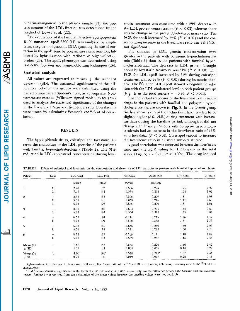
The hypolipidemic drugs, colestipol and lovastatin, al- tered the catabolism of the LDL particles of the patients with familial hypercholesterolemia (Table 2). The 31% reduction in LDL cholesterol concentration during lova-
statin treatment was associated with a 29% decrease in the LDL protein concentration (P < 0.02), whereas there was no change in the proteidcholesterol mass ratio. The FCR for apoB increased by 22% ( P < 0.02) and the cor- responding increase in the liverlheart ratio was 8% (N.S., not significant).
The changes in LDL protein concentration were greater in the patients with polygenic hypercholesterole- mia (Table 3) than in the patients with familial hyper- cholesterolemia. The decrease in LDL protein brought about by lovastatin treatment was 41% (P < 0.001). The FCR for LDL apoB increased by 31% during colestipol treatment and by 37% (P < 0.01) during lovastatin ther- apy. The FCR for LDL-apoB showed a negative correla- tion with the LDL cholesterol level in both patient groups (Fig. 1; in the total series r = -0.86; P < 0.001).
The individual responses of the livedheart ratio to the drugs in the patients with familial and polygenic hyper- cholesterolemia are shown in Fig. 2. In the former group the liverlheart ratio of the technetium label tended to be slightly higher (8%, N.S.) during treatment with lovasta- tin than during the baseline period, although it did not change significantly. Patients with polygenic hypercholes- terolemia had an increase in the liverlheart ratio of 15% with lovastatin (P < 0.01). Colestipol tended to increase the livedheart ratio in all three subjects studied.
A good correlation was observed between the liverlheart ratio and the FCR values for LDL-apoB in the total series (Fig. 3; r = 0.80; P < 0.001). The drug-induced
TABLE 2. Effects of colestipol and lovastatin on the composition and clearance of LDL particles in patients with familial hypercholesterolemia
Patient Drug LDL-Chol LDL-Yrot Prot/Chol ApoB-FCR LIH Ratio LIL Ratio
mmol/l mg/dl mdms pooldday
6
C 7.48 L 7.16
- 8.34 c 5.39 L 6.04
- 8.38 L 4.92
- 6.55 I, 4.03
- 6.36 L 4.30
152 142
156 121 126
180 107
118 109
166 84
0.536 0.574
0. SO6 0.659 0.556
0.603 0.506
0.531 0.520
0.656 0.521
0.224 0.234
0.200 0.216 0.209
0.231 0.300
0.275 0.338
0.249 0.285
1.25 1.24
1.31 1.47 1.31
1.65 1.85
1.16 1 34
1.39 1.60
1.92 2.06
1.98 2.60 2.01
2.80 3.07
1.59 2.36
2.93 2.26
- 8.72 172 0.519 0.191 I .48 2.82 L 5.20 103 0.539 0.267 1.45 2.36
Mean ( 5 ) - 7.67 158 0.563 0.229 1.40 2.42 +_ SD 1.12 24 0.064 0.035 0.18 0.27
Mean (5) L 4.90h 106" 0.528 0.280" 1.52 2.41 + SD 0.79 15 0.019 0.047 0.22 0.18
Abbreviations: C, colestipol; L, lovastatin; L/H ratio, livedheart ratio of the qy""T~-LDL distribution; L/L ratio, livedlung ratio of the "'"Tc-LDL
and ' denote statistical significance at the levels of P < 0.05 and P < 0.001, respectively, for the difference between the baseline and the lovastatin distribution.
values. Patient 1 was omitted from the calculation of the mean values because the baseline values were not available.
1978 Journal of Lipid Research Volume 34, 1993
by guest, on July 14, 2018w
ww
.jlr.orgD
ownloaded from

TABLE 3. Effects of colestipol and lovastatin on the composition and clearance of LDL particles in patients with polygenic hypercholesterolemia
LIL Ratio LtH Ratio Patient Drug LDL-Chol LDL-Prot ProtlChol ApoB-FCR
mmol/l mg/dl mdmg pools/day
7
8
9
10
11
12
13
14
- 4.35 L 3.67
1.45 3.06 0.292 1.44 2.69
- 6.41 137 0.535 0.234 1.62 2.48 L 3.23 59 0.537 0.338 1.81 2.90
- 5.31 93 0.521 0.268 1.73 2.99 C 2.75 41 0.528 0.327 1.83 2.98 L 2.48 42 0.442 0.414 1.94 3.57
- 5.22 107 0.583 0.218 1.49 2.57 C 4.02 75 0.506 0.309 1.89 3.28 L 2.98 75 0.669 0.342 1.78 2.62
- 5.43 111 0.520 0.261 1.40 2.64 L 3.79 91 0.697 0.330 1.68 2.76
- 4.72 93 0.515 0.300 1.51 2.24 L 3.30 73 0.541 0.324 1.68 2.55
- 5.93 126 0.548 0.235 1.40 2.27 L 3.28 70 0.584 0.302 1.66 2.82
- 6.62 147 0.565 0.240 1.45 3.25 L 3.46 58 0.491 0.401 1.97 3.60
Mean (8) - 5.50 116 0.541 0.251 1.51 2.69 + SD 0.79 21 0.025 0.028 0.11 0.13
Mean (8) L 3.27" 67b 0.566 0.343b 1.74' 2.94' i SD 0.41 16 0.092 0.043 0.17 0.15
Abbreviations: C , colestipol; L, lovastatin. "P < 0,001; bP < 0.01; 'P < 0.05: denote statistical significance for the difference between the baseline and lovastatin values.
change in the livedheart ratio showed a good correlation with the corresponding change in the FCR for LDL-apoB (Fig. 4; in the total series r = -0.69; P < 0.004). Fur- thermore, the livedheart ratio showed a negative correla-
0 0 0.1 0.2 0.3 0.4 0.5
Fractional catabolic rate for LDL-B (poolslday)
Fig. 1. Correlation between the fractional catabolic rate for LDL and LDL-cholesterol concentration. Squares, patients with familial hyper- cholesterolemia; circles, patients with polygenic hypercholesterolemia. Open symbols are observations during the diet period and closed sym- bols are results obtained during the drug treatments. 7 = -0.86; P < 0.001.
tion with the LDL cholesterol concentration ( r = -0.66;
For comparison, the livedlung ratios are also given in Tables 2 and 3. A good correlation was observed between the livedheart ratio and the livedlung ratio in the total
P c 0.001).
3
n L
L m 3) s L 0 >
.- * *
.
.- I
2 .o
1.8
1.6
1.4
1.2 '
Polygenic I I I I I I
B C L B C L
Fa m i I ia I 1 .o
Fig. 2. Effects of colestipol and lovastatin on the hepatic uptake of LDL particles, estimated by determination of the livedheart ratio of the radioactive label 24 h after an intravenous injection of technetium- labeled LDL. Squares, patients with familial hypercholesterolemia; cir- cles, patients with polygenic hypercholesterolemia. B, baseline period; C, colestipol treatment period; L, lovastatin treatment period.
Kervinen et al. Lovastatin and hepatic LDL uptake in humans 1979
by guest, on July 14, 2018w
ww
.jlr.orgD
ownloaded from

series ( r = 0.78; P < 0.001) as well as in the patients with familial hypercholesterolemia ( r = 0.79; P < 0.001) and patients with polygenic hypercholesterolemia ( r =
0.63; P < 0.006). Lovastatin tended to reduce the production rate of
LDL-apoB in both the patients with familial hyper- cholesterolemia and patients with polygenic hypercholes- terolemia, but the drug-induced change was statistically significant only in the total series (from 14.2 f 1.0 mg/kg per day to 12.0 mg/kg per day, P < 0.025).
DISCUSSION
The results show that the in vivo hepatic removal of LDL, presumably representing LDL receptor activity, can be estimated in humans using imaging isotope tech- niques, as used previously to demonstrate the biodistribu- tion of technetium-labeled LDL particles in vivo in experimental animals (8, 25) and in patients with myelo- proliferative diseases (26). The incorporation of radio- actively labeled LDL particles into atherosclerotic lesions has recently been used to visualize the degree of vascular disease in patients (27) and the uptake of LDL by tendon xanthomas in hypercholesterolemic subjects (28). In the present cases, the hepatic uptake of technetium-labeled LDL particles was determined and compared with the fractional catabolic rate, which is usually used as an esti- mate for the rate of LDL clearance through the LDL receptors.
The close correlation between the uptake of 99mT~- labeled LDL particles and the FCR for LDL suggests that the technetium scanning method may be a good indicator of LDL catabolism. It has been shown previously that
0.4 c a 4
I I I I I I 1
1.0 1.2 1.4 1.6 1.8 2 .0
Liverlheart ratio
Fig. 3. Correlation between hepatic uptake of LDL particles (liver/ heart ratio) and fractional catabolic rate for LDL-apoB. Symbols as in Fig. 1 . 7 = 0.80; P < 0.001.
m . U h OS2 r v1 - 0 0 a cz 0 LL
-1
-1
v
gi 0.1
n c 0 OD c m c
.-
0 0
a a
0 0.2 0.4 0.6 Change in livedheart ratio
Fig. 4. Correlation between the lovastatin-induced changes in hepatic uptake of LDL particles (livedheart ratio) and fractional catabolic rate for LDL-apoB. Squares, patients with familial hypercholesterolemia; circles, patients with polygenic hypercholesterolemia. 7 = 0.69, P < 0.004.
99mTc-labeled LDL particles act in a manner similar to tyramine cellobiose-labeled LDL and are accumulated quantitatively after uptake into tissues (29). Furthermore, 99mTc-labeled LDL is recognized by the LDL receptor just as well as lZ5I-labeled LDL (25). We were not able here to introduce a third isotope to measure the uptake of LDL particles through the receptor-mediated pathway, but it is known that this usually parallels the total catabolism of LDL (2, 3). It is therefore probable that the present accumulation of technetium label into the liver reflects the activity of hepatic LDL receptors (4).
The fractional catabolic rate for LDL was seen here to increase during drug treatment in both groups, although the increase was lower in the patients with familial hyper- cholesterolemia. An increase in the FCR for LDL has previously been observed in patients with familial hyper- cholesterolemia treated with lovastatin (30), but no increase during treatment with the same drug has been observed in patients with moderate primary hypercholes- terolemia (31). The different response of the patients with polygenic hypercholesterolemia in the present case may be due to the higher dose of lovastatin (80 mg/day) and higher pretreatment cholesterol levels. In this study, the patients with familial hypercholesterolemia were some- what younger than those with polygenic hypercholesterol- emia. The higher age of patients with polygenic hyper- cholesterolemia may have reduced the difference in the FCR for LDL between the groups, as there is an estab- lished effect of age on the FCR for LDL (32, 33).
The drug-induced increase in the hepatic uptake of LDL, with subsequent lowering of LDL cholesterol, sug- gests that the major part of the effect of bile acid binding resins or lovastatin on serum cholesterol concentration in
1980 Journal of Lipid Research Volume 34, 1993
by guest, on July 14, 2018w
ww
.jlr.orgD
ownloaded from

humans is also mediated through enhancement of the hepatic LDL receptor activity (34, 35), although lovasta- tin also reduces the synthesis rate of LDL-apoB as ob- served here in accordance with previous studies (36, 37). The importance of the LDL receptors in the liver as regu- lators of serum LDL cholesterol level is further empha- sized by the fact that the drug-induced increase in hepatic LDL receptor activity is more accentuated in the patients with polygenic hypercholesterolemia than in those with familial hypercholesterolemia, as the latter have a lower capacity to synthesize LDL receptors.
The major benefit of the present method for estimating the hepatic uptake of LDL particles is its rapidity. The method does not obviate the isolation of LDL particles or labeling of the LDL with an isotope, but 14-day blood sampling can be avoided. Scanning of the thorax and upper abdomen with a computerized gamma camera could, in the future, be performed immediately after in- travenous injection of the 99"Tc-labeled LDL preparation in order to obtain a background image of the distribution of the label in the circulation, i.e., the amount of blood residing in the liver. A repeat scan 24 h later would then enable the increase in the livedheart ratio to be cal- culated. m This work was supported by grants from the Sigrid JusClius Foundation and the Medical Council of the Academy of Finland and was carried out under a contract with the Finnish Life In- surance Companies. We are indebted to Dr. Kimmo Kontula (Second Department of Medicine, University of Helsinki) for the analysis of FH-North Karelia mutation in one of our pa- tients. The skillful technical assistance of Ms. Leena Saikko, Irma Piri, and Saija Kortetjarvi is gratefully acknowledged. The study drugs were provided by Merck, Sharp & Dohme Research Laboratories. Manuscript received 29 April 1991 and in revisedfonn 23 June 1993.
REFERENCES
1.
2.
3.
4.
5.
Brown, M. S., and J. L. Goldstein. 1983. Lipoprotein receptors in the liver. Control signals for plasma cholesterol traffic. J. Clin. Invest. 72: 743-747. Kesaniemi, Y. A., J. L. Witztum, and U. P. Steinbrecher. 1983. Receptor-mediated catabolism of low density lipopro- tein in man. Quantitation using glucosylated low density lipoprotein. J. Clin. Invest. 71: 950-959. Steinbrecher, U. P., J. L. Witztum, Y. A. Kesaniemi, and R. L. Elam. 1983. Comparison of glucosylated low density lipoprotein with methylated or cyclohexanedione-treated low density lipoprotein in the measurement of receptor- independent low density lipoprotein catabolism. J. Clin. Invest. 71: 960-964. Brown, M. S., and J. L. Goldstein. 1984. How LDL recep- tors influence cholesterol and atherosclerosis. Sci. Am. 251:
Kesaniemi, Y. A,, G. L. Vega, and S. M. Grundy. 1982. Ki- netics of apolipoprotein B in normal and hyperlipidemic man. Review of current data. In Lipoprotein Kinetics and
52-60.
Modelling. M. Berman, S. M. Grundy, and B. V. Howard, editors. Academic Press, New York. 181-205.
6. Rudling, M. J., E. Reihntr, K. Einarsson, S. Ewerth, and B. Angelin. 1990. Low density lipoprotein receptor-binding activity in human tissues: quantitative importance of hepatic receptors and evidence for regulation of their ex- pression in vivo. Proc. Natl. Acad. Sci. USA. 87: 3469-3473. Reihntr, E., M. Rudling, D. Sthlberg, L. Berglund, S. Ewerth, I. Bjorkhem, K. Einarsson, and B. Angelin. 1990. Influence of pravastatin, a specific inhibitor of HMG-CoA reductase, on hepatic metabolism of cholesterol. N. Engl. J. Med. 323: 224-228.
8. Lees, R. S., H. D. Garabedian, A. M. Lees, D. J. Schumacher, A. Miller, L. Isaacsohn, A. Derksen, and H. W. Strauss. 1985. Technetium-99m low density lipo- proteins: preparation and biodistribution. J. Nucl. Med. 26:
9. Rifkind, B. M., and P. Segal. 1983. Lipid Research Clinics Program Reference Values for Hyperlipidemia and Hypo- lipidemia. J. Am. Med. Assoc. 2 5 0 1869-1872.
10. Aalto-Setda, K., E. Helve, P. T. Kovanen, and K. Kontula. 1989. The Finnish type of low density lipoprotein receptor gene mutation (FH-Helsinki) deletes exons encoding the carboxyterminal part of the receptor and creates an internalization-defective phenotype. J. Clin. Invest. 84:
11. Aalto-Setala, K., U-M. Koivisto, T. A. Miettinen, H. Gylling, Y. A. Keskiemi, M. Savolainen, K. Pyorda, T. Ebeling, I. Mononen, H. Turtola, J. Viikari, and K. Kontula. 1992. Prevalence and geographical distribution of major LDL receptor gene rearrangements in Finland. J. Intern. Med. 23 1 : 2 2 7 - 23 4.
12. Savolainen, M. J., T. Korhonen, K. Aalto-Setda, K. Kontula, and Y. A. Kesaniemi. 1991. Screening of patients with severe hypercholesterolaemia for a prevalent LDL receptor mutation. Hum. Genet. 87: 125-128.
13. Koivisto, U-M., H. Turtola, K. Aalto-Set Frants, P. T. Kovanen, A-C. Syvanen, and K. Kontula. 1992. The familial hypercholesterolemia (FH) North Kare- ha mutation of the low density lipoprotein receptor gene deletes 7 nucleotides of exon-6 and is a common cause of FH in Finland. J. Clin. Invest. 90: 219-228.
14. Soria, L. F., E. H. Ludwig, H. R. G. Clarke, G. L. Vega, S. M. Grundy, and B. J. McCarthy. 1989. Association be- tween a specific apolipoprotein mutation and familial defec- tive apolipoprotein B-100. Proc. Natl. Acad. Sci. USA. 86:
Lindgren, F. T., L. C. Jensen, R. D. Wills, and N. K. Freeman. 1969. Flotation rates, molecular weights and hydrated densities of low-density lipoproteins. Lipids. 4: 337-344.
16. McFarlane, A. S. 1958. Efficient trace-labelling of proteins with iodine. Nature (London) 182: 53-57.
17. Bilheimer, D. W., S. Eisenberg, and R. I. Levy. 1972. The metabolism of very low density lipoprotein proteins. I. Pre- liminary in vitro and in vivo observations. Biochim. Biophys. Acta. 260 212-221.
18. Kesaniemi, Y. A., and S. M. Grundy. 1982. Significance of low density lipoprotein production in the regulation of plasma cholesterol level in man. J. Clin. Invest. 70: 13-22.
19. Atsma, S. E., H. J. M. Kempen, W. Nieuwenhuizen, F. M. van't Hooft, and E. K. J. Pauwels. 1991. Partial characteri- zation of low density lipoprotein preparations isolated from fresh and frozen plasma after radiolabeling by seven differ- ent methods. J. Lipid Res. 32: 173-181.
20. Matthews, C. M. E. 1957. The theory of tracer experiments
7.
1056-1062.
499-505.
587-592. 15.
Kervinen et al. Lovastatin and hepatic LDL uptake in humans 1981
by guest, on July 14, 2018w
ww
.jlr.orgD
ownloaded from

21.
22.
23.
24.
25.
26.
27.
28.
with iodine 131-labeled plasma proteins. Phys. Med, Bid. 2:
Manual of Laboratory Operations. 1974. Lipid Research Clinics Program. I. Lipid and Lipoprotein Analysis. Dept. of Health, Education, and Welfare publication (NIH) 75-628. National Institutes of Health. Lowry, 0. H., N. J. Rosebrough, A. L. Farr, and R. J. Randall. 1951. Protein measurement with the Folin phenol reagent. J. Bid. Chm. 193: 265-275. Tybjaerg-Hansen, A., J. Gallagher, J. Vincent, P. Talmud, A. M. Dunning, M. Seed, A. Hamsten, S. E. Humphries, and N. B. Myant. 1990. Familial defective apolipoprotein B-100: detection in the United Kingdom and Scandinavia, and clinical characteristics of ten cases. Atherosclerosis. 80:
Menzel, H-J., and G. Utermann. 1986. Apolipoprotein E phenotyping from serum by Western blotting. Electrophoresis.
Vallabhajosula, S., M. Paidi, J. J. Badimon, N-A. Le, S. J. Goldsmith, V. Fuster, and H. N. Ginsberg. 1988. Radio- tracers for low density lipoprotein biodistribution studies in vivo: technetium-99m low density lipoprotein versus radio- iodinated low density lipoprotein preparations. J. Nucl. Med. 29: 1237-1245. Vallabhajosula, S., H. S. Gilbert, S. J. Goldsmith, M. Paidi, M. M. Hanna, and H. N. Ginsberg. 1989. Low-density lipoprotein (LDL) distribution shown by 99mtechnetium- LDL imaging in patients with myeloproliferative diseases. Ann. Intern. Med. 110: 208-213. Lees, A. M., R. S. Lees, F. J. Schoen, J. L. Isaacsohn, A. J. Fischman, K. A. McKusick, and H. W. Strauss. 1988. Im- aging human atherosclerosis with 99mTc-labeled low den- sity lipoproteins. Arteriosclerosis. 8: 461-470. Ginsberg, H. N., S. J. Goldsmith, and S. Vallabhajosula. 1990. Noninvasive imaging of 99mtechnetium-labeled low density lipoprotein uptake by tendon xanthomas in hyper- cholesterolemic patients. Arteriosclerosis. 10: 256-262.
36-53.
235-242.
7: 492-494.
29.
30.
31.
32.
33.
34.
35.
36.
37.
Lees, A. M., and R. S. Lees. 1991. 99mTechnetium-labeled low density lipoprotein: receptor recognition and intra- cellular sequestration of radiolabel. J. Lipid Res. 32: 1-8. Bilheimer, D. W., S. M. Grundy, M. S. Brown, and J. L. Goldstein. 1983. Mevinolin and colestipol stimulate receptor-mediated clearance of low density lipoprotein from plasma in familial hypercholesterolemia hetero- zygotes. Proc. Natl. Acad. Sci. USA. 80: 4124-4128. Grundy, S. M., and G. L. Vega. 1985. Influence of mevino- lin on metabolism of low density lipoproteins in primary moderate hypercholesterolemia. J Lipid Res. 26:
Grundy, S. M., G. L. Vega, andD. W. Bilheimer. 1985. Ki- netic mechanisms determining variability in low density lipoprotein levels and rise with age. Arteriosclerosis. 5: 623-630. Ericsson, S., M. Eriksson, S. Vitols, K. Einarsson, L. Berglund, and B. Angelin. 1991. Influence of age on the metabolism of plasma low density lipoproteins in healthy males. J. Clin. Invest. 87: 591-596. Shepherd, J., C. J. Packard, S. Bicker, T. D. V. Lawrie, and H. G. Morgan. 1980. Cholestyramine promotes receptor- mediated low-density-lipoprotein catabolism. N. Engl. J Med. 302: 1219-1222. Slater, H. R., C. J. Packard, S. Bicker, and J. Shepherd. 1980. Effects of cholestyramine on receptor-mediated plasma clearance and tissue uptake of human low density lipoproteins in the rabbit. J Biol. Chm. 255: 10210-10213. Vega, G. L., C. East, and S. M. Grundy. 1988. Lovastatin therapy in familial dyslipoproteinemia: effects on kinetics of apolipoprotein B. Atherosclerosis. 70: 131-143. Arad, Y., R. Ramakrishnan, and H. N. Ginsberg. 1990. Lovastatin therapy reduces low density lipoprotein apoB levels in subjects with combined hyperlipidemia by reduc- ing the production of apoB-containing lipoproteins: impli- cations for the pathophysiology of apoB production. J. Lipid Res. 31: 567-582.
1464-1475.
1982 Journal of Lipid Research Volume 34, 1993
by guest, on July 14, 2018w
ww
.jlr.orgD
ownloaded from