toxin-antitoxin systems in eukaryotic cells: strategies ... · toxins review heterologous...
TRANSCRIPT
toxins
Review
Heterologous Expression of Toxins from BacterialToxin-Antitoxin Systems in Eukaryotic Cells:Strategies and Applications
Chew Chieng Yeo 1,*, Fauziah Abu Bakar 2, Wai Ting Chan 3, Manuel Espinosa 3 andJennifer Ann Harikrishna 2,*
1 Biomedical Research Centre, Faculty of Medicine, Universiti Sultan Zainal Abidin, Medical Campus,Jalan Sultan Mahmud, 20400 Kuala Terengganu, Malaysia
2 Centre for Research in Biotechnology for Agriculture (CEBAR) and Institute for Biological Sciences,Faculty of Science, University of Malaya, 50603 Kuala Lumpur, Malaysia; [email protected]
3 Molecular Microbiology and Infection Biology, Centro de Investigaciones Biológicas,Consejo Superior de Investigaciones Cientificas, Ramiro de Maeztu 9, 28040 Madrid, Spain;[email protected] (W.T.C.); [email protected] (M.E.)
* Correspondence: [email protected] (C.C.Y.); [email protected] (J.A.H.);Tel.: +609-627-5506 (C.C.Y.); +603-7967-5896 (J.A.H.)
Academic Editor: Anton MeinhartReceived: 17 January 2016; Accepted: 15 February 2016; Published: 19 February 2016
Abstract: Toxin-antitoxin (TA) systems are found in nearly all prokaryotic genomes and usuallyconsist of a pair of co-transcribed genes, one of which encodes a stable toxin and the other, its cognatelabile antitoxin. Certain environmental and physiological cues trigger the degradation of the antitoxin,causing activation of the toxin, leading either to the death or stasis of the host cell. TA systems have avariety of functions in the bacterial cell, including acting as mediators of programmed cell death, theinduction of a dormant state known as persistence and the stable maintenance of plasmids and othermobile genetic elements. Some bacterial TA systems are functional when expressed in eukaryoticcells and this has led to several innovative applications, which are the subject of this review. Here,we look at how bacterial TA systems have been utilized for the genetic manipulation of yeasts andother eukaryotes, for the containment of genetically modified organisms, and for the engineering ofhigh expression eukaryotic cell lines. We also examine how TA systems have been adopted as animportant tool in developmental biology research for the ablation of specific cells and the potentialfor utility of TA systems in antiviral and anticancer gene therapies.
Keywords: toxin-antitoxin systems; genetic manipulation; gene containment; cell ablation;high expression cell lines; gene therapy; antiviral therapy; anticancer therapy
1. Introduction: An Overview of Bacterial Toxin-Antitoxin Systems
Toxin-antitoxin (TA) systems are nearly ubiquitous genetic modules in bacterial and archaealgenomes. They generally comprise a pair of genes coding for a stable toxin and its cognate labileantitoxin. Under normal growth conditions, the toxin is prevented from exerting its lethal effect by theantitoxin. However, environmental stresses usually cause a drastic drop in the levels of the unstableantitoxin in the cell due mainly to degradation by endogenous proteases. This leads to activation ofthe remaining toxin which, in turn, causes either cell death or cell stasis [1–5].
TA systems were initially discovered encoded within bacterial plasmids where they function tomediate plasmid maintenance and stability through the postsegregational killing of any plasmid-freedaughter cells that arise within a population. The bacterial hosts became “addicted” to the presence of
Toxins 2016, 8, 49; doi:10.3390/toxins8020049 www.mdpi.com/journal/toxins

Toxins 2016, 8, 49 2 of 16
these TA-encoding plasmids and thus, TA systems were also termed as addiction modules [6,7].Chromosomal homologues of these plasmid-encoded addiction modules were first reported inEscherichia coli and the chromosomal mazEF system, comprising the mazE-encoded antitoxin andthe mazF-encoded toxin, was postulated to mediate programmed bacterial cell death under nutrientstarvation conditions [8]. This apparent altruistic killing was envisaged to enable part of the bacterialpopulation to survive during adverse conditions, reflecting a multicellular facet of bacteria [8–10].Research into other chromosomal TA systems in E. coli, namely the relBE system and chpAIK(a mazEF homologue), appeared to indicate an alternative role. Overexpression of the relE- andchpAK-encoded toxins in E. coli induces a bacteriostatic condition with severe inhibition of translation,but subsequent induction of expression of the respective cognate antitoxins relB and chpAI fully reversesthe toxin-induced stasis [11,12]. relBE and other similar TA systems were proposed to function as partof the general stress response of bacteria by regulating the global level of translation and together withthe trans-translation ssrA system, function in the quality control of gene expression [1,13]. However,with increasing numbers of novel TA systems being discovered, their biological functions haveexpanded, mirroring their genetic diversity. TA systems have been implicated in various other cellularprocesses such as the formation of persister cells leading to antibiotic tolerance [14,15], as anti-addictionmodules [16], in protection against invading bacteriophages [17,18], as stabilization modules for largemobile genetic elements such as superintegrons and genomic islands [19,20], in biofilm formation [21]and in virulence of pathogenic bacteria [22–24].
TA systems have so far been broadly classified into five different types, designated types I–V, basedon the characteristics of the antitoxin and the mechanisms by which they counteract the toxins [4,5,19].In type I TA systems, the antitoxin is an antisense RNA that binds to the toxin mRNA, preventing itstranslation [25]. In type II TA systems, both antitoxin and toxin are proteins and the antitoxin functionsby direct binding with the toxin, usually blocking its active site [2]. As for type III TA systems, theantitoxin is an RNA that functions by direct binding, with the toxin protein leading to the formation ofa non-lethal protein-RNA complex [26]. In type IV systems, both antitoxins and toxins are proteins butunlike in type II systems, the antitoxins and toxins of type IV systems do not directly interact witheach other. Rather, the antitoxin binds to the target of the toxin to prevent the toxin from exerting itslethal effect [27]. Finally, in type V systems, the antitoxin is a protein with ribonuclease activity thatcleaves the toxin mRNA and thus prevents the synthesis of the toxin [28]. Nevertheless, a potentiallynew class of TA system (a possible type VI) was recently discovered in the form of the SocAB systemfrom Caulobacter crescentus [29]. Both the SocB toxin and the SocA antitoxin are proteins but in this case,the SocB toxin is the unstable partner due to its susceptibility to the endogenous ClpXP protease. TheSocA antitoxin functions as an essential ClpXP adaptor for the SocB toxin, promoting its degradationand thus abolishing its lethality [30]. To date, TA systems belonging to types I and II are the mostabundant in prokaryotes with type II TAs being the most well-characterized [5,19].
TA toxins target a wide variety of essential cellular structures and processes such as membraneintegrity, cell wall synthesis, DNA replication, ribosome assembly and translation factors, withRNA cleavage being the most prevalent mode of action [3,23]. The near ubiquity of TA systemsin prokaryotes and the potential for triggering latent intracellular molecular timebombs, especiallyin pathogenic bacteria, led to several interesting avenues of research for the use of TA systems astargets for novel antibacterial compounds [5,19,31]. TA systems have also been harnessed as toolsin molecular biology, such as for the positive selection of clones containing inserted DNA fragmentsin cloning vectors. The ccdB toxin gene of the ccdAB TA system from the E. coli F plasmid has beensuccessfully used in a number of cloning vectors where the toxin gene is inactivated upon insertion offoreign DNA, enabling only insert-containing clones to survive and grow [19,32]. With the discoverythat some of these bacterial TA systems can be expressed and are functional in eukaryotic cells [33–35],several interesting applications have been proposed and developed. In this mini-review, we will lookat several strategies used for the heterologous expression of bacterial TA genes in eukaryotic systemsand their potential applications in biotechnology and molecular biology.

Toxins 2016, 8, 49 3 of 16
2. Expression of TA Systems in Yeasts: Applications
2.1. TA Systems as Tools for Containment in Yeasts
The increasing use of genetically modified organisms (GMOs) in various bioprocesses necessitatesappropriate containment strategies to address concerns over the accidental release of these GMOs intothe environment, or in cases where the deliberate release of the GMOs into the environment are requiredfor biotechnological applications (such as bioremediation, bioleaching and biopesticides) [36,37].Two main strategies that have been utilized are passive and active containment systems [36]. In passivecontainment systems, the GMO is engineered such that it cannot synthesize an essential compound;thus, when these auxotrophic GMOs accidentally escape from the bioreactor or environment wherethe compound is provided, the organisms will likely die. Recently, “genetically-recoded” bacteria havebeen engineered whereby the expression of several essential genes is totally reliant on the supply ofexogenously supplied synthetic amino acids, resulting in the death of these cells in an environmentlacking these synthetic compounds [38,39]. However, such synthetic auxotrophy systems requireextensive genome-wide engineering and hence, may not be technologically and economically practicalfor eukaryotic systems at this point in time. In active containment systems, the GMO is engineeredwith a controllable lethal function that will not interfere with normal cellular processes until a specificenvironmental cue is triggered. TA systems have been utilized as the controllable lethal function inseveral such active containment strategies for bacteria and these have been recently reviewed [36,40].
The E. coli relBE TA system was demonstrated to be functional in the yeast Saccharomyces cerevisiaeand was proposed as a containment system for genetically modified yeast [33]. The pYES2 yeastexpression vector was used for this purpose with two recombinant plasmids constructed: pKP727 withthe relE toxin gene under the control of the GAL1 promoter; and pKP1006 with relE controlled by theGAL1 promoter and the relB antitoxin gene under the control of the MET25 promoter [33]. Expressionof relE in yeast cells transformed with pKP727 and induced with galactose showed clear inhibitoryeffects, whereas in yeast cells that were transformed with pKP1006 and induced with galactose inthe absence of methionine (for the co-expression of both relE and relB), higher growth rates wereobserved, albeit lower than normal [33]. This indicated that the RelE toxin is lethal in S. cerevisiae andits toxicity could be somewhat neutralized by co-expression of the RelB antitoxin. However, sincethat research was published in 2000 [33], there have not been any follow-up reports on the utility ofrelBE or any other TA system for the containment of genetically modified yeasts. A general strategythat could be used to engineer such a TA-based containment system is depicted in Figure 1. OtherTA systems have been shown to be functional in S. cerevisiae and this includes the kis-kid TA systemfrom the E. coli plasmid R1 [35] and ε-ζ from plasmid pSM19035 of Streptococcus pyogenes [41]. Thekis-kid genes were expressed in S. cerevisiae using a similar system to relBE in that the kis antitoxin wasexpressed from a MET25 promoter (i.e., induced expression in the presence of methionine), whereasthe kid toxin was under the control of the CUP1 promoter (induced expression in the presence of Cu2+)in an integrative pRS303-derived plasmid recombinant [35]. The ε-ζ genes were, however, expressedby vectors designed for a commercial yeast two-hybrid system (Matchmaker Two-Hybrid System kitfrom Clontech). Toxicity of the ζ toxin in S. cerevisiae was demonstrated, as was its neutralization byits cognate ε antitoxin and interaction between the toxin and antitoxin proteins [41]. The RelE andKid toxins are endoribonucleases [13,42] whereas the ζ toxin functions by blocking bacterial cell wallsynthesis by phosphorylating uridine diphosphate-N-acetylglucosamine (UNAG), a key intermediatein peptidoglycan synthesis [43]. In S. cerevisiae, the nucA gene from the Gram-negative bacteria Serratiamarcescens has been successfully used to construct a conditional lethal system for containment byplacing the nucA gene under the control of the glucose-repressed S. cerevisiae alcohol dehydrogenase-2ADH2 gene promoter [44]. It was proposed that the small size of the S. marcescens nuclease (26 kDa)facilitates its entry into the yeast nucleus [44] and it is likely to be the same situation for the smaller~12 kDa RelE and Kid endoribonuclease toxins. The lethal mechanism for the ζ toxin in yeast is still
Toxins 2016, 8, 49 4 of 16
unknown although it could act to inhibit the synthesis of the yeast cell wall much like in bacteria, asUNAG is a component for the biosynthesis of chitin in yeast [45].
Toxins 2016, 8, 49 4 of 16
of the yeast cell wall much like in bacteria, as UNAG is a component for the biosynthesis of chitin in
yeast [45].
Two new microbial “kill switches” were recently engineered based on the CcdB and MazF TA
toxins (along with the EcoRI restriction endonuclease) for the containment of genetically modified
bacteria. These switches, designated “Deadman” and “Passcode” are modular and flexible
(customizable), can be conveniently transferred to other bacterial strains [46] and would therefore be
useful in various biotherapeutic and industrial applications. These toxin‐based switches should also
be functional in yeasts and could likewise be adopted for the containment of genetically modified yeasts.
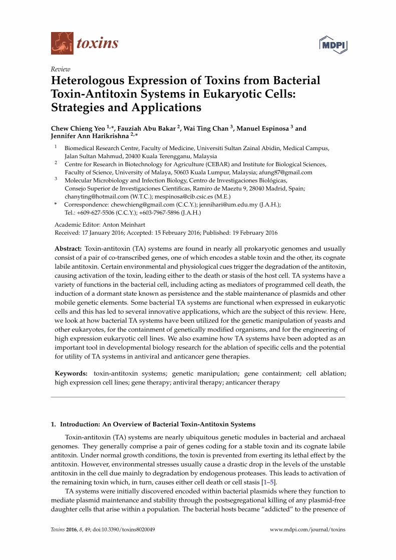
Figure 1. General strategy utilizing a bacterial TA system to engineer a containment system for
genetically modified yeasts. The antitoxin‐coding gene (depicted by a green arrow) and the
toxin‐coding gene (red arrow) are cloned under the control of two separate promoters (depicted as
yellow and blue triangles) within the same yeast vector. Under controlled conditions (such as in a
fermenter), the presence of a specific signal (which can either be a certain media constituent or
nutrients such as glucose and specific amino acids), the antitoxin is expressed whereas expression of
the toxin gene is repressed (indicated by orange “+” and “−“ signs, respectively). Any leaky expression
of the toxin gene (depicted as a dotted arrow) would be countered by the continual expression of the
antitoxin. However, in the absence of the specific signal, such as when the genetically modified yeast
has escaped into the environment, transcription of the antitoxin gene will be repressed whereas the
toxin gene will be actively transcribed, thus killing the escaped organism.
2.2. TA Systems as Tools for the Genetic Manipulation of Yeasts
Expression of another endoribonuclease toxin, MazF, from the tightly controlled, methanol‐inducible
AOX1 promoter in the methylotrophic yeast Pichia pastoris, was found to be lethal. This was used as
the basis of a novel method for unmarked genetic modification of P. pastoris using mazF as a
counter‐selectable marker [47]. A modular plasmid, pKSCTMF, was constructed consisting of an
AOX1 promoter‐mazF expression cassette and a Zeocin‐resistance gene as a dominant selection
marker flanked by direct repeats from segments of the CYC1 transcriptional terminator (TT), as
recombination sites, along with multiple restriction sites to permit subcloning of fragments
homologous to the P. pastoris genome for disruption [47]. P. pastoris was first transformed with a
linearized recombinant pKSCTMF construct and selected on medium supplemented with Zeocin.
Transformants with the correct disruption were determined by phenotype and/or PCR analysis.
Subsequently, selection for recombination of the two CYC1 TT repeats was performed by growing
Figure 1. General strategy utilizing a bacterial TA system to engineer a containment system forgenetically modified yeasts. The antitoxin-coding gene (depicted by a green arrow) and the toxin-codinggene (red arrow) are cloned under the control of two separate promoters (depicted as yellow andblue triangles) within the same yeast vector. Under controlled conditions (such as in a fermenter),the presence of a specific signal (which can either be a certain media constituent or nutrients such asglucose and specific amino acids), the antitoxin is expressed whereas expression of the toxin gene isrepressed (indicated by orange “+” and “´“ signs, respectively). Any leaky expression of the toxingene (depicted as a dotted arrow) would be countered by the continual expression of the antitoxin.However, in the absence of the specific signal, such as when the genetically modified yeast has escapedinto the environment, transcription of the antitoxin gene will be repressed whereas the toxin gene willbe actively transcribed, thus killing the escaped organism.
Two new microbial “kill switches” were recently engineered based on the CcdB and MazFTA toxins (along with the EcoRI restriction endonuclease) for the containment of geneticallymodified bacteria. These switches, designated “Deadman” and “Passcode” are modular and flexible(customizable), can be conveniently transferred to other bacterial strains [46] and would therefore beuseful in various biotherapeutic and industrial applications. These toxin-based switches should also befunctional in yeasts and could likewise be adopted for the containment of genetically modified yeasts.
2.2. TA Systems as Tools for the Genetic Manipulation of Yeasts
Expression of another endoribonuclease toxin, MazF, from the tightly controlled,methanol-inducible AOX1 promoter in the methylotrophic yeast Pichia pastoris, was found tobe lethal. This was used as the basis of a novel method for unmarked genetic modification of P. pastorisusing mazF as a counter-selectable marker [47]. A modular plasmid, pKSCTMF, was constructedconsisting of an AOX1 promoter-mazF expression cassette and a Zeocin-resistance gene as a dominantselection marker flanked by direct repeats from segments of the CYC1 transcriptional terminator(TT), as recombination sites, along with multiple restriction sites to permit subcloning of fragmentshomologous to the P. pastoris genome for disruption [47]. P. pastoris was first transformed with a
Toxins 2016, 8, 49 5 of 16
linearized recombinant pKSCTMF construct and selected on medium supplemented with Zeocin.Transformants with the correct disruption were determined by phenotype and/or PCR analysis.Subsequently, selection for recombination of the two CYC1 TT repeats was performed by growingthe transformants on methanol medium plates that would enable only transformants with a singleCYC1 TT segment inserted into the disrupted site to survive [47], i.e., effectively disrupting the gene ofinterest and subsequently evicting the selectable markers. This would enable the disrupted strainsto be amenable for a second or multiple genetic modifications using the same MazF-ZeoR cassette(marker recycling). This method also enabled knock-in of a gene of interest as well as site-directedmutagenesis at the native locus [47].
A variant of this method was utilized to enable disruption of certain genes that were less amenableto gene replacement in P. pastoris such as OCH1 that encodes α-1,6-mannosyltransferase and forwhich gene replacement occurs at frequencies of <1% even with flanking arms that are longer than1 kb [48]. Here, the AOX1 promoter-mazF cassette was cloned into an episomal “helper” plasmid thatalso contained a full copy of the OCH1 gene, providing a functional backup of the OCH1 gene forP. pastoris before deletion to avoid compromising the fitness of the yeast cells due to possible loss offunction. P. pastoris carrying this helper plasmid was then used as the host for conventional OCH1 genedisruption using a Zeocin-resistance gene [48]. Once replacement of the chromosomal OCH1 genewith the Zeocin-disrupted copy had been validated, these strains were grown in medium containingmethanol to induce expression of mazF. This exerts a strong selection pressure for the strains to losethe episomal “helper” plasmid. The same strategy was used to successfully delete the KU70 and SGS1genes in P. pastoris, and increasing their targeting efficiencies [48].
3. TA Systems as Cell Ablation Tools
3.1. Cell Ablation for the Containment of Genetically Modified Plants
Transgenic crops have become integrated into modern agriculture and are increasingly adoptedworldwide. Besides having useful traits such as herbicide resistance and pest tolerance, transgenicplants have also served as a platform for the large-scale production of recombinant pharmaceuticalproteins (also known as biopharming) and industrial enzymes [49]. One of the major concerns of thewide-scale adoption of transgenic crops is their accidental spread into the environment leading tocontamination of the human food chain. This concern is not without precedent as several episodes ofaccidental release or contamination have occurred, leading to a negative perception of GM crops in theeyes of the public [49,50]. In addition to physical containment methods, biological containmentstrategies such as plastid transformation, male sterility and genetic use restriction technologies(GURTs) have been proposed or developed to confine the potential spread of transgenic crops [51–53].Engineered sterility of transgenic plants is not only helpful in preventing their pollen spread intonatural ecosystems, it is also useful for the development of male sterile parents for hybrid seedproduction [54] in self-pollinated crops, for the removal of allergenic pollen and for the production ofseedless varieties of fruit and vegetable crops [55]. To achieve sterility, a toxin gene is placed under thecontrol of a tissue-specific promoter that enables the toxin to be expressed in certain parts of the plantanther and/or pistil.
One of the earliest and successfully applied bacterial toxin genes that has been used for the specificablation of the plant’s reproductive organs leading to sexual sterility is Barnase, a small 12.4-kDa RNaseof 110 amino acid residues produced by Bacillus amyloliquefaciens that also synthesizes its antidote, asmall protein called Barstar (89 amino acid residues) that binds to Barnase and sterically blocks theBarnase active site in a one-to-one noncovalent interaction [56]. Barnase has been used since the 1990’sto engineer male, female or bisexual sterility in various transgenic plants by placing the barnase geneunder the control of a tissue-specific promoter, which enables it to be expressed in certain parts of theplant anther and/or pistil [56–61]. Expression of barnase leads to the destruction of the reproductiveorgans, thereby conferring sterility to the transgenic plants. Although not a TA system, Barnase-Barstar
Toxins 2016, 8, 49 6 of 16
is illustrative of the utility of a bacterial regulatory module where the toxic effect can be rescued by thespecific counterpart; in the case of sexually sterile (barnase expressing) plants, this has been appliedby the expression of barstar in “fertility restorer” plant lines, when viable pollen or seed is requiredfor hybrid seed production [62]. Unlike in TA systems, the genes for Barnase and Barstar are not inan operon and are located in different loci in the B. amyloliquefaciens genome, suggestive of distinctregulation of their expression [56]. However, this is not a barrier to similar application of TA systems,since the toxin and antitoxin can be expressed as independent units in a transgenic context and in factthis permits differing expression levels, for example, stronger expression of the “restorer” moleculewhen required [62]. Other bacterial or bacteriophage toxins or enzymes that have been applied fornegative selection in plants include the expression of the restriction endonuclease EcoRI in tobaccopollen [63] and cytosine deaminase from E. coli and the dipththeria A-chain toxin (DT-A) subunit fromcorynephages of Corynebacterium diptheriae for gene targeting in rice [64].
Toxins 2016, 8, 49 6 of 16
B. amyloliquefaciens genome, suggestive of distinct regulation of their expression [56]. However, this
is not a barrier to similar application of TA systems, since the toxin and antitoxin can be expressed as
independent units in a transgenic context and in fact this permits differing expression levels, for
example, stronger expression of the “restorer” molecule when required [62]. Other bacterial or
bacteriophage toxins or enzymes that have been applied for negative selection in plants include the
expression of the restriction endonuclease EcoRI in tobacco pollen [63] and cytosine deaminase from
E. coli and the dipththeria A‐chain toxin (DT‐A) subunit from corynephages of Corynebacterium diptheriae
for gene targeting in rice [64].
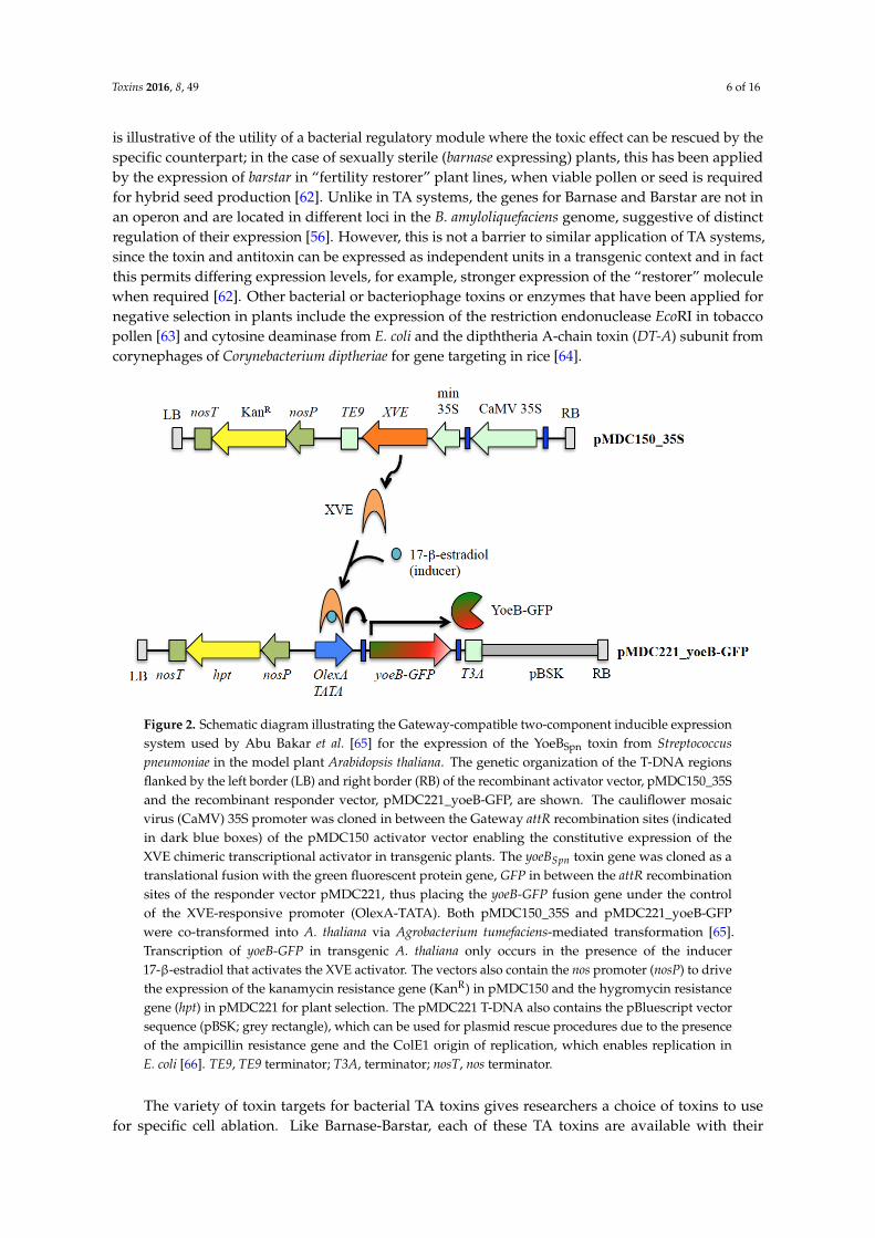
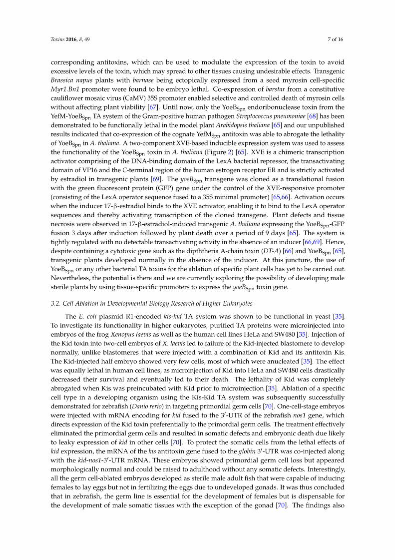
Figure 2. Schematic diagram illustrating the Gateway‐compatible two‐component inducible expression
system used by Abu Bakar et al. [65] for the expression of the YoeBSpn toxin from Streptococcus pneumoniae
in the model plant Arabidopsis thaliana. The genetic organization of the T‐DNA regions flanked by the
left border (LB) and right border (RB) of the recombinant activator vector, pMDC150_35S and the
recombinant responder vector, pMDC221_yoeB‐GFP, are shown. The cauliflower mosaic virus (CaMV)
35S promoter was cloned in between the Gateway attR recombination sites (indicated in dark blue
boxes) of the pMDC150 activator vector enabling the constitutive expression of the XVE chimeric
transcriptional activator in transgenic plants. The yoeBSpn toxin gene was cloned as a translational fusion
with the green fluorescent protein gene, GFP in between the attR recombination sites of the responder
vector pMDC221, thus placing the yoeB‐GFP fusion gene under the control of the XVE‐responsive
promoter (OlexA‐TATA). Both pMDC150_35S and pMDC221_yoeB‐GFP were co‐transformed into
A. thaliana via Agrobacterium tumefaciens‐mediated transformation [65]. Transcription of yoeB‐GFP in
transgenic A. thaliana only occurs in the presence of the inducer 17‐β‐estradiol that activates the XVE
activator. The vectors also contain the nos promoter (nosP) to drive the expression of the kanamycin
resistance gene (KanR) in pMDC150 and the hygromycin resistance gene (hpt) in pMDC221 for plant
selection. The pMDC221 T‐DNA also contains the pBluescript vector sequence (pBSK; grey rectangle),
which can be used for plasmid rescue procedures due to the presence of the ampicillin resistance gene
and the ColE1 origin of replication, which enables replication in E. coli [66]. TE9, TE9 terminator; T3A,
terminator; nosT, nos terminator.
The variety of toxin targets for bacterial TA toxins gives researchers a choice of toxins to use for
specific cell ablation. Like Barnase‐Barstar, each of these TA toxins are available with their
corresponding antitoxins, which can be used to modulate the expression of the toxin to avoid
excessive levels of the toxin, which may spread to other tissues causing undesirable effects.
Transgenic Brassica napus plants with barnase being ectopically expressed from a seed myrosin
cell‐specific Myr1.Bn1 promoter were found to be embryo lethal. Co‐expression of barstar from a
constitutive cauliflower mosaic virus (CaMV) 35S promoter enabled selective and controlled death
of myrosin cells without affecting plant viability [67]. Until now, only the YoeBSpn endoribonuclease
Figure 2. Schematic diagram illustrating the Gateway-compatible two-component inducible expressionsystem used by Abu Bakar et al. [65] for the expression of the YoeBSpn toxin from Streptococcuspneumoniae in the model plant Arabidopsis thaliana. The genetic organization of the T-DNA regionsflanked by the left border (LB) and right border (RB) of the recombinant activator vector, pMDC150_35Sand the recombinant responder vector, pMDC221_yoeB-GFP, are shown. The cauliflower mosaicvirus (CaMV) 35S promoter was cloned in between the Gateway attR recombination sites (indicatedin dark blue boxes) of the pMDC150 activator vector enabling the constitutive expression of theXVE chimeric transcriptional activator in transgenic plants. The yoeBSpn toxin gene was cloned as atranslational fusion with the green fluorescent protein gene, GFP in between the attR recombinationsites of the responder vector pMDC221, thus placing the yoeB-GFP fusion gene under the controlof the XVE-responsive promoter (OlexA-TATA). Both pMDC150_35S and pMDC221_yoeB-GFPwere co-transformed into A. thaliana via Agrobacterium tumefaciens-mediated transformation [65].Transcription of yoeB-GFP in transgenic A. thaliana only occurs in the presence of the inducer17-β-estradiol that activates the XVE activator. The vectors also contain the nos promoter (nosP) to drivethe expression of the kanamycin resistance gene (KanR) in pMDC150 and the hygromycin resistancegene (hpt) in pMDC221 for plant selection. The pMDC221 T-DNA also contains the pBluescript vectorsequence (pBSK; grey rectangle), which can be used for plasmid rescue procedures due to the presenceof the ampicillin resistance gene and the ColE1 origin of replication, which enables replication inE. coli [66]. TE9, TE9 terminator; T3A, terminator; nosT, nos terminator.
The variety of toxin targets for bacterial TA toxins gives researchers a choice of toxins to usefor specific cell ablation. Like Barnase-Barstar, each of these TA toxins are available with their
Toxins 2016, 8, 49 7 of 16
corresponding antitoxins, which can be used to modulate the expression of the toxin to avoidexcessive levels of the toxin, which may spread to other tissues causing undesirable effects. TransgenicBrassica napus plants with barnase being ectopically expressed from a seed myrosin cell-specificMyr1.Bn1 promoter were found to be embryo lethal. Co-expression of barstar from a constitutivecauliflower mosaic virus (CaMV) 35S promoter enabled selective and controlled death of myrosin cellswithout affecting plant viability [67]. Until now, only the YoeBSpn endoribonuclease toxin from theYefM-YoeBSpn TA system of the Gram-positive human pathogen Streptococcus pneumoniae [68] has beendemonstrated to be functionally lethal in the model plant Arabidopsis thaliana [65] and our unpublishedresults indicated that co-expression of the cognate YefMSpn antitoxin was able to abrogate the lethalityof YoeBSpn in A. thaliana. A two-component XVE-based inducible expression system was used to assessthe functionality of the YoeBSpn toxin in A. thaliana (Figure 2) [65]. XVE is a chimeric transcriptionactivator comprising of the DNA-binding domain of the LexA bacterial repressor, the transactivatingdomain of VP16 and the C-terminal region of the human estrogen receptor ER and is strictly activatedby estradiol in transgenic plants [69]. The yoeBSpn transgene was cloned as a translational fusionwith the green fluorescent protein (GFP) gene under the control of the XVE-responsive promoter(consisting of the LexA operator sequence fused to a 35S minimal promoter) [65,66]. Activation occurswhen the inducer 17-β-estradiol binds to the XVE activator, enabling it to bind to the LexA operatorsequences and thereby activating transcription of the cloned transgene. Plant defects and tissuenecrosis were observed in 17-β-estradiol-induced transgenic A. thaliana expressing the YoeBSpn-GFPfusion 3 days after induction followed by plant death over a period of 9 days [65]. The system istightly regulated with no detectable transactivating activity in the absence of an inducer [66,69]. Hence,despite containing a cytotoxic gene such as the dipththeria A-chain toxin (DT-A) [66] and YoeBSpn [65],transgenic plants developed normally in the absence of the inducer. At this juncture, the use ofYoeBSpn or any other bacterial TA toxins for the ablation of specific plant cells has yet to be carried out.Nevertheless, the potential is there and we are currently exploring the possibility of developing malesterile plants by using tissue-specific promoters to express the yoeBSpn toxin gene.
3.2. Cell Ablation in Developmental Biology Research of Higher Eukaryotes
The E. coli plasmid R1-encoded kis-kid TA system was shown to be functional in yeast [35].To investigate its functionality in higher eukaryotes, purified TA proteins were microinjected intoembryos of the frog Xenopus laevis as well as the human cell lines HeLa and SW480 [35]. Injection ofthe Kid toxin into two-cell embryos of X. laevis led to failure of the Kid-injected blastomere to developnormally, unlike blastomeres that were injected with a combination of Kid and its antitoxin Kis.The Kid-injected half embryo showed very few cells, most of which were anucleated [35]. The effectwas equally lethal in human cell lines, as microinjection of Kid into HeLa and SW480 cells drasticallydecreased their survival and eventually led to their death. The lethality of Kid was completelyabrogated when Kis was preincubated with Kid prior to microinjection [35]. Ablation of a specificcell type in a developing organism using the Kis-Kid TA system was subsequently successfullydemonstrated for zebrafish (Danio rerio) in targeting primordial germ cells [70]. One-cell-stage embryoswere injected with mRNA encoding for kid fused to the 31-UTR of the zebrafish nos1 gene, whichdirects expression of the Kid toxin preferentially to the primordial germ cells. The treatment effectivelyeliminated the primordial germ cells and resulted in somatic defects and embryonic death due likelyto leaky expression of kid in other cells [70]. To protect the somatic cells from the lethal effects ofkid expression, the mRNA of the kis antitoxin gene fused to the globin 31-UTR was co-injected alongwith the kid-nos1-31-UTR mRNA. These embryos showed primordial germ cell loss but appearedmorphologically normal and could be raised to adulthood without any somatic defects. Interestingly,all the germ cell-ablated embryos developed as sterile male adult fish that were capable of inducingfemales to lay eggs but not in fertilizing the eggs due to undeveloped gonads. It was thus concludedthat in zebrafish, the germ line is essential for the development of females but is dispensable forthe development of male somatic tissues with the exception of the gonad [70]. The findings also

Toxins 2016, 8, 49 8 of 16
demonstrated that TA proteins could be applied for highly specific ablation of targeted eukaryoticcells, hence a potentially important tool in developmental studies.
The kis-kid genes were also cloned into expression vectors and transfected into human cell lines toinvestigate whether independent transcriptional control of the TA genes would enable regulated cellkilling or survival in human cells [35]. In one of the recombinant constructs, the kid toxin gene wasplaced under the control of the constitutive cytomegalovirus (CMV) early promoter while transcriptionof the kis antitoxin gene was controlled from a tetracycline-repressible promoter in which the presenceof the tetracycline analogue, doxycycline, would lead to transcriptional repression in HeLa TetOffcells [35]. The kis and kid genes were cloned in a tail-to-tail orientation. When Kid was expressedwithout Kis in the transfected HeLa TetOff cell lines (i.e., in the presence of doxycycline), cell deathwas widespread beyond three days, with total cell death after 15 days. Such a lethal effect was notobserved in the absence of doxycycline (i.e., co-expression of Kid and Kis), indicating that inhibitionof cell proliferation can be modulated in human cells through independent transcriptional controlof kis and kid [35]. Similar observations were reported earlier for experiments conducted with theE. coli-encoded RelE toxin in a human osteosarcoma cell line, TREx-U2OS [34]. In this case, the relEgene was cloned under the control of a Tet promoter-operator and in the TREx-U2OS cells, the Tetrepressor is constitutively expressed. Hence, in the transfected TREx-U2OS cells, expression of relE isinduced by addition of tetracycline. Expression of RelE was indeed detrimental to cell growth withestimation of less than 1 in 108 cells surviving the expression of the toxin and metabolic activity asmeasured using the MTT assay, showing an obvious decline 12 h after tetracycline induction [34].However, Yamamoto et al. [34] did not show if co-expression of the cognate RelB antitoxin was able tocounteract the toxic effects of RelE expression. The authors did indicate that cells that were induced forRelE expression showed morphological changes that are characteristic of apoptosis such as membranebudding, reduction in cell volume, chromatin condensation and fragmentation. DNA laddering,typical of caspase-activated DNase was also shown [34]. HeLa cells expressing the Kid toxin were alsodemonstrated to undergo apoptosis through propidium iodide and Annexin-V staining, which are earlymarkers of apoptosis, along with the characteristic morphological changes [35]. A more extensive studywas carried out by Shimazu et al. [71], who showed that expression of the E. coli MazF toxin in humanT-Rex-293 cells resulted in cellular mRNA degradation, inhibition of protein synthesis and inductionof apoptosis with activation of the caspase-3 executioner caspase. The pathway that was utilized toactivate apoptosis triggered by MazF-induced mRNA cleavage, was also elucidated resulting in theidentification of the BH3-only proapoptotic protein NBK/BIK as a mediator of apoptosis induced byadenovirus infection [71].
4. TA Systems as Tools for Overproduction of Heterologous Proteins in Eukaryotic Cells
The strength and stability of transgene expression in transfected mammalian cell lines dependsmainly on the chromosomal integration site, which occurs mostly at random. A lot of time andeffort is required to screen and identify stable transfected clones that highly overexpress the gene ofinterest [72]. Nehlsen et al. [73] developed a method that utilized the Kis-Kid TA system to enable amore efficient selection and enrichment of mammalian cells that highly express recombinant genes.The initial step in this strategy is to stably establish in transfected CHO-K1 cells the ability for controlledexpression of the kid toxin gene, which was placed under Tet-dependent transactivation control. Cellswere cultivated in the presence of doxycycline (Dox) to protect the cells from the lethality of Kid.Cell clones were evaluated for viability of growth in the presence and absence of Dox with clonesselected that showed normal growth in the presence of Dox and at least 80% cell death when grownwithout Dox for 10 days [73]. These cells were then transfected with a plasmid from which the gene ofinterest and the kis antitoxin gene were transcriptionally coupled using an internal ribosome entrysite (IRES) that enabled a bicistronic arrangement in eukaryotic cells. Expression data from threedifferent transgenes (luciferase, eGFP and an IgG antibody, the genes of which were driven by an SV40promoter), showed that transfectants that expressed the Kid toxin steadily increased their transgene

Toxins 2016, 8, 49 9 of 16
expression over several weeks (up to 100-fold increase for the IgG antibody), whereas in the absence oftoxin expression, transgene expression in the transfectants dropped over the same period of time [73].The increased expression within the pools of kid-transfected cells was possibly due to a selectionprocess for highly expressing clones created by random integration of the transgene-kis cassette. Cellswith reduced expression levels are likely eliminated or are over-grown. Thus, to apply this system inother cells, the lethal effect of kid or any other TA toxin expression needs to be validated in the cellline of interest. Furthermore, coupled expression of the gene of interest with the kis or other antitoxingenes must be achieved [73].
5. TA Systems in Gene Therapy
5.1. Antiviral Gene Therapy
The development of TA systems for application in antiviral gene therapy is seemingly feasible, ashas been demonstrated in several elegant studies, and strategies to employ them as possible drugswere thoroughly described previously [31]. As most of the toxins of TA systems are ribonucleases,this would have potential in particular for the control of RNA viruses. To exploit TA systems asantivirals, specificity has become a major concern, as while bacterial endoribonuclease toxins usuallycleave at specific RNA sequences or sites, they are not cell-specific. Activation of toxin effect through aviral promoter in response to its specific protein, and activation through cleavage by a specific viralprotease, are two rather clever approaches to explicitly tackle the viral-infected cells. The first approachis exemplified by the Tat (transactivator of transcription) protein, which is a viral regulator proteinproduced during the early stage of HIV-1 infection by HIV-1 viruses. The Tat protein is essentialin HIV infection as HIV-1 mutants lacking a functional tat gene are not able to proliferate. The Tatregulator protein will bind to the specific long terminal repeat (LTR), termed the transactivationresponse (TAR) sequence, that consequently induces the production of various HIV-1 proteins forinfection purposes [74]. Thus, therapy using a construct with a toxin gene placed downstream of a TARsequence in a retroviral vector, during the early stage of HIV-1 infection, should result in activationof expression of the toxin gene upon the binding of the Tat regulator protein to the TAR promotersequence. MazF is one of the most well-studied TA system toxins, and the E. coli-encoded MazFfunctions as an endoribonuclease that cleaves mRNA specifically at ACA codons [75]. The E. coli mazFgene itself harbors nine ACA codons, and can be engineered to be void of ACA sequences withoutaltering the amino acid sequences, to prevent self-cleavage and yet maintain the toxic effect. As theHIV RNA contains over 240 ACA sequences, it is thus a very good target for MazF. It was shown thatwhen a human T lymphoid line CEM-SS, that is highly prone to HIV infection, was transduced with anHIV-1-LTR-regulated MazF recombinant plasmid and then infected with HIV-1 IIIB, the replication ofthe virus was thwarted, as HIV-1 IIIB p24 could not be detected in the culture medium [74]. In addition,the CD4 level was also not affected. Fortunately, although MazF was also able to cleave cellular mRNA,the levels of induced MazF did not seem to cause serious cell damage and thus normal cellular growthwas maintained. Similar results were also observed in an experiment whereby the Tat-dependentMazF expression system was performed in rhesus macaque primary CD4+ T cells from monkeys thatwere infected with the suppressed chimerical virus simian/human immunodeficiency virus SHIV89.6P [74].
In general, the shift of a patient from chronic phase to AIDS phase is caused by the continuousgrowth of the HIV virus and the body’s suppressed immune system that fails to protect the infectedcells, which subsequently lead to decreases in CD4+ T cells. Therefore, the inhibition of viral growthby the MazF-based therapy to control and protect the cells from HIV viruses and to maintain theimmune system is somewhat important. To examine the persistence and the in vivo safety of theMazF-transduced autologous CD4+ T cells (herein called MazF-Tmac cells), cynomolgus macaqueprimary CD4+ T cells were first transduced with the HIV-1-LTR-regulated MazF recombinant plasmid,then infused into the autologous monkeys, and several parameters were monitored for more than half

Toxins 2016, 8, 49 10 of 16
a year [76]. As a result, even though the levels of MazF-Tmac cells in the peripheral blood graduallydecreased, their level was still significantly detected throughout the entire experimental period andMazF-Tmac cells were also detected in the lymphoid tissues and the spleen. Strikingly, no lesionswere observed and antibodies against MazF were also not detected. Moreover, the gene-modifiedcells harvested from the monkeys more than half a year post-infusion were still able to inhibit thereplication of SHIV 89.6P [76]. These combined results reflect the persistency and safety of thispromising MazF-based anti-HIV virus approach.
Besides Tat-dependent activation of the MazF toxin, another interesting approach made use of viralproteases, which cleave at specific cleavage sites, to activate the toxin to explicitly target the infectedcells. A non-structural serine protease (NS3)-activated MazF system was constructed, in which NS3-4Ais a hepatitis C virus (HCV) protein that is important in the replication of HCV. The NS3-activatedMazF construct was designed by fusing the NS3 protease cleavage site in between MazF and thetruncated C-terminal of MazE, which is the cognate antitoxin of MazF, and thus the resulting productsneutralize the toxic effect of MazF. MazF can be activated by incubating the inert proteins with NS3protease that cleaves the linker in between the MazE and MazF proteins, thereby releasing the MazFtoxin [77]. The same principle applies to other viral proteases such as HIV-1 protease and factor Xa [77].When a similar construct of the NS3-activated MazF system, termed “zymoxin”, was placed in HEK293T-REx cells that harbored the tetracycline-inducible NS3–4A constructs, NS3-mediated activation ofMazF that inhibited cellular protein synthesis was observed. However, unlike the MazF-Tmac cells,cytotoxic effects were observed although this was with low levels of NS3 [78]. Thus, the dosage ofMazF needs be fine-tuned to eliminate its harmfulness to human cells to make this antiviral approachmore feasible.
5.2. Anticancer Gene Therapy
Although TAs are absent in eukaryotic cells, TA toxins have been found to inhibit growth inSaccharomyces cerevisiae and Arabidopsis thaliana (see above). More interestingly, these toxins wereable to trigger apoptosis in human cells [34,35]. This finding has opened new avenues to explorethe feasibility of using TAs as tools to develop anti-tumor drugs [79,80]. The hypothesis was basedon the finding that the toxins Kid from plasmid R1 [81], and MazF from the chromosome of E. coli(reviewed by [82]) were able to induce apoptosis in eukaryotic cells. It was found that induction ofapoptosis by MazF toxin in human cells was dependent of the BAK pro-apoptotic protein and itsupstream regulator NBK/BIK; cells defective in BAK, however, did not exhibit apoptosis but MazFwas still able to cause total inhibition of protein synthesis [71]. Even though not many studies onthe activity of prokaryotic toxins on oncogenic cells have been reported, it has been recently shownthat toxins VapC 22 (from the chromosome of Mycobacterium tuberculosis H37Rv), and PasB (fromplasmid pTF-FC2 from Thiobacillus ferroxidans) exhibit pro-apoptotic activity in diverse human cancercell lines [83]. However, most of these studies have been conducted using cell-cultures (i.e., underin vitro conditions) transfected with plasmids harboring genes encoding the bacterial toxin [84]. Then,there are a number of remaining questions to be tackled, such as how to target cancer cells with TAswhile avoiding potential harmfulness to the normal cells? Are there activator-regulatory proteinspresent in oncogenic cells that could be used to mimic the antiviral therapy mentioned above? And last,but not least, what ways can be envisaged to deliver the desired anti-cancer toxin into tumor cells andnot into healthy cells?
Stable expression of a foreign gene integrated within the chromosome of a mammalian cell woulddepend greatly on a number of factors, but especially on the region where integration occurred, on thesurrounding DNA context (adjacent genes, DNA structure), and on the promoter used to express thetransgene. In many instances, integration occurs randomly if no targeted site has been chosen. Thismakes it very tedious and difficult to detect the cells that have integrated the transgene. Consequently,a targeted integration site would be generally favored. An excellent and imaginative approach totackle the above questions and to achieve stable expression of heterologous genes in eukaryotic cells is
Toxins 2016, 8, 49 11 of 16
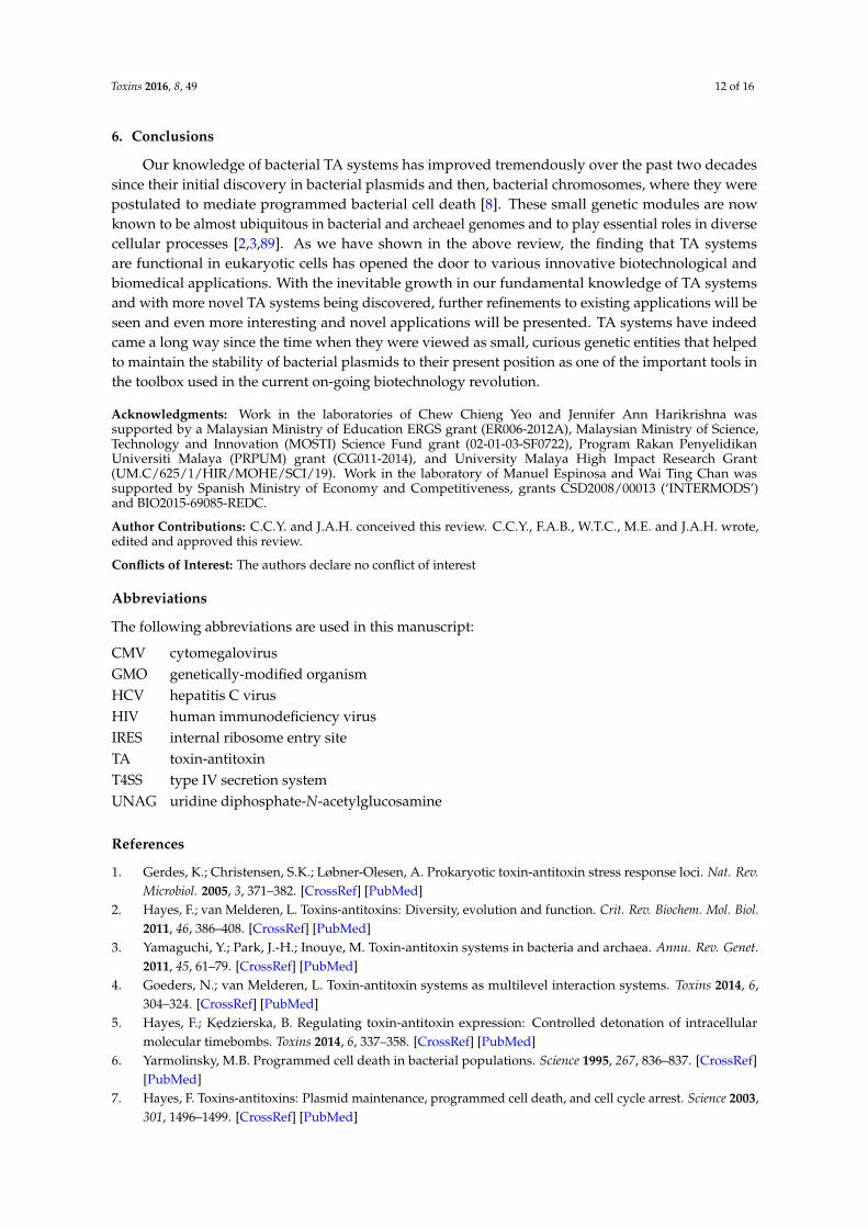
proposed using the Agrobacterium tumefaciens Ti plasmid machinery, or any other T4 secretion system(T4SS) protein complex, to specifically integrate the desired genetic information within any eukaryoticchromosome [85]. T4SS is used by bacteria to translocate and transport DNA-protein complexes froma donor to a recipient cell (Figure 3). This strategy has been used to develop gene cassettes that makeuse of the site-specific recombinase and integrase ability of some bacterial plasmid-encoded relaxases(proteins devoted to conjugative transfer between bacteria or between bacteria and eukaryotes).In some cases, it has been shown that the target sequence for the relaxase to perform a strand transferreaction could be as short as 17 nucleotides, and furthermore, the relaxase can integrate the transferredDNA into the nucleus of eukaryotic cells if it finds sequences homologous to its target in the recipientcells [86]. We speculate that the relaxase activity could be also used to efficiently integrate the desiredgenes into eukaryotic chromosomes, provided some stretches of homologous DNA are cloned withinthe incoming plasmid DNA [86,87]. Employment of these cassettes is predicted to function in anefficient manner for gene therapy with specific targeting into the recipient chromosome, rather thanrandom integration [88]. If the incoming plasmid harbors a toxin-encoding gene and some specifictumor-related DNA region, we could envisage that, at least as a preliminary experimental approach,integration of the toxin gene into the desired chromosome region of tumor cells would be feasible.If, in addition, the toxin gene is cloned under the control of any oncogenic-specific promoter, orany inducible promoter that can be used in eukaryotic cells, then we could also consider that sometargeted-vectors could be used to employ toxins as specific anti-cancer tools.
Toxins 2016, 8, 49 11 of 16
into the recipient chromosome, rather than random integration [88]. If the incoming plasmid harbors
a toxin‐encoding gene and some specific tumor‐related DNA region, we could envisage that, at least
as a preliminary experimental approach, integration of the toxin gene into the desired chromosome
region of tumor cells would be feasible. If, in addition, the toxin gene is cloned under the control of
any oncogenic‐specific promoter, or any inducible promoter that can be used in eukaryotic cells, then we
could also consider that some targeted‐vectors could be used to employ toxins as specific anti‐cancer tools.
Figure 3. Possible way to deliver an RNase toxin into the chromosome of a tumor cell.
The plasmid‐encoded relaxase (depicted in pink) harbors a toxin gene (denoted as a red arrow), and
a DNA stretch homologous to the chromosome of the recipient cell (shown as green lines). The relaxase
recognizes and initiates transfer from the donor (bacterial) cell by means of the protein‐DNA complex
formed by the relaxase, auxiliary proteins (green), and oriT (shadowed). This complex is pumped into
the recipient tumor cell by the coupling protein (CP, shown in orange) and the T4SS export system
(in blue). The relaxase‐DNA enters into the nucleus and integrates its cargo transgene into the
chromosome of the recipient cell. Induction of the toxin leads to RNA cleavage and tumor cell stasis.
6. Conclusions
Our knowledge of bacterial TA systems has improved tremendously over the past two decades
since their initial discovery in bacterial plasmids and then, bacterial chromosomes, where they were
postulated to mediate programmed bacterial cell death [8]. These small genetic modules are now
known to be almost ubiquitous in bacterial and archeael genomes and to play essential roles in
diverse cellular processes [2,3,89]. As we have shown in the above review, the finding that TA
systems are functional in eukaryotic cells has opened the door to various innovative biotechnological
and biomedical applications. With the inevitable growth in our fundamental knowledge of TA
systems and with more novel TA systems being discovered, further refinements to existing
applications will be seen and even more interesting and novel applications will be presented.
TA systems have indeed came a long way since the time when they were viewed as small, curious
genetic entities that helped to maintain the stability of bacterial plasmids to their present position as
one of the important tools in the toolbox used in the current on‐going biotechnology revolution.
Figure 3. Possible way to deliver an RNase toxin into the chromosome of a tumor cell.The plasmid-encoded relaxase (depicted in pink) harbors a toxin gene (denoted as a red arrow),and a DNA stretch homologous to the chromosome of the recipient cell (shown as green lines). Therelaxase recognizes and initiates transfer from the donor (bacterial) cell by means of the protein-DNAcomplex formed by the relaxase, auxiliary proteins (green), and oriT (shadowed). This complex ispumped into the recipient tumor cell by the coupling protein (CP, shown in orange) and the T4SSexport system (in blue). The relaxase-DNA enters into the nucleus and integrates its cargo transgeneinto the chromosome of the recipient cell. Induction of the toxin leads to RNA cleavage and tumorcell stasis.
Toxins 2016, 8, 49 12 of 16
6. Conclusions
Our knowledge of bacterial TA systems has improved tremendously over the past two decadessince their initial discovery in bacterial plasmids and then, bacterial chromosomes, where they werepostulated to mediate programmed bacterial cell death [8]. These small genetic modules are nowknown to be almost ubiquitous in bacterial and archeael genomes and to play essential roles in diversecellular processes [2,3,89]. As we have shown in the above review, the finding that TA systemsare functional in eukaryotic cells has opened the door to various innovative biotechnological andbiomedical applications. With the inevitable growth in our fundamental knowledge of TA systemsand with more novel TA systems being discovered, further refinements to existing applications will beseen and even more interesting and novel applications will be presented. TA systems have indeedcame a long way since the time when they were viewed as small, curious genetic entities that helpedto maintain the stability of bacterial plasmids to their present position as one of the important tools inthe toolbox used in the current on-going biotechnology revolution.
Acknowledgments: Work in the laboratories of Chew Chieng Yeo and Jennifer Ann Harikrishna wassupported by a Malaysian Ministry of Education ERGS grant (ER006-2012A), Malaysian Ministry of Science,Technology and Innovation (MOSTI) Science Fund grant (02-01-03-SF0722), Program Rakan PenyelidikanUniversiti Malaya (PRPUM) grant (CG011-2014), and University Malaya High Impact Research Grant(UM.C/625/1/HIR/MOHE/SCI/19). Work in the laboratory of Manuel Espinosa and Wai Ting Chan wassupported by Spanish Ministry of Economy and Competitiveness, grants CSD2008/00013 (‘INTERMODS’)and BIO2015-69085-REDC.
Author Contributions: C.C.Y. and J.A.H. conceived this review. C.C.Y., F.A.B., W.T.C., M.E. and J.A.H. wrote,edited and approved this review.
Conflicts of Interest: The authors declare no conflict of interest
Abbreviations
The following abbreviations are used in this manuscript:
CMV cytomegalovirusGMO genetically-modified organismHCV hepatitis C virusHIV human immunodeficiency virusIRES internal ribosome entry siteTA toxin-antitoxinT4SS type IV secretion systemUNAG uridine diphosphate-N-acetylglucosamine
References
1. Gerdes, K.; Christensen, S.K.; Løbner-Olesen, A. Prokaryotic toxin-antitoxin stress response loci. Nat. Rev.Microbiol. 2005, 3, 371–382. [CrossRef] [PubMed]
2. Hayes, F.; van Melderen, L. Toxins-antitoxins: Diversity, evolution and function. Crit. Rev. Biochem. Mol. Biol.2011, 46, 386–408. [CrossRef] [PubMed]
3. Yamaguchi, Y.; Park, J.-H.; Inouye, M. Toxin-antitoxin systems in bacteria and archaea. Annu. Rev. Genet.2011, 45, 61–79. [CrossRef] [PubMed]
4. Goeders, N.; van Melderen, L. Toxin-antitoxin systems as multilevel interaction systems. Toxins 2014, 6,304–324. [CrossRef] [PubMed]
5. Hayes, F.; Kedzierska, B. Regulating toxin-antitoxin expression: Controlled detonation of intracellularmolecular timebombs. Toxins 2014, 6, 337–358. [CrossRef] [PubMed]
6. Yarmolinsky, M.B. Programmed cell death in bacterial populations. Science 1995, 267, 836–837. [CrossRef][PubMed]
7. Hayes, F. Toxins-antitoxins: Plasmid maintenance, programmed cell death, and cell cycle arrest. Science 2003,301, 1496–1499. [CrossRef] [PubMed]

Toxins 2016, 8, 49 13 of 16
8. Aizenman, E.; Engelberg-Kulka, H.; Glaser, G. An Escherichia coli chromosomal “addiction module”regulated by guanosine 31,51-bispyrophosphate: A model for programmed bacterial cell death. Proc. Natl.Acad. Sci. USA 1996, 93, 6059–6063. [CrossRef] [PubMed]
9. Engelberg-Kulka, H.; Glaser, G. Addiction modules and programmed cell death and antideath in bacterialcultures. Annu. Rev. Microbiol. 1999, 53, 43–70. [CrossRef] [PubMed]
10. Engelberg-Kulka, H.; Amitai, S.; Kolodkin-Gal, I.; Hazan, R. Bacterial programmed cell death andmulticellular behavior in bacteria. PLoS Genet. 2006, 2. [CrossRef] [PubMed]
11. Pedersen, K.; Christensen, S.K.; Gerdes, K. Rapid induction and reversal of a bacteriostatic condition bycontrolled expression of toxins and antitoxins. Mol. Microbiol. 2002, 45, 501–510. [CrossRef] [PubMed]
12. Christensen, S.K.; Mikkelsen, M.; Pedersen, K.; Gerdes, K. RelE, a global inhibitor of translation, is activatedduring nutritional stress. Proc. Natl. Acad. Sci. USA 2001, 98, 14328–14333. [CrossRef] [PubMed]
13. Christensen, S.K.; Gerdes, K. RelE toxins from bacteria and Archaea cleave mRNAs on translating ribosomes,which are rescued by tmRNA. Mol. Microbiol. 2003, 48, 1389–1400. [CrossRef] [PubMed]
14. Keren, I.; Shah, D.; Spoering, A.; Kaldalu, N.; Lewis, K. Specialized persister cells and the mechanism ofmultidrug tolerance in Escherichia coli. J. Bacteriol. 2004, 186, 8172–8180. [CrossRef] [PubMed]
15. Lewis, K. Persister cells. Annu. Rev. Microbiol. 2010, 64, 357–372. [CrossRef] [PubMed]16. De Bast, M.S.; Mine, N.; van Melderen, L. Chromosomal toxin-antitoxin systems may act as antiaddiction
modules. J. Bacteriol. 2008, 190, 4603–4809. [CrossRef] [PubMed]17. Hazan, R.; Engelberg-Kulka, H. Escherichia coli mazEF-mediated cell death as a defense mechanism that
inhibits the spread of phage P1. Mol. Genet. Genomics 2004, 272, 227–234. [CrossRef] [PubMed]18. Blower, T.R.; Evans, T.J.; Przybilski, R.; Fineran, P.C.; Salmond, G.P.C. Viral evasion of a bacterial suicide
system by RNA-based molecular mimicry enables infectious altruism. PLoS Genet. 2012, 8. [CrossRef][PubMed]
19. Unterholzner, S.J.; Poppenberger, B.; Rozhon, W. Toxin-antitoxin Systems: Biology, identification, andapplication. Mob. Genet. Elem. 2013, 3. [CrossRef] [PubMed]
20. Szekeres, S.; Dauti, M.; Wilde, C.; Mazel, D.; Rowe-Magnus, D.A. Chromosomal toxin-antitoxin loci candiminish large-scale genome reductions in the absence of selection. Mol. Microbiol. 2007, 63, 1588–1605.[CrossRef] [PubMed]
21. Wang, X.; Wood, T.K. Toxin-antitoxin systems influence biofilm and persister cell formation and the generalstress response. Appl. Environ. Microbiol. 2011, 77, 5577–5583. [CrossRef] [PubMed]
22. Mutschler, H.; Meinhart, A. ε/ζ Systems: Their role in resistance, virulence, and their potential for antibioticdevelopment. J. Mol. Med. 2011, 89, 1183–1194. [CrossRef] [PubMed]
23. Bertram, R.; Schuster, C.F. Post-transcriptional regulation of gene expression in bacterial pathogens bytoxin-antitoxin systems. Front. Cell. Infect. Microbiol. 2014, 4. [CrossRef] [PubMed]
24. Ren, D.; Walker, A.N.; Daines, D.A. Toxin-antitoxin loci vapBC-1 and vapXD contribute to survival andvirulence in nontypeable Haemophilus influenzae. BMC Microbiol. 2012, 12. [CrossRef] [PubMed]
25. Fozo, E.M.; Hemm, M.R.; Storz, G. Small toxic proteins and the antisense RNAs that repress them.Microbiol. Mol. Biol. Rev. 2008, 72, 579–589. [CrossRef] [PubMed]
26. Blower, T.R.; Short, F.L.; Rao, F.; Mizuguchi, K.; Pei, X.Y.; Fineran, P.C.; Luisi, B.F.; Salmond, G.P.C.Identification and classification of bacterial Type III toxin-antitoxin systems encoded in chromosomaland plasmid genomes. Nucleic Acids Res. 2012, 40, 6158–6173. [CrossRef] [PubMed]
27. Masuda, H.; Tan, Q.; Awano, N.; Wu, K.-P.; Inouye, M. YeeU enhances the bundling of cytoskeletal polymersof MreB and FtsZ, antagonizing the CbtA (YeeV) toxicity in Escherichia coli. Mol. Microbiol. 2012, 84, 979–989.[CrossRef] [PubMed]
28. Wang, X.; Lord, D.M.; Cheng, H.-Y.; Osbourne, D.O.; Hong, S.H.; Sanchez-Torres, V.; Quiroga, C.; Zheng, K.;Herrmann, T.; Peti, W.; et al. A new type V toxin-antitoxin system where mRNA for toxin GhoT is cleaved byantitoxin GhoS. Nat. Chem. Biol. 2012, 8, 855–861. [CrossRef] [PubMed]
29. Markovski, M.; Wickner, S. Preventing bacterial suicide: A novel toxin-antitoxin strategy. Mol. Cell 2013, 52,611–612. [CrossRef] [PubMed]
30. Aakre, C.D.; Phung, T.N.; Huang, D.; Laub, M.T. A bacterial toxin inhibits DNA replication elongationthrough a direct interaction with the β sliding clamp. Mol. Cell 2013, 52, 617–628. [CrossRef] [PubMed]
31. Chan, W.T.; Balsa, D.; Espinosa, M. One cannot rule them all: Are bacterial toxins-antitoxins druggable?FEMS Microbiol. Rev. 2015, 39, 522–540. [CrossRef] [PubMed]

Toxins 2016, 8, 49 14 of 16
32. Stieber, D.; Gabant, P.; Szpirer, C.Y. The art of selective killing: Plasmid toxin/antitoxin systems and theirtechnological applications. Biotechniques 2008, 45, 344–346. [CrossRef] [PubMed]
33. Kristoffersen, P.; Jensen, G.B.; Gerdes, K.; Piskur, J. Bacterial toxin-antitoxin gene system as containmentcontrol in yeast cells. Appl. Environ. Microbiol. 2000, 66, 5524–5526. [CrossRef] [PubMed]
34. Yamamoto, T.M.; Gerdes, K.; Tunnacliffe, A. Bacterial toxin RelE induces apoptosis in human cells. FEBS Lett.2002, 519, 191–194. [CrossRef]
35. De la Cueva-Méndez, G.; Mills, A.D.; Clay-Farrace, L.; Díaz-Orejas, R.; Laskey, R.A. Regulatable killing ofeukaryotic cells by the prokaryotic proteins Kid and Kis. EMBO J. 2003, 22, 246–251. [CrossRef] [PubMed]
36. García, J.L.; Díaz, E. Plasmids as tools for containment. Microbiol. Spectr. 2014, 2. [CrossRef]37. Torres, B.; Jaenecke, S.; Timmis, K.N.; García, J.L.; Díaz, E. A dual lethal system to enhance containment of
recombinant micro-organisms. Microbiology 2003, 149, 3595–3601. [CrossRef] [PubMed]38. Mandell, D.J.; Lajoie, M.J.; Mee, M.T.; Takeuchi, R.; Kuznetsov, G.; Norville, J.E.; Gregg, C.J.; Stoddard, B.L.;
Church, G.M. Biocontainment of genetically modified organisms by synthetic protein design. Nature 2015,518, 55–60. [CrossRef] [PubMed]
39. Rovner, A.J.; Haimovich, A.D.; Katz, S.R.; Li, Z.; Grome, M.W.; Gassaway, B.M.; Amiram, M.; Patel, J.R.;Gallagher, R.R.; Rinehart, J.; et al. Recoded organisms engineered to depend on synthetic amino acids. Nature2015, 518, 89–93. [CrossRef] [PubMed]
40. Kroll, J.; Klinter, S.; Schneider, C.; Voss, I.; Steinbüchel, A. Plasmid addiction systems: Perspectives andapplications in biotechnology. Microb. Biotechnol. 2010, 3, 634–657. [CrossRef] [PubMed]
41. Zielenkiewicz, U.; Kowalewska, M.; Kaczor, C.; Ceglowski, P. In vivo interactions between toxin-antitoxinproteins epsilon and zeta of streptococcal plasmid pSM19035 in Saccharomyces cerevisiae. J. Bacteriol. 2009,191, 3677–3684. [CrossRef] [PubMed]
42. Muñoz-Gómez, A.J.; Lemonnier, M.; Santos-Sierra, S.; Berzal-Herranz, A.; Díaz-Orejas, R. RNase/anti-RNaseactivities of the bacterial parD toxin-antitoxin system. J. Bacteriol. 2005, 187, 3151–3157. [CrossRef] [PubMed]
43. Mutschler, H.; Gebhardt, M.; Shoeman, R.L.; Meinhart, A. A novel mechanism of programmed cell deathin bacteria by toxin-antitoxin systems corrupts peptidoglycan synthesis. PLoS Biol. 2011, 9. [CrossRef][PubMed]
44. Balan, A.; Schenberg, A.C.G. A conditional suicide system for Saccharomyces cerevisiae relying on theintracellular production of the Serratia marcescens nuclease. Yeast 2005, 22, 203–212. [CrossRef] [PubMed]
45. Cabib, E.; Farkas, V.; Kosik, O.; Blanco, N.; Arroyo, J.; McPhie, P. Assembly of the yeast cell wall: Crh1pand Crh2p act as transglycosylases in vivo and in vitro. J. Biol. Chem. 2008, 283, 29859–29872. [CrossRef][PubMed]
46. Chan, C.T.Y.; Lee, J.W.; Cameron, D.E.; Bashor, C.J.; Collins, J.J. “Deadman” and “Passcode” microbial killswitches for bacterial containment. Nat. Chem. Biol. 2016, 12, 82–86. [CrossRef] [PubMed]
47. Yang, J.; Jiang, W.; Yang, S. MazF as a counter-selectable marker for unmarked genetic modification of Pichiapastoris. FEMS Yeast Res. 2009, 9, 600–609. [CrossRef] [PubMed]
48. Chen, Z.; Sun, H.; Li, P.; He, N.; Zhu, T.; Li, Y. Enhancement of the gene targeting efficiency ofnon-conventional yeasts by increasing genetic redundancy. PLoS ONE 2013, 8. [CrossRef] [PubMed]
49. Murphy, D.J. Improving containment strategies in biopharming. Plant Biotechnol. J. 2007, 5, 555–569.[CrossRef] [PubMed]
50. Kamle, S.; Ali, S. Genetically modified crops: Detection strategies and biosafety issues. Gene 2013, 522,123–132. [CrossRef] [PubMed]
51. Sang, Y.; Millwood, R.J.; Neal Stewart, C., Jr. Gene use restriction technologies for transgenic plantbioconfinement. Plant Biotechnol. J. 2013, 11, 649–658. [CrossRef] [PubMed]
52. Zhang, X.; Wang, D.; Zhao, S.; Shen, Z. A double built-in containment strategy for production of recombinantproteins in transgenic rice. PLoS ONE 2014, 9. [CrossRef] [PubMed]
53. Lombardo, L. Genetic use restriction technologies: A review. Plant Biotechnol. J. 2014, 12, 995–1005. [CrossRef][PubMed]
54. Kempe, K.; Rubtsova, M.; Gils, M. Split-gene system for hybrid wheat seed production. Proc. Natl. Acad.Sci. USA 2014, 111, 9097–9102. [CrossRef] [PubMed]
55. Medina, M.; Roque, E.; Pineda, B.; Cañas, L.; Rodriguez-Concepción, M.; Beltrán, J.P.; Gómez-Mena, C.Early anther ablation triggers parthenocarpic fruit development in tomato. Plant Biotechnol. J. 2013, 11,770–779. [CrossRef] [PubMed]
Toxins 2016, 8, 49 15 of 16
56. Ulyanova, V.; Vershinina, V.; Ilinskaya, O. Barnase and binase: Twins with distinct fates. FEBS J. 2011, 278,3633–3643. [CrossRef] [PubMed]
57. Mariani, C.; de Beuckeleer, M.; Truettner, J.; Leemans, J.; Goldberg, R.B. Induction of male sterility in plantsby a chimaeric ribonuclease gene. Nature 1990, 347, 737–741. [CrossRef]
58. Goldman, M.H.; Goldberg, R.B.; Mariani, C. Female sterile tobacco plants are produced by stigma-specificcell ablation. EMBO J. 1994, 13, 2976–2984.
59. Beals, T.P.; Goldberg, R.B. A novel cell ablation strategy blocks tobacco anther dehiscence. Plant Cell 1997, 9,1527–1545. [CrossRef] [PubMed]
60. Gardner, N.; Felsheim, R.; Smith, A.G. Production of male- and female-sterile plants through reproductivetissue ablation. J. Plant Physiol. 2009, 166, 871–881. [CrossRef] [PubMed]
61. Kobayashi, K.; Munemura, I.; Hinata, K.; Yamamura, S. Bisexual sterility conferred by the differentialexpression of barnase and barstar: A simple and efficient method of transgene containment. Plant Cell Rep.2006, 25, 1347–1354. [CrossRef] [PubMed]
62. Bisht, N.C.; Jagannath, A.; Augustine, R.; Burma, P.K.; Gupta, V.; Pradhan, A.K.; Pental, D. Effectiverestoration of male-sterile (barnase) lines requires overlapping and higher levels of barstar expression:A multi-generation field analysis in Brassica juncea. J. Plant Biochem. Biotechnol. 2014, 24, 393–399. [CrossRef]
63. Millwood, R.J.; Moon, H.S.; Poovaiah, C.R.; Muthukumar, B.; Rice, J.H.; Abercrombie, J.M.; Abercrombie, L.L.;Green, W.D.; Stewart, C.N. Engineered selective plant male sterility through pollen-specific expression of theEcoRI restriction endonuclease. Plant Biotechnol. J. 2015. [CrossRef] [PubMed]
64. Iida, S.; Terada, R. Modification of endogenous natural genes by gene targeting in rice and other higherplants. Plant Mol. Biol. 2005, 59, 205–219. [CrossRef] [PubMed]
65. Bakar, F.A.; Yeo, C.C.; Harikrishna, J.A. Expression of the Streptococcus pneumoniae yoeB chromosomal toxingene causes cell death in the model plant Arabidopsis thaliana. BMC Biotechnol. 2015, 15. [CrossRef][PubMed]
66. Brand, L.; Horler, M.; Nuesch, E.; Vassalli, S.; Barrell, P.; Yang, W.; Jefferson, R.A.; Grossniklaus, U.;Curtis, M.D. A versatile and reliable two-component system for tissue-specific gene induction in arabidopsis.Plant Physiol. 2006, 141, 1194–1204. [CrossRef] [PubMed]
67. Borgen, B.H.; Thangstad, O.P.; Ahuja, I.; Rossiter, J.T.; Bones, A.M. Removing the mustard oil bomb fromseeds: Transgenic ablation of myrosin cells in oilseed rape (Brassica napus) produces MINELESS seeds.J. Exp. Bot. 2010, 61, 1683–1697. [CrossRef] [PubMed]
68. Nieto, C.; Cherny, I.; Khoo, S.K.; de Lacoba, M.G.; Chan, W.T.; Yeo, C.C.; Gazit, E.; Espinosa, M.The yefM-yoeB toxin-antitoxin systems of Escherichia coli and Streptococcus pneumoniae: Functional andstructural correlation. J. Bacteriol. 2007, 189, 1266–1278. [CrossRef] [PubMed]
69. Zuo, J.; Niu, Q.-W.; Chua, N.-H. An estrogen receptor-based transactivator XVE mediates highly induciblegene expression in transgenic plants. Plant J. 2000, 24, 265–273. [CrossRef] [PubMed]
70. Slanchev, K.; Stebler, J.; de la Cueva-Méndez, G.; Raz, E. Development without germ cells: The role of thegerm line in zebrafish sex differentiation. Proc. Natl. Acad. Sci. USA 2005, 102, 4074–4079. [CrossRef][PubMed]
71. Shimazu, T.; Degenhardt, K.; Nur-E-Kamal, A.; Zhang, J.; Yoshida, T.; Zhang, Y.; Mathew, R.; White, E.;Inouye, M. NBK/BIK antagonizes MCL-1 and BCL-XL and activates BAK-mediated apoptosis in responseto protein synthesis inhibition. Genes Dev. 2007, 21, 929–941. [CrossRef] [PubMed]
72. Browne, S.M.; Al-Rubeai, M. Selection methods for high-producing mammalian cell lines. Trends Biotechnol.2007, 25, 425–432. [CrossRef] [PubMed]
73. Nehlsen, K.; Herrmann, S.; Zauers, J.; Hauser, H.; Wirth, D. Toxin-antitoxin based transgene expression inmammalian cells. Nucleic Acids Res. 2010, 38. [CrossRef] [PubMed]
74. Chono, H.; Matsumoto, K.; Tsuda, H.; Saito, N.; Lee, K.; Kim, S.; Shibata, H.; Ageyama, N.; Terao, K.;Yasutomi, Y.; et al. Acquisition of HIV-1 resistance in T lymphocytes using an ACA-specific E. coli mRNAinterferase. Hum. Gene Ther. 2011, 22, 35–43. [CrossRef] [PubMed]
75. Zhang, Y.; Zhang, J.; Hoeflich, K.P.; Ikura, M.; Qing, G.; Inouye, M. MazF cleaves cellular mRNAs specificallyat ACA to block protein synthesis in Escherichia coli. Mol. Cell 2003, 12, 913–923. [CrossRef]
76. Chono, H.; Saito, N.; Tsuda, H.; Shibata, H.; Ageyama, N.; Terao, K.; Yasutomi, Y.; Mineno, J.; Kato, I. In vivosafety and persistence of endoribonuclease gene-transduced CD4+ T cells in cynomolgus macaques forHIV-1 gene therapy model. PLoS ONE 2011, 6. [CrossRef] [PubMed]
Toxins 2016, 8, 49 16 of 16
77. Park, J.-H.; Yamaguchi, Y.; Inouye, M. Intramolecular regulation of the sequence-specific mRNAinterferase activity of MazF fused to a MazE fragment with a linker cleavable by specific proteases.Appl. Environ. Microbiol. 2012, 78, 3794–3799. [CrossRef] [PubMed]
78. Shapira, A.; Shapira, S.; Gal-Tanamy, M.; Zemel, R.; Tur-Kaspa, R.; Benhar, I. Removal of hepatitis Cvirus-infected cells by a zymogenized bacterial toxin. PLoS ONE 2012, 7. [CrossRef] [PubMed]
79. De la Cueva-Méndez, G. Systems and Methods for Diminishing Cell Growth and Inducing Selective Killingof Target Cells. WO2013037504 A4, 7 June 2013.
80. Preston, M.A.; Pimentel, B.; Bermejo-Rodríguez, C.; Dionne, I.; Turnbull, A.; de la Cueva-Méndez, G.Repurposing a prokaryotic toxin-antitoxin system for the selective killing of oncogenically stressed humancells. ACS Synth. Biol. 2015. [CrossRef] [PubMed]
81. Bravo, A.; de Torrontegui, G.; Díaz, R. Identification of components of a new stability system of plasmid R1,ParD, that is close to the origin of replication of this plasmid. Mol. Gen. Genet. 1987, 210, 101–110. [CrossRef][PubMed]
82. Yamaguchi, Y.; Inouye, M. Type II toxin-antitoxin loci: The mazEF Family. In Prokaryotic Toxin-Antitoxins;Gerdes, K., Ed.; Springer-Verlag: Berlin, Germnay, 2013; pp. 107–136.
83. Wieteska, Ł.; Skulimowski, A.; Cybula, M.; Szemraj, J. Toxins vapC and pasB from prokaryotic TA modulesremain active in mammalian cancer cells. Toxins 2014, 6, 2948–2961. [CrossRef] [PubMed]
84. De la Cueva-Méndez, G.; Pimentel, B. Biotechnological and medical exploitation of toxin-antitoxin genesand their components. In Prokaryotic Toxin-Antitoxins; Gerdes, K., Ed.; Springer: Berlin, Germany, 2013;pp. 341–360.
85. Llosa, M.; Zupan, J.; Baron, C.; Zambryski, P. The N- and C-terminal portions of the agrobacterium VirB1protein independently enhance tumorigenesis. J. Bacteriol. 2000, 182, 3437–3445. [CrossRef] [PubMed]
86. Draper, O.; César, C.E.; Machón, C.; de la Cruz, F.; Llosa, M. Site-specific recombinase and integrase activitiesof a conjugative relaxase in recipient cells. Proc. Natl. Acad. Sci. USA 2005, 102, 16385–16390. [CrossRef][PubMed]
87. Llosa, M.; Roy, C.; Dehio, C. Bacterial type IV secretion systems in human disease. Mol. Microbiol. 2009, 73,141–151. [CrossRef] [PubMed]
88. González-Prieto, C.; Agúndez, L.; Linden, R.M.; Llosa, M. HUH site-specific recombinases for targetedmodification of the human genome. Trends Biotechnol. 2013, 31, 305–312. [CrossRef] [PubMed]
89. Gerdes, K. Prokaryotic Toxin-Antitoxins, 1st ed.; Springer-Verlag: Berlin, Germnay, 2013.
© 2016 by the authors; licensee MDPI, Basel, Switzerland. This article is an open accessarticle distributed under the terms and conditions of the Creative Commons by Attribution(CC-BY) license (http://creativecommons.org/licenses/by/4.0/).