towards improved diagnosis of neglected zoonotic …campbell/resources/wpw13w-1-s2.0-s...trematode...
TRANSCRIPT

Acta Tropica 141 (2015) 161–169
Contents lists available at ScienceDirect
Acta Tropica
journa l homepage: www.e lsev ier .com/ locate /ac ta t ropica
Towards improved diagnosis of neglected zoonotic trematodes usinga One Health approach!
Maria Vang Johansena,∗, Tore Lierb, Paiboon Sithithawornc
a Section for Parasitology and Aquatic Diseases, Department of Veterinary Disease Biology, Faculty of Health and Medical Sciences, University ofCopenhagen, Denmarkb Department of Microbiology and Infection Control, University Hospital of North Norway, Norwayc Department of Parasitology and Liver Fluke and Cholangiocarcinoma Research Center, Faculty of Medicine, Khon Kaen University, Thailand
a r t i c l e i n f o
Article history:Available online 22 July 2013
Keywords:DiagnosisTrematode zoonosesNeglected Tropical DiseasesOne Health
a b s t r a c t
Reaching the goal of control, elimination and eradication of the Neglected Tropical Disease in a fore-seeable future provides significant challenges at the ground level especially regarding helminthiasis.Helminths are still mainly diagnoses by egg identification in stool, methods with low sensitivity and formost species low specificity. Cross-sectoral collaboration with regard to zoonoses is almost non-existingand cross-validation by inter-laboratory evaluation of diagnostic tests is not a common practice. The aimof this review was to elucidate the dilemma of helminth diagnosis using zoonotic trematodes as exam-ples. Much progress has been made improving the diagnostic sensitivity of Opisthorchis and Clonorchisusing DNA-based techniques but the specificity of these tests is still a challenge due to the many mostcommon but neglected intestinal trematodes. The burden of these diseases and ways to control themremains to be elucidated. Although efficacious drugs are available, the effectiveness of mass drug admin-istration remains to be assessed. The importance of animal reservoirs and ways to control the diseasesin animals are yet unknown. Diagnostic challenges regarding Schistosoma japonicum and Schistosomamekongi include the many light infections and the persisting influx from the animal reservoirs. The sensi-tivity of the faecal based techniques suited morbidity control but will be insufficient for elimination of thehelminths. More accurate diagnostic tools are required and new algorithms for detection and progressionof helminth elimination will be needed. Standardized inter-laboratory test validation, inter-sectoral col-laboration and establishment of an international One Health diagnostic platform, sharing best practiceson diagnosis of helminth zoonoses, could all significantly contribute to control and elimination of thesediseases.
© 2013 The Authors. Published by Elsevier B.V. All rights reserved.
1. Introduction – from microscopes to molecular bands
In the second WHO report on Neglected Tropical Diseases (NTD),a detailed roadmap is outlined for control, elimination and possibleeradication of the 17 identified NTD before 2020. The roadmap isa very promising effort initiating concerted action against the NTDwhich currently affects nearly 2 billion people Worldwide (WHO,2013). The strategy combines five intervention strategies (i.e. pre-ventive chemotherapy, intensive case management, vector control,
! This is an open-access article distributed under the terms of the CreativeCommons Attribution License, which permits unrestricted use, distribution andreproduction in any medium, provided the original author and source are credited.
∗ Corresponding author at: Department of Veterinary Disease Biology, Faculty ofHealth and Medical Sciences, University of Copenhagen, Thorvaldsensvej 57, DK-1871 Frederiksberg C, Denmark. Tel.: +45 35 33 14 38.
E-mail addresses: [email protected], [email protected], [email protected](M.V. Johansen).
improving water and sanitation and including veterinary publichealth) of which preventive chemotherapy is being promoted asthe most important for many of the NTD (WHO, 2013). However,the report also urges the need to refine strategies, and develop newtools and algorithms as the outlined plan causes major challengesfor most existing systems. A central prerequisite for control of theNTD is accurate diagnosis and in the case of the neglected zoonoses,techniques for animal diagnosis also need to be available but aspointed out in the report by WHO (2010) major challenges existregarding proper diagnostic and assessment tools for many of theNTD. Zoonotic trematodes like foodborne zoonotic trematodes andAsian schistosomiasis serve as good examples. Major knowledgegaps exist regarding the burden of these zoonoses and for the food-borne zoonotic trematodes even species identification is a problem.Without these, identification of the problem as well as assessmentof any intervention effect will remain a challenge (Fig. 1).
Zoonotic trematodes, like most helminths, are diagnosedtoday in man and animals primarily by microscopy of helmintheggs in stool; a method introduced in the late 19th century.
0001-706X/$ – see front matter © 2013 The Authors. Published by Elsevier B.V. All rights reserved.http://dx.doi.org/10.1016/j.actatropica.2013.07.006

162 M.V. Johansen et al. / Acta Tropica 141 (2015) 161–169
Develop and validate diagnostic tools for
human and animal useto:
1. Identify disease organism2. Assess distribution and important risk factors 3. Determine
disease and societal burden
4. Identify efficacious control tools
5. Assess cost-effectiveness of pilot control programmes
Monitor evidence-based surveillance & control programmes
Fig. 1. Illustration of essential steps from parasite identification to evidence-basedsurveillance and control. Each step will require development and validation ofappropriate diagnostic tools for both human and animal hosts.
Immunological tests, numerous and diverse, are generally less usedand less validated. Only in very recent years have molecular tech-niques been introduced and are now being optimized and validatedacross country borders. As the clinical manifestations for zoonotictrematodes are non-pathognomonic, clinical diagnosis makes lit-tle sense. The lack of a reliable gold standard has been anothermajor drawback in diagnostic advancements of these parasites.As the new tests are being evaluated against a suboptimal goldstandard, the outcome of the assessment remains questionable(Fig. 2). This has been an on-going debate while trying to introduceserological tests assessing specific antibodies as well as molecu-lar methods. While some attention has been paid to develop andimprove diagnostic tools for human zoonotic trematodes, the diag-nostic ante mortem opportunities for animal zoonotic trematodesis almost nonexisting despite a growing recognition of their impor-tance in control and eventually eradication of these diseases. In thefollowing an update on the diagnostic advancements and presentchallenges are presented for three groups of neglected zoonosesamong the NTD.
1.1. Opisthorchiasis and clonorchiasis
The foodborne zoonotic trematodes consist of liver, lung andintestinal flukes of which the most important species parasitiz-ing humans are the fishborne zoonotic trematodes Opisthorchisviverrini, Opisthorchis felineus and Clonorchis sinensis (Keiser and
Fig. 2. Example of combinations of test results. The accuracy of a new test (here PCR)will be wrongly perceived as low if a test with low sensitivity (here microscopy) orlow specificity (here antibody detection) are used as gold standard.
Utzinger, 2009; Sithithaworn et al., 2012). Distribution of theseliver flukes is focal in nature and restricted to areas where thefirst and second intermediate hosts are abundant. In addition, theirdistribution ranges are related to habit of eating raw, pickle, orundercooked fish and fishery products particularly in SoutheastAsia (Grundy-Warr et al., 2012). Distribution ranges of these liverflukes do generally not overlap for example O. viverrini is knownto occur in Thailand, Lao PDR, Cambodia and Vietnam while C.sinensis is endemic in China, Korea, Taiwan, Vietnam and part ofRussia (Sithithaworn et al., 2012). O. felineus confine to Eurasia inthe former Soviet Union, Kazakhstan, the Ukraine and Europe suchas Italy and Germany (Pozio et al., 2013). Recent outbreak of O.felineus in Bolsena and Bracciano lakes in central Italy demonstratedan on-going maintenance of life cycle of this trematode in animalreservoir hosts in Europe (De Liberato et al., 2011).
The currently available diagnostic methods for liver fluke detec-tion are still far from ideal and significant problems are seen inareas with low prevalence, light infections and co-infection withother trematodes. In addition, diagnosis problem persists due towidespread use of chemotherapy for parasite control and this mayincrease proportion of light infection individuals. In the followingdifferent approaches will be presented and discussed.
Conventional faecal examination as well as recovery of adultworms is still the gold standard diagnoses of O. viverrini, O. felineusand C. sinensis. Eggs can be detected either in faeces, bile from naso-biliary or percutaneous transhepatic biliary drainage (PTBD) duringtreatment of bile duct obstruction, or duodenal fluid. Adult wormsmay be encountered from expulsion chemotherapy (Ramsay et al.,1989; Elkins et al., 1991; Radomyos et al., 1994; Joo and Bang,2005) or more accurately from the liver at post mortem exami-nation (Sithithaworn et al., 1991). Faecal examination is routinelyused for diagnosis of liver fluke infections due to non-invasivenessand ease of sample collection. Commonly used techniques includethe modified formalin ether (or ethyl acetate) concentration tech-nique (FECT) (Elkins et al., 1990), the modified Kato-Katz thicksmear (Hong et al., 2003), and Stoll’s dilution egg count technique(Viyanant et al., 1983). These classical coprological techniques allhave major problems regarding specificity and sensitivity.
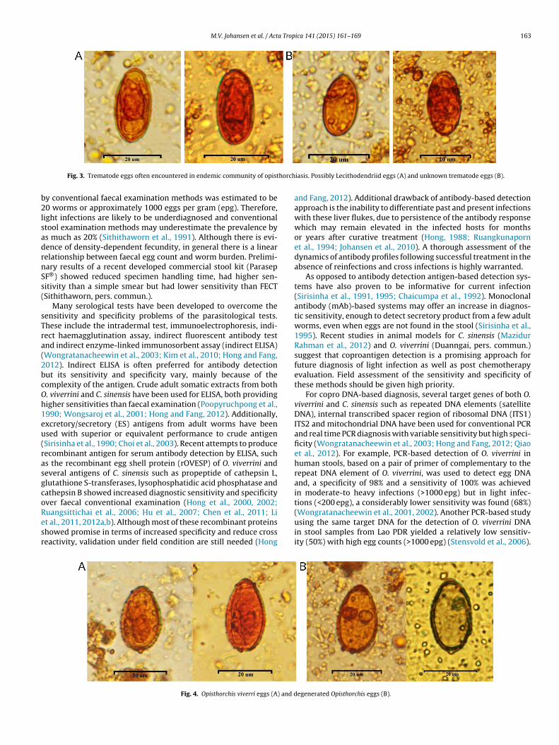
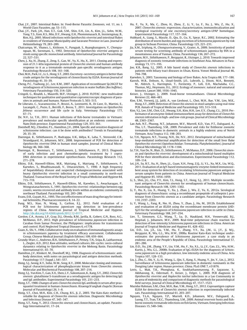
Under light microscopy, the eggs of the liver flukes are char-acterized by rough and thick egg shells, and are similar in shapeand size to several species of food-borne trematodes belonging tothe families Opisthorchiidae, Heterophyidae and Lecithodendriidae(Figs. 3 and 4). The latter two families of trematodes are collectivelyreferred to as minute intestinal flukes (MIF) because of the smallsize of the adult worms compared to the liver flukes (Kaewkes,2003; Chai et al., 2005; De and Le, 2011). Eggs of Opisthorchiidaeand Heterophyidae are very similar while eggs of Lecithodendriidaedo have smooth egg shall and less distinct shoulder in comparisonwith other two groups (Kaewkes, 2003). The marked morphologi-cal resemblance of MIF eggs with those of O. viverrini and C. sinensisincreases the probability of a false positive diagnosis, with con-comitant decrease in diagnostic specificity.
As the parasitological techniques rely on egg excretion, theycannot detect infection during prepatency, and they have poorsensitivity in light infections, in old chronic infections, followingtreatment, or when the bile duct is obstructed causing intermit-tent egg excretion. Repeated examinations are needed to improvediagnostic sensitivity of faecal examination. Three consecutiveKato-Katz thick smears were reported to be more sensitive than asingle examination by FECT (Lovis et al., 2009). However, even withrepeated stool examination using a standardized method like FECT,discrepancy remains between egg count and worm detection. Forexample in an autopsy study, adult O. viverrini were recovered from113 of 139 (81.2%) livers but faecal examinations of the same indi-viduals determined that only 86 cases (67%) were egg positive byFECT (Sithithaworn et al., 1991). From this study, the detection limit
M.V. Johansen et al. / Acta Tropica 141 (2015) 161–169 163
Fig. 3. Trematode eggs often encountered in endemic community of opisthorchiasis. Possibly Lecithodendriid eggs (A) and unknown trematode eggs (B).
by conventional faecal examination methods was estimated to be20 worms or approximately 1000 eggs per gram (epg). Therefore,light infections are likely to be underdiagnosed and conventionalstool examination methods may underestimate the prevalence byas much as 20% (Sithithaworn et al., 1991). Although there is evi-dence of density-dependent fecundity, in general there is a linearrelationship between faecal egg count and worm burden. Prelimi-nary results of a recent developed commercial stool kit (ParasepSF®) showed reduced specimen handling time, had higher sen-sitivity than a simple smear but had lower sensitivity than FECT(Sithithaworn, pers. commun.).
Many serological tests have been developed to overcome thesensitivity and specificity problems of the parasitological tests.These include the intradermal test, immunoelectrophoresis, indi-rect haemagglutination assay, indirect fluorescent antibody testand indirect enzyme-linked immunosorbent assay (indirect ELISA)(Wongratanacheewin et al., 2003; Kim et al., 2010; Hong and Fang,2012). Indirect ELISA is often preferred for antibody detectionbut its sensitivity and specificity vary, mainly because of thecomplexity of the antigen. Crude adult somatic extracts from bothO. viverrini and C. sinensis have been used for ELISA, both providinghigher sensitivities than faecal examination (Poopyruchpong et al.,1990; Wongsaroj et al., 2001; Hong and Fang, 2012). Additionally,excretory/secretory (ES) antigens from adult worms have beenused with superior or equivalent performance to crude antigen(Sirisinha et al., 1990; Choi et al., 2003). Recent attempts to producerecombinant antigen for serum antibody detection by ELISA, suchas the recombinant egg shell protein (rOVESP) of O. viverrini andseveral antigens of C. sinensis such as propeptide of cathepsin L,glutathione S-transferases, lysophosphatidic acid phosphatase andcathepsin B showed increased diagnostic sensitivity and specificityover faecal conventional examination (Hong et al., 2000, 2002;Ruangsittichai et al., 2006; Hu et al., 2007; Chen et al., 2011; Liet al., 2011, 2012a,b). Although most of these recombinant proteinsshowed promise in terms of increased specificity and reduce crossreactivity, validation under field condition are still needed (Hong
and Fang, 2012). Additional drawback of antibody-based detectionapproach is the inability to differentiate past and present infectionswith these liver flukes, due to persistence of the antibody responsewhich may remain elevated in the infected hosts for monthsor years after curative treatment (Hong, 1988; Ruangkunapornet al., 1994; Johansen et al., 2010). A thorough assessment of thedynamics of antibody profiles following successful treatment in theabsence of reinfections and cross infections is highly warranted.
As opposed to antibody detection antigen-based detection sys-tems have also proven to be informative for current infection(Sirisinha et al., 1991, 1995; Chaicumpa et al., 1992). Monoclonalantibody (mAb)-based systems may offer an increase in diagnos-tic sensitivity, enough to detect secretory product from a few adultworms, even when eggs are not found in the stool (Sirisinha et al.,1995). Recent studies in animal models for C. sinensis (MazidurRahman et al., 2012) and O. viverrini (Duanngai, pers. commun.)suggest that coproantigen detection is a promising approach forfuture diagnosis of light infection as well as post chemotherapyevaluation. Field assessment of the sensitivity and specificity ofthese methods should be given high priority.
For copro DNA-based diagnosis, several target genes of both O.viverrini and C. sinensis such as repeated DNA elements (satelliteDNA), internal transcribed spacer region of ribosomal DNA (ITS1)ITS2 and mitochondrial DNA have been used for conventional PCRand real time PCR diagnosis with variable sensitivity but high speci-ficity (Wongratanacheewin et al., 2003; Hong and Fang, 2012; Qiaoet al., 2012). For example, PCR-based detection of O. viverrini inhuman stools, based on a pair of primer of complementary to therepeat DNA element of O. viverrini, was used to detect egg DNAand, a specificity of 98% and a sensitivity of 100% was achievedin moderate-to heavy infections (>1000 epg) but in light infec-tions (<200 epg), a considerably lower sensitivity was found (68%)(Wongratanacheewin et al., 2001, 2002). Another PCR-based studyusing the same target DNA for the detection of O. viverrini DNAin stool samples from Lao PDR yielded a relatively low sensitiv-ity (50%) with high egg counts (>1000 epg) (Stensvold et al., 2006).
Fig. 4. Opisthorchis viverri eggs (A) and degenerated Opisthorchis eggs (B).

164 M.V. Johansen et al. / Acta Tropica 141 (2015) 161–169
Improvement of DNA preparation using cetyltrimethylammoniumbromide to remove PCR inhibitors raises the sensitivity to be 79%(Duenngai et al., 2008). Interestingly, in this study, PCR-positivetests were identified in several parasite negative cases by theparasitological method (29%), indicating its advantage in diagnosisof individuals with light infection. Additional O. viverrini-specificprimer pair was established for the PCR test and the sensitivity ofthe test was 10–12 ng of adult worm DNA, and three metacercariaein a fish sample (Parvathi et al., 2008). Additionally, species-specificPCR tests to distinguish between the species of liver fluke are nowavailable for O. viverrini (Ando et al., 2001; Wongratanacheewinet al., 2001), O. felineus (Pauly et al., 2003) and C. sinensis (Le et al.,2006). By using real-time PCR, quantification and early detection ofC. sinensis infection has also been demonstrated (Kim et al., 2009;Rahman et al., 2011). The loop-mediated isothermal amplification(LAMP) method was recently established for detection of O. viver-rini and C. sinensis and was reported to yield higher sensitivity thanconventional PCR method (Cai et al., 2010; Arimatsu et al., 2012; Leet al., 2012). These molecular diagnostic techniques are also valu-able for food safety inspection as they can be used to detect otherlife stages of the parasites e.g. in fish products (Parvathi et al., 2007,2008; Cai et al., 2010). A few recent studies have also been able todifferentiate liver and intestinal trematodes based on copro DNA-based detection (Lovis et al., 2009; Thaenkham et al., 2011). It isanticipated that these molecular diagnostic tests, due to their highspecificity, can play significant roles in anthelminthic drug efficacyevaluations, rigorous monitoring of re-infection patterns, as wellas in the recognition of a potentially new endemic range of theliver flukes in Southeast Asia (Touch et al., 2009; Traub et al., 2009,Duenngai et al., 2013). However, proper validation of the coproDNA-based methods is required.
There is no doubt that molecular methods will contribute sig-nificantly towards higher sensitivity and specificity of trematodezoonoses diagnosis, but further simplification and validation ofthe tests as well as considerations of cost will be needed. Addi-tionally, the importance of animal reservoirs in transmission andmaintaining the infection needs urgent attendance but with thehelp of molecular diagnostic tools, the major problem of speciesidentification should at least partly be solved.
1.2. Intestinal trematode zoonoses
Intestinal trematodes infecting humans comprise a group of70 species belonging to 14 different families, of which more than35 are zoonotic (Chai, 2005). Among the intestinal flukes, Stel-lantchasmus falcatus, Haplorchis spp. and Procerovum spp. have beenreported to cause severe health problems in humans (WHO, 1995).These trematodes have in the past been largely overlooked, hencebasic information about human and animal prevalence, transmis-sion, their veterinary and public health burden and impact, as wellas efficacy and effectiveness of control options remains to be elu-cidated. One of the main reasons for this lack of knowledge isthe continuous use of copro-microscopy tests for human diagnosiswhich fail to differentiate eggs from those of the liver flukes but alsobecause most often humans harbour multiple species infections(Chai et al., 2005; Steinmann et al., 2010).
The importance of animal reservoirs in the transmission ofintestinal trematodes has received almost no attention and no stan-dardized diagnostic tests have been established for this group ofparasites. In a single study in Vietnam, Lan Anh et al. (2009) found11 MIF species in 448 domestic animals (cats, dogs and pigs) ofwhich 10 were reported for the first time in Vietnam. Of the 448examined animals all harboured more than one species of MIF.From the study it was concluded that domestic animals most likelyplay a major role in maintaining the transmission of the intestinaltrematodes.
Several copro-microscopy tests in various modifications havebeen used to diagnose intestinal trematode eggs. The Kato-Katzthick smear and the formalin-ether/ethyl acetate concentrationtechniques have been used routinely for human diagnosis (WHO,2009). The Kato-Katz technique was also evaluated for veterinaryuse by Nguyen et al. (2008) but did not prove adequate. Nguyenet al. (2009) found a technique combining filtration and sedimen-tation useful for qualitative and quantitative detection of the smalltrematode eggs in domestic animals. The FLOTAC technique, a mod-ified McMaster flotation method, has also been tested for detectionof food-borne trematodes in both human and animal hosts (Cringoliet al., 2010; WHO, 2009), However, none of the methods fulfil therequirements of sensitivity and specificity (Sindberg et al., 2013).The lack of identification to species level by any copro-microscopytest is a major problem which calls for more specific methodologies.
Reliable diagnosis of the intestinal flukes requires recovery andidentification of adult worms expelled from egg positive hosts.As most of the adult flukes are less than 2 mm long, recovery istedies and identification includes special staining and morphologi-cal characterization. This does not apply to routine diagnostics butis very useful as a reference method validating new tests. Adulttrematodes may easily be missed due to their size and especially ifinfection intensity is low. As an alternative to use worm recoveryas reference test it has been suggested to use faecal samples spikedwith eggs from species of interest as reference (Thaenkham et al.,2007).
Many of the characterized intestinal trematodes reside in thecrypt of the villi in the small intestine, where they are attachedto the mucosa and cause ulceration, inflammation and fibrosis.The main symptoms are diarrhoea, nausea, vomiting and colickyabdominal pain, all vague and non-pathognomonic symptoms,which cannot be used for proper diagnosis. However, for many ofthe trematodes, not even the life cycle is described and neitherare the clinical manifestations (Chai et al., 2005).
Immunodiagnosis has so far been targeted towards liver flukesand intestinal trematodes have been attached as ‘the unavoidablecontamination’. No antibody or antigen test has been developedwhich can provide an accurate diagnosis of the different intestinaltrematodes (WHO, 2009).
Recently, several human copro-DNA-based techniques havebeen developed for liver flukes and tested against intestinal flukes(Stensvold et al., 2006; Duenngai et al., 2008; Lovis et al., 2009;Sato et al., 2009; Traub et al., 2009). Sato et al. (2009) and Traubet al. (2009) concluded that their PCR method was not able to dis-tinguish O. viverrini from Haplorchis taichui in mixed infections andgenerally the tests captured many more O. viverrini positive sam-ples than H. taichui. Sato et al. (2010) hypothesized that fluctuatingegg excretion from MIF could be a reason for the poor result. In avery recent experimental study, Nissen et al. (2013) showed thatthe egg excretion of Haplorchis pumilio in foxes declined to verylow levels following a high excretion in the acute phase. This anti-fecundity phenomenon does most likely apply to most MIF whichquestions the use of tests based on egg excretion. Despite millionsof people and animals being infected with MIF Worldwide the diag-nosis of these helminths remains to be developed, not only for thesake of being a differential problem but also because at least someof them cause significant health problems (Chai, 2007).
1.3. Asian zoonotic schistosomiasis
Control strategies, mainly based on mass drug administrationto humans, have been carried through for decades in most ofthe areas where Schistosoma japonicum and Schistosoma mekongiare still endemic. Hence the prevalence and infection intensity areoften low, and parts of the human population have persistingantibodies from previous infections. This is a huge challenge

M.V. Johansen et al. / Acta Tropica 141 (2015) 161–169 165
for the diagnostics. This dilemma has been discussed in a paperby Zhou et al. (2011), which also gives an overview of relevantstudies.
Measuring antibodies in serum has many qualities making itsuitable for use in control programmes and has been used exten-sively (Zhu, 2005). They are relatively cheap and easy to perform,even in large quantities. They are also often perceived as havinghigh sensitivity even in low intensity infections. Hence, a high pro-portion of those who are infected will have a positive test. They alsonullify the problem of egg shedding variability in stool. The maindrawback is a slow rate of sero-conversion after treatment, thoughthis rate varies between studies, even with a given assay (Alarconde Noya et al., 1992; Rabello et al., 1997; Whitty et al., 2000; Zhu,2005). Antibody assays may also cross react with other infections(Xu et al., 2011). Some authors argue that these shortcomings canbe minimized by the use of purified antigens or detection of cer-tain immunoglobinisotypes (Doenhoff et al., 2004; Hamilton et al.,1998). However, none of these assays have made it into routineuse.
There is an overwhelming number of different antibody assaysavailable, differing in antigen, purity of the antigen and choice ofassay system (Hamilton et al., 1998; Wu, 2002). The assays in usein large scale in recent years are indirect hemagglutination assay(IHA) and ELISA with soluble egg antigen (SEA). Variations in exper-imental conditions in clinical studies, and not the least a lack of areliable diagnostic gold standard in low intensity infections resultsin highly variable data regarding test properties for immunodiag-nostic assays (Zhou et al., 2011). Two meta analyses on clinicalstudies of IHA and ELISA have been published the last two years(Wang et al., 2012; Zhu et al., 2010). As expected, they showed avery wide distribution in test properties, but the analysis by Wanget al. (2012) calculated the merged sensitivity and specificity to be76% and 73% for IHA and 85% and 50% for ELISA, respectively.
As a disease is successfully controlled, the disease prevalencein that area will decrease, but so will the positive predictive valueof the diagnostic tests. Zhou et al. (2007b) and Lin et al. (2008b)used comprehensive stool examination to evaluate IHA and ELISAin field conditions. The prevalence was 7–19% in the four examinedvillages. Using the data from the two studies, the positive predictivevalue were 17–31% for the IHA and 10–25% for the ELISA, respec-tively. This means that 25% or less of those with a positive ELISAtest and 31% or less of those with a positive IHA test were indeedinfected. These figures are representative for similar studies in lowprevalence settings (Zhou et al., 2011). They illustrate why someauthors point out that ELISA and IHA assays currently in use are notsuitable for making decisions on treatment in individuals. Insteadsome argue that the proportion of positives in a village can be usedto determine interventions on village or area level. There is how-ever no consensus on this matter (Doenhoff et al., 2004; Lin et al.,2008b; Wu, 2002).
In the past decade a number of rapid antibody detection assaysusing dipsticks or test cards have been developed. They have theadvantage of easily being field-applicable and the possibility forquick decisions regarding further diagnosis and treatment. Sincethese assays make use of the same antigens (primarily SEA) as theconventional assays, they are prone to have the same problems withcross reactivity and persisting antibodies. Available kits includedipstick dye immunoassay (DDIA), colloidal dye immunofiltrationassay (CDIFA) and dot immuno-gold filtration assay (DIGFA) (Wenet al., 2005; Xiao et al., 2005; Xu et al., 2011; Zhu et al., 2005).
Detection of antigens secreted from the parasite is in principlean interesting diagnostic alternative, as it demonstrates the pres-ence of the parasite directly. Studied antigens include the adultworm antigens circulating anodic antigen (CAA) and circulatingcathodic antigen (CCA), but also Sj31/32 and most recently solu-ble egg antigen (Lei et al., 2011; Van’t Wout et al., 1992; Wu, 2002).
The paucity of papers on antigen detection of S. japonicum may inpart be due to the results of a Chinese collaborative study from 1996(Guan and Shi, 1996; Wu, 2002). Here 13 antigen detection systemswere tested in parallel on a heterogeneous sera panel. The speci-ficity was 90% or higher for nine of the assays, but only three of theassays had sensitivity above 60%, with the highest being 81%. Mostantigen assays use sera instead of stool. Except for this advantage,it seems that antigen detection hitherto has failed to demonstratea significantly superior advantage compared to even a moderatenumber of Kato-Katz slides. This may change as new antigens andnew assays are discovered. A urine antigen test for CCA is now com-mercialized, but has yet to prove useful in S. japonicum endemicareas (Bergquist, 2013).
Microscopy of eggs in stool, using Kato-Katz thick smears havebeen a major method for 30 years. It is simple, cheap and provides aquantitative result as epg. However, with the Kato-Katz techniquethe examined amount of stool is small, and the main criticismagainst this method is the low sensitivity in low intensity infections.Yu et al. (1998) examined duplicate smears from seven consecutivestool samples in two villages. The proportion of individuals with atleast one smear positive for S. japonicum increased from 42% to 68%and from 17% to 36% when one and seven stool samples were used,respectively. Lin et al. (2008a) examined a village for two consec-utive years, each year using a total of six smears from two stoolsamples. The prevalence increased without a clear levelling-offfrom one to six smears. If a single smear or three smears had beenused, the prevalence would have been underestimated by 55% and25%, respectively. In both studies the underestimation was highestin those with lowest infection intensity.
Hatching test is used for diagnosis in both humans and animals,mostly in China. It utilizes a large stool sample, usually 30–50 g,and hence has potentially high sensitivity. However, the methodseems difficult to standardize due to both biotic and abiotic fac-tors. Variations in such factors may in part be the reason for theinconsistency in diagnostic performance in different studies andeven within the same study (Yu et al., 2007). Hatching tests is timeconsuming and has not consistently shown to have higher sensi-tivity than Kato-Katz in humans (Hubbard et al., 2002; Zhou et al.,1998, 2011).
The search for more sensitive and specific diagnostic tests hasprompted research into the use of molecular methods like PCR andthe related LAMP. Several molecular targets have been evaluated inboth serum and stool samples, using artificial samples and animalmodels. However, clinical studies are scarce and hence it is too earlyto tell whether the promising initial results can be conveyed toclinical use (Fung et al., 2012; Lier et al., 2009; Xu et al., 2009). Thesensitivity should be substantially better than Kato-Katz to justifythe increased cost.
Asian schistosomiasis is a zoonosis, but there are very fewstudies on diagnostic methods in animals compared to humans.Hatching test has been used to a large degree in China, but alsoKato-Katz and a number of filtration/sedimentation tests (Carabinet al., 2005; Liu et al., 2012; Wang et al., 2005). Immunodiagnos-tic tests like ELISA have also been studied to some extent (Chenget al., 2007; Li et al., 2012a,b; Peng et al., 2008). In general, the testsused in animals seem to be prone to the same inadequacy as theirhuman counterparts when infection intensity is low. Two studieshave shown very superior sensitivity for PCR compared to Kato-Katz and hatching test in buffalo stool (Gordon et al., 2012; Wuet al., 2010). Gordon et al. (2012) found a formalin-ethyl acetatesedimentation technique to be equally sensitive as a PCR method.
A reliable diagnostic strategy for diagnosing low intensityAsian schistosomiasis infections in humans and animals are verywished for. Effective evaluation of new strategies and tests arehindered by the lack of an accurate and precise gold standardtests.

166 M.V. Johansen et al. / Acta Tropica 141 (2015) 161–169
2. From identification to elimination
The progress in development of sensitive and specific diagnostictools for zoonotic trematodes has been slow, fragmented, incom-plete and generally not properly validated. The need for low costand simplicity has been the driving force rather than accuracy,which has kept these parasites neglected. Hence, routine diag-nosis in most places remains with insensitive and non-specificparasitological tests for humans and no diagnosis for the animalreservoirs. From a strategic control point of view it might be rele-vant to group the zoonotic trematodes as they can all be targeted bythe anthelmintic praziquantel but with the new WHO goal to elimi-nate and eventually eradicate these diseases (WHO, 2012), accuracyof test results at species level becomes crucial. There are severaloptions which could be explored. First and foremost there is aneed to establish an Internet-based platform for diagnosis of trema-tode zoonoses across disciplines and sectors. Stratified and specificguidelines should be agreed upon. As illustrated in Fig. 1, manydifferent diagnostic/assessment tools will be needed dependingon the assessment step. The platform should gather ‘best practice’on each of the steps for both humans and animals. As the eggs ofmost zoonotic trematodes are indistinguishable, and clinical symp-toms are non-pathognomonic, the development of tele-diagnosticmight be a significant challenge. Another way, which is increas-ingly being used, is to collect specimens containing parasite DNAfrom the host of interest, either man or animal or from the envi-ronment, and send them to specialized reference laboratories forspecies or even strain identification. Along with the assessment ofwhole genome sequences of trematode zoonoses, laboratories canoffer fast and accurate diagnosis within days of delivery. This wouldovercome the present major hindering of lack of proper goldenstandards and the indistinguishable co-infections. For many diag-nostic purposes this would however, be unrealistically expensive,hence different algorithms are needed. For each of the steps indi-cated in Fig. 1, the level of accuracy and precision needed should beguiding the algorithm. Also, as a consequence of progress in controlof zoonotic trematodes, fewer cases with lower infection intensitiesare expected, which would influence the choice of tests (Bergquistet al., 2009). Combination of tests is a well-known way of improv-ing sensitivity and specificity despite a limited budget. For example,epidemiological surveys for schistosomiasis in China uses an initialscreening with IHA or dip-stick ELISA is followed by Kato-Katz ifantibody positive (Zhou et al., 2007a). In order to improve test sen-sitivity and specificity further, the Kato-Katz could be replaced bymolecular methods or enzyme-linked immunoelectrotransfer blotassay (EITB) (Lier et al., 2009; Xu et al., 2011). Since the majority ofsamples will be negative, it may be possible to save cost by poolingsamples and only if the mixture is positive, the individual compo-nents of the mixture would need to be tested. This strategy is widelyapplied in meat inspection and could easily be used in serologicaland molecular methods (Jia et al., 2009; Van et al., 2012).
3. Conclusion and the way forward
Because of the complexity of the life cycle of zoonotic trema-todes which involve snail intermediate hosts, animal reservoirhosts as well as human, control and even more so eliminationand eradication of these parasitic zoonoses will require a trueOne Health approach (Karesh et al., 2012). Although still not fullyagreed on, the American Veterinary Medical Association definesOne Health as: “The collaborative efforts of multiple disciplinesworking locally, nationally and globally to attain optimal healthfor people, animals and our environment” (Okello et al., 2011). Thisdefinition is in essence what is needed if zoonoses are to be con-trolled. Isolated sector specific control efforts against zoonoses, like
e.g. mass drug administration to human populations in endemicsettings with substantial animal reservoirs, will most likely havelittle or no impact especially in the long term.
When moving from morbidity control to transmission con-trol dominated by populations with low intensity and persistingantibodies from previous infections or cross-reactions from otherinfections, diagnostic priorities change from simplicity and low costto high accuracy. One way to achieve higher sensitivity and speci-ficity could be to use a combination of tests. The first test shouldbe simple, cheap but with high sensitivity. An antibody detec-tion test is a likely choice. Since some false positive cases may betolerated, high specificity should be prioritized in the second testof those found positive in the first. Molecular methods or highlyspecific antigen or antibody detection assays should replace copro-microscopy tests.
Molecular tools for species specific diagnosis of zoonotic trema-todes exist, but consensus of the algorithm to move from the optionof individual diagnosis to eradication of this group of NeglectedTropical Diseases has yet to be initiated. Major challenges lies aheadas accuracy of test costs. Unlike Rapid Diagnostic Tests (RDT) formalaria which play important roles for case management and con-trol, particularly when good quality microscopy-based diagnosisis unavailable, similar rapid tests for diagnosis of the neglectedzoonotic trematodes are scarce and insufficient data are currentlyavailable to derive an evidence-based algorithm of different diag-nostic methods under field condition. For disease control purpose,analysis of cost-effectiveness between mass or target-group treat-ment without the need for diagnosis versus selective treatmentfor control of parasitic disease has been raised (Evans and Guyatt,1995; Rodgers et al., 2006). If, however, the public health sectorand the veterinary and agricultural sectors could join forces andestablish a One Health diagnostic platform, sharing best practiceson diagnosis of trematode zoonoses or even all neglected zoonoses,much would be gained. Guidance via telecommunication, diagno-sis at international central DNA-based One Health laboratories, anddevelopment of algorithms for diagnostic progression dependingon the control step, will be three central elements for the successof the outlined zoonoses control.
Acknowledgements
The support from Higher Education Research Promotion andNational Research University Project of Thailand, Office of theHigher Education Commission, through health cluster (SHeP-GMS),Thailand to PS is acknowledged.
References
Alarcon de Noya, B., Spencer, L., Noya, O., 1992. Pre- and post-treatment immune-diagnostic evaluation in human schistosomiasis mansoni. Memórias do InstitutoOswaldo Cruz 87 (Suppl. 4), 271–276.
Ando, K., Sithithaworn, P., Nuchjungreed, C., Tesana, S., Srisawangwong, T.,Limviroj, W., Chinzei, Y., 2001. Nucleotide sequence of mitochondrial CO I andribosomal ITS II genes of Opisthorchis viverrini in northeast Thailand. SoutheastAsian Journal of Tropical Medicine and Public Health 32 (Suppl. 2), 17–22.
Arimatsu, Y., Kaewkes, S., Laha, T., Hong, S.J., Sripa, B., 2012. Rapid detection ofOpisthorchis viverrini copro-DNA using loop-mediated isothermal amplification(LAMP). Parasitology International 61, 178–182.
Bergquist, R., 2013. Good things are worth waiting for. American Journal of TropicalMedicine and Hygiene 88, 409–410.
Bergquist, R., Johansen, M.V., Utzinger, J., 2009. Diagnostic dilemmas in helminthol-ogy: what tools to use and when? Trends in Parasitology 25, 151–156.
Carabin, H., Balolong, E., Joseph, L., McGarvey, S.T., Johansen, M.V., Fernandez, T.,Willingham, A.L., Olveda, R., 2005. Estimating sensitivity and specificity of afaecal examination method for Schistosoma japonicum infection in cats dogs,water buffaloes, pigs, and rats in Western Samar and Sorsogon Provinces, ThePhilippines. International Journal for Parasitology 35, 1517–1524.
Cai, X.Q., Xu, M.J., Wang, Y.H., Qiu, D.Y., Liu, G.X., Lin, A., Tang, J.D., Zhang, R.L.,Zhu, X.Q., 2010. Sensitive and rapid detection of Clonorchis sinensis infection infish by loop-mediated isothermal amplification (LAMP). Parasitology Research106, 1379–1383.
M.V. Johansen et al. / Acta Tropica 141 (2015) 161–169 167
Chai, J.Y., 2007. Intestinal flukes. In: Food-Borne Parasitic Zoonoses, vol. 11, no. I.World Class Parasites, pp. 53–115.
Chai, J.Y., Park, J.H., Han, E.T., Guk, S.M., Shin, E.H., Lin, A., Kim, J.L., Sohn, W.M.,Yong, T.S., Eom, K.S., Min, D.Y., Hwang, E.H., Phommmasack, B., Insisiengmay, B.,Rim, H.J., 2005. Mixed infections with Opisthorchis viverrini and intestinal flukesin residents of Vientiane Municipality and Saravane Province in Laos. Journal ofHelminthology 79, 283–289.
Chaicumpa, W., Ybanez, L., Kitikoon, V., Pungpak, S., Ruangkunaporn, Y., Chongsa-nguan, M., Sornmani, S., 1992. Detection of Opisthorchis viverrini antigens instools using specific monoclonal antibody. International Journal for Parasitology22, 527–531.
Chen, J., Xu, H., Zhang, Z., Zeng, S., Gan, W., Yu, X., Hu, X., 2011. Cloning and expres-sion of 21.1-kDa tegumental protein of Clonorchis sinensis and human antibodyresponse to it as a trematode–nematode pan-specific serodiagnosis antigen.Parasitology Research 108, 161–168.
Choi, M.H., Park, I.C., Li, S., Hong, S.T., 2003. Excretory–secretory antigen is better thancrude antigen for the serodiagnosis of clonorchiasis by ELISA. Korean Journal ofParasitology 41, 35–39.
Cheng, P.C., Tsaihong, J.C., Lee, K.M., 2007. Application of recombinant Sjc26GST forserodiagnosis of Schistosoma japonicum infection in water buffalo (Bos buffelus).Veterinary Parasitology 150, 314–320.
Cringoli, G., Rinaldi, L., Maurelli, M.P., Utzinger, J., 2010. FLOTAC: new multivalenttechniques for qualitative and quantitative copromicroscopic diagnosis of para-sites in animals and humans. Nature Protocols 5, 503–515.
De Liberato, C., Scaramozzino, P., Brozzi, A., Lorenzetti, R., Di Cave, D., Martini, E.,Lucangeli, C., Pozio, E., Berrilli, F., Bossu, T., 2011. Investigation on Opisthorchisfelineus occurrence and life cycle in Italy. Veterinary Parasitology 177,67–71.
De, N.V., Le, T.H., 2011. Human infections of fish-borne trematodes in Vietnam:prevalence and molecular specific identification at an endemic commune inNam Dinh province. Experimental Parasitology 129, 355–361.
Doenhoff, M.J., Chiodini, P.L., Hamilton, J.V., 2004. Specific and sensitive diagnosis ofschistosome infection: can it be done with antibodies? Trends in Parasitology20, 35–39.
Duenngai, K., Sithithaworn, P., Rudrappa, U.K., Iddya, K., Laha, T., Stensvold, C.R.,Strandgaard, H., Johansen, M.V., 2008. Improvement of PCR for detection ofOpisthorchis viverrini DNA in human stool samples. Journal of Clinical Micro-biology 46, 366–368.
Duenngai, K., Boonmars, T., Sithithaworn, J., Sithithaworn, P., 2013. Diagnosisof early infection and post chemotherapeutic treatment by 1 copro-DNA detection in experimental opisthorchiasis. Parasitology Research 112,271–278.
Elkins, D.B., Haswell-Elkins, M.R., Mairiang, E., Mairiang, P., Sithithaworn, P.,Kaewkes, S., Bhudhisawasdi, V., Uttaravichien, T., 1990. A high frequencyof hepatobiliary disease and suspected cholangiocarcinoma associated withheavy Opisthorchis viverrini infection in a small community in north-eastThailand. Transactions of the Royal Society of Tropical Medicine and Hygiene 84,715–719.
Elkins, D.B., Sithithaworn, P., Haswell-Elkins, M., Kaewkes, S., Awacharagan, P.,Wongratanacheewin, S., 1991. Opisthorchis viverrini: relationships between eggcounts, worms recovered and antibody levels within an endemic community innortheast Thailand. Parasitology 102, 283–288.
Evans, D.B., Guyatt, H.L., 1995. The cost effectiveness of mass drug therapy for intesti-nal helminths. Pharmacoeconomics 8, 14–22.
Fung, M.S., Xiao, N., Wang, S., Carlton, E.J., 2012. Field evaluation of aPCR test for Schistosoma japonicum egg detection in low-prevalenceregions of China. American Journal of Tropical Medicine and Hygiene,http://dx.doi.org/10.4269/ajtmh.2012.12-0177.
Gordon, C.A., Acosta, L.P., Gray, D.J., Olveda, R.M., Jarilla, B., Gobert, G.N., Ross, A.G.,McManus, D.P., 2012. High prevalence of Schistosoma japonicum infection inCarabao from Samar Province, the Philippines: implications for transmissionand control. PLoS Neglected Tropical Diseases 6, e1778.
Guan, X., Shi, Y., 1996. Collaborative study on evaluation of immunodiagnostic assaysin schistosomiasis japonica by treatment efficacy assessment. CollaborationGroup. Chinese Medical Journal (English Edition) 109, 659–664.
Grundy-Warr, C., Andrews, R.H., Sithithaworn, P., Petney, T.N., Sripa, B., Laithavewat,L., Ziegler, A.D., 2012. Raw attitudes, wetland cultures, life-cycles: socio-culturaldynamics relating to Opisthorchis viverrini in the Mekong Basin. ParasitologyInternational 61, 65–70.
Hamilton, J.V., Klinkert, M., Doenhoff, M.J., 1998. Diagnosis of schistosomiasis: anti-body detection, with notes on parasitological and antigen detection methods.Parasitology 117 (Suppl.), S41–S57.
Hong, S.J., Seong, K.Y., Sohn, W.M., Song, K.Y., 2000. Molecular cloning and immuno-logical characterization of phosphoglycerate kinase from Clonorchis sinensis.Molecular and Biochemical Parasitology 108, 207–216.
Hong, S.J., Yun Kim, T., Gan, X.X., Shen, L.Y., Sukontason, K., Kang, S.Y., 2002. Clonorchissinensis: glutathione S-transferase as a serodiagnostic antigen for detecting IgGand IgE antibodies. Experimental Parasitology 101, 231–233.
Hong, S.T., 1988. Changes of anti-Clonorchis sinensis IgG antibody in serum after praz-iquantel treatment in human clonorchiasis. Kisaengch’unghak chapchi (KoreanJournal of Parasitology) 26, 1–8.
Hong, S.T., Choi, M.H., Kim, C.H., Chung, B.S., Ji, Z., 2003. The Kato-Katz method isreliable for diagnosis of Clonorchis sinensis infection. Diagnostic Microbiologyand Infectious Disease 47, 345–347.
Hong, S.T., Fang, Y., 2012. Clonorchis sinensis and clonorchiasis, an update. Parasito-logy International 61, 17–24.
Hu, F., Yu, X., Ma, C., Zhou, H., Zhou, Z., Li, Y., Lu, F., Xu, J., Wu, Z., Hu, X.,2007. Clonorchis sinensis: expression, characterization, immunolocalization andserological reactivity of one excretory/secretory antigen-LPAP homologue.Experimental Parasitology 117, 157–164.
Hubbard, A., Liang, S., Maszle, D., Qiu, D., Gu, X., Spear, R.C., 2002. Estimating thedistribution of worm burden and egg excretion of Schistosoma japonicum by riskgroup in Sichuan Province, China. Parasitology 125, 221–231.
Jia, X.M., Sriplung, H., Chongsuvivatwong, V., Geater, A., 2009. Sensitivity of pooledserum testing for screening antibody of schistosomiasis japonica by IHA in amountainous area of Yunnan, China. Parasitology 136, 267–272.
Johansen, M.V., Sithithaworn, P., Bergquist, R., Utzinger, J., 2010. Towards improveddiagnosis of zoonotic trematode infections in Southeast Asia. Advances in Para-sitology 73, 171–195.
Joo, K.R., Bang, S.J., 2005. A bile based study of Clonorchis sinensis infections inpatients with biliary tract diseases in Ulsan, Korea. Yonsei Medical Journal 46,794–798.
Kaewkes, S., 2003. Taxonomy and biology of liver flukes. Acta Tropica 88, 177–186.Karesh, W.B., Dobson, A., Lloyd-Smith, J.O., Lubroth, J., Dixon, M.A., Bennett,
M., Aldrich, S., Harrington, T., Formenty, P., Loh, E.H., Machalaba, C.C.,Thomas, M.J., Heymann, D.L., 2012. Ecology of zoonoses: natural and unnaturalhistories. Lancet 380, 1936–1945.
Keiser, J., Utzinger, J., 2009. Food-borne trematodiases. Clinical MicrobiologyReviews 22, 466–483.
Kim, E.M., Verweij, J.J., Jalili, A., van Lieshout, L., Choi, M.H., Bae, Y.M., Lim, M.K.,Hong, S.T., 2009. Detection of Clonorchis sinensis in stool samples using real-timePCR. Annals of Tropical Medicine and Parasitology 103, 513–518.
Kim, Y.J., Lee, S.M., Choi, G.E., Hwang, S.H., Kim, H.H., Lee, E.Y., Chang, C.L., 2010. Per-formance of an enzyme-linked immunosorbent assay for detection of Clonorchissinensis infestation in high- and low-risk groups. Journal of Clinical Microbiology48, 2365–2367.
Lan Anh, N.T., Phuong, N.T., Johansen, M.V., Murrell, K.D., Van, P.T., Dalsgaard, A.,Thu, L.T., Thamsborg, S.M., 2009. Prevalence and risks for fishborne zoonotictrematode infections in domestic animals in a highly endemic area of NorthVietnam. Acta Tropica 112, 198–203.
Le, T.H., Nguyen, N.T., Truong, N.H., De, N.V., 2012. Development of mitochondrialloop-mediated isothermal amplification for detection of the small liver flukeOpisthorchis viverrini (Opisthorchiidae; Trematoda; Platyhelminthes). Journal ofClinical Microbiology 50, 1178–1184.
Le, T.H., Van De, N., Blair, D., Sithithaworn, P., McManus, D.P., 2006. Clonorchis sinen-sis and Opisthorchis viverrini: development of a mitochondrial-based multiplexPCR for their identification and discrimination. Experimental Parasitology 112,109–114.
Lei, J.H., Su, B.T., Xu, H., Shen, J.L., Guan, X.H., Feng, Z.Q., Li, Y.L., Xu, M.X., Liu, W.Q.,2011. Evaluation of an IgY-based immunomagnetic enzyme-linked immunosor-bent assay system for detection of circulating Schistosoma japonicum antigen inserum samples from patients in China. American Journal of Tropical Medicineand Hygiene 85, 1054–1059.
Li, S., Shin, J.G., Cho, P.Y., Kim, T.I., Hong, S.T., Hong, S.J., 2011. Multiple recombi-nant antigens of Clonorchis sinensis for serodiagnosis of human clonorchiasis.Parasitology Research 108, 1295–1302.
Li, Y., Hu, X., Liu, X., Huang, Y., Xu, J., Zhao, J., Wu, Z., Yu, X., 2012a. Serologicaldiagnosis of clonorchiasis: using a recombinant propeptide of cathepsin L pro-teinase from Clonorchis sinensis as a candidate antigen. Parasitology Research110, 2197–2203.
Li, Y., Wang, L., Fang, R., Nie, H., Zhou, Y., Zhao, J., Hu, M., 2012b. Establishmentand evaluation of an iELISA using the recombinant membrane protein LHD-Sj23 for the serodiagnosis of Schistosoma japonicum infection in cattle in China.Veterinary Parasitology 188, 247–254.
Lier, T., Simonsen, G.S., Wang, T., Lu, D., Haukland, H.H., Vennervald, B.J.,Hegstad, J., Johansen, M.V., 2009. Real-time polymerase chain reaction fordetection of low-intensity Schistosoma japonicum infections in China. AmericanJournal of Tropical Medicine and Hygiene 81, 428–432.
Lin, D.D., Liu, J.X., Liu, Y.M., Hu, F., Zhang, Y.Y., Xu, J.M., Li, J.Y., Ji, M.J.,Bergquist, R., Wu, G.L., Wu, H.W., 2008a. Routine Kato-Katz technique under-estimates the prevalence of Schistosoma japonicum: a case study in anendemic area of the People’s Republic of China. Parasitology International 57,281–286.
Lin, D.D., Xu, J.M., Zhang, Y.Y., Liu, Y.M., Hu, F., Xu, X.L., Li, J.Y., Gao, Z.L., Wu, H.W.,Kurtis, J., Wu, G.L., 2008b. Evaluation of IgG-ELISA for the diagnosis of Schisto-soma japonicum in a high prevalence, low intensity endemic area of China. ActaTropica 107, 128–133.
Liu, J., Zhu, C., Shi, Y., Li, H., Wang, L., Qin, S., Kang, S., Huang, Y., Jin, Y., Lin, J., 2012.Surveillance of Schistosoma japonicum infection in domestic ruminants in theDongting Lake region, Hunan province, China. PLoS ONE 7, e31876.
Lovis, L., Mak, T.K., Phongluxa, K., Soukhathammavong, P., Sayasone, S.,Akkhavong, K., Odermatt, P., Keiser, J., Felger, I., 2009. PCR diagnosis ofOpisthorchis viverrini and Haplorchis taichui infections in a Lao Community inan area of endemicity and comparison of diagnostic methods for parasitologicalfield surveys. Journal of Clinical Microbiology 47, 1517–1523.
Mazidur Rahman, S.M., Choi, M.H., Bae, Y.M., Hong, S.T., 2012. Coproantigen captureELISA for detection of Clonorchis sinensis infection in experimentally infectedrats. Parasitology International 61, 203–207.
Nguyen, T.L.A., Nguyen, T.P., Murrell, K.D., Johansen, M.V., Dalsgaard, A.,Luong, T.T., Tran, T.K.C., Thamsborg, S.M., 2009. Animal reservoir hosts and fish-borne zoonotic trematode infections on fish farms, Vietnam. Emerging InfectiousDiseases 15, 540–546.

168 M.V. Johansen et al. / Acta Tropica 141 (2015) 161–169
Nguyen, T.L.A., Nguyen, T.P., Giang, H.H., Luong, T.T., Johansen, M.V., Murrell, K.D.,Thamsborg, S.M., 2008. Evaluation of techniques for detection of small trema-tode eggs in faeces of domestic animals. Veterinary Parasitology 156, 346–349.
Nissen, S., Thamsborg, S.M., Kania, P.W., Leifsson, P.S., Dalsgaard, A., Johansen, M.V.,2013. Population dynamics and host reactions in young foxes following exper-imental infection with the minute intestinal fluke, Haplorchis pumilio. Parasites& Vectors 6, 4, 1–10.
Okello, A.L., Gibbs, P.J., Vandersmissen, A., Welburn, S.C., 2011. One Health andthe neglected zoonoses: turning rhetoric into reality. Veterinary Record 169,281–285.
Parvathi, A., Sanath Kumar, H., Kenchanna Prakasha, B., Lu, J., Xu, X., Hu, W.,Feng, Z., Karunasagar, I., 2007. Clonorchis sinensis: development and evaluationof a nested polymerase chain reaction (PCR) assay. Experimental Parasitology115, 291–295.
Parvathi, A., Umesha, K.R., Kumar, S., Sithithaworn, P., Karunasagar, I., 2008. Develop-ment and evaluation of a polymerase chain reaction (PCR) assay for the detectionof Opisthorchis viverrini in fish. Acta Tropica 107, 13–16.
Pauly, A., Schuster, R., Steuber, S., 2003. Molecular characterization and differenti-ation of opisthorchiid trematodes of the species Opisthorchis felineus (Rivolta,1884) and Metorchis bilis (Braun, 1790) using polymerase chain reaction. Para-sitology Research 90, 409–414.
Peng, S.Y., Lee, K.M., Tsaihong, J.C., Cheng, P.C., Fan, P.C., 2008. Evaluation ofrecombinant fructose-1,6-bisphosphate aldolase ELISA test for the diagnosis ofSchistosoma japonicum in water buffaloes. Research in Veterinary Science 85,527–533.
Rabello, A.L., Garcia, M.M., Pinto da Silva, R.A., Rocha, R.S., Katz, N., 1997. Humoralimmune responses in patients with acute Schistosoma mansoni infection whowere followed up for two years after treatment. Clinical Infectious Diseases 24,304–308.
Poopyruchpong, N., Viyanant, V., Upatham, E.S., Srivatanakul, P., 1990. Diagnosis ofopisthorchiasis by enzyme-linked immunosorbent assay using partially purifiedantigens. Asian Pacific Journal of Allergy Immunology 8, 27–31.
Pozio, E., Armignacco, O., Ferri, F., Gomez Morales, M.A., 2013. Opisthorchis felineus,an emerging infection in Italy and its implication for the European Union. ActaTropica 126, 54–62.
Qiao, T., Zheng, P.M., Ma, R.H., Luo, X.B., Luo, Z.L., 2012. Development of a real-timePCR assay for the detection of Clonorchis sinensis DNA in gallbladder bile andstone samples from patients with cholecystolithiasis. Parasitology Research 111,1497–1503.
Radomyos, P., Radomyos, B., Tungtrongchitr, A., 1994. Multi-infection withhelminths in adults from northeast Thailand as determined by post-treatmentfecal examination of adult worms. Tropical Medicine and Parasitology 45,133–135.
Rahman, S.M., Bae, Y.M., Hong, S.T., Choi, M.H., 2011. Early detection and estima-tion of infection burden by real-time PCR in rats experimentally infected withClonorchis sinensis. Parasitology Research 109, 297–303.
Ramsay, R.J., Sithithaworn, P., Prociv, P., Moorhouse, D.E., Methaphat, C., 1989.Density-dependent fecundity of Opisthorchis viverrini in humans, based on fae-cal recovery of flukes. Transactions of the Royal Society of Tropical Medicine andHygiene 83, 241–242.
Rodgers, M., Nixon, J., Hempel, S., Aho, T., Kelly, J., Neal, D., Duffy, S.,Ritchie, G., Kleijnen, J., Westwood, M., 2006. Diagnostic tests and algorithms usedin the investigation of haematuria: systematic reviews and economic evaluation.Health Technology Assessment 3–4, 259.
Ruangkunaporn, Y., Akai, P.S., Chongsa-nguan, M., Sri-Yeythong, S., Kitikoon, V.,Chaicumpa, W., 1994. Changes in serum antibodies to Opisthorchis viverriniin humans and hamsters following treatment of opisthorchiasis. Asian PacificJournal of Allergy and Immunology 12, 83–84.
Ruangsittichai, J., Viyanant, V., Vichasri-Grams, S., Sobhon, P., Tesana, S., Upatham,E.S., Hofmann, A., Korge, G., Grams, R., 2006. Opisthorchis viverrini: identificationof a glycine–tyrosine rich eggshell protein and its potential as a diagnostic toolfor human opisthorchiasis. International Journal for Parasitology 36, 1329–1339.
Sato, M., Thaenkham, U., Dekumyoy, P., Waikagul, J., 2009. Discrimination of O. viver-rini C. sinensis, H. pumilio and H. taichui using nuclear DNA-based PCR targetingribosomal DNA ITS regions. Acta Tropica 109, 81–83.
Sato, M., Pongvongsa, T., Sanguankiat, S., Yoonuan, T., Dekumyoy, P.,Kalambaheti, T., Keomoungkhoun, M., Phimmayoi, I., Boupha, B., Moji, K.,Waikagul, J., 2010. Copro-DNA diagnosis of Opisthorchis viverrini and Haplorchistaithui infection in an endemic area of Lao PDR. Southeast Asian Journal ofTropical Medicine and Public Health 41, 28–35.
Sindberg, D., Nissen, S., Lan Anh, N.T., Johansen, M.V., 2013. Evaluation of four copro-logical techniques for detection of small trematode eggs in dog faeces, Vietnam.Veterinary Parasitology 195, 192–197.
Sirisinha, S., Chawengkirttikul, R., Haswell-Elkins, M.R., Elkins, D.B., Kaewkes, S.,Sithithaworn, P., 1995. Evaluation of a monoclonal antibody-based enzymelinked immunosorbent assay for the diagnosis of Opisthorchis viverrini infec-tion in an endemic area. American Journal of Tropical Medicine and Hygiene 52,521–524.
Sirisinha, S., Chawengkirttikul, R., Sermswan, R., Amornpant, S., Mongkolsuk, S.,Panyim, S., 1991. Detection of Opisthorchis viverrini by monoclonal antibody-based ELISA and DNA hybridization. American Journal of Tropical Medicine andHygiene 44, 140–145.
Sirisinha, S., Sahassananda, D., Bunnag, D., Rim, H.J., 1990. Immunological analysisof Opisthorchis and Clonorchis antigens. Journal of Helminthology 64, 133–138.
Sithithaworn, P., Andrews, R.H., Nguyen, V.D., Wongsaroj, T., Sinuon, M., Oder-matt, P., Nawa, Y., Liang, S., Brindley, P.J., Sripa, B., 2012. The current status
of opisthorchiasis and clonorchiasis in the Mekong Basin. Parasitology Inter-national 61, 10–16.
Sithithaworn, P., Tesana, S., Pipitgool, V., Kaewkes, S., Pairojkul, C., Sripa, B.,Paupairoj, A., Thaiklar, K., 1991. Relationship between faecal egg count andworm burden of Opisthorchis viverrini in human autopsy cases. Parasitology 102,277–281.
Stensvold, C.R., Saijuntha, W., Sithithaworn, P., Wongratanacheewin, S.,Strandgaard, H., Ornbjerg, N., Johansen, M.V., 2006. Evaluation of PCRbased coprodiagnosis of human opisthorchiasis. Acta Tropica 97, 26–30.
Steinmann, P., Du, Z.W., Utzinger, J., Zhou, X.N., 2010. Multiparasitism: a neglectedreality on global, regional and local scale. Advances in Parasitology 73, 21–50.
Thaenkham, U., Phuphisut, O., Pakdee, W., Homsuwan, N., Sa-nguankiat, S., Waik-agul, J., Nawa, Y., Dung do, T., 2011. Rapid and simple identification of humanpathogenic heterophyid intestinal fluke metacercariae by PCR-RFLP. Parasito-logy International 60, 503–506.
Thaenkham, U., Visetsuk, K., Dung, D.T., Waikagul, J., 2007. Discrimination ofOpisthorchis viverrini from Haplorchis taichui using COI sequence marker. ActaTropica 103, 26–32.
Touch, S., Komalamisra, C., Radomyos, P., Waikagul, J., 2009. Discovery of Opisthorchisviverrini metacercariae in freshwater fish in southern Cambodia. Acta Tropica111, 108–113.
Traub, R.J., Macaranas, J., Mungthin, M., Leelayoova, S., Cribb, T., et al., 2009.A new PCR-based approach indicates the range of Clonorchis sinensis nowextends to Central Thailand. PLoS Neglected Tropical Diseases 3 (1), e367,http://dx.doi.org/10.1371/journal.pntd.0000367.
Van, T.T., Miller, J., Warshauer, D.M., Reisdorf, E., Jernigan, D., Humes, R., Shult, P.A.,2012. Pooling nasopharyngeal/throat swab specimens to increase testing capac-ity for influenza viruses by PCR. Journal of Clinical Microbiology 50, 891–896.
Van’t Wout, A.B., De Jonge, N., Tiu, W.U., Garcia, E.E., Mitchell, G.F., Deelder, A.M.,1992. Schistosome circulating anodic antigen in serum of individuals infectedwith Schistosoma japonicum from the Philippines before and after chemotherapywith praziquantel. Transactions of the Royal Society of Tropical Medicine andHygiene 86, 410–413.
Wang, T.P., Vang, J.M., Zhang, S.Q., Wang, F.F., Wu, W.D., Zhang, G.H., Pan, X.P.,Ju, Y., Ørnbjerg, N., 2005. Transmission of Schistosoma japonicum by humansand domestic animals in the Yangtze River valley, Anhui province, China. ActaTropica 96, 198–204.
Wang, W., Li, Y., Li, H., Xing, Y., Qu, G., Dai, J., Liang, Y., 2012. Immunodiagnosticefficacy of detection of Schistosoma japonicum human infections in China: a metaanalysis. Asian Pacific Journal of Tropical Medicine 5, 15–23.
Wen, L.Y., Chen, J.H., Ding, J.Z., Zhang, J.F., Lu, S.H., Yu, L.L., Shen, L.Y., Wu, G.L.,Zhou, X.N., Zheng, J., 2005. Evaluation on the applied value of the dot immuno-gold filtration assay (DIGFA) for rapid detection of anti-Schistosoma japonicumantibody. Acta Tropica 96, 142–147.
Whitty, C.J., Mabey, D.C., Armstrong, M., Wright, S.G., Chiodini, P.L., 2000. Presen-tation and outcome of 1107 cases of schistosomiasis from Africa diagnosed ina non-endemic country. Transactions of the Royal Society of Tropical Medicineand Hygiene 94, 531–534.
Viyanant, V., Brockelman, W.Y., Lee, P., Ardsungnoen, S., Upatham, E.S., 1983. A com-parison of a modified quick-Kato technique and the Stoll dilution method forfield examination for Opisthorchis viverrini eggs. Journal of Helminthology 57,191–195.
WHO, 1995. Control of foodborne trematode infections. WHO Technical Report No.849., pp. 1–157.
WHO, 2009. Position paper on food-borne trematode (FBT) infections. In: WHOexpert meeting on food-borne trematode infections and taeniasis/cysticercosisin Vietntiane, Lao People’s Democratic Republic, 12–16 October, 2009. WorldHealth Organisation, Geneva.
WHO, 2010. First WHO report on neglected tropical diseases. Working to over-come the global impact of neglected tropical diseases (WHO/HTM/NTD/2009.2).World Health Organization, Geneva.
WHO, 2012. Accelerating work to overcome the global impact of neglected trop-ical diseases: a roadmap for implementation (WHO/HTM/NTD/2012.1). WorldHealth Organization, Geneva.
WHO, 2013. Sustaining the drive to overcome the global impact of neglected tropicaldiseases. World Health Organization, Geneva http://bit.ly/Wd5LXs
Wongratanacheewin, S., Pumidonming, W., Sermswan, R.W., Maleewong, W., 2001.Development of a PCR-based method for the detection of Opisthorchis viverriniin experimentally infected hamsters. Parasitology 122, 175–180.
Wongratanacheewin, S., Pumidonming, W., Sermswan, R.W., Pipitgool, V., Malee-wong, W., 2002. Detection of Opisthorchis viverrini in human stool specimens byPCR. Journal of Clinical Microbiology 40, 3879–3880.
Wongratanacheewin, S., Sermswan, R.W., Sirisinha, S., 2003. Immunology andmolecular biology of Opisthorchis viverrini infection. Acta Tropica 88,195–207.
Wongsaroj, T., Sakolvaree, Y., Chaicumpa, W., Maleewong, W., Kitikoon, V.,Tapchaisri, P., Chongsa-nguan, M., Cross, J.H., 2001. Affinity purified oval anti-gen for diagnosis of opisthorchiasis viverrini. Asian Pacific Journal of AllergyImmunology 19, 245–258.
Wu, G., 2002. A historical perspective on the immunodiagnosis of schistosomiasisin China. Acta Tropica 82, 193–198.
Wu, H.W., Qin, Y.F., Chu, K., Meng, R., Liu, Y., McGarvey, S.T., Olveda, R.,Acosta, L., Ji, M.J., Fernandez, T., Friedman, J.F., Kurtis, J.D., 2010. High preva-lence of Schistosoma japonicum infection in water buffaloes in the Philippinesassessed by real-time polymerase chain reaction. American Journal of TropicalMedicine and Hygiene 82, 646–652.

M.V. Johansen et al. / Acta Tropica 141 (2015) 161–169 169
Xiao, X., Wang, T., Ye, H., Qiang, G., Wei, H., Tian, Z., 2005. Field evaluation of a rapid,visually-read colloidal dye immunofiltration assay for Schistosoma japonicum forscreening in areas of low transmission. Bulletien of World Health Organization83, 526–533.
Xu, J., Peeling, R.W., Chen, J.X., Wu, X.H., Wu, Z.D., Wang, S.P., Feng, T., Chen,S.H., Li, H., Guo, J.G., Zhou, X.N., 2011. Evaluation of immunoassays for the diag-nosis of Schistosoma japonicum infection using archived sera. PLoS NeglectedTropical Diseases 5, e949.
Xu, J., Rong, R., Zhang, H.Q., Shi, C.J., Zhu, X.Q., Xia, C.M., 2009. Sensitive and rapiddetection of Schistosoma japonicum DNA by loop-mediated isothermal amplifi-cation (LAMP). International Journal for Parasitology 40, 327–331.
Yu, J.M., de Vlas, S.J., Jiang, Q.W., Gryseels, B., 2007. Comparison of the Kato-Katztechnique, hatching test and indirect hemagglutination assay (IHA) for the diag-nosis of Schistosoma japonicum infection in China. Parasitology International 56,45–49.
Yu, J.M., de Vlas, S.J., Yuan, H.C., Gryseels, B., 1998. Variations in fecal Schistosomajaponicum egg counts. American Journal of Tropical Medicine and Hygiene 59,370–375.
Zhou, H., Ross, A.G., Hartel, G.F., Sleigh, A.C., Williams, G.M., McManus, D.P., Luo, X.S.,He, Y., Li, Y.S., 1998. Diagnosis of schistosomiasis japonica in Chinese schoolchil-dren by administration of a questionnaire. Transactions of the Royal Society ofTropical Medicine and Hygiene 92, 245–250.
Zhou, X.N., Guo, J.G., Wu, X.H., Jiang, Q.W., Zheng, J., Dang, H., Wang, X.H.,Xu, J., Zhu, H.Q., Wu, G.L., Li, Y.S., Xu, X.J., Chen, H.G., Wang, T.P., Zhu, Y.C.,Qiu, D.C., Dong, X.Q., Zhao, G.M., Zhang, S.J., Zhao, N.Q., Xia, G., Wang, L.Y.,Zhang, S.Q., Lin, D.D., Chen, M.G., Hao, Y., 2007a. Epidemiology of schistoso-miasis in the People’s Republic of China, 2004. Emerging Infectious Diseases 13,1470–1476.
Zhou, Y.B., Yang, M.X., Wang, Q.Z., Zhao, G.M., Wei, J.G., Peng, W.X., Jiang, Q.W.,2007b. Field comparison of immunodiagnostic and parasitological techniquesfor the detection of schistosomiasis japonica in the People’s Republic of China.American Journal of Tropical Medicine and Hygiene 76, 1138–1143.
Zhou, Y.B., Zheng, H.M., Jiang, Q.W., 2011. A diagnostic challenge for schistosomia-sis japonica in China: consequences on praziquantel-based morbidity control.Parasites & Vectors 4, 194.
Zhu, H., Yu, C., Xia, X., Dong, G., Tang, J., Fang, L., Du, Y., 2010. Assessing the diagnosticaccuracy of immunodiagnostic techniques in the diagnosis of schistosomiasisjaponica: a meta-analysis. Parasitology Research 107, 1067–1073.
Zhu, Y.C., 2005. Immunodiagnosis and its role in schistosomiasis control in China: areview. Acta Tropica 96, 130–136.
Zhu, Y.C., Socheat, D., Bounlu, K., Liang, Y.S., Sinuon, M., Insisiengmay, S., He,W., Xu, M., Shi, W.Z., Bergquist, R., 2005. Application of dipstick dye immunoas-say (DDIA) kit for the diagnosis of schistosomiasis mekongi. Acta Tropica 96,137–141.