topographical anatomy and morphometry of the temporal bone
TRANSCRIPT

Folia Morphol. Vol. 68, No. 1, pp. 13–22
Copyright © 2009 Via MedicaISSN 0015–5659
www.fm.viamedica.plO R I G I N A L A R T I C L E
13
Address for correspondence: J. Wysocki, Laboratory of Clinical Anatomy of the Head and Neck, Institute of Physiology and Pathology ofHearing, Kajetany, Mokra 17, 05–830 Nadarzyn, Poland, e-mail: [email protected]
Topographical anatomy and morphometryof the temporal bone of the macaqueJ. Wysocki1Clinic of Otolaryngology and Rehabilitation, II Medical Faculty, Warsaw Medical University, Poland, Kajetany,Nadarzyn, Poland2Laboratory of Clinical Anatomy of the Head and Neck, Institute of Physiology and Pathology of Hearing,Poland, Kajetany, Nadarzyn, Poland
[Received 7 July 2008; Accepted 10 October 2008]
Based on the dissections of 24 bones of 12 macaques (Macaca mulatta),a systematic anatomical description was made and measurements of the cho-sen size parameters of the temporal bone as well as the skull were taken.Although there is a small mastoid process, the general arrangement of themacaque’s temporal bone structures is very close to that which is observed inhumans. The main differences are a different model of pneumatisation andthe presence of subarcuate fossa, which possesses considerable dimensions.The main air space in the middle ear is the mesotympanum, but there are alsoadditional air cells: the epitympanic recess containing the head of malleusand body of incus, the mastoid cavity, and several air spaces on the floor ofthe tympanic cavity. The vicinity of the carotid canal is also very well pneuma-tised and the walls of the canal are very thin. The semicircular canals arerelatively small, very regular in shape, and characterized by almost the samedimensions. The bony walls of the labyrinth are relatively thin. (Folia Morphol2009; 68, 1: 13–22)
Key words: anatomy, inner ear, macaque, middle ear, temporalbone
INTRODUCTIONThe temporal bone containing the vestibuloco-
chlear organ is significant in the pathology of hu-mans and animals. The middle and inner ear of manyanimals, including the macaque [2, 7], are used aslaboratory models in various research projects. Theanatomical description of the ear structures in ani-mals, as found in veterinary anatomy textbooks, isscarce, schematic, and does not go beyond a roughenumeration of the structures often with no dataon topography and variableness. This work is a con-tinuation of a program of investigation of the tem-poral bones of laboratory animals [16]. The aim ofundertaking this study has been to come up witha systematic anatomic description of the temporal
bone, including the topographic aspects for clinicalneeds in the selected animal species.
MATERIAL AND METHODSThe material included 24 heads (12 males and
12 females) of macaques (Macaca mulatta, Linnae-us, 1758) provided by the Department of NormalAnatomy at the Medical University in Warsaw.Macaques were imported in the 1950s from Indiafor testing polio vaccinations and were conveyed tothe Department. The whole specimens had beenpreviously fixed in 10% formaldehyde solution. Forthe purposes of this study, the heads were isolatedand then the temporal bones prepared under anoperating microscope. Anatomical description and

14
Folia Morphol., 2009, Vol. 68, No. 1
measurements of the temporal bone selected sizeparameters were made. Measurements of the mid-dle and inner ear structures were made using aneyepiece (ocular) with 0.05 mm to 0.1 mm accuracygauge (depending on the size of the objective used).Neither thorough investigation of the interior of thecochlea nor measurements of its scalae had beenperformed and previous studies dealing with thisproblem are available [14]. The dimensions of theskull and the dimensions of the whole temporal bonewere made with linear callipers with an accuracy of1 mm. The full length of the skull was measured asthe distance between the opistocranion and pros-thion points. The length of the neurocranial basiswas evaluated as the distance between the hormi-on and opisthion points. The Student-t test was usedfor the evaluation of differences between gendersand body sides.
RESULTSThe morphological description is illustrated with
figures, and the measurements are presented inTables 1–3. The acquired numerical values (data) arepresented combined (together) for males and fe-males as well as for right and left side of the body,since no statistically significant differences depend-ing on sex or the side of the body were found.
Morphology of the temporal bone
The temporal bone of the macaque is composedof three parts: squamous, tympanic and petrous,which in adult individuals are combined totally intoone part. The temporal bone borders with the oc-cipital bone caudally, rostrally with the sphenoid andzygomatic bones, and with the parietal from thedorsal side (Fig. 1).
The squamous part is flat, low, and elongatedin the antero-posterior direction. It has sharp endsin the anterior and posterior part, which intussus-cept between the sphenoid and parietal as wellas occipital and parietal bones, respectively. Zy-gomatic and postglenoid processes come out fromthe squamous part. A not very prominent articu-lar tubercle and a mandibular fossa are locatedbetween the epiphyseal parts of the processes. Thetemporal canal and its external opening, the post-glenoid foramen, are not always found and arerather small.
The tympanic part has the form of a wide, in-complete ring, opened in the dorsal part, comple-mented in this place by a fragment of the squamouspart. Then, together with the squamous part, itforms the external acoustic meatus (Fig. 2).
The petrous part of the temporal bone is elon-gated, slender, and has the shape of a regular four
Table 1. Results of measurements of selected size parameters of the macaque’s temporal bone.All values are in millimetres. Arithmetical means with ranging below
Structure Parameter Value
Whole skull Full length of skull 116.17 (105.0–137.0)Full length of neurocranial basis 62.67 (56.0–73.0)
Whole bone Full length of bone 44.92 (32.0–53.0)Length of petrous part 29.4 (26.1–32.7)Height of petrous part 11.28 (9.2–14.7)Length of external auditory meatus 9.3 (7.4–12.5)
External auditory pore and its vicinity Height 3.08 (2.2–5.2)Width 2.97 (2.2–3.6)Distance between stylomastoid foramen 10.41 (7.4–15.1)and posterior wall of external auditory meatus
Internal auditory meatus Height 2.87 (2.0–3.4)Width 3.12 (2.2–3.8)Length 4.64 (4.1–5.8)
Subarcuate fossa Height 3.26 (2.4–4.3)Width 2.97 (2.3–3.4)Depth 4.21 (3.1–5.2)

15
J. Wysocki, The temporal bone of the macaque...
A.
B.
parietal pyramid with sharp edges (Fig. 3). The apexof the pyramid wedges between the basilar part ofthe occipital bone and the body and greater wingof the sphenoid bone. The base of the pyramid fac-es caudally and laterally and changes into a flat,narrow ending intussuscepted between the occipi-tal and parietal bone. The dorsal part of the pyra-mid, in its caudal part, is smooth, flat, and formsa little mastoid process. In the anterior part, its sur-
face is irregular and develops a short, wide, andsharply tipped styloid process. The external open-ing of the internal carotid artery canal, stylomas-toid foramen, and the opening of the musculotubalcanal are situated here as well (Figs. 2, 3).
There are several important structures visible onthe walls of the pyramid. The medial wall of thepetrous part has two large recesses (depressions) inthe central 1/3 (Fig. 3). The rostral recess constitutes
Table 2. Results of measurements of selected size parameters of tympanic cavity of the macaque’s temporal bone.All values are in millimetres. Arithmetical means with the ranging below
Structure Parameter Value
Auditory ossicles Malleus — full length 4.93 (4.35–5.25)Incus — full length 3.42 (3.25–3.65)Stapes — full height 1.76 (1.15–2.05)
Tympanic cavity Full height of tympanic cavity 8,78 (7.1–10.6)Full length of tympanic cavity 6.63 (5.8–9.3)Height of epitympanic recess 2.28 (1.4–3.8)Length of epitympanic recess 4.18 (3.2–5.8)
Mastoid cavity Length 5.23 (4.2–6.8)Height 4.86 (3.4–5.8)Width 2.34 (1.8–3.2)
Distance between round window and 11.48 (9.2–16.5)internal orifice of Eustachian tubeDistance between round window 3.31 (2.8–4.3)and internal carotid artery
Eustachian tube Full length of its bony part 6.92 (6.1–8.9)
Table 3. Results of measurements of selected parameters characterizing magnitude of internal ear of the macaque.All values are in millimetres. Arithmetical means with ranging below
Structure Parameter Value
Semicircular canals Superior Vertical diameter 4.68 (4.25–5.35)Horizontal diameter 4.17 (3.15–5.0)
Posterior Vertical diameter 4.19 (3.25–4.85)Horizontal diameter 4.12 (3.75–4.8)
Lateral Vertical diameter 4.18 (3.8–5.1)Horizontal diameter 4.08 (3.65–4.85)
Cochlea Diameter of base of cochlea 5.28 (4.25–5.85)Height 2.87 (2.4–3.65)
Oval window Height 0.72 (0.65–0.85)Width 1.68 (1.45–1.95)
Round window Height 1.29 (1.0–1.65)Width 1.31 (1.05–2.1)

16
Folia Morphol., 2009, Vol. 68, No. 1
the internal acoustic meatus, and the caudal, thesubarcuate fossa. The petrous crest does not appearon the superior border of the petrous part as it doesin other animals.
The internal acoustic meatus is a short, bony ca-nal. It runs obliquely from the anterior and medialpart caudally and sideward, forming abouta 45-degree angle with the axis of the pyramid. Thesubarcuate fossa holding the cerebellum flocculusis a slightly bigger recess surrounded caudally anddorsally by a bony prominence, in which the superi-or semicircular canal runs. Directly inferiorly and cau-dally from the internal acoustic meatus lies theexternal opening of the cochlear canaliculus, and di-
rectly inferiorly and caudally from the entrance tothe subarcuate fossa is the external opening of thevestibular aqueduct. Both openings are veiled bysmall bony overhangs. In the antero-inferior andpostero-inferior part of the medial wall of the pe-trous part lie rather shallow grooves for the inferiorpetrosal sinus and the sigmoidal sinus, respectively.The superior petrosal sinus groove occupies the lat-eral 1/3 of the superior border of the petrous part.The groove for the inferior petrosal sinus runs alongthe facing borders of the basilar part of the occipi-tal and temporal bones. A prominent groove for thesigmoid sinus ends in the considerable jugular fora-men. Sometimes the internal opening of the mas-toid emissary vein is found close to it.
The dorsal wall of the petrous part in the macaque,which is called the anterior wall in human anatomy,has openings of lesser and greater petrosal nerve ca-nals in its central part, and the trigeminal impressioncan be found rostrally from them. A shallow groovefor the petrosquamous sinus runs towards a rathersmall (and not always found in the macaque) postgle-noid foramen in the posterior wall of the petrous part.The internal opening of the internal carotid canal isvisible at the apex of the petrous part.
Middle ear
The tympanic membrane separates the externalacoustic meatus from the tympanic cavity. Its shape,
Figure 2. Skull base of the macaque; 1 — vomer; 2 — foramenmagnum; 3 — mastoid process; 4 — inferior wall of externalauditory meatus; 5 — postglenoid process; 6 — articulartubercle; 7 — external opening of carotid canal; 8 — apex ofpyramid; 9 — right opening of choanae.
Figure 1. Localization of the macaque’s temporal bone in the neuroc-ranium and viscerocranium. Lateral wall of the skull from the rightside. One millimetre gauge on the figure; 1 — external acoustic pore;2 — squamous part of temporal bone; 3 — zygomatic arch; 4 —articular tubercle; 5 — postglenoid process; 6 — hypoglossal canal;7 — foramen magnum; 8 — stylomastoid foramen.
Figure 3. Medial wall of petrous part of macaque’s left temporalbone and its closest vicinity. View from posterior part of cranialcavity. Macerated bones; 1 — petrosquamous sinus; 2 — hiatusfor greater petrosal nerve; 3 — entrance to facial canal in sole ofinternal acoustic meatus; 4 — accessory venous foramina; 5 —sulcus of inferior petrous sinus; 6 — jugular foramen; 7 — hypo-glossal canal; 8 — mastoid emissary (internal opening); 9 —external opening of vestibular aqueduct; 10 — external openingof cochlear canaliculus; 11 — area of cochlear nerve; 12 —interior of subarcuate fossa.

17
J. Wysocki, The temporal bone of the macaque...
viewed from the lateral side, is elliptic, with the long-er diagonal running from the anterior and superiorcaudally and inferiorly. The dimensions of the ellipsevary slightly and on average are 4.51 ¥ 5.54 mm.The angle between the tympanic membrane planeand the horizontal plane is about 50–60 degrees,and the angle formed with the sagittal plane is about50 degrees. Both angles are opened rostrally.
Similarly to humans, the macaque’s middle earconsists of the tympanic cavity, mastoid cavity, andauditory tube. The shape of the tympanic cavity canbe described as a biconcave lens embraced in a smallcuboid block, in which the wide surfaces form later-
al and medial walls, and the rest of the walls aremuch narrower. The width of the cavity is greatestin the superior part and, unlike in humans, also inthe inferior part.
The lateral wall of the tympanic cavity is formedmainly by the tympanic membrane, from the supe-rior by the petrous part and from the inferior bytympanic part. The quite well-defined superior partof the tympanic cavity is the epitympanic recess(a term drawn from human anatomy, absent in an-imal anatomy). The lateral bony wall of the recess isthick and lies deep under the initial part of the zy-gomatic process. It corresponds to the lateral wallof the “attic” (clinical term) in humans.
The medial wall of the tympanic cavity has themost diversiform carving. The promontory formsa distinct, centrally located prominence of that wall(Figs. 4–6). The caudoventral edge of the promon-tory curves dorsally and caudally, forming a promi-nent lateral labium of the entrance (aditus) to thefossa of the round window. The apex of the prom-ontory is visible by looking in the axis of the exter-nal acoustic meatus.
The oval window has a beanlike shape. The infe-rior border of the oval window is slightly concave;the superior is convex and seems to be hollower,owing to the underlain prominence of the facial ca-nal. The round window is slightly oval and extendscaudally and a little laterally. The round window issituated in a rather deep small cavity called the nicheor the fossa of the round window.
Figure 6. Macaque’s left tympanic cavity interior with auditoryossicles inside. Widened external acoustic meatus. Maceratedbone; 1-milimeter scale bar; 1 — handle of malleus; 2 — longlimb of incus; 3 — incudostapedial joint; 4 — bony trabecula onposterior wall of tympanic cavity; 5 — stylomastoid foramen;6 — round window niche; 7 — promontory; 8 — tympanicopening of auditory tube.
Figure 5. View of macaque’s left tympanic cavity from side ofexternal acoustic pore which has been widened for better insight.Macerated bone; 1-milimeter scale bar; 1 — promontory;2 — prominence of facial canal; 3 — oval window; 4 — roundwindow niche.
Figure 4. View of lateral left wall of macaque’s skull and skullbase from ventroposterior side. Opened mastoid process andremoved inferior and lateral wall of the tympanic cavity; 1 — articulartubercle; 2 — mandibular fossa; 3 — superior wall of externalacoustic meatus; 4 — mastoid air cells; 5 — round windowniche; 6 — foramen magnum; 7 — internal opening of righthypoglossal canal; 8 — left jugular foramen; 9 — carotid canal;10 — semicanal for auditory tube; 11 — oval window niche;12 — opened facial canal.

18
Folia Morphol., 2009, Vol. 68, No. 1
The round window niche has a sphenoidal shapewith the apex at the round window membrane andthe base at the entrance (aditus) to the niche. Theniche is deeper in the posterior and shallower in theanterior. Posteriorly from the round window fossalies a concavity, which, by analogy with human anat-omy, would be called the tympanic sinus. It under-mines the facial nerve canal and the pyramidal em-inence from the ventral side.
Directly over the oval window lies the horizontalsegment of the facial nerve canal, forming a dis-tinct bony eminence on the medial wall of the tym-panic cavity — the prominence of the facial canal.At the level of the canal, but rostrally from the ovalwindow, lies an opening (foramen) with irregularborders, intended for the tendon of the tensor tym-pani. The prominence of the lateral semicircular ca-nal is situated caudally and dorsally from the facialnerve canal, at the place of passage from the tym-panic to the mastoid cavity. It curves superiorly andposteriorly, limiting the fossa of incus dorsally.
The anterior part of medial wall of the tympaniccavity is smooth and elongates into the bony canalof the auditory tube in its superior part. The bonypart of the auditory tube lies directly laterally fromthe internal carotid canal. In some cases the inter-nal carotid artery may be visible in the canal itselfbecause the canal walls are thin and often incom-plete, having small natural defects called dehiscenc-es. Dorsally from the auditory tube canal runs thesemicanal and farther the canal for the tensor tym-pani muscle. Dorsally from these structures, on themedial wall of tympanic cavity and on the borderwith its superior wall (the tegmental wall), there isa relatively deep bony niche already belonging tothe epitympanic recess.
The anterior wall of the tympanic cavity, the wallof the carotid artery, is very thin. It is strongly pneuma-tised in the macaque by a few tympanic cells (Fig. 4).It is perforated by the mentioned tympanic open-ing of the auditory tube and the tensor tympani sem-icanals. In the vicinity of these structures there isa prominence formed by the carotid artery canal,highly visible on all the studied bones, as it has onlya thin bony wall around itself. Here, the entrance toa series of pneumatic cells of the anterior part ofpyramid is located.
The inferior wall of the tympanic cavity is alsopneumatised. Depending on the degree of pneuma-tisation, the space between the capsule of the bonylabyrinth and the inferior tympanic wall may havea different thickness. The petrous bulla is the larg-
est of pneumatic cells in this part of the temporalbone. It spreads from the posterior wall of the tym-panic cavity to the top of the pyramid. In the supe-rior wall of the bulla, and then in the inferior wall ofthe hypotympanum, runs the tympanic canaliculuswith the tympanic nerve inside. The bulla is usuallyso big that it undermines the labyrinth as well asthe carotid and musculo-tubal canals.
The posterior wall (the mastoid wall) is very nar-row, reaches superiorly to the lateral semicircularcanal, and inferiorly turns into the inferior wall with-out any visible border. The facial nerve canal formsa distinct prominence on the posterior wall, sur-rounded by an array (range) of pneumatic cells.A small bony process, a pyramidal eminence, lieslaterally from the canal, on the level of the oval win-dow. It holds the stapedial muscle. A small openingof the chorda tympani canaliculus lies more lateral-ly and below. The largest of all the pneumatic cellsof the pyramid posterior part is the mastoid cavity.It has four walls: anterior, posterior, medial, andinferior, through which it communicates with therest of the pneumatic cells of the mastoid processand the epitympanum. The prominence of the lat-eral semicircular canal lies on the medial wall. Thesuperior wall of the tympanic cavity is the tegmen(cover) of the tympanic cavity, which also forms partof the middle cranial fossa.
The interior of the macaque’s tympanic cavity,similarly to that of humans, can be divided into threespaces: hypotympanum, mesotympanum and epi-tympanum. The epitympanum, i.e. the epitympanicrecess of the tympanic cavity, is located in the supe-rior area of tympanic cavity. It is medially limited bythe prominence of the lateral semicircular canal andfacial canal, laterally by a bony wall above the tym-panic ring (by analogy with human anatomy or asa clinical term, the wall may be called the “attic”).This space contains the body of incus and the headof malleus together with their ligaments (Fig. 6).The epitympanic recess occupies about 1/3 of themiddle ear space. Caudally, it communicates withthe mastoid cavity — the largest air cell pneumatis-ing the mastoid process and the posterior part ofthe temporal bone pyramid.
The remaining parts of the malleus i.e. the neckof malleus (to which the tendon of the tensor tym-pani is fixed), the handle of malleus, and the longlimb of incus are located in the largest middle spaceof the tympanic cavity (mesotympanum). There isalso the stapes, with its footplate situated in theoval window (Fig. 6).

19
J. Wysocki, The temporal bone of the macaque...
Auditory ossicles with their structure and pro-portions resemble human ones; however, themalleus possesses a prominent muscular processthat is absent in humans (Figs. 6, 7).
The facial nerve canal, also known as the facialcanal, originates in the fundus of the internal acous-tic meatus and ends at the stylomastoid foramen.Unlike in humans, its wall does not have any naturaldefects — dehiscences. Three parts can be distin-guished in the course of the facial nerve canal: laby-rinthine, tympanic, and mastoid. In the labyrinthinepart, the nerve runs between the basal turn of thecochlea and the superior semicircular canal, andappears on the anterior wall of the vestibule. Thelabyrinthine part of the facial canal ends in the placewhere it approaches the medial wall of the tympan-ic cavity and forms a distinct geniculum at an acuteto almost right angle. Here lies the geniculate gan-glion and here originates the first of the intratem-poral branches of the facial nerve — the greaterpetrosal nerve. The second part of the facial nervecanal, the tympanic part, lies in the labyrinthine wallof the tympanic cavity, between the prominence ofthe lateral semicircular canal and the oval window.In this part, the nerve runs obliquely, caudally, andinferiorly, up to the area of the pyramid eminence inthe posterior wall of the tympanic cavity. Here thecanal passes into its mastoid part, which runs ina gentle arch towards the stylomastoid foramen. Inthe mastoid part, the facial nerve gives back the sta-pedial nerve and the chorda tympani. The tympanicchord emerges from the facial nerve just before itleaves the stylomastoid foramen. It runs between thehandle of malleus and the long limb of incus, to leave
through a small opening in the anterior wall, on theborder with the lateral. The opening lies above thetensor tympani canal and is separated from it bya recess on the medial wall. The recess has the shapeof an ellipse, elongated in the antero-posterior direction.
The internal carotid artery in the macaque runsin a bony canal in the temporal bone at its full length(Fig. 4). The carotid canal participates in the forma-tion of the anterior wall of the tympanic cavity, andis therefore called the carotid wall. The canal runs ina gentle arch, with the convexity heading caudallyand slightly to the side. The vicinity of the canal isstrongly pneumatised, so the canal is somehow sus-pended in a “cribriform” porous base. The internalcarotid artery closely neighbours with other impor-tant structures of the temporal bone. Laterally fromit lie the musculotubal canal, and medially, the ex-ternal acoustic meatus and the cochlea.
Bony labyrinth
The bony labyrinth, the location of the vestibu-locochlear organ, is situated in the central part ofthe pyramid. It is oblong, and its long axis is in linewith the axis of the pyramid and equals about10 mm. The bony labyrinth includes the vestibule,the cochlea, and the semicircular canals. The vestibuleis an approximately oviform, 2 ¥ 3 mm cavity, andsix walls can be distinguished in it. The lateral wallseparates the vestibule from the tympanic cavity,and in its great part is occupied by the oval window(Fig. 5). The medial wall, with the most complicatedcarving, has recesses including the utricle, the sac-cule and the initial part of the cochlear duct. Theanterior wall is narrow and very thin and neighboursthe facial canal in its labyrinthine part. In its inferiorpart, the anterior wall has an opening called the el-liptic foramen, which leads to the scala vestibuli.The semicircular canals enter and leave the vestibulein its superior and posterior walls. The posterior wallhas an opening leading to the ampulla of the pos-terior semicircular canal. The superior wall has fouropenings leading to the semicircular canals: the twoanterior openings lead to the ampullar parts of thelateral semicircular canal (which lies more laterally)and to the superior canal (which lies more medial-ly); the two posterior openings lead to the non-am-pullar end of the lateral semicircular canal and tothe common limb of the superior and posterior (alsonon-ampullar) semicircular canals. The inferior wallis formed by the spiral lamina.
The semicircular canals lie caudally and superior-ly from the vestibule. Their geometry is irregular.
Figure 7. Isolated and disjoined auditory ossicles of macaque’sright ear; 1-milimeter scale bar; 1 — lateral process of malleus;2 — head of malleus; 3 — muscular process of malleus; 4 —handle of malleus; 5 — long limb of incus; 6 — body of incus;7 — short limb of incus; 8 — base of stapes; 9 — head of stapes.
20
Folia Morphol., 2009, Vol. 68, No. 1
All three bony canals have a more or less ellipticalshape and they are a little flattened in the trans-verse section. The semicircular canals originate andend in the vestibule. Therefore, each of them hastwo openings (apertures), but because the anteriorand posterior canal have a common limb, there arein total 5 not 6 openings in the vestibule.
The lateral canal is visible in the medial wall ofthe tympanic cavity where, as mentioned, it formsits own prominence. The prominence is most dis-tinct close to the aditus to the mastoid antrum(Figs. 5, 6). A little farther down, the canal sinks intothe surroundings and disappears among the air cells.The plane of the canal runs strongly obliquely fromthe anterior and superior caudally, and forms anangle of approximately 45 degrees with the longaxis of the oval window, thus it is situated moreslantwise than in humans. Caudally from the prom-inence of the lateral semicircular canal lies an areadeprived of bigger cells, constituting the base of thesubarcuate fossa, and caudally from it there is anarea corresponding to the sigmoid sinus, located inthe cranial cavity. The superior semicircular canal lim-its the entrance (aditus) to the subarcuate fossa onthe posterior wall of the pyramid. The posterior ca-nal is difficult to find because it is not distinguish-able by any topographic point either on the surfaceor inside the bone. It runs slightly arched aroundthe external orifice of the vestibular aqueduct andafter joining the superior canal as the common limbembraces inferiorly the entrance (aditus) to the sub-arcuate fossa.
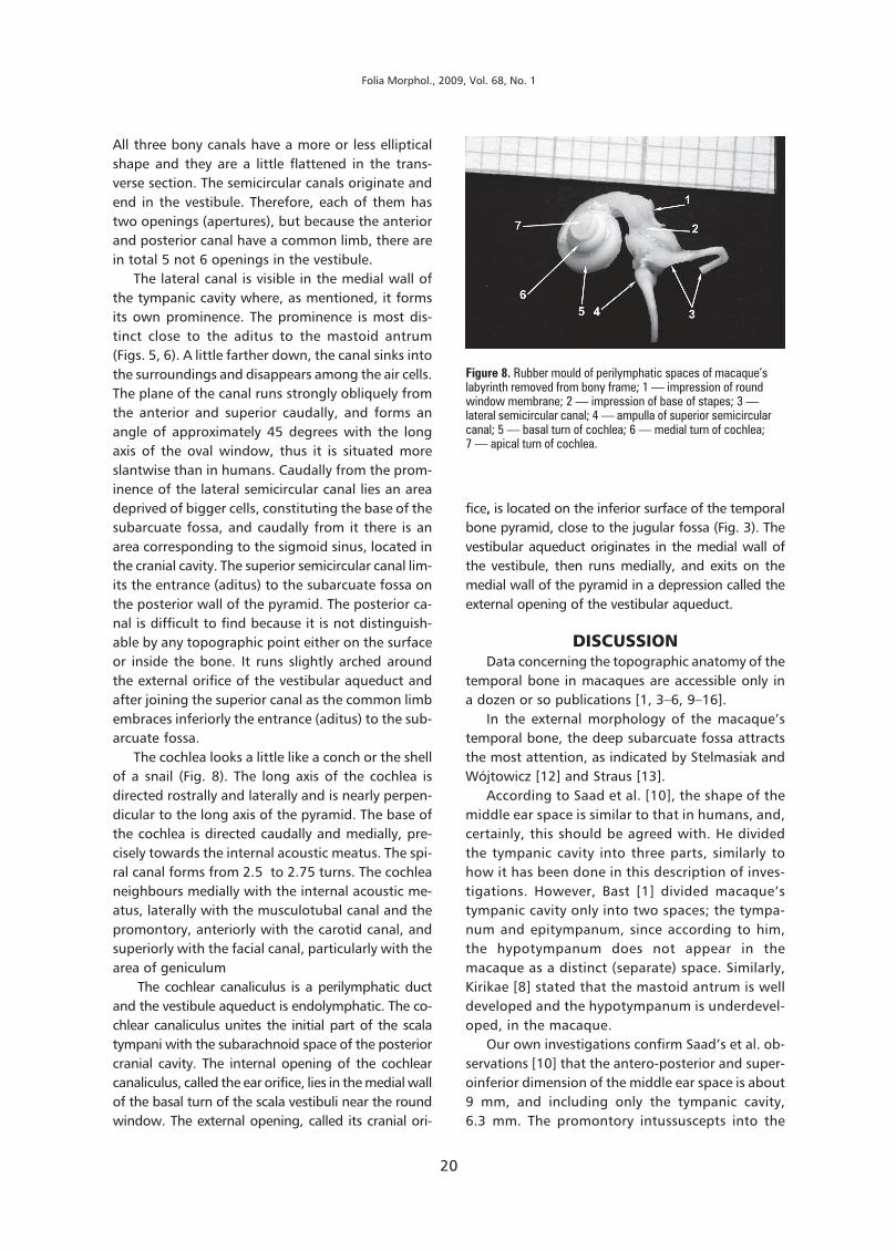
The cochlea looks a little like a conch or the shellof a snail (Fig. 8). The long axis of the cochlea isdirected rostrally and laterally and is nearly perpen-dicular to the long axis of the pyramid. The base ofthe cochlea is directed caudally and medially, pre-cisely towards the internal acoustic meatus. The spi-ral canal forms from 2.5 to 2.75 turns. The cochleaneighbours medially with the internal acoustic me-atus, laterally with the musculotubal canal and thepromontory, anteriorly with the carotid canal, andsuperiorly with the facial canal, particularly with thearea of geniculum
The cochlear canaliculus is a perilymphatic ductand the vestibule aqueduct is endolymphatic. The co-chlear canaliculus unites the initial part of the scalatympani with the subarachnoid space of the posteriorcranial cavity. The internal opening of the cochlearcanaliculus, called the ear orifice, lies in the medial wallof the basal turn of the scala vestibuli near the roundwindow. The external opening, called its cranial ori-
fice, is located on the inferior surface of the temporalbone pyramid, close to the jugular fossa (Fig. 3). Thevestibular aqueduct originates in the medial wall ofthe vestibule, then runs medially, and exits on themedial wall of the pyramid in a depression called theexternal opening of the vestibular aqueduct.
DISCUSSIONData concerning the topographic anatomy of the
temporal bone in macaques are accessible only ina dozen or so publications [1, 3–6, 9–16].
In the external morphology of the macaque’stemporal bone, the deep subarcuate fossa attractsthe most attention, as indicated by Stelmasiak andWójtowicz [12] and Straus [13].
According to Saad et al. [10], the shape of themiddle ear space is similar to that in humans, and,certainly, this should be agreed with. He dividedthe tympanic cavity into three parts, similarly tohow it has been done in this description of inves-tigations. However, Bast [1] divided macaque’stympanic cavity only into two spaces; the tympa-num and epitympanum, since according to him,the hypotympanum does not appear in themacaque as a distinct (separate) space. Similarly,Kirikae [8] stated that the mastoid antrum is welldeveloped and the hypotympanum is underdevel-oped, in the macaque.
Our own investigations confirm Saad’s et al. ob-servations [10] that the antero-posterior and super-oinferior dimension of the middle ear space is about9 mm, and including only the tympanic cavity,6.3 mm. The promontory intussuscepts into the
Figure 8. Rubber mould of perilymphatic spaces of macaque’slabyrinth removed from bony frame; 1 — impression of roundwindow membrane; 2 — impression of base of stapes; 3 —lateral semicircular canal; 4 — ampulla of superior semicircularcanal; 5 — basal turn of cochlea; 6 — medial turn of cochlea;7 — apical turn of cochlea.

21
J. Wysocki, The temporal bone of the macaque...
tympanic cavity, narrowing it down to a width of1 mm. As far as the shape and dimensions of thetympanic membrane are concerned, there are somecontroversies indicated by investigators. Accordingto Saad et al. [10], the macaque’s tympanic membraneis almost a round disc with both its diameters equalto about 5.8 mm.
Also according to Saad et al. [10], the narrowexternal acoustic meatus (diameter 2.5–3 mm) wid-ens medially. One should agree with this statementwhich indicates that this meatus is relatively verylong and potentially makes all surgical interventionsthrough this approach more difficult. On the otherhand, the vicinity of the meatus with the mastoidantrum, and through the atrium, with the lateralsemicircular canal, allows for relatively easy accessto the latter without causing any major surgical in-jury.
According to Saad et al. [10], the oval window isbeanlike in shape and measures about 1.8 by 0.5 mm,which is in accordance with our observations. How-ever, the value of the oval window height given bySaad is lower in comparison with our observations.The round window is a little longer in its horizontaldiameter than in the vertical, measuring 1.5 mm and1.2 mm, respectively, which is close to own mea-surements.
Our own observations concerning the vestibularaqueduct and cochlear canaliculus are in accordancewith Neiger’s [9] description.
The spiral canal of cochlea, according to previ-ous studies, amounts to an average of 21 mm [14].
The course of the carotid canal is quite similar asin humans. According to Sikorska-Piwowska et al. [11],the external opening of the canal measures 1.11––2.4 mm (average 2 mm). The carotid canal in themacaque runs in two phases: the ascending part andthe horizontal part. The angle between themamounts to about 80–119 degrees (average 89.6 de-grees).
Some authors underline the good pneumatisa-tion of the macaque’s temporal bone. The air cellsspread from the mastoid process to the apex of thepyramid including the tegmen of the tympanic cav-ity and its fundus [6, 10].
It is impossible to agree with the statement thatthere is no subiculum of promontory in the macaque;therefore, the access to the round window is easy [6].In my opinion, the access is easy because there is nopromontory bridge, thus there is no natural limit(border) of the tympanic sinus superiorly: as it flat-tens and gradually declines dorsally.
According to Saad et al. [10], the malleus isabout 6 mm, and the author gives separately thedimension of the head (2 mm) and the handle(4 mm). It is a little more than the dimension cal-culated in these studies. The difference probablycomes from overlaying of the head and handle di-mensions measured, in a way twofold, by Saadet al. [10]. The size of the body of incus is 3 ¥ 2.5 mm.The long limb is 2 mm long, which gives a highervalue, by around 4–5 mm, as the total length ofthe incus, compared with our measurements. Thefull length of the stapes given by Saad et al. [10]is about 2 mm, falling into the upper range of valuesobtained in this investigation.
The mastoid atrium, mastoid cells, petrous bul-la, and apex of the pyramid cells belong to the sys-tem of the middle ear air cells. It is worth notingthat none of the investigators mentions the clearlyisolated (distinct) mastoid antrum as an element ofthe middle ear in the macaque. Indirectly related tothis is a mention by Saad et al. [10], who noticedthat 2/3 of the 9 mm long middle ear is occupied bythe tympanic cavity. The pneumatic cells are unusu-ally developed, are a different size and shape, andare surrounding and fixing the carotid canal with itsbony walls, which, in a way, is suspended amongthem like inside a tracery construction designed byan engineer. Saad et al. [10] called one of them thepneumatic cell of the pyramid, because it is particu-larly big and accompanies the auditory tube canalalong its full length; however, it was encounteredonly once during this study.
The macaque’s auditory tube has many similari-ties with that of humans, as indicated earlier [3, 4].
The temporal canal and the postglenoid foramendo not always appear in the macaque, as confirmedearlier in our own investigations [15].
In conclusion, it should be stated that a num-ber of evident similarities in the structure of thetemporal bone in the macaque and in humansallows us to ascertain that the morphology ofboth species is based on an identical spatial plan.Knowledge of those spatial relations enables safeplanning of experiments on the middle or inter-nal ear in the macaque. Substantial differencesbetween both species depend on different pneu-matisation of the temporal bone, morphologyand length of the external acoustic meatus, andthin bony walls of the labyrinth, which makes theuse of the macaque’s temporal bone as a modelfor experiments and trainings in otosurgery im-possible.

22
Folia Morphol., 2009, Vol. 68, No. 1
REFERENCES1. Bast TH (1971) The eye and ear. In: Hartman CG, Straus LW
eds. The anatomy of the rhesus monkey (Macaca mu-latta). Hafner Publ Comp, New York, pp. 339–358.
2. Burnett PA, Miller JM, Mangham CA (1984) Intra-auralreflexes elicited by a cochlear prosthesis in monkeys.Hear Res, 16: 175–180.
3. Doyle WJ, Rood SR (1979) Anatomy of the auditorytube and related structures in the rhesus monkey (Maca-ca mulatta). Acta Anat (Basel), 105: 209–225.
4. Doyle W, Rood SR (1980) Comparison of the anatomyof the auditory tube in the rhesus monkey (Macaca mu-latta) and man implications for physiologic modeling.Am J Otol, 89: 49–57.
5. Doyle WJ, Webster DB (1991) Neonatal conductive hearingloss does not compromise brainstem auditory function andstructure in rhesus monkeys. Hear Res, 54: 145–151.
6. Kelemen G (1968) Non-experimental aural pathologyin rhesus monkeys (Macaca mulatta). Acta Otolaryn-gol, 66: 399–408.
7. Kesser BW, Hashisaki GT, Spindel JH, Ruth RA, Scheld WM(1999) Time course of hearing loss in an animal modelof pneumococcal meningitis. Otolaryngol Head NeckSurg, 120: 628–637.
8. Kirikae I (1963) Physiology of the middle ear. Arch Oto-laryngol, 78: 317–323.
9. Neiger M (1968) Zur Morphologie und Physiologie desAquaeductus cochleae. Experimentelle und morpholo-gische Untersuchungen beim Rhesusaffen. Experimen-telle Untersuchung über die Herkunft der Perilymphe.Forstchr. Hals-Nasen-Ohrenheilk, 15: 113–126.
10. Saad MM, Doyle WJ, Gest TR (1982) Morphology ofthe middle ear in the rhesus monkey (Macaca Mulat-ta). Acta Anat, 112: 117–130.
11. Sikorska-Piwowska Z, Kukwa A, Łukaszewska-Otto H,Kozłowski P, Kryst-Widźgowska T, Giza A (1985) Thecarotid canal in rhesus monkey (Cercopithecidae).Folia Morphol, 44: 33–38.
12. Stelmasiak M, Wójtowicz Z (1972) Doły i jamy czaszkiu Macacus rhesus. Folia Morphol, 31: 1–10.
13. Straus WL (1960) The subarcuate fossa in primates. AnatRec, 138: 93–103.
14. Wysocki J (2001) Dimensions of the vestibular and tym-panic scalae of the cochlea in selected mammals. HearRes, 161: 1–9.
15. Wysocki J (2002) The morphology of the temporal ca-nal and postglenoid foramen with reference to the sizeof the jugular foramen in man and selected kinds ofanimals. Folia Morphol, 61: 199–208.
16. Wysocki J (2008) Topographical anatomy and measure-ments of selected parameters of the rat temporal bone.Folia Morphol, 67: 111–119.