topic 7 the discovery of dna & its roles october 7-14, 2005 biology 1001
TRANSCRIPT

Topic 7The Discovery of DNA
& Its RolesOctober 7-14, 2005
Biology 1001

Introduction to DNA DNA is deoxyribonucleic acid, a polymer of the four
nucleotide monomers adenine, guanine, cytosine, & thymine Arranged as a double helix
DNA is the molecule of heredity DNA is precisely replicated during cell division Each cell in the organism has a copy and a copy is transmitted from
parent to offspring
DNA encodes the amino acid sequences of all the proteins in an organism All biochemical, anatomical, physiological, and behavioural
characteristics of organisms are at least partially determined by DNA

Science as a process
A series of classic experiments elucidated the molecular basis of inheritance
Griffith (1928) – The genetic material transforms bacteria
Avery & MacLeod (1944) – The transforming agent is DNA
Hershey & Chase (1952) – DNA is the hereditary material of viruses
Watson & Crick (1953) – Modeled the 3D structure of DNA
Meselson & Stahl (1958) – DNA replication is semi-conservative
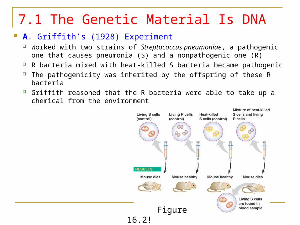
7.1 The Genetic Material Is DNA A. Griffith’s (1928) Experiment
Worked with two strains of Streptococcus pneumoniae, a pathogenic one that causes pneumonia (S) and a nonpathogenic one (R)
R bacteria mixed with heat-killed S bacteria became pathogenic The pathogenicity was inherited by the offspring of these R bacteria Griffith reasoned that the R bacteria were able to take up a chemical from
the environment
Figure 16.2!
7.1 The Genetic Material Is DNA Griffith called this process transformation, but he did not
know what the transforming chemical was
Avery and colleagues spent 14 years testing various chemicals from the S bacterial remains to see which would transform nonpathogenic bacteria into pathogenic ones (R into S) Only DNA worked
Their discovery was met with considerable skepticism because it was widely held that proteins were a better candidate for the genetic material
The question than became whether this finding was generalizable to other organisms, or specific to bacteria
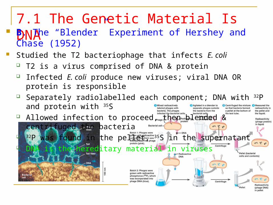
7.1 The Genetic Material Is DNA B. The “Blender” Experiment of Hershey and Chase (1952) Studied the T2 bacteriophage that infects E. coli
T2 is a virus comprised of DNA & protein Infected E. coli produce new viruses; viral DNA OR protein is responsible Separately radiolabelled each component; DNA with 32P and protein with 35S Allowed infection to proceed, then blended & centrifuged the bacteria 32P was found in the pellet, 35S in the supernatant DNA is the hereditary material in viruses

7.2 The Discovery of the Model of DNA What was known to Watson & Crick
DNA is the hereditary material DNA is a polymer of nucleotides
Nucleotides contain a pentose sugar (deoxyribose), a phosphate group, and a nitrogenous base (A,C,G, or T)
A & G are purines, C & T are pyrimidines The sugars and phosphates form the backbone of
the polymer and give it directionality The proportion of each base varies from species
to species – first evidence of diversity of DNA But in each species, the proportion of adenine
equals that of thymine, and the proportion of guanine equals that of cytosine
These are Chargaff’s rules A=T and G=C

7.2 The Discovery of the Model of DNA The challenge was to devise a 3D structure that would account
for DNA’s role in inheritance Watson had also seen Rosalind Franklin’s X-ray diffraction data
suggesting that the molecule was helical and wide enough to accommodate two strands of DNA – a double helix
Franklin reasoned that the sugar phosphate backbone faced outward, allowing the hydrophobic bases to occupy the interior of the molecule
What remained to be determined was the specific pairing of bases holding the two strands together

7.2 The Discovery of the Model of DNA
• In 1953, Watson and Crick reported their model in a one page paper to Nature entitled Molecular Structure of Nucleic Acids– Its key feature was the arrangement of bases between the two strand
strands of the helix
– Watson and Crick proposed that adenine paired with thymine, and guanine with cytosine, with hydrogen bonds between bases holding together the two strands of the helix
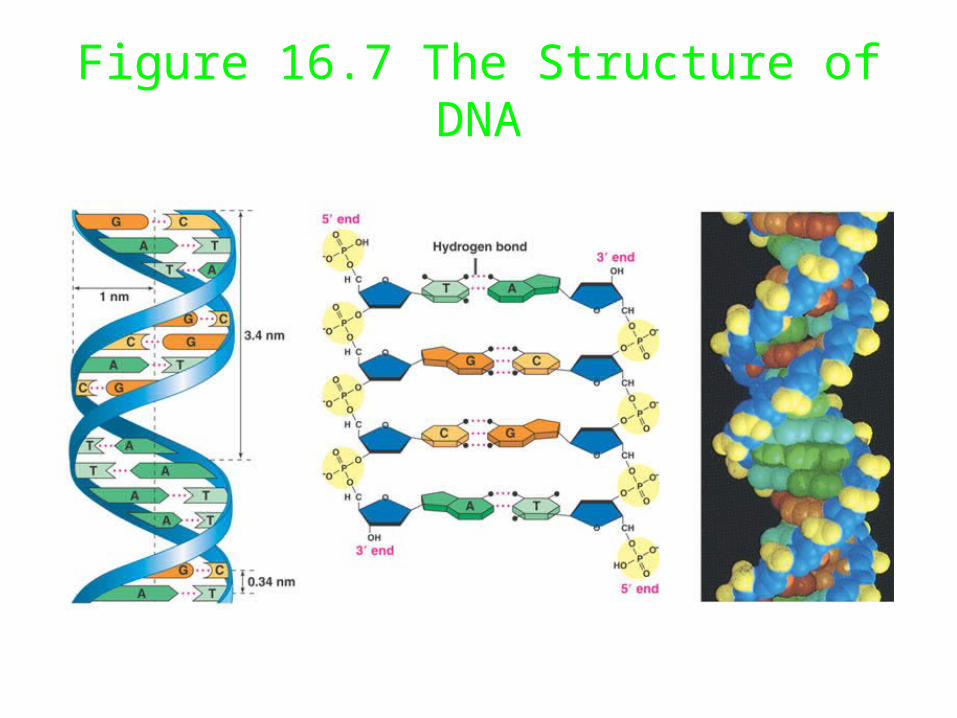
Figure 16.7 The Structure of DNA
Review - The 3D Structure of DNA
• Features of the model
– Two strands of DNA arranged in a double-helix, antiparallel with the bases inward and the sugar phosphate backbone outward
– The two strands are held together by hydrogen bonding between base pairs, A with T and C with G
– The helix is “right-handed” with a 3.4 nm spacing between adjacent turns of the helix, and a 0.34 nm spacing between adjacent base pairs
– The model fits the X-ray diffraction data and Chargaff’s rules