to print ---- pathguy memory aid
DESCRIPTION
group of images and pictures to aid memoryTRANSCRIPT
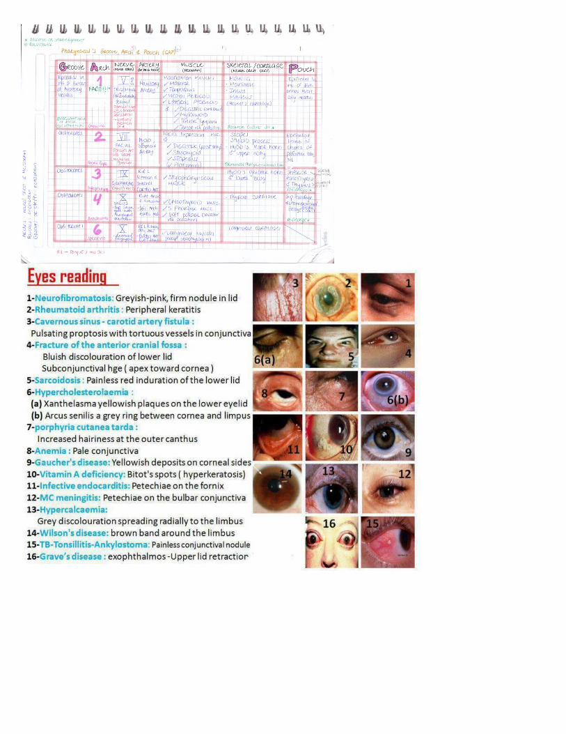
Neurofibromatous type 1(Von Recklinghausen's Disease) Autosomal dominat Findings: cafe-au-lait spots(french-coffe with milk pigmented birth marks), neural tumors, Lisch nodules (pigmcnletl iris hamartomas). Also marked by skeletal disorders (e.g., scoliosis), optic pathway gliomas, pheochromocytoma, and T tumor susceptibility. On long arm of chromosome 17 mutated NF-gene; 17 letters in von Recklinghausen
Neurofibromatosis type 1(Von Recklinghausen's Disease) 1)Lisch nodules (pigmcnletl iris hamartomas)2)optic pathway gliomas
Neurofibromatosis type 2(Autosomal dominat inheritance)Bilaleral acoustic neuroma, juvenile cataracts. NF 2 gene on chromosome 22;type 2 = 22.

Tuberous sclerosis(inheritance: AD)Findings: facial lesions (adenoma sebaceum), hypopigmenled "ash leaf spots" on skin, cortical and retinal hamartomas, seizures, mental retardation. renal cysts and renal angiomyolipomas, cardiac rhabdomyomas, ↑ incidence of astrocytomas. Incomplete penetrance, variable presentation.
von Hippel-Lindau disease(Inheritance: AD)Findings: hemangioblastomas of retina/cerebellum/medulla; about half of affectedindividuals develop multiple bilateral renal cell carcinomas and other tumors.Associated with deletion of VHL , gene (tumor suppressor) on chromosome 3 (3p).Results in constitutive expression of HIF (transcription factor) and activation ofangiogenic growth factors. Vou Hippel-Lindau = 3 words for chromosome 3.
Sturge-Weber syndromeCongenital vascular disorder that affects capillary sized blood vessels.with port-wine stains (aka nevus flammeus-"birthmark", typically in V1(ophlhalmicbranch of cranial nerve V (trigeminal)ophthalmic distribution; ipsilateral leptomeningeal angiomas(AVM)-pia mater vessels overlying occipilal and parietal lobes, pheochromocytomas.Can cause glaucoma, seizures, hemiparesis, and mental retardation. Occurs sporadically.

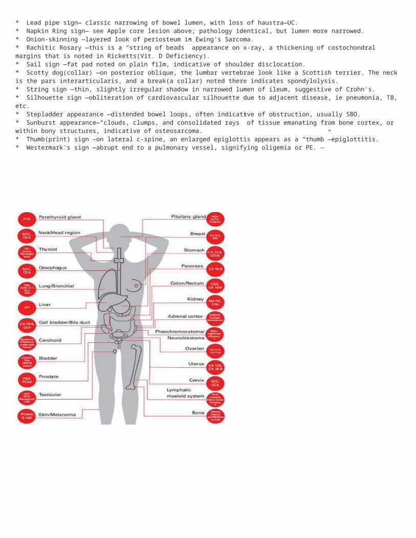
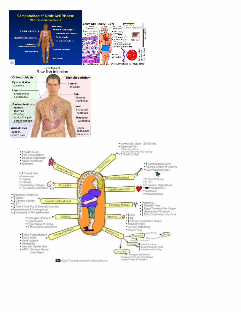
MEN
Multiple endocrine neoplasias (MEN) Autosomal Dominant-Several distinct syndromes (1, 2A, 2B) characterized by familial tumors of endoerineglands, including those of the pancreas, parathyroid, pituitary, thyroid, and adrenal medulla. MEN 2Aand 2B are associated with ret gene.
MEN 1 (Wermer's syndrome)-Parathyroid lumors Pituitary tumors (prolactin or GH) Pancreatic endocrine tumors —Zollinger-Ellison syndrome, insulinomas, VIPomas. glucagonomas (rare) Commonly presents with kidney stones andstomach ulcersMnemonic:MEN I = 3 P's (Pancreas, Pituitary, and Parathyroid) MEN 2A (Sipple's syndrome) Medullary thyroid carcinoma (secretes calcilonin)Pheochromacytoma, Parathyroid lumorsMnemonic: MEN2A = 2P's 1 Pheochromocytoma(neuroendocrine tumor of the medulla of the adrenal glands) and Parathyroid.MEN 2B Medullary thyroid carcinoma (secretes calcilonin),PheochromocylomaOral/intestinal gauglioneuromatosis (associated with marfanoid habitus)
Mnemonic: MEN 2B = 1 P MEN 2A
MEN 1
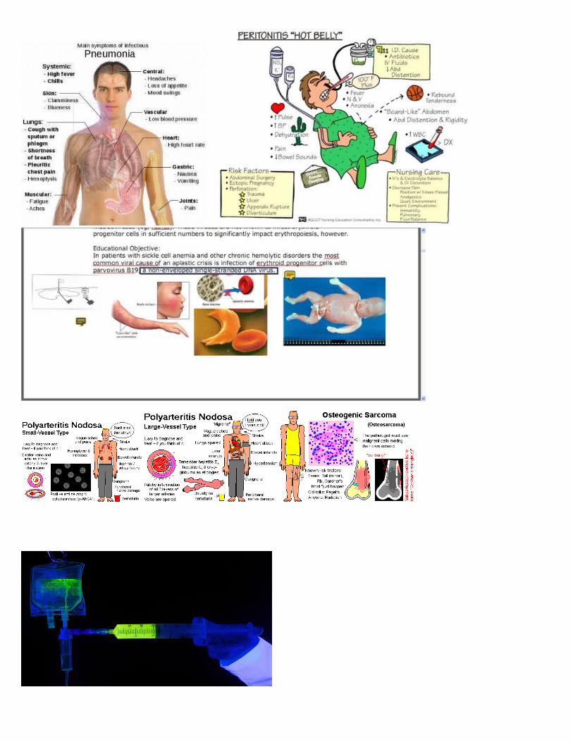
Hereditary Hemorrhagic telangiectasia (Osler Weber-Rendu syndrome)Autosomal dominant inherited disorder of blood vessels, Findings: telangietiasia, recurrent epistaxis. skin discollorations, arteriovenous malformations (AVMs)
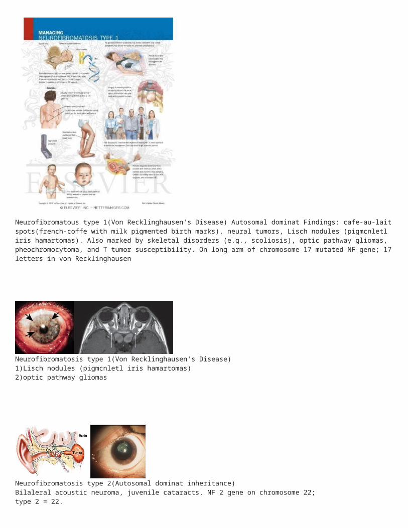
Familial hypercholeslerelemia (hyperlipidemia type ll A)-Autosomal dominatElevated LDL due lo defective or absent LDL receptor. Heterozygotes (1:500) have cholesterol = 300 mg/dL. Homozygotes (very rare) have cholesterol=700+ mg/dL, severe atherosclerotic disease early in life, and tendon xanthomas (classically in the Achilles tendon); MI may develop before age 20.
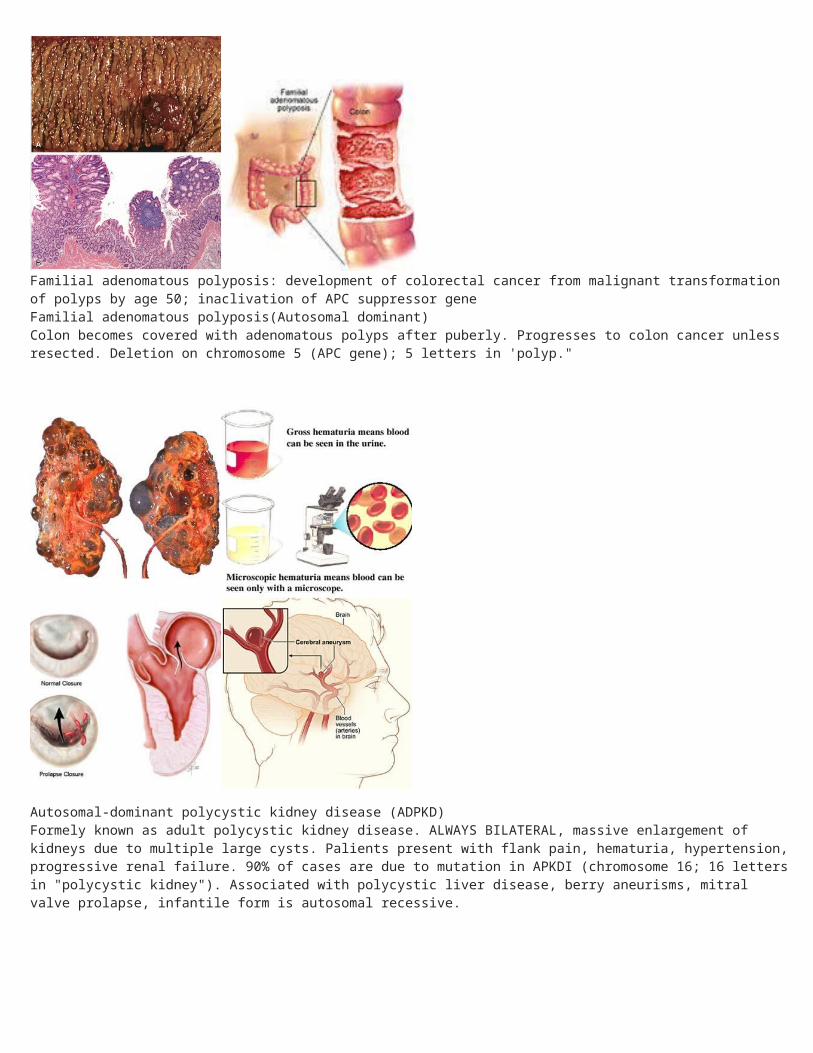
Familial adenomatous polyposis: development of colorectal cancer from malignant transformation of polyps by age 50; inaclivation of APC suppressor geneFamilial adenomatous polyposis(Autosomal dominant)Colon becomes covered with adenomatous polyps after puberly. Progresses to colon cancer unless resected. Deletion on chromosome 5 (APC gene); 5 letters in 'polyp."
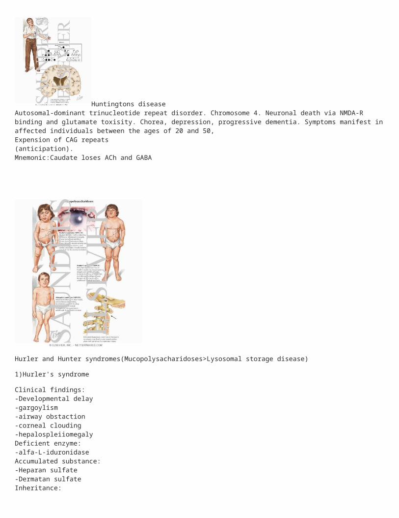
Autosomal-dominant polycystic kidney disease (ADPKD)Formely known as adult polycystic kidney disease. ALWAYS BILATERAL, massive enlargement of kidneys due to multiple large cysts. Palients present with flank pain, hematuria, hypertension, progressive renal failure. 90% of cases are due to mutation in APKDI (chromosome 16; 16 letters in "polycystic kidney"). Associated with polycystic liver disease, berry aneurisms, mitral valve prolapse, infantile form is autosomal recessive.
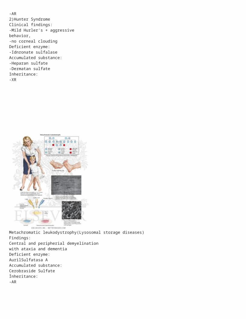
Huntingtons diseaseAutosomal-dominant trinucleotide repeat disorder. Chromosome 4. Neuronal death via NMDA-R binding and glutamate toxisity. Chorea, depression, progressive dementia. Symptoms manifest in affected individuals between the ages of 20 and 50,Expension of CAG repeats(anticipation). Mnemonic:Caudate loses ACh and GABA
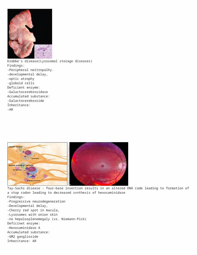
Hurler and Hunter syndromes(Mucopolysacharidoses>Lysosomal storage disease)
1)Hurler's syndrome
Clinical findings:-Developmental delay-gargoylism-airway obstaction-corneal clouding-hepalospleiiomegalyDeficient enzyme:-alfa-L-iduronidaseAccumulated substance:-Heparan sulfate-Dermatan sulfateInheritance:-AR2)Hunter SyndromeClinical findings:-Mild Hurler's + aggressivebehavior, -no corneal cloudingDeficient enzyme:-Idnronate sulfalaseAccumulated substance:-Heparan sulfate-Dermatan sulfateInheritance:-XR
Metachromatic leukodystrophy(Lysosomal storage diseases)Findings:Central and peripherial demyelinationwith ataxia and dementiaDeficient enzyme:AurilSulfatasa AAccumulated substance:Cerobraside Sulfateİnheritance:-AR
Krabbe's disease(Lysosomal storage diseases)Findings:-Peripheral neitropalhy.-developmental delay,-optic atrophy-globoid cellsDeficient enzyme:-GalactocerebrosidaseAccumulated substance:-Galactocerebrosideİnheritance:-AR
Tay-Sachs disease - four-base insertion results in an altered DNA code leading to formation of a stop codon leading to decreased svnthesis of hexosaminidaseFindings--Progressive neurodegeneration-Developmental delay,-Cherry red spot in macula,-Lysosomes with onion skin-no hepalosplenomegaly (vs. Niemann-Pick)Deficinet enzyme:-Hexosaminidase AAccumulated substance:-GM2 gangliosideInheritance: AR
Niemann-Pick disease(Lysosomal storage diseases)Findings:-Progressive neurodegeneration. -hepatosplenomegaly-cherry red spot on macula.- foam cellsDeficient enzyme:-Sphingomyelinase↓Accumulated substance:-Sphingomyelin ↑Inheritance: AR

Gaucher's disease(most common Lysosomal storage diseases)Findings:-Hepatosplenomegaly-aseptic necrosis of femur.-bone crises. Gaucher's cells(macrophages that look likecrumpled tissue paper)Deficient enzyme:-Beta-glucocerebrosidaseAccumulated substarate:-GlucocerebrosideInheritance:-AR(Autosomal ressesive) Gaucher's cells (macrophages that look like crumpled tissue paper)
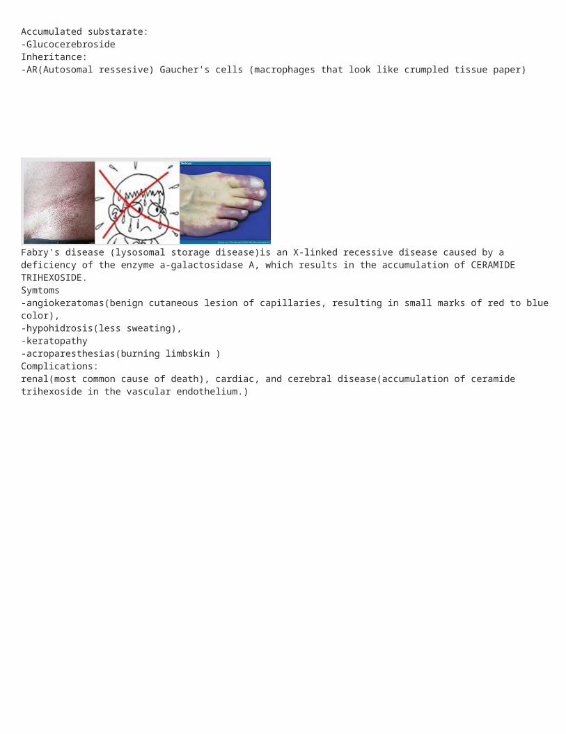
Fabry's disease (lysosomal storage disease)is an X-linked recessive disease caused by a deficiency of the enzyme a-galactosidase A, which results in the accumulation of CERAMIDE TRIHEXOSIDE. Symtoms-angiokeratomas(benign cutaneous lesion of capillaries, resulting in small marks of red to blue color), -hypohidrosis(less sweating), -keratopathy-acroparesthesias(burning limbskin ) Complications:renal(most common cause of death), cardiac, and cerebral disease(accumulation of ceramide trihexoside in the vascular endothelium.)
Classic Radiology Signs for All hope u like it :)
* Apple core lesion or Napkin Ring sign ¬¬¬- in right hip synovial chondromatosis ... same can be viewed in colonic Ca.... seems like eaten apple.* Bamboo Spine —fused spinal segments with their syndesmophytes look, on radiographs, similar to bamboo stalks—classically associated with ankylosing spondylitis.* Bird's Beak— noted on Upper GI with contrast, a dilated upper/middle esophagus with an abrupt taper to exceptionally narrowed lumen, typical of achalasia.* Boot-shaped Heart —due to RVH, the LV is lifted above the edge of the diaphragm, forming the “toe” of the boot. Classic for Tetralogy of Fallot.* Bat's Wing/Butterfly —this appearance on CXR is classically associated with CHF and resultant pulmonary edema.* Cobblestone appearance— this sign is produced on barium studies due to ulcerative pockets, usually in the terminal ileum, indicative of Crohn's.* Codman's Triangle —a triangle on plain film of extremities that signifies reactive bone, classically associated with osteosarcoma, or other infectious/hemorrhagic process that causes periosteal elevation.* Coin lesion— solitary pulmonary nodule; may be cancer or granuloma.* Cookie Cutter lesions— metastatic lesions to bone cortex, or Paget's.* Crescent sign— classic sign of avascular necrosis, femoral head.* Egg-on-a-string— a large, ovoid-shaped heart on newborn CXR, classically signifying complete transposition of the great vessels with intact ventricular septum.* Ground glass— a “white-out” on CXR, usually PCP pneumonia or ARDS.* Hampton's Hump— a peripheral triangle, usually near pleural edges, classically PE.* Honeycomb lung— used to describe any pathologic process that causes radiographic appearance of multiple small, thick-walled cystic spaces; e.g. pulmonary fibrosis.* Lead pipe sign— classic narrowing of bowel lumen, with loss of haustra—UC.* Napkin Ring sign— see Apple core lesion above; pathology identical, but lumen more narrowed.* Onion-skinning —layered look of periosteum in Ewing's Sarcoma.* Rachitic Rosary —this is a “string of beads” appearance on x-ray, a thickening of costochondral margins that is noted in Ricketts(Vit. D Deficiency).
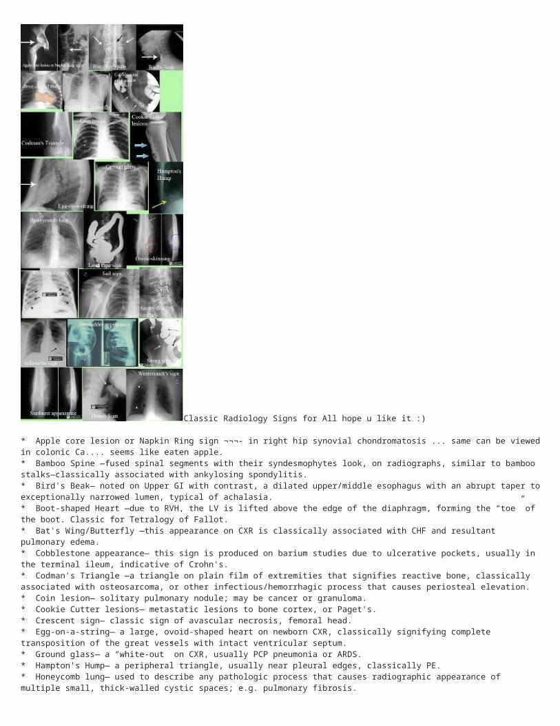
* Sail sign —fat pad noted on plain film, indicative of shoulder disclocation.* Scotty dog(collar) —on posterior oblique, the lumbar vertebrae look like a Scottish terrier. The neck is the pars interarticularis, and a break(a collar) noted there indicates spondylolysis.* String sign —thin, slightly irregular shadow in narrowed lumen of ileum, suggestive of Crohn's.* Silhouette sign —obliteration of cardiovascular silhouette due to adjacent disease, ie pneumonia, TB, etc.* Stepladder appearance —distended bowel loops, often indicative of obstruction, usually SBO.* Sunburst appearance—“clouds, clumps, and consolidated rays” of tissue emanating from bone cortex, or within bony structures, indicative of osteosarcoma.* Thumb(print) sign —on lateral c-spine, an enlarged epiglottis appears as a “thumb”—epiglottitis.* Westermark's sign —abrupt end to a pulmonary vessel, signifying oligemia or PE. —