to or through the skin: a rational design of transdermal
TRANSCRIPT

University of Texas at El Paso University of Texas at El Paso
ScholarWorks@UTEP ScholarWorks@UTEP
Open Access Theses & Dissertations
2020-01-01
To Or Through The Skin: A Rational Design Of Transdermal Drug To Or Through The Skin: A Rational Design Of Transdermal Drug
Delivery Delivery
Isaac Giovanni Deaguero University of Texas at El Paso
Follow this and additional works at: https://scholarworks.utep.edu/open_etd
Part of the Biomedical Commons
Recommended Citation Recommended Citation Deaguero, Isaac Giovanni, "To Or Through The Skin: A Rational Design Of Transdermal Drug Delivery" (2020). Open Access Theses & Dissertations. 2955. https://scholarworks.utep.edu/open_etd/2955
This is brought to you for free and open access by ScholarWorks@UTEP. It has been accepted for inclusion in Open Access Theses & Dissertations by an authorized administrator of ScholarWorks@UTEP. For more information, please contact [email protected].
TO OR THROUGH THE SKIN: A RATIONAL DESIGN
OF TRANSDERMAL DRUG DELIVERY
ISAAC GIOVANNI DEAGUERO
DOCTORAL PROGRAM IN BIOMEDICAL ENGINEERING
APPROVED:
Md Nurunnabi, Ph.D., Chair
Thomas Boland, Ph.D.
Taslim Al-Hilal, Ph.D.
Fatima Z. Alshbool, Ph.D.
_________________________
Stephen Crites, Ph.D.
Dean of the Graduate School

DEDICATION
To my mother
who used to sit with me when I was a child to watch Sesame Street.
I only sat there and watched the show because I wanted to be near you.
This simple act inadvertently taught me the awesome power of an education
and started me on the path

TO OR THROUGH THE SKIN: A RATIONAL DESIGN
OF TRANSDERMAL DRUG DELIVERY
By
ISAAC GIOVANNI DEAGUERO B.S.
DISSERTATION
Presented to the Faculty of the Graduate School of
The University of Texas at El Paso
in Partial Fulfillment
of the Requirements
for the Degree of
DOCTOR OF PHILOSOPHY
DEPARTMENT OF METALLURGICAL MATERIALS AND BIOMEDICAL ENGINEERING
THE UNIVERSITY OF TEXAS AT EL PASO
May 2020

iv
ACKNOWLEDGEMENTS
I would like to thank my committee members for their patience and consultation.
Specifically, Dr. Thomas Boland, who saw potential in someone who had passion, but lacked
direction, and honed him into someone who can make the world better. For that I am eternally
grateful. Thank you Dr. Fatima Alshbool for encouraging me to be the best person I can be. Your
confidence in me is not in vain. I represent the derivation of your teachings and do my best in that
manner. Thank you Dr. Taslim Al-Hilal, for being approachable, direct, and straightforward. These
qualities often go unnoticed, but these innate portions of your character constantly guide me in the
right direction.
I want to thank my mentor Dr. Md Nurunnabi, who has constantly gone above and beyond
to offer assistance in countless ways. You are a perennial source of positive motivation and endless
knowledge, who has given me the confidence to unabatingly move forward with my projects. I
thank God that you have blessed me by allowing you into my life. You do so much more than your
job requires, and I am incredibly fortunate to call you a mentor, colleague, and friend. You are
what I strive to emulate.
To my lab mates Huda, Jade, and Chris; An anagram of the word “dissertation” is
“satirist”. You have all taught me that this is not a coincidence.
To my family, who have always believed and encouraged me to be the best I can be. I could
not ask for more. I am so lucky. My grandmother, Linda; you constantly remind me that I am my
fathers’ son, and that he has always been with me; I needed that reminder on many occasions. I
think he would have liked that his son would carry his last name with the preceding title of Doctor.
v
Thank you, Ms. Porth, my 3rd grade teacher, for pulling me aside one day and telling me
that you believed I was the smartest student she had ever had. That one sentence changed the way
I looked at myself and set me on this path. Thank you, Mr. Flickinger, for giving me a failing grade
on the first paper I ever turned into your class. You wrote the words “Step up or step out” and
taught me that simply being smart not good enough; effort is what makes the difference in life.
Thank you, Andrew Collins, for teaching me that there is a time to be weak and vulnerable,
and that it is nothing to be ashamed of. Thank you, Melvyn Naidas, for teaching me the real
definition of true strength. Both of you in many similar and different ways taught me how to be a
man.
To my friends, especially Brian, Joe, Matthew, and Sabin, I would not have been able to
do this without all of you. You all know me better than myself sometimes and have offered a
guiding light in my darkest hrs. I did not do this myself; we did it together.
Thank you, Rex, for reminding me that if I was gone, there would be no one to take care
of you. You saved my life.
There are so many people who I have not mentioned individually here who have shaped
who I am, and in-turn have made me a better person. I have only named a few who immediately
come to mind, but there are countless more.
Robert Goddard once wrote "It is difficult to say what is impossible - for the dream of
yesterday is the hope of today and the reality of tomorrow"[1]. This frame of mind is not to be
taken lightly. When combined with his unbridled tenacity, his technology propelled mankind to
the moon. His naive dreams became our reality.

vi
As a member of the millennial generation, my inspirations have not been founded in 19th
century scientists, but rather of those based in the 23rd. Fiction, specifically the television show
“Star Trek” opened my eyes to an optimistic future, where a small rectangular handheld device
known as a “tricorder” could be used to communicate anywhere, translate, and even diagnose
disease. Sadly, the smartphone was already a well-established tool of my youth, so I had to look
for ulterior messages within the subtext of Gene Roddenberry’s world. I found my inspiration in
Bones.
Leonard “Bones” McCoy was the ships doctor. Though, he did not practice medicine all
that often. His character would usually shine a laser or apply a salve that would almost
instantaneously heal the wounds of those in need. The power to heal was not based on years of
practice to improve mending abilities, rather Bones himself was simply a man with incredible
power; the power flowed through his hands from the tools he utilized. Technology was his only
limit. Through this realization, I inadvertently discovered my tricorder.
A doctor is limited by the technology of his generation. Even the most well-meaning
physicians in history were restricted in their practice by their rudimentary understanding of
concepts we now find to be the foundations of medicine. Examples like antibiotics, blood pressure,
germ theory, or even something as simple as sanitary technique, were so beyond the realm of
understanding, even as early as 150 years ago, that death rates when compared to today’s standards
would be considered abhorrent. It is the responsibility of modern researchers to ensure that our
practices today are viewed in the future with the same distaste as we see within our ancestors. This
can only be accomplished through modest and gradual advances in technology which will further
our understanding of the natural world. My personal contribution to this progress may be

vii
considered somewhat ambitious, focusing primarily on the largest organ of the human body, the
skin.
To answer these questions is by no means a small task; it is an endeavor that has consumed
many researchers, myself included. However, incremental progress, inspiration through the dream
world of science fiction, and the relentless optimism of those seeking a better tomorrow has led to
several discoveries that offer those with ambition and yearning, the keys to overcoming our current
limitations, and opens a window with a view of a brighter future with better treatments and
possibilities just over the horizon of our current moment.
“If you know the enemy and know yourself, your victory will not be in doubt; if you know
Heaven and know Earth, you may make your victory complete” -Sun Tzu
Most importantly of all, thank you to our God, the spiritual creator with many names.
Thank you for the gift of life; I do my best to not waste something so precious.

viii
ABSTRACT:
The skin is the largest organ in the body and plays many crucial roles through diverse
functions ranging from protection, temperature regulation, energy storage via fat deposits, and is
used to communicate with our surroundings. Various methods have been attempted to deliver
drugs to target areas of the skin in effective dosages by a variety of means with limited success.
Recently, advances in transdermal drug delivery have offered avenues previously unattainable
through conventional means. Some of these new delivery methods have been reviewed, explored
and offer promising results with the potential to carve a new avenue in the way therapeutic
substances may be consumed.
In one study, a novel miconazole-ufosome nano carrier was characterized, the formulation
was optimized in approximately a <300 nm diameter lipid carrier, and at the same time achieved
an entrapment efficiency of 62%. In-vitro permeation studies displayed significant targeted
transdermal drug delivery to the site of infection in higher amounts than conventional marketed
creams, and antifungal studies showed that the current formulation greatly enhanced the efficacy
of antifungal activity when compared to standalone miconazole while displaying low toxicity and
high biocompatibility.
In another study, we have developed a combination of choline and octenoic acid ionic
liquid with an experimental anticancer BCI-2 inhibitor, ABT-263 (navitoclax) to combat
melanoma by delivering the drug directly to the site of infection. This formula was characterized
and optimized for loading capacity, size, and stability. Cell internalization was achieved at higher
levels due to the penetrative capabilities of the ionic liquid, while toxicity remained low. Skin
permeation studies displayed significantly higher penetration into the target site, displaying
capabilities previously unattainable for non-invasive transdermal drug delivery.

ix
TABLE OF CONTENTS
Page
DEDICATION................................................................................................................................ii
ACKNOWLEDGEMENTS............................................................................................................iv
ABSTRACT.................................................................................................................................viii
TABLE OF CONTENTS................................................................................................................ix
LIST OF TABLES..........................................................................................................................xi
LIST OF FIGURES.......................................................................................................................xii
CHAPTER 1
1. INTRODUCTION ......................................................................................................................1
2.1 TO THE SKIN: WAYS WE DELIVER THERAPY TO THE SKIN LAYERS......................9
2.2 THROUGH THE SKIN: WAYS WE DELIVER THERAPY TO THE BLOODSTREAM..23
BIBLIOGRAPHY/LIST OF REFERENCES ...............................................................................29
CHAPTER 2
1. ABSTRACT ............................................................................................................................. .39
2. INTRODUCTION.....................................................................................................................40
3. MATERIALS & METHODS....................................................................................................44
4. RESULTS & DISCUSSION.....................................................................................................50

x
5. CONCLUSIONS.......................................................................................................................57
BIBLIOGRAPHY/LIST OF REFERENCES ...............................................................................59
CHAPTER 3
1. ABSTRACT ............................................................................................................................. .65
2. INTRODUCTION ............................................................................................................. .......66
3. MATERIALS & METHODS....................................................................................................67
4. RESULTS & DISCUSSION.................................................................................................... .73
5. CONCLUSIONS.......................................................................................................................82
BIBLIOGRAPHY/LIST OF REFERENCES ...............................................................................83
DISSERTATION CONCLUSIONS .............................................................................................86
CURRICULUM VITA .................................................................................................................87

xi
LIST OF TABLES:
Table 1.1: Overview of Transdermal Drug Delivery Systems…………………………………..25
Table 2.1: Display of the various ratios of miconazole, cholesterol, and sodium oleate used in
each formulation. Miconazole was used as the determinant anchor to maintain consistency with
testing, and to offer a support when comparing with Actavis brand marketed cream…………...44
Table 2.2: Skin samples used for diffusion studies. Samples were taken from the same locatio2n
(abdomen) and with similar scarring (severe), however the age, gender, and ethnicity were
different between the samples. This was done purposely to determine if the intake of miconazole
vesicles would act differently when alternative factors were introduced, and to compare these
potential differences with those of a marketed cream (Actavis). ………………………………..47
Table 2.3: Each petri dish was examined for 48 hr by observing the amount of dead space between
the drug and fungal colony. The total area of the petri dish was measured using ImageJ software
by NIH and using CAD software35. The ImageJ calculation has been tabulated………………... 49

xii
LIST OF FIGURES:
Figure 1.1. Skin diseases and their potential origin locations within the various layers…………..3
Figure 1.2. Design of micro-reservoir transdermal patch………………………………………..13
Figure 1.3. Transdermal delivery approach offers one of the most convenient ways of drug
delivery to or through the skin. The proposed ionic liquid mediated transdermal delivery is very
unique in terms of biosafety and the ability to transport to the dermis of the layer of the skin…….16
Figure 2.1. Miconazole loaded nano-vesicles offer a higher delivery of drug content through the
epidermis to the target site of fungal infection. The high lipid content of the nano-vesicles allows
the drug to be carried through the stratum corneum and offers a previously unattainable avenue of
drug delivery in high content for lipophilic nano-sized drugs…………………………………….43
Figure 2.2. Size, zeta potential, and encapsulation efficiency amongst the various ratios
of miconazole:cholesterol:sodium oleate. (A) Size distribution and (B) zeta potential remained
relatively similar, however high variation between vesicles eliminated consideration for 1:1:1 and
1:2:2 due to an inordinate amount of vesicles stretching beyond the nano-range. (C) Encapsulation
efficiency amongst the various ratios eliminated several formulations from consideration due to
poor performance in relation to drug delivery, while two ratios (1:0.5:1 and 1:1:0.5) conversely
displayed exceptional loading potential. All data are presented as mean ± SE (n = 3)……………51
Figure 2.3. Viscosity testing was performed at various temperatures to determine optimal
temperature during administration, as well as to see if any major change occurred at temperatures
similar to contact with human skin. Viscosity remained relatively constant throughout the
spectrum, maintaining between 355-405 CPS. All data are presented as mean ± SE (n = 3)……...52

xiii
Figure 2.4. (A) Diagram of the Permegear Franz-diffusion cell system. PBS is warmed to 37°C
before being pumped into the donor compartment, underneath the skin, and finally resides in the
scintillation vial, which was changed hourly. (B1, B2) UV-Vis comparing the amount of
miconazole located within the Stratum Corneum and lower dermal layers after 24 hrs in
the Permegear diffusion system. (C1, C2) Hourly readings of the PBS collected scintillation vials,
identifying the amount of drug that may pass completely through the skin and into the bloodstream
within 24 hrs. All data are presented as mean ± SE (n = 6)………………………………………..54
Figure 2.5. Fungal growth inhibition using free miconazole, as well as nano-vesicles at 1% and
2% miconazole. PBS was used as a control. (A) Significantly higher levels of inhibition were
expressed using nano-vesicles as opposed to free miconazole, supporting the notion that nano-
vesicles act as an inhibition enhancer. (B) Around 20% and 40% fungal colonies were inhibited
with the 1% and 2% miconazole nano-vesicles treatment respectively. All data are presented as
mean ± SE (n = 8)………………………………………………………………………………...56
Figure 2.6. Cell survival rates at varying time points. 2% miconazole nano-vesicles displayed
lower cell viability at all time points when compared with 1% miconazole nano-vesicle and 1%
free miconazole. All data are presented as mean ± SE (n = 9)…………………………………….57
Figure 3.1. The IL/navitoclax formulation was evaluated to determine size, polydispersity, zeta
potential, and maximum load…………………………………………………………………….73
Figure 3.2. Polydispersity of navitoclax loaded IL varies at different time points within a 10-hour
span. Increased time displayed higher diversity as well, displaying instability in the
suspension………………………………………………………………………………………..75

xiv
Figure 3.3. Potential of ionic liquid to enhance cellular uptake of a therapeutic molecule. The
feasibility study was conducted with Coumarin 6, a hydrophobic small molecule with fluorescence
properties, as a model molecule. The images show enhanced uptake of coumarin 6 by the A375
cell when co-incubated with ionic liquid compared to free coumarin 6, upon 4 hr of incubation...76
Figure 3.4. Cytotoxicity of ionic liquid, navitoclax and formulation was investigated with A375
cell upon incubation for various time points. Ionic liquid itself does not show any sign of toxicity
whereas both free navitoclax and ionic liquid containing navitoclax show 70% cell death after 6hr
of co-incubation. Data represent mean±SD, where n=6………………………………………….78
Figure 3.5. To investigate the diffusion ability of ionic liquid and potential as transdermal drug
delivery carrier, we have performed a diffusion study with pig skin. The results show absorbance
values of coumarin 6 within the stratum corneum is as much as 2 folds higher than PBS and free
coumarin 6. Data represent mean±SD, where n=6………………………………………………79
Figure 3.6. To investigate the diffusion ability of ionic liquid potential as transdermal drug
delivery carrier, we have performed a diffusion study with pig skin. The results show
that absorbance readings of coumarin 6 within the epidermis layer is as much as 3 folds higher
than PBS and free coumarin 6. Data represent mean±SD, where n=6……………………………80
Figure 3.7. To investigated diffusion ability of ionic liquid and potential as transdermal drug
delivery carrier, we have investigated diffusion study with pig skin. The results show that
concentration of coumarin 6 within the dermis layer is 2 folds higher than Stratum Corneum and 5
folds higher than epidermis. Data represent mean±SD, where n=6……………………………...81
Figure 3.8. The photograph shows coumarin 6 in different layers of the skin after diffusion…….82

1
CHAPTER 1:
A REVIEW OF CURRENT SPECIFIC DISEASES AND THERAPIES
Introduction
The skin is the most easily accessible organ in the body, however due to the strong diffusive
properties of the skin, access to this avenue for drug delivery offers a plethora of challenges that
are all too often circumvented through painful or minimally effective pathways [1] This limited
scope of options has been persistent since the dawn of our more current forms of modern medicine.
Recently however, several novel approaches have been published with assorted levels of success
[2]. Often, due to toxic drug effects, targeted drug delivery must be implemented to ensure proper
amounts of therapy is delivered to a specific area, while also minimizing contact with areas not
affected by the disease. Hepatoxic materials must not only avoid direct contact with liver cells, but
also must avoid systemic circulation [3]. Hemotoxic substances must avoid contact with blood
altogether, severely limiting delivery options [4].
This skin serves a variety of purposes that includes temperature regulation, energy storage
via fat deposits, and preventing crossing the toxic substances across the skin. Most importantly,
skin acts as a barrier, protecting vital organs and processes from exterior threats, and is absolutely
necessary to maintain a healthy internal homeostatic environment.
The outermost layer of the skin is composed of a thick layer of protein known as keratin.
This building block is joined by a moist layer of fat which is interwoven between protein deposits.
This layer is known as the stratum corneum, also called the “horny layer” or in shorthand, the SC
layer. Below this layer is the lucidum, granulosum, spinosum, basale, and the dermis. Each deeper

2
layer considered easier to penetrate than the previous due to the looser configuration of the cells
[5]. This proprietary structure delineates the SC as the main culprit in terms of penetration. The
lifespan of a human keratin cell ranges depending on age from 14-90 days, being slowly pushed
out over time, offering some recompense in terms of vulnerability.
This understanding naturally leads one to ask the question: What is to happen to the body
when its protector is the system that is damaged? Aside from minor lacerations, burns, or
intrusions, the answer is often death. The skin being the largest organ of the human body makes it
a prime target for attempted infiltration. The unforgiving nature of nature itself, has honed the
organ into one that offers much resilience, adaptivity, and strength when presented with adversity.
However, skin is not invincible. Like all organs, the passage of time through the slow
breakdown of DNA through methylation and telomere shortening eventually takes a serious toll
on the barrier. Protection from harmful radiation becomes less effective, elasticity is hindered and
therefore sections no longer bounce back into shape as easily [6]. Tumors fester, and the
malignancy of cancer welcomes death through uncontrolled growth. Other genetic betrayal can
manifest in this organ as well, through the continuous creation of scars from scleroderma, which
can be classified as anything from a minor inconvenience, to a death sentence [7]. External threats
such as fungus, can penetrate, feed, and attack vulnerable areas [8].
Often these areas are quite difficult to treat, mainly due to the diffusive properties of the
skin, which only allow. The skin has the ability to protect itself from external threats, even if the
invader is not necessarily dangerous. Depravity is not a prerequisite for exclusion, so even the
most desirable medicines and treatments are refused admittance through the barrier. This
indiscriminate behavior has been the culprit for healers since the creation of the first salve. How
does one get to or through the skin? How does one create treatments that target something that

3
nature designed specifically to be impenetrable? How can we save the barrier that has a primary
function of protecting us from the exact type of physical invader we hope to emulate?
In this dissertation, an amalgamation of various delivery methods has been
comprehensively gathered to determine the state of targeted drug delivery within the current scope
of understanding. Several challenging diseases, such as melanoma, skin cancer, and scleroderma,
have been lain out as examples of the criteria necessary to structure new targeted drug delivery
systems specifically residing in skin layers. These layers often require the delivery of high
molecular weight drugs which may have undesirable effects in off-target areas, while concurrently
requiring a high dosage for maximum efficacy [9]. These diseases emphasize many of the
challenges and realistic expectations that will drive therapies out of the laboratory, and into the
market.
1.1 Skin disease and their location within the various layers
Figure 1.1: Skin diseases and their potential origin locations within the various layers.

4
Basal Cell Carcinoma
Basal Cell Carcinoma (BSC) is one of the most common types of cancers. The cancer
begins in the basal cells, a cell within the lowest layers of the epidermis, which is responsible for
the production of new skin cells [10]. Most cases of BSC are contracted by long term exposure to
UV radiation or sun exposed parts of the body. People with fair skin are more than likely to contract
it more than other races. Most cases of basal cell carcinoma occur in older adults beyond the age
of 45, but BSC can also be seen in younger people who are exposed to abnormal amounts of
radiation from the sun, such as seasonal farmers, truck drivers, and others that spend copious
amounts of time in direct sunlight [11]. BSC is mostly found on the head and neck, however other
body parts such as genitals can be infected as well [12]. More than 3 million cases are reported in
the United States annually. The primary therapies used to treat BSC is by the use of targeted
radiation, copious application of topical drugs, and surgical excision [13].
Squamous Cell Carcinoma
Squamous Cell Carcinoma (SCC) is also a common form of skin cancer [14]. This cancer
develops in the squamous cells in the middle and outer layers of the skin. Squamous cells can be
found all throughout the body, so the disease can occur anywhere. The cancer is not usually life
threatening but it can be aggressive if left untreated. Most cases of SCC results from high levels
of UV light from tanning beds or prolonged time in the sunlight [15]. All races are able to contract
Squamous Cell Carcinoma, Caucasians are more likely to contract the disease. More than 1 million
cases per year are diagnosed with 15,000 annual deaths. Treatment of SCC is almost identical to
BCC [16].

5
Melanoma
Malignant melanoma, commonly referred to simply as melanoma, is relatively the most
serious type of skin cancer when compared to BSC and SCC. This cancer develops in the cells that
produce melanin. Melanoma can form outside of conventional skin areas, with reports of
melanoma originating in the eyes, under the nails, in the mouth, and rarely in the digestive tract or
other internal organs. Melanoma can be diagnosed in areas of the skin that are not exposed to the
sun, such as spaces between the toes and the genitals [17]. As for the other causes of skin cancers,
exposure to UV light and sunlight can be the cause for the cancer [18]. The risk of melanoma in
people under 40 is increasing, especially in women [19]. Melanomas are harder to recognize on
people with darker skin. It is still a common factor that Caucasian people have a higher risk of
contracting the cancer [20]. More than 200,000 US cases are reported per year. Melanoma can be
treated depending on the stage of the cancer. In treating stage 1, the melanoma is removed
(excision) and the sample is sent to a lab for histological analysis. In other cases, the melanoma
can be treated by the use of anti-tumor medication such as imiquimod or Mohs surgery, which is
essentially an exploratory excision process that leads to large scarring [21]. The choice of treatment
method is largely dependent on the infected area. Treating stage 1 melanoma starts with the
excision of the infected area, moving on to a sentinel lymph node biopsy [22]. If the SLNB comes
back negative, there is no further treatment. If the SLNB is positive, removal of the lymph nodes
will take place or monitoring of the lymph nodes and surrounding areas for growth will be
suggested. Additional treatment can be provided with targeted chemotherapeutics [23]. Treating
stage 2 melanoma approaches the same steps as treating stage 1, but with more aggressive steps to
ensure no further spread of the cancer [24]. In stage 3 of melanoma, the cancer has already infected
the lymph nodes, the removal of the tumor and the lymph nodes will happen as well as in the other

6
stages. Targeted therapeutic agents and/or radiation can be used to prevent the cancer from
recurring [25]. Depending on the location of the cancer, there are different treatment options. If
tumors are found in the lymph nodes or just under the skin, they can be removed or injections of
T-VEC vaccine [26] Basile Calmette-Guerin vaccine, or interleukin can be directly injected into
the melanoma, as well as radiation [27]. For melanomas on the arms and legs, an option could be
infusing the limp with chemotherapy [28]. When treating stage 4 melanoma, which is also known
as metastases stage, the cancer has already spread through the body and lymph nodes, the tumors
are sometimes removed depending on the placement, if they are cancerous, and the quantity of
individual tumors. Immunotherapeutic drugs (checkpoint inhibitors) are used in those, whose
cancer does not have BRAF gene changes. The use of the immunotherapeutic drugs allows for the
tumor to shrink but there are serious side effects [29]. If the cancer cells have BRAF cell changes,
then immunotherapy drugs such as pembrolizumab can be used to shrink the tumor or have a
greater chance of shrinking them. Chemotherapy is commonly used in patients that have stage 4;
the cancer shrinks but there is a possibility that the tumors can continue to grow that increases risk
of spreading the cancer to other organs including liver, and kidney.
Scleroderma
There are two different types of scleroderma: localized and systemic. Localized
scleroderma only affects the skin, causing the skin to become thick and hard. Systemic scleroderma
is a more serious type of the disease that can affect skin, muscles, joints, blood vessels, lungs,
heart, and other organs. Scleroderma is a rare disease, but 75,000 to 100,000 people in the United
States are currently affected [30]. Twins and those with a family history of scleroderma or
autoimmune disease are more likely to be afflicted [31]. There is no actual known cause for

7
scleroderma, but genetic factors and certain chemical exposure may play a factor in the
development of the disease.
No two cases are alike, so the identification of the stage, type, and which organs are
involved all play a factor in deciding proper treatment [32]. Although no cases are alike, the main
features are the similar; inflammation, autoimmunity, vascular disease, and tissue fibrosis are all
indicators of the disease [33]. Inflammation medications are used to treat the symptoms of the
specific case in order to alleviate pain, and as a direct form of pain management. These anti-
inflammatory medication treatments can treat the various forms of arthritis, myositis, and other
serositis based problems [34]. Immunosuppressive therapy is the most popular approach for
symptom alleviation. The most promising drugs are mycophenolate and cyclophosphamide in
treating aggressive cases of scleroderma [35]. Drug therapy of vascular disease allows for the drugs
to treat the medium and small arteries that the disease infects [36]. Treatment of the vascular disease
is crucial in order to control the disease and to prevent organ damage. Anti-fibrotic agents are used
to reduce collagen production or reduce tissue collagen [37]. Colchicine, PABA, dimethyl
sulfoxide, and D-penicillamine are the common drugs used to treat excess collagen being produced
due to scleroderma [38, 39]. Experts believe that these drugs do not have enough potency to take
effect or benefit the patient. The search for new drugs to treat the fibrotic reaction is one of the
most active areas of research for scleroderma. Navitoclax, a chemotherapeutic agent used to treat
melanoma, is one of these potential promising agents. However, the high molecular weight and
hemotoxic qualities of the drug make conventional delivery methods unsustainable in effective
quantities.

8
Tinea Corporis
Tinea Corporis, commonly known as ringworm, is a rash contracted by a fungal infection.
This dermatophyte feeds directly on the keratin layer of skin. The contagious fungal infection is
spread by a parasite that lives on the outer layer of the skin. Ringworm can be spread from direct
contact from humans, animals, objects, and the soil. people who live in warm climates, work with
animals or participate in contact sports (such as wrestling or martial arts) are more likely to contract
the diseases than others living a more secluded lifestyle. This common disease affects more than
3 million US cases per year, but globally many mild cases are not reported. Ringworm can usually
be treated with non-prescription antifungal creams, lotions, or powders. These azoles are used in
order to treat fungal infections without having to see a physician, however patient compliance is
very low. Most antifungal medications are over the counter and easy to access. The typical
application period is multiple times a day for 4 or more weeks. This leads to a lack of patient
compliance, and eventually consistent recurrences of the infection which leads to more medical
issues, and/or scarring of the affected area.
Athletes foot is a common fungal infection that usually starts between the toes. People
whose feet become sweaty from tightfitting shoes are more likely to contract the infection. The
infection is caused by the same type of fungus that causes ringworm. Men are at higher risk of
contracting athletes’ foot than women. Frequently wearing sweaty socks or tightfitting shoes
allows for the fungus to grow due to damp conditions. Poor hygiene and sharing infected linens
can cause the fungus to spread from person to person. more than 3 million US cases are reported
annually.
Although there are countless skin infections and diseases, the aforementioned ailments tend
to offer a diverse insight into the various forms of treatment necessary to adequately produce

9
proper treatment regiments, with respect to time, area, infection, and levels of seriousness
associated with various diseases. With these targets in mind, one must realize that a large toolbox
of potential treatments is vital for the proper implementation of disease control; ones that may
target a wide variety of areas, and allow proper penetration, as well as accurate and precise targeted
drug delivery.
To the skin
Various strategies have been attempted for targeted drug delivery to the skin with mixed levels of
success, but with each offering limitations to counter certain obstacles. We will examine each of
these potential drug delivery methods, as well as their advantages and disadvantages. Each
historically, has their fair share of trials and errors.
Transdermal patch
This form of topical treatment allows for specific areas of the skin to be treated. There are
four different types of transdermal patch delivery systems; membrane permeation-
controlled systems, matrix diffusion-controlled systems, adhesive dispersion-type systems, and
Microreservoir types Transdermal delivery allows for the medication to move through the skin,
usually by ensuring sustained contact through synthetic patches held in place on the skin by glue.
This uncomfortable and inconvenient form of delivery is necessary to ensure permeation of the SC
layer. These patches release a specific dose of medication through the skin, usually controlling
drug release through a hydrogel. The advantages to using this type of system include enabling the
10
avoidance of gastrointestinal absorption, consistent dosage rather than peaks in medication levels
commonly associated with other forms of delivery, and the capacity to immediately terminate drug
dosage (and potentially effects) via immediate removal of the patch. Disadvantages include
potential irritation of the skin, a lack of universal consistency to skin types when considering
adherence, discomfort in certain areas of application, high cost per dose, and the inability to
administer high amounts of the drug into the bloodstream due to the size of the patch and the low
amount of drug that can be present on each individual patch. Importantly, the major limitation to
transdermal patch is that it welcomes a very high potential chance for leakage [40], concluding that
the administration of treatment may not be sufficient; as well as the fact that individuals may be
allergic to the adhesive material. These patches release a specific dose of medication through the
skin, usually controlling drug release through a hydrogel.
Membrane permeable control system
A membrane permeable control-transdermal drug delivery system (TDDS) of isosorbide
dinitrate (ISDN), the model drug is prepared and mixed with polyvinyl alcohol aqueous gel. The
membrane consists of ethylene vinyl copolymer membrane and acrylic adhesive [55]. These
membranes have some advantages: (i) Prolonged action owing to sustained drug release [56]. (ii)
Similar drug delivery both in vivo and in vitro model [57]. (iii) Prevention of high plasma
concentration in case of skin damage [58]. Advantages of this system are only for high permeable
drugs, but these functions have limited application. This form of delivery is desirable for a wide
variety of drug applications. Function achieved is independent of skin condition and application
site. In this application, skin permeation rate of the drug should be high enough to allow systematic
pharmacological application. High skin penetration can be achieved by the use of penetration

11
enhancer such as ethanol, medium chain ester, or Azone [59]. However high levels of penetration
through the use of this method may cause inflammation. If the skin barrier function is remarkably
decrease due to inflammation or placement over a scar, drug delivery rate will be still equal to the
permeation rate through the controlling membrane and the plasma concentration should be
maintain below the toxic concentration [55], however this may manipulate the dosage. Another
transdermal patch application has been created entirely out of collagen. One major advantage of
this system is that collagen membrane is very skin friendly and significantly reduces skin damage.
However, collagen membranes could not be used as such for an application without stabilizing it
by crosslinking [60]. With traditional crosslinking agents like glutaraldehyde, the potential to cause
some adverse side effect is raised. To overcome such limitations, a polycationic biopolymer
chitosan form stable composite with collagen was created [61]. In this study, authors propose
collagen-chitosan membrane for transdermal drug delivery using the lipophilic drug nifedipine,
and a hydrophilic drug, propranolol hydrochloride (prop-HC1) [60].
Matrix diffusion control system
Verapamil hydrochloride is a calcium ion influx inhibitor. It is widely used in the treatment
of angina, hypertension, and supraventricular tachyarrhythmias [62]. It is suitable for transdermal
delivery owing to its low molecular weight (491.07) and low melting point (144˚ c). Transdermal
patches of verapamil hydrochloride were prepared using four different polymers (individual and
combination): Eudragit RL100 (ERL100), Eudragit RS100 (ERS100), hydroxypropyl
methylcellulose 15 cps (HPMC), and ethyl cellulose (EC), of varying degrees of hydrophilicity
and hydrophobicity. Authors use both water-soluble polymers like hydroxypropyl methylcellulose
(HPMC) and water insoluble polymers, ERL 100, ERS 100, and ethylcellulose (EC), were

12
selected as carriers into which the drug was incorporated [63]. In another study, authors prepared,
designed, and evaluated matrix diffusion controlled transdermal patches of verapamil
hydrochloride. Dexibuprofen is chemically (2S)-2-[4-(2-ethylpropyl) phenyl] propanoic acid and
used as an NSAID for management of pain and inflammation associated osteoarthritis. In this
study, matrix transdermal patches prepared with dexibuprofen exhibited better drug permeation
[64]. In another study authors demonstrate a matrix diffusion patch containing rosin: Eudragit RL
PM in the ratio 6:4 with 5%w/v of camphor achieved the desired objectives of transdermal drug
delivery systems, which according to the study were overcoming of first pass metabolism effects,
extended release, and reduced frequency of administration [65].
Micro reservoir type system
The drug reservoir is made of a homogenous dispersion of drug particles suspended in an
unleachable viscous liquid medium (e.g. Silicon fluids) to form a paste like suspension, gel, or a
clear solution of drug in a releasable solvent (e. G. Ethanol) [66]. The drug reservoir formed is
sandwiched between a rate controlling membrane and backing laminate. The rate controlling
membrane can be nonporous so that the drug is released by diffusing directly through the material,
or the material may contain fluid filled micropores in which case the drug may additionally diffuse
through the fluid, thus filling the pores [67]. In the case of nonporous membrane, the rate of passage
of drug molecules depends on the solubility of the drug in the membrane as well as the thickness.
The drug reservoir is formed by suspending the drug solids in an aqueous solution of water
miscible drug solubilizer e.g. Polyethylene glycol. The drug suspension is homogenously
dispersed by a high shear mechanical force in lipophilic polymer, forming thousands of
unleachable microscopic drug reservoirs (micro reservoirs) [62]. This system is exemplified by the

13
development of a nitrodisc. The micro reservoir type transdermal system is shown in the figure 1.2
below:
Figure 1.2: Design of micro-reservoir transdermal patch
Adhesive dispersion type system
The drug and other selected excipients, if any, are directly incorporated into the organic
solvent-based pressure sensitive adhesive solution, mixed, cast as a thin film, and dried to
evaporate the solvents leaving a dried adhesive matrix film containing the drug and excipients [69].
This drug in an adhesive matrix is sandwiched between release liner and backing layer. The drug-
in-adhesive patch may be single or multi-layered. The multi-layered system is different from single
layer in that it adds another layer of drug-in-adhesive, usually separated by a membrane [70]. This
serves to adhere the components of the patch together along with adhering the patch to the skin.
The adhesive must possess enough adhesion property so that the TDDS should remain in place for
an extended period of time. Pressure sensitive adhesives are commonly used for transdermal
patches to adhere to the skin. Commonly used adhesives are silicone [69], polyisobutylenes, and
poly acrylate-based adhesives [71].

14
The mechanism of the transdermal patch has been stated by Prausnitz and Langer (vii) that
these patch-based transdermal drug delivery systems have yet to achieve its potential as an
alternative to oral and hypodermic injection. With that stated, much work is still expected to take
place in this field to improve upon these techniques for delivery.
Electrophoresis
Electrophoresis is a method used to separate DNA fragments as well as other
macromolecules (RNA) according to the size of their particles. A current is run through a gel that
contains the molecules that are needed to be separated. The molecules will travel through the gel
at different speeds and directions which allows for them to be separated from each other. The size
of the molecule depends on how the gel will react based on speed and direction. In order for the
molecules to be placed, the gel must be placed in a gel box. The box is hooked up to a positive and
negative electrode which allows for a current to take place. Once turned on, the current flows
through the wells, the gel is running. The gel running towards the positive electrode shows the
fragments of the molecules, as well as the lengths.
This same type of technology can be utilized on the skin by placing electrodes on the outer layer
of skin. This electric effect may allow small molecules to travel through the skin layer through the
pores, or by accelerating movement through the lipo-sections of the keratin layer. This process
may be effective with low molecular weight drugs, however these size limits are far from its
detractions. This type of electrical shock may permanently alter the skin, causing hair loss,
inflammation, and destruction of the skin pores.

15
Ultrasound
Ultrasound therapy has been a beneficial use for therapeutic purposes since the 1950s.
Ultrasound therapies were originally used for the treatment of tendinitis and bursitis. As utilization
became more advanced, the use of ultrasound therapy became beneficial for various alternative
ventures such as uterine fibroid ablation, cataract removal, surgical tissue cutting, hemostasis,
transdermal drug delivery, and in the acceleration of bone fracture healing. Therapeutic ultrasound
therapy uses short bursts to deliver beneficial energy to the tissues. Ultrasound heat induced
therapy uses the absorption of ultrasonic energy in the tissues. Temperature elevations are usually
kept at low to limit the temporal average intensities as well as exposure to the heat. Applications
of heat induced ultrasonic therapy use long durations of heat with unfocused beams or higher
intensity focused bursts. Unfocused heating commonly used in physical therapy is used to treat
highly absorbable tissues such as the bones or tendons. The use of the unfocused heating is to
produce healing without injury.
Therapeutic ultrasound uses an electric signal and a handheld transducer. The transducer is applied
with a coupling gel and moved into a circular motion over the injured area. The transducer is
commonly used to treat bursitis or tendonitis to accelerate healing. The coupling gel can also be
beneficial to the area of treatment, allowing for the transport of the compound into the skin.
Commonly used drugs such as lidocaine or cortisol have been used during ultrasonic therapy.
Although there is a chance of burns from the ultrasound therapy, it is still considered to be a benefit
for patients as well as being effective. Ultrasound therapy has been founded to be proficient
because of its capability to focus energy several millimeters away from the transducer plane.
Before using ultrasound therapy, it is important to accurately determine the location of the
treatment. As a transdermal delivery system, ultrasound works in a similar manner as

16
electrophoresis, and has similar benefits. Ultrasound heating can cause irreversible tissues changes
based on the heating, coagulative necrosis, and tissue vaporization.
Figure 1.3: Transdermal delivery approach offers one of the most convenient ways of drug
delivery to or through the skin. The proposed ionic liquid mediated transdermal delivery is very
unique in terms of biosafety and the ability to transport to the dermis of the layer of the skin.
Light based therapies
Photoactivated approaches to transdermal therapy have attracted interest among several
research communities. Light based drug delivery has the ability to enable precise control in a
highly spatial and temporal manner. The spiral and temporal manner allows for a controlled release
of drug molecules. Several photo responsive systems use ultraviolet (UV) or visible light (400-700
nm) to undergo multiple reaction pathways, such as photocleavage and photoisomerization[73].
Although UV light is used mostly in light activated cancer therapies, UV light (<400 nm) has poor
tissue penetration and high phototoxicity. The potential issues of using UV light has brought upon
the idea of near infrared (NIR) light (700-1000nm) which has been proven to have distinct
advantages amongst cancer therapies. All of the NIR advantages, such as the deep penetration and

17
low cell damage are important for cancer diagnosis and therapies[73]. This type of activation is
useful in drug delivery systems because the manual targeting of drug activation may allow more
precise control of drug delivery underneath the skin after insertion into systemic circulation via
injection or oral introduction. This method could potentially significantly lower
UV light
The commonly used light sources in photoinduced chemotherapy mainly include light
energy at UV or short wavelength range.[52]. There are different kinds of approaches that can be
taken with using UV or short wavelength light energy; photoactivation through photocaged
approach and photoisomerization. The first strategy towards the photoactivated therapy is the
photolysis of photocaged molecules, in which a beam of light will be utilized to activate a
photocleavable functional group that blocks the activities of therapeutic molecules.[52]. Using
photoactivation through the photocaged method achieves the use of a controlled delivery release
system of molecules in a living system. Several photocaged systems are based on chemical groups
such a o-nitro benzyl, pyridylmethyl ester, and coumaroyl ester to regulate biomolecule activities
such as real time monitoring cell trafficking and controlled release of therapeutic molecules. Such
chemically photocaged approaches have also been documented for rational design of
photoactivable bioluminescent probes for the remote control of optical imaging in living
subjects.[52]. Using UV light controlled photocaged strategy has been promising in the usage of
bioimaging but can also be promising for the controlled delivery of specific anticancer regents
including doxorubicin, paclitaxel, and calprotectin.

18
Photoisomerization is a reversible strategy that uses the chemical groups to link with the
carrier system. Using UV exposure, photosensitive moieties could undergo reversible cis-trans
light switches and result in the conformational exchanges between inactive and active states and
thus controlled the drug release from the carriers[52]. Photoisomerization molecules include
azobenzene, spiropyran, and dithienylethene have been studied to be used in conjugation with UV
light triggered drug delivery and imaging.
Visible light:
UV light is the most popular strategy for photo-induced therapeutics[74].UV light has had
initial success in its field but also has widely accepted disadvantages such as poor ability of tissue
penetration, and often have biological damage. In terms that UV light has these disadvantages,
longer wavelength exposure at visible range can be used in order to cause less damage to the
tissues. Visible light may serve as an alternative option to more damaging wavelengths and has
been proposed for light activated drug delivery. Photo responsive organic molecules including
vitamin B12 derivatives, ruthenium complexes, and dithiocarbonates have been reported to be of
use for visible light- triggered release. Several functionalized nanostructures used in drug delivery
are activated by visible light[76].
Near infrared light
Lights with short wavelengths may bare enough energy to induce disruption of chemical
structures by either breaking a covalent bod of causing a cis-trans isomerization but despite their
promising photoactivation applications, there are still some cons; UV light has poor capability of

19
tissue penetration, high autofluorescence background, and potential phototoxicity. The
development of a more reliable alternative that has a high depth tissue penetration and more precise
of photoactivation reaction as well as minimum cellular damage is needed. NIR (700-1000nm)
shows unique advantages toward deep tissue penetration, low fluorescence background and limited
photodamage, the exact opposite of UV light therapy.
Photoactivated drug delivery and bioimaging using NIR light
Photoactivated delivery strategies could enhance therapeutic quality by preventing
nontargeted effect through the spatial and temporal precision of light irradiation. For short
wavelength they can bear enough energy to induce disruption of chemical structures but despite
their promising photoactivation applications, they are shown to have limited tissue penetrating
capability, high autofluorescence, and potential phototoxicity. NIR enables a high tissue
penetration and more precise control of photoactivation reaction as well as minimal cell damage.
NIR provides advantages toward deep tissue penetration, low fluorescence background and limited
photo damage. One common use of NIR is to utilize photosensitive nanoplatforms, converting NIR
light irradiation into UV and spatiotemporally triggering the release of payload reagents into living
systems. Photothermal effect may cause the temperature to increase caused by the light transduced
heat to trigger the drug release from nanostructures to tumor sites.
NIR triggered to therapeutic agents UCNPs can serve as phototherapeutic platforms for the
controlled delivery of drugs using NIR. NIR regions could absorb photoenergy and convert them
to thermal energy. The heat generated by the photoenergy will increase temperatures in the
surroundings which can induce the rapid release of the drug molecules. Gold related nanomaterials

20
have the capability to absorb NIR light and can produce photothermal conversion. Gold based
nanoparticles can be modified with thermosensitive polymers (liposomes, mesoporous silica
shells, and red blood cells have the ability to absorb NIR light and produce photothermal
conversion.
Light mediated therapeutic strategies have recently emerged as a promising and effective
tool to precisely treat cancer as effectively as possible. Using light mediated therapeutic strategies
allows for a patient to receive exceptional care without the high costs commonly associated with
some transdermal therapies. When it comes to cancers, precise surgery and accurate medical
diagnosis is necessary, but with light-based drug delivery medical professionals can do so with
minimal invasiveness. NIR has displayed several notable advantages relating to precision and
trauma, such as deep penetration, low autofluorescence in living cells and tissues, and minimimal
photodamage[73]. Due to the minimized photodamage, the light therapy also has the advantage to
be used more often. Deep penetration allows for the light to go into the skin deeper and have a
more precise control than UV or visible light. The cells can absorb light, too much
autofluorescence can cause the image to be hard to read but with using NIR, low autofluorescence
allows for the cell to be easily read.
Although light mediated therapies have achieved initial success, the conventional strategies
use short wavelengths which can have some undesired drawbacks. UV and visible light both suffer
from light scattering, limited tissue penetration depth, and potential phototoxicity to off-target
damage[75]. UV light cannot travel long distances into the skin in order to treat the cancer
effectively. When using light mediated therapies, the cells can absorb the light which can cause
the cell to absorb too much light or involve the autofluorescence to scatter outside of the cell. High
autofluorescence can cause the image to be hard to read or locate the cell that is being treated.

21
Phototoxicity or photosensitivity; a chemically induced skin irrational from the light can be seen
when using UV or visible light. UV or visible light are not the only types of light mediated
therapies that can have negative side effects but NIR as well. NIR also has some clinical
drawbacks; low water solubility, troublesome synthetic procedures, and limited accumulation
abilities in targeted areas.
Lipid Nanoencapsulation
Another recent advancement has been the prospect of nanoencapsulation of drugs in lipid-
based vesicles for stratum corneum penetration. This strategy has shown promising results in the
delivery of azoles, which are antifungal agents used to treat tinea-based diseases such as jock itch,
athletes’ foot, and ringworm. The drugs are encapsulated in a vesicle containing cholesterol,
sodium oleate, and the antifungal agent. This is accomplished through a combination of
mechanical forces, including intense sonication, and a thorough washing of the product to ensure
safe removal of the more toxic materials such as chloroform and methanol. Upon completion of
this process, the nanovesicles can be added to a variety of creams or gels for simple application
Efficacy of these nanovesicles has been most promising, delivering 2-3x more of the drug to the
target layer and due to the lipid encapsulation, allows the drug to reside in the infected zone for an
extended period of time. This additional time and concentration allow for fewer applications,
driving down the typical 50-60 necessary doses to potentially far less, which would significantly
improve patient compliance and concurrently lower the amount of recurring infections.
The only major disadvantage for this type of application is the molecular size limit that comes
naturally with this type of application. The stratum corneum skin layer consists of hard keratin
22
surrounded by a patchwork “brick and mortar” type of configuration where lipids encompass the
mortar. This mortar will accept other lipids for transport through the layer but must adhere to a
low molecular weight limit.
Ionic liquid
Recent studies involving new avenues have shown promising results involving the use of
ionic liquids for transdermal drug delivery [50]. Macromolecule permeability is increased when
ionic liquids are present. The liquid extracts high amounts of lipids from the SC layer of the skin
and allows for very large molecules, some exceeding 160kda, to permeate the skin. This new
avenue offers promising new alternatives for transdermal drug delivery, one that does not require
nano-level breakdown or encapsulation of drugs, simplifying and expediting what would otherwise
be an endeavor with a meager payoff.
Comparing to IL/drug methods, patients may no longer need to be wary of having to face
particles pressuring their way into the vessel, that will obstruct from a mixture of high pressure
plus nanoparticles crystal formation. Ultimately, the aim for IL/drug method to replication or
mimic these methods can reshape the future of medicinal application where patients no longer
need to fear treatment that is necessary for their pain, cancer treatment, or addiction.
Presenting the flexibility of IL/drug treatment. The ideal mechanism of IL is very simply, as
previously stated macromolecule permeability is increased when ionic liquid is present. To support
this claim in one study [50], the researchers concluded that the IL induces lipid extraction, followed
by skin lipids replaced with IL that allows water to mediate faster through skin pores. Therefore,
from the SC layer of the skin; molecules exceeding 160kDa may permeate through the skin.

23
Further experiments in the near future could open IL potential in controlling how deep a particle
is permitted to travel. As previously mentioned, IL can penetrate through the SC layer, this
potentially can open the discussion if IL can be controlled to deliver drug treatments to the venous
system as well.
IL has convenient applications in vitro, in vivo, and fluorescently-labeled dextrans to study
permeability for both experiment and theoretical [50]. With that said, TDD can successfully deliver
only a few hundred Daltons in molecular mass, versus as previously mentioned IL potentially
exceeding 160kDa. As drug therapy passes through the IL, their molecular structure will not be
compromised[47], due to controlling stability. With these known advantages, high-molecular
weight hydrophobic drugs such as navitoclax and other hemotoxic substances now have a delivery
method that can circumvent systemic circulation [Figure 1.3]. Some studies have reported slight
skin inflammation at the site of application, but no long-term damage has been observed.
1.2 Through the skin: Ways we deliver therapy to the bloodstream
Intravenous injection
Intravenous injection is one of the most popular forms of transdermal drug delivery, which
involves inserting a hollow and sharp metal rod directly into a vein through the skin. The drug is
injected directly into the vein, and dosage is controlled via pressure, as well as the gauge of the
rod, which allows for precise drug concentration in the plasma. Advantages such as immediate
access to the circulatory system, easy routes for patients that cannot tolerate oral medications, and
instant drug action via systemic circulation, make IV injections one of the most popular forms of

24
drug delivery in hospital settings. Despite the advances, there are major disadvantages in regard to
this form of delivery; dispersing the medication too quickly can cause speed shock, and once
infused, the side effects cannot be reversed. This type of danger offers little recompense for
mistakes and can easily lead to serious injury that may ultimately lead to extensive tissue damage,
high pressure injuries, and tearing an aorta or vein. Extravasation can occur when the medication
leaks into the surrounding tissue due to excess pressure. There can also be the negative side effects
of physical, chemical, and therapeutic incompatibly with systemic drug circulation. Many of these
factors can inevitably lead to death. Due to the high possibility of infection, these types of delivery
systems are rarely seen outside of hospital settings, and patient compliance in home settings is low
due to the pain and required precision associated with injections. Though it accomplished its goal
of delivering treatment to patient, by a professionally trained individual is required to understand
how the needle must be administered upon a patient’s skin, with the bolus into the subcutis [49]
Intra-arterial injection
Under certain circumstances with analgesic, antibiotic, antifungal, and anticancer drugs in
an effort to deliver a high concentration of the drug to a particular tissue, the administration of
Intra-arterial injection is a viable option. This method requires the drug treatment to enter the
cerebrospinal fluid via injection; then from the subarachnoid space of the spinal cord it will bypass
the blood-brain barrier, allowing drugs with poor penetration easy access to the central nervous
system. Advantages to using this method include good absorption, a more rapid onset than oral
and rectal routes, and offers a long duration of action. This particular type of injection requires an
experienced professional for proper administration. One study [72]presented intra-arterial drug

25
injection for treating lethal brain diseases. The idea of IA can only be effective when three criteria
is met; (i) when there is low regional blood flow, (ii) high regional extraction, and (iii) high
systemic clearance [72]. Some disadvantages to using this method include the somewhat common
unpredictable absorption, and the painful injections may cause serious swelling or bruising that
may lead to further complications. Previously it has been reported that an accident involving intra-
arterial drug injection caused a sporadic blood vessel deformation[44]. Conclusions drawn from
the authors determined that IA drug injection may cause serious vessel injury, deforming vessels
from the drug’s particles being large enough to disrupt these vessels post-injection.
Table 1.1: Overview of Transdermal Drug Delivery Systems.
Form of Delivery Type of Disease Pros Cons
Ultrasound • Bursitis
• Tendonitis
• Muscle strains
• Sprains
• Joint contracture
• Reduce swelling,
inflammation, and
promote bone
healing
• Accelerate bone
healing time
• Promote tissue
healing
• Noninvasive
• Cannot be used
over cancerous
areas
• Cannot be used on
children, near the
eyes, around the
heart, in people
with vascular
problems

26
Topical • Ring worm
• Athletes foot
• Psoriasis
• Acne
• Eczema
• Fewer risks of
gastrointestinal
difficulties
• Easy to administer
• Irritation
• May not adhere to
all skin types
IV Injection • Cancers
• Heart failure
• Crohns disease
• Immune deficiencies
• Rheumatoid arthritis
• Sclerosis
• Direct access to the
circulatory system
• Instant drug action
• High drug
concentrations
• Instant drug
termination
• Errors with IV
mixing
• Adsorption
• Speed shock
• Chemical phlebitis
• Extravasation of
agent
Electrophoresis • Seizure control
• Transdermal
• Electrically
modulated
• Non-toxic
• Various buffers
can be used, easily
made and sets
quickly,
• High percentage
gels may not set
evenly,
• low percentage
gels may break,
• does not have a
uniform pore size
• permanent damage

27
UV light • Eczema
• Psoriasis
• Vitiligo
• Lymphoma
• Possess higher
energy
• Activates
therapeutic agents
• Noninvasive
• Precise control
• Ageing of the skin
• Skin darkening
• Increased risk to
other types of
cancers
• Limited tissue
penetration
• Potential
photodamage
Visible light • Cancers
• Acne
• Seasonal disorders
• Neurodermitis
• Eczema
• Controllable
operation
• Precision
• Lower expenditure
• Noninvasive
• Potential
photodamage
• Limited light
penetration depth
NIR light • Osteoarthritis
• Tendinitis
• Cancers
• Minimized
photodamage
• Deep penetration
• Low
autofluorescence
• Noninvasive
• Low water
solubility
• Limited
accumulation
properties

28
Lipid
Nanoencapsulation
• Antifungal
agents
• Higher levels
of drug
concentration
• 500Da
molecular
weight limit.
• Must be
encapsulated
in lipids
Ionic Liquid • Antifungal
agents
• Anticancer
therapy
• 160+kDa
molecules
• Slight
inflammation

29
References:
1. Chuong, C M, et al. “What Is the 'True' Function of Skin?” Experimental Dermatology, U.S.
National Library of Medicine, Apr. 2002, www.ncbi.nlm.nih.gov/pmc/articles/PMC7010069/.
2. Maghraby, Gamal M.M. El, et al. “Mechanisms of Action of Novel Skin Penetration Enhancers:
Phospholipid versus Skin Lipid Liposomes.” International Journal of Pharmaceutics, Elsevier, 30
Sept. 2005, www.sciencedirect.com/science/article/pii/S0378517305005673?via%3Dihub.
3. Ishak, K., & Zimmerman, H. (1987). Hepatotoxic Effects of the Anabolic/Androgenic Steroids.
Seminars in Liver Disease, 7(03), 230–236. doi:10.1055/s-2008-1040579
4. Dameshek, William. “Hemotoxic Reactions to Drugs.” Taylor & Francis, Tufts College
Medical School, Boston, 6 July 2016,
www.tandfonline.com/doi/abs/10.1080/00325481.1954.11711709?journalCode=ipgm20&.
5. H, Trommer, and Neubert R.H.H. “ Overcoming the Stratum Corneum: The Modulation of
Skin Penetration.” Karger Publishers, 2006, www.karger.com/Article/Abstract/91978.
https://doi.org/10.1159/000091978
6. Ryu, Hyo Sub, et al. “Influence of Age and Regional Differences on Skin Elasticity as Measured
by the Cutometer®.” Wiley Online Library, John Wiley & Sons, Ltd, 19 Mar. 2008,
onlinelibrary.wiley.com/doi/full/10.1111/j.1600-0846.2008.00302.x.
7. MB, Kahaleh, et al. “A Modified Scleroderma Skin Scoring Method.” Europe PMC, 30 Sept.
1986, europepmc.org/article/med/3791722.

30
8. Bruck, Harold M. “Opportunistic Fungal Infection of the Burn Wound With Phycomycetes and
Aspergillus.” Archives of Surgery, American Medical Association, 1 May 1971,
jamanetwork.com/journals/jamasurgery/article-abstract/572017.
9. Roberts, Andrew W, et al. “Substantial Susceptibility of Chronic Lymphocytic Leukemia to
BCL2 Inhibition: Results of a Phase I Study of Navitoclax in Patients with Relapsed or Refractory
Disease.” Journal of Clinical Oncology : Official Journal of the American Society of Clinical
Oncology, American Society of Clinical Oncology, 10 Feb. 2012,
www.ncbi.nlm.nih.gov/pmc/articles/PMC4979082/.
10. “Skin Health.” Oregon State University, Linus Pauling Institute, 2 Jan. 2020,
lpi.oregonstate.edu/mic/health-disease/skin-health.
11. Leffell, David J. “Aggressive-Growth Basal Cell Carcinoma in Young Adults.” Archives of
Dermatology, American Medical Association, 1 Nov. 1991,
jamanetwork.com/journals/jamadermatology/article-abstract/553590.
12. Kikuchi, Mamoru, et al. “Giant Basal Cell Carcinoma Affecting the Lower Abdominal,
Genital and Bilateral Inguinal Regions.” British Journal of Plastic Surgery, Churchill Livingstone,
2 Oct. 2002, www.sciencedirect.com/science/article/abs/pii/S0007122602938695.
13. Rowe, Dan E., et al. “Mohs Surgery Is the Treatment of Choice for Recurrent (Previously
Treated) Basal Cell Carcinoma.” Wiley Online Library, John Wiley & Sons, Ltd, 21 June 2013,
onlinelibrary.wiley.com/doi/abs/10.1111/j.1524-4725.1989.tb03249.x.
14. Alam, Murad, and Desiree Ratner. “Cutaneous Squamous-Cell Carcinoma: NEJM.” New
England Journal of Medicine, 29 Mar. 2001,
www.nejm.org/doi/full/10.1056/NEJM200103293441306.

31
15. Boyd, Alan S., et al. “Basal Cell Carcinoma in Young Women: An Evaluation of the
Association of Tanning Bed Use and Smoking.” Journal of the American Academy of
Dermatology, Mosby, 19 May 2003,
www.sciencedirect.com/science/article/pii/S0190962202261666.
16. Pignon, JP, et al. “Chemotherapy Added to Locoregional Treatment for Head and Neck
Squamous-Cell Carcinoma: Three Meta-Analyses of Updated Individual Data.” The Lancet,
Elsevier, 1 Apr. 2005, www.sciencedirect.com/science/article/pii/S0140673600900114.
17. M, Morhle, and Hafner H.M. “Is Subungual Melanoma Related to TraumaMohrle.” Karger
Publishers, 2002, www.karger.com/Article/Abstract/63354.
18. Brash, D E, et al. “A Role for Sunlight in Skin Cancer: UV-Induced p53 Mutations in
Squamous Cell Carcinoma.” PNAS, National Academy of Sciences, 15 Nov. 1991,
www.pnas.org/content/88/22/10124.short.
19. “Melanoma Skin Cancer Statistics.” American Cancer Society, American Cancer Society, 14
Aug. 2019, www.cancer.org/cancer/melanoma-skin-cancer/about/key-statistics.html.
20. Gandini, Sara, et al. “Meta-Analysis of Risk Factors for Cutaneous Melanoma: II. Sun
Exposure.” European Journal of Cancer, Pergamon, 21 Dec. 2004,
www.sciencedirect.com/science/article/pii/S0959804904008330.
21. Zitelli, John A., et al. “Mohs Micrographic Surgery for Melanoma.” Dermatologic Clinics,
Elsevier, 26 June 2018, www.sciencedirect.com/science/article/abs/pii/S0733863518305710.
22. Swanson, N. A. “Mohs Surgery. Technique, Indications, Applications, and the Future.”
Archives of Dermatology, vol. 119, no. 9, 1983, pp. 761–773., doi:10.1001/archderm.119.9.761.

32
23. Bajetta, Emilio, et al. “Metastatic Melanoma: Chemotherapy.” Seminars in Oncology, W.B.
Saunders, 1 Oct. 2005, www.sciencedirect.com/science/article/pii/S009377540250289X.
24. Bajetta, Emilio, et al. “Metastatic Melanoma: Chemotherapy.” Seminars in Oncology, W.B.
Saunders, 1 Oct. 2005, www.sciencedirect.com/science/article/abs/pii/S009377540250289X.
25. Overgaard, Jens. “Radiation Treatment of Malignant Melanoma.” International Journal of
Radiation Oncology*Biology*Physics, Elsevier, 22 Dec. 2009,
www.sciencedirect.com/science/article/pii/0360301680902011.
26. Andtbacka, R.H.I., Ross, M., Puzanov, I. et al. Patterns of Clinical Response with Talimogene
Laherparepvec (T-VEC) in Patients with Melanoma Treated in the OPTiM Phase III Clinical Trial.
Ann Surg Oncol 23, 4169–4177 (2016). https://doi.org/10.1245/s10434-016-5286-0
27. Fagone, Paolo, et al. “Identification of Novel Chemotherapeutic Strategies for Metastatic
Uveal Melanoma.” Nature News, Nature Publishing Group, 17 Mar. 2017,
www.nature.com/articles/srep44564/.
28. Balch, Charles M. “Cutaneous Melanoma: Prognosis and Treatment Results Worldwide.”
Wiley Online Library, John Wiley & Sons, Ltd, 22 Aug. 2006,
onlinelibrary.wiley.com/doi/abs/10.1002/ssu.2980080611.
29. Balch, Charles M. “Cutaneous Melanoma: Prognosis and Treatment Results Worldwide.”
Europe PMC, 1992, europepmc.org/article/med/2245418.
30. Medsger, Thomas A., and Alfonse T. Masi. “Epidemiology of Systemic Sclerosis
(Scleroderma).” Annals of Internal Medicine, American College of Physicians, 1 May 1971,
annals.org/aim/article-abstract/685358/epidemiology-systemic-sclerosis-scleroderma.

33
31. F, De Keyser, et al. “Occurrence of Scleroderma in Monozygotic Twins.” Europe PMC, The
Journal of Rheumatology, 31 Aug. 2000, europepmc.org/article/MED/11607924.
32. Sapadin, Allen N. “Treatment of Scleroderma.” Archives of Dermatology, American Medical
Association, 1 Jan. 2002, jamanetwork.com/journals/jamadermatology/article-
abstract/478648%C2%A0.
33. Zulian, F, et al. “Juvenile Localized Scleroderma: Clinical and Epidemiological Features in
750 Children. An International Study.” OUP Academic, Oxford University Press, 20 Dec. 2005,
academic.oup.com/rheumatology/article/45/5/614/2899430.
34. Atzeni, Fabiola, et al. “Switching Rheumatoid Arthritis Treatments: An Update.”
Autoimmunity Reviews, Elsevier, 22 Jan. 2011,
www.sciencedirect.com/science/article/abs/pii/S1568997211000061.
35. Ginzler, Ellen M., et al. “Mycophenolate Mofetil or Intravenous Cyclophosphamide for Lupus
Nephritis.” New England Journal of Medicine, vol. 353, no. 21, 2005, pp. 2219–2228.,
doi:10.1056/nejmoa043731.
36. Bravo, Yanauri, et al. “Netrin-1 and Kidney Injury. II. Netrin-1 Is an Early Biomarker of Acute
Kidney Injury.” American Journal of Physiology-Renal Physiology, 1 Aug. 2007,
journals.physiology.org/doi/full/10.1152/ajprenal.00507.2007.
37. Bravo, Yanauri, et al. “Netrin-1 and Kidney Injury. II. Netrin-1 Is an Early Biomarker of Acute
Kidney Injury.” American Journal of Physiology-Renal Physiology, 1 Aug. 2007,
journals.physiology.org/doi/full/10.1152/ajprenal.00507.2007.

34
38. Falanga, Vincent, and Thomas A Medsger. “D-Penicillamine in the Treatment of Localized
Scleroderma.” Archives of Dermatology, American Medical Association, 1990,
jamanetwork.com/journals/jamadermatology/article-abstract/551732.
39. Tuffanelli, Denny L. “A Clinical Trial With Dimethyl Sulfoxide in Scleroderma.” Archives of
Dermatology, American Medical Association, 1 June 1966,
jamanetwork.com/journals/jamadermatology/article-abstract/529840.
40. Pastore, M. N., Kalia, Y. N., Horstmann, M., & Roberts, M. S. (2015). Transdermal patches:
history, development and pharmacology. British journal of pharmacology, 172(9), 2179–2209.
https://doi.org/10.1111/bph.13059
41. Victor B. Stolberg. (2009) Historical Images and Reviews of Substance Use and Substance
Abuse in the Teaching of Addiction Studies. Journal of Teaching in the Addictions 8:1-2, pages
65-83.
42. Dailiana H Z, Kotsaki D, Varitimidis S, Moka S, Bakarozi M, Oikonomou K, Malizos N K.
(2008). Injection injuries: seemingly minor injuries and major consequences. Hippokratia. (33- 36)
43. Ipaktchi, K., Ipaktchi, R., Niederbichler, A.D. et al. Unrecognized hand ischemia after
intraarterial drug injection: successful management of a "near miss" event. Patient Saf Surg 2, 32
(2008). https://doi.org/10.1186/1754-9493-2-32
44. Rai KM, Rao KS, Maudar KK. ACCIDENTAL INTRA-ARTERIAL DRUG INJECTION: A
Case Report. Med J Armed Forces India. 1997;53(2):137–139. doi:10.1016/S0377-
1237(17)30686-X
45. Bottros, M. M., & Christo, P. J. (2014). Current perspectives on intrathecal drug delivery.
Journal of pain research, 7, 615–626. https://doi.org/10.2147/JPR.S37591

35
46. Prausnitz, M. R., & Langer, R. (2008). Transdermal drug delivery. Nature biotechnology,
26(11), 1261–1268. https://doi.org/10.1038/nbt.1504
47. Shamshina, J. L., Barber, P. S., & Rogers, R. D. (2013). Ionic liquids in drug delivery. Expert
Opinion on Drug Delivery, 10(10), 1367–1381. doi:10.1517/17425247.2013.808185
48. Caroline Lake, FANZCA FRCA B Pharm, Christina L Beecroft, FRCA FDS RCS,
Extravasation injuries and accidental intra-arterial injection, Continuing Education in Anaesthesia
Critical Care & Pain, Volume 10, Issue 4, August 2010, Pages 109–113,
https://doi.org/10.1093/bjaceaccp/mkq018
49. Jin, J. F., Zhu, L. L., Chen, M., Xu, H. M., Wang, H. F., Feng, X. Q., Zhu, X. P., & Zhou, Q.
(2015). The optimal choice of medication administration route regarding intravenous,
intramuscular, and subcutaneous injection. Patient preference and adherence, 9, 923–942.
https://doi.org/10.2147/PPA.S87271
50. Qi, Q. M., & Mitragotri, S. (2019). Mechanistic study of transdermal delivery of
macromolecules assisted by ionic liquids. Journal of Controlled Release.
doi:10.1016/j.jconrel.2019.08.029
51. Overview of Therapeutic Ultrasound Applications and Safety Considerations Douglas Miller,
Nadine Smith, [...], and American Institute of Ultrasound in Medicine Bioefects Committee
52. Photoactivated drug delivery and bioimaging Yanmei Yang,1* Jing Mu2 and Bengang
Xing2,3*
53. A graphene quantum dot photodynamic therapy agent with high singlet oxygen generation
54. Recent Advances of Light-Mediated Theranostics
55. T. Seki, T. Kawaguchi, K. Sugibayashi, K. Juni, Y. Morimoto, Membrane permeation-
controlled transdermal delivery system design. Influence of controlling membrane and adhesive

36
on skin permeation of isosorbide dinitrate., Chem. Pharm. Bull. (Tokyo). 38 (1990) 740–743.
https://doi.org/10.1248/cpb.38.740.
56. M. Yamada, K. Sakuda, Analysis of almost-periodic distributed feedback slab waveguides
via a fundamental matrix approach, Appl. Opt. 26 (1987) 3474–3478.
https://doi.org/10.1364/AO.26.003474.
57 T. Okano, M. Miyajima, F. Komada, G. Imanidis, S. Nishiyama, S.W. Kim, W.I. Higuchi,
Control of drug concentration-time profiles in vivo by zero-order transdermal delivery systems, J.
Control. Release. 6 (1987) 99–106. https://doi.org/https://doi.org/10.1016/0168-3659(87)90067-
8.
58. T. Seki, K. Sugibayashi, K. Juni, Y. Morimoto, Percutaneous absorption enhancer applied
to membrane permeation-controlled transdermal delivery of nicardipine hydrochloride., Drug
Des. Deliv. 4 (1989) 69–75.
59. K. Sato, K. Sugibayashi, Y. Morimoto, Effect and mode of action of aliphatic esters on the
in vitro skin permeation of nicorandil, Int. J. Pharm. 43 (1988) 31–40.
https://doi.org/https://doi.org/10.1016/0378-5173(88)90055-5.
60. D. Thacharodi, K. Panduranga Rao, Collagen-chitosan composite membranes controlled
transdermal delivery of nifedipine and propranolol hydrochloride, Int. J. Pharm. 134 (1996) 239–
241. https://doi.org/https://doi.org/10.1016/0378-5173(96)04453-5.
61. S. Hirano, H. Seino, Y. Akiyama, I. Nonaka, Chitosan: A Biocompatible Material for Oral
and Intravenous Administrations BT - Progress in Biomedical Polymers, in: C.G. Gebelein, R.L.
Dunn (Eds.), Springer US, Boston, MA, 1990: pp. 283–290. https://doi.org/10.1007/978-1-4899-
0768-4_28.

37
62. M.R. Prausnitz, R. Langer, Transdermal drug delivery, Nat. Biotechnol. 26 (2008) 1261–
1268. https://doi.org/10.1038/nbt.1504.
63. V. Kusum Devi, S. Saisivam, G.R. Maria, P.U. Deepti, Design and Evaluation of Matrix
Diffusion Controlled Transdermal Patches of Verapamil Hydrochloride, Drug Dev. Ind. Pharm.
29 (2003) 495–503. https://doi.org/10.1081/DDC-120018638.
64. P.J. Culligan, K. Kubik, M. Murphy, L. Blackwell, J. Snyder, A randomized trial that
compared povidone iodine and chlorhexidine as antiseptics for vaginal hysterectomy., Am. J.
Obstet. Gynecol. 192 (2005) 422–425. https://doi.org/10.1016/j.ajog.2004.08.010.
65. S. Lakshmana Prabu, A. Shirwaikar, A. Shirwaikar, A. Kumar, A. Jacob, Design and
evaluation of matrix diffusion controlled transdermal patches of Diltiazem hydrochloride, Ars
Pharm. 49 (2008) 211–227.
66. H. Marwah, T. Garg, A.K. Goyal, G. Rath, Permeation enhancer strategies in transdermal
drug delivery., Drug Deliv. 23 (2016) 564–578. https://doi.org/10.3109/10717544.2014.935532.
67. L.K. Omray, S. Kohli, A.J. Khopade, S. Patil, A. Gajbhiye, G.P. Agrawal, Development of
mesophasic microreservoir-based transdermal drug delivery system of propranolol, Indian J.
Pharm. Sci. 70 (2008) 578–584. https://doi.org/10.4103/0250-474X.45394.
68. R. Mali, S. Pattan, S. Nirmal, A. Vaykole, A. Shaikh, PREPARATIO A EVALUATIO OF
IBUPROFE TRA PATCH, (2011) 623–637.
69. B. Dasht Bozorg, K.A. Banga, Effect of Different Pressure-Sensitive Adhesives on
Performance Parameters of Matrix-Type Transdermal Delivery Systems, Pharm. . 12 (2020).
https://doi.org/10.3390/pharmaceutics12030209.

38
70. F. Cilurzo, C.G.M. Gennari, P. Minghetti, Adhesive properties: a critical issue in transdermal
patch development, Expert Opin. Drug Deliv. 9 (2012) 33–45.
https://doi.org/10.1517/17425247.2012.637107.
71. A. Cantor, D. Wirtanen, Novel Acrylate Adhesives for Transdermal Drug Delivery, 26
(2002).
72. Joshi S, Ellis JA, Emala CW. Revisiting intra-arterial drug delivery for treating brain
diseases or is it "déjà-vu, all over again"? Journal of Neuroanaesthiology and Critical Care. 2014
May;1(2):108-115.
73. Yang Y, Mu J, Xing B, Photoactivated drug delivery and bioimaging WIREs Nanomed
Nanobiotechnol 2016. doi: 10.1002/wnan.1408
74. Ai X, Mu J, Xing B, Recent Advances of Light-Mediated Theranostics, Vol. 6, issue 13,
Doi 10.7150
75. Dcona MM, Mitra D, Goehe RW, Gewirtz DA, Lebman DA, Hartman MCT. Photocaged
permeability: a new strategy for controlled drug release. Chemical Communications 2012
https://doi.org/10.1039/C2CC30819C
76. Yuan Q, Zhang Y, Chen T, Lu D, Zhao Z, Zhang X, Li Z, Yan C-H, Tan W. Photon-
manipulated drug release from a mesoporous nanocontainer controlled by azobenzene-modified
nucleic acid. ACS Nano 2012, 6:6337–6344.
77. Shell TA, Lawrence DS. Vitamin B12: a tunable, long wavelength, light-responsive
platform for launching therapeutic agents. Acc Chem Res 2015, 48:2866–2874.

39
CHAPTER 2
NANO-VESICLE BASED ANTI-FUNGAL FORMULATION SHOWS
HIGHER STABILITY, SKIN DIFFUSION, BIOSAFETY AND ANTI-
FUNGAL EFFICACY IN VITRO
Abstract
Opportunistic fungal infections are responsible for over 1.5 million deaths per year. This
has created a need for highly effective antifungal medication to be as potent as possible. In this
study, we improved the efficacy of a common (over-the-counter) OTC antifungal skin medication,
miconazole, by encapsulating nano-molecules of the drug in cholesterol/sodium oleate nano-
vesicles. These nano-vesicles were characterized to optimize their size, zeta potential,
polydispersity index and encapsulation efficiency. Furthermore, these nano-vesicle were
compared to a conventional miconazole-based commercially available cream to determine
potential improvement via permeation through the stratum corneum, cytotoxicity, and antifungal
capabilities. Our results found that the vesicle size was within the nano range (~300 nm), with
moderate polydispersity and stability. When compared with the commercially available cream,
Actavis, as well as free miconazole, the miconazole nano-vesicle formulation displayed enhanced
fugal inhibition by a factor of 3 or more when compared to free miconazole. Furthermore, with
smaller NP sizes, higher percentages of miconazole may be delivered, further enhancing the
efficacy of miconazole’s antifungal capability. Cytotoxicity studies conducted with human dermal
fibroblast cell confirms its biosafety and biocompatibility as cell survival rate was observed 2 folds
higher in nano-vesicle formulation than free miconazole. This formulation has the potential to treat

40
fungal infections through increasing the retention time in the skin, improving the treatment
approach, and by enhancing the efficacy by use of the nanovesicles.
1. Introduction
Globally, 300 million people are inflicted with a serious fungal infection annually1. Of
those infected, 25 million will face the possibility of losing their sight1. Fungal infections are the
cause of over 1.5 million deaths per year1. Most of these deaths are caused by opportunistic
infections acting on already immunocompromised individuals, such as those diagnosed with
HIV/AIDS, influenza, cancer, COPD, asthma, tuberculosis, or those currently taking
immunosuppressants for a variety of medical reasons2. Essentially, these fungal infections prey on
societies most vulnerable, and therefore, a high degree of significance must be placed to ensure
that current therapies are achieving maximum efficacy.
Tinea Corporis, a dermatophyte commonly known as ringworm, is one of these
opportunistic infections that plagues many individuals, especially the immunocompromised3.This
type of disease subsides deep within the skin, using these layers as a barrier to protect itself from
external threats, while it manifests and spreads beneath the stratum corneum, wreaking havoc on
the body and if left untreated in an immunocompromised adult may eventually lead to serious
illness or death4. Tinea Corporis is a pathogenic fungus that is often found on all areas of
mammalian skin, and can be seen on the surface layers, often creating a distinctive ring pattern,
and is especially prominent on those with weakened immune systems, such as those infected with
HIV/AIDS 5.
Superficial skin infections are often difficult to treat and require the use of systemically
dispersed drugs to properly attack the site of infection6. This is primarily due to the makeup of the
41
skin, which consists of a protective outer layer of keratin and lipids known as the Stratum Corneum
(SC) layer. This layer is the primary defense against foreign invaders and allows very few
molecules to pass through into the deeper layers of the skin7. Due to its protective nature, passing
foreign material through this layer is quite challenging, and requires specific size and outer makeup
to pass to the lower layers of skin8.
Miconazole is an imidazole antifungal agent commonly used to treat various fungal
infections of the skin and vagina9. It is available in tablet, cream, ointment, and suppository form,
and may also be delivered intravenously in some circumstances. This drug is currently listed on
the World Health Organization’s list of essential medicines: those which are considered to be the
most effective and safe medicines necessary to meet the major and minor needs of a health
system10. This drug is also extremely affordable, being one of the most cost-effective antifungal
agents available over the counter, and available in virtually every country worldwide11.
Miconazole is not considered toxic due to its high LD50 and is especially attractive due to its low
EC5011. However, miconazole is water-insoluble, making it difficult to deliver to the target area
in large quantities beneath the skin12.
This blaring issue involving miconazole has led to most creams and conventional
treatments to be limited to 1-2% drug with an abundance of lipid fillers to transport the drug to the
target site13. On average, for a typical ringworm infection, miconazole cream must be applied twice
daily, for 4 weeks to eliminate the infection9. This means that a conventional treatment requires
almost 60 applications over the course of a month. Due to this issue, patient compliance is low and
leads to recurring infections that may last years in an otherwise healthy person. Continuous
manifestation may lead to scarring at the site of infection and can both spread and infect new hosts
during this time.

42
This study attempts to remedy this plethora of issues by securing adequate transport of
miconazole to the site of infection [Figure 2.1], while still involving a familiar route of skin entry
through conventional transdermal skin application. Lipid encapsulation has been shown to enhance
delivery while also increasing efficacy of antifungal drugs14. Previous studies involving the use of
a combination of cholesterol and sodium oleate with proper ratios have been successful in
transdermal drug delivery, while maintaining respectable drug encapsulation efficiency15
Understanding the proper ratios of miconazole, cholesterol, and sodium oleate, as well as
measuring vesicle size, polydispersity index, zeta potential, and encapsulation efficiency are
paramount to understanding overall drug efficacy, and were intimately studied to ensure the best
combination to battle infection would be properly utilized for further study.
Additionally, proper measurement through the skin layers was of prime importance16,
especially when compared to Actavis creams, and across a spectrum of in-vitro human skin types
to ensure result consistency17. In-vitro cytotoxicity and efficacy against dermal cell line and
potential fungi, respectively18 was also required to showcase the efficacy of lipid-enhanced
miconazole nanoparticles when compared to free miconazole.

43
Figure 2.1. Miconazole loaded nano-vesicles offer a higher delivery of drug content through the
epidermis to the target site of fungal infection. The high lipid content of the nano-vesicles allows
the drug to be carried through the stratum corneum and offers a previously unattainable avenue of
drug delivery in high content for lipophilic nano-sized drugs.

44
2. Materials and Methods
2.1. Materials
Miconazole was purchased from MP Biomedicals (Solon, OH, USA). Actavis (miconazole
2%) was purchased from a local pharmacy store in El Paso, TX, USA. Cholesterol was purchased
from Alfa Aesar (Ward Hill, MA, USA). Cadaver skin for permeation and skin diffusion studies
was obtained from Zen-Bio Inc., (Research Triangle Park, NC, USA). Deionized water (resistivity
of 18.2 MΩ) used for all experiments was obtained from in-house Milli-Q® IQ 7000 Ultrapure
Water System (EMD Millipore, Bedford, MA, USA). Sodium oleate was purchased from TCI
America (Portland, OR, USA). Methanol (ACS grade), PBS (pH 7.4), and chloroform were
procured from Fisher Chemicals (Fair Lawn, NJ, USA).
Table 2.1: Display of the various ratios of miconazole, cholesterol, and sodium oleate used in each
formulation. Miconazole was used as the determinant anchor to maintain consistency with testing,
and to offer a support when comparing with Actavis brand marketed cream
2.2. Preparation of Nano-vesicles
Miconazole loaded nano-vesicles were prepared by using a thin-film hydration method
previously used in a similar azole study15. Miconazole, cholesterol, and sodium oleate were
dissolved in 10 mL of methanol and chloroform in a 1:1 ratio solution. This solution was placed
in a 15 mL conical tube and left in an ultrasonic bath until completely dissolved, approximately

45
10 min. The solution was then moved to a 50 mL beaker and left to stir at 100 rpm overnight to
ensure complete evaporation of the solvents. The resulting thin film was rehydrated using 5 mL of
PBS (pH 7.4) for 2 hr. Residual amounts of methanol and chloroform may be unnecessarily toxic
to off-target cells, and therefore these solvents had to be completely removed. The nano-vesicles
were washed by placing the solution in an ultracentrifuge and were spun at 15,000 rpm for 1 hr.
The supernatant was replaced with PBS, vortexed until dispersed, and the nano-vesicles were then
washed again using the same process for a second time. Finally, the formed vesicular dispersion
was sonicated for 6 min to de-clump the mass of lipids, and to ensure the nano-vesicles retained
similar sizes. Various ratios of miconazole, sodium oleate, and cholesterol [Table 1] were prepared
to determine ideal amounts to maximize entrapment efficiency19. All samples were produced at
least in triplicate. All experiments were also performed at least in triplicate.
2.3. Determination of Vesicle Size, Polydispersity Index, and Zeta Potential
Vesicle size and polydispersity index were determined by using dynamic light scattering (DLS)
and zeta potential was determined by electrophoretic mobility20. 100 µL of nano-vesicles were
diluted in 10 mL of de-ionized water and measurements were taken using a Malvern Zetasizer
(Nano series ZS90, Malvern, Worchestershire, UK) at 25°C.
2.4. Determination of Entrapment Efficiency
Entrapment efficiency (EE) of the formulations was determined using an ultracentrifugation
method21 using a Optima XPN-90 Ultracentrifuge (Beckman Coulter Life Sciences, Indianapolis,
IN, USA) at 15,000 rpm for 4 hr at 4°C. After centrifugation, the supernatant was discarded to

46
remove any unentrapped miconazole. 10 mL of methanol was added to the lipid precipitate, and
then bath sonicated for 1 hr, and left for 12 hr in a shaking water bath (25°C at 100 rpm) to
completely extract the entrapped miconazole. The resultant solution was centrifuged at 15000 rpm
for 1 hr at 4°C to separate the methanol lipid layer. The supernatant was then diluted, and the
concentration of miconazole was determined using a Shimadzu UV-1800 spectrophotometer
(Shimadzu Corporation, Kyoto, Japan), and encapsulation efficiency was determined by using the
following formula:
Entrapment Efficiency = (Miconazole remaining in vesicles) x 100/ (Initial miconazole)
2.5. Determination of Viscosity
Viscosity testing was determined by using a fungilab Viscolead One (Fungilab, Hauppuage
NY, USA) following manufacturers instructions for a 200 mL sample. The sample was placed in
a cold-water bath and then tested at 5 °C, 24 °C, and 37 °C. These temperatures were chosen to
determine if there would be significant changes in the formulation viscosity when at temperatures
relating to cold storage, ambient temperature, and the average temperature of human skin.
2.6. In Vitro Permeation Study
Skin preparation studies were performed using skin harvested from two human sources
provided by the ZenBio corporation (ZenBio, East Triangle Park, NC, USA) listed below:
Table 2.2: Skin samples used for diffusion studies. Samples were taken from the same location
(abdomen) and with similar scarring (severe), however the age, gender, and ethnicity were

47
different between the samples. This was done purposely to determine if the intake of miconazole
vesicles would act differently when alternative factors were introduced, and to compare these
potential differences with those of a marketed cream (Actavis).
Ethnicity Gender Age BMI Stretch
marks
Location
Caucasian Male 24 29.9 Severe Abdomen
African/Black Female 43 26.9 Severe Abdomen
Various skin sources were used to determine any potential discrepancies in permeation due
to sex or ethnicity, although few were expected22. Skin samples were stored in -20 °C until needed,
in which they were thawed for 24 hr in 4 °C and then left to equilibrate at 24 °C for 20 min, and
then lightly shaved to remove any hair, if any. The skin was then soaked in 150 mL of PBS (pH
7.4) for 20 min for rehydration. The skin was then cut into squares approximately 0.7-inch x 0.7
inch to fit into the ILC07 automated flow-through system (Permegear Inc. Hellertown, PA, USA).
The skin was then placed individually in one of the 7 donor/receptor compartments.
These receptor compartments were locked in place with a small lower chamber that
pumped PBS (pH 7.4) directly underneath the skin at 37 °C at a flow rate of 4 mL/hr to simulate
typical flow conditions of the human body23. The pump used was a multi-channel peristaltic pump
IPC (Ismatec, Zurich, Switzerland) and the heating source was a Julabo BC4 circulating water bath
(Julabo Pumps, Seelbach, Germany). The liquid that flowed through the chambers was collected
in 20 mL scintillation vials, and changed at various time points (0.5, 1, 2, 3, 4, 5, 6, 7, 8, 12, 16,
20 and 24 hr) to determine how much drug penetrated the final skin layer and could potentially
enter the bloodstream. This process is presented in Figure 2.4A.

48
2.7. Separation of the SC Layer
In order to quantify the amount of drug left in the skin, the Stratum Corneum layer of skin
was removed from the inner layers using a tape strip and scrape method24. The skin was carefully
detached from the cells and placed on a flat metal surface. The skin was gently dabbed to remove
excess formulation, and a piece of scotch tape was pressed against the skin to ensure maximum
adherence. This strip was removed with light force and discarded. An additional piece of tape was
placed on the skin, and the SC was then forcefully ripped from the other layers quickly in a single
pull. This process was repeated 10 times. Additionally, a surgical scalpel was used to carefully
scrape away and collect any leftover SC left after the tape stripping. The tape strips, along with
the SC, were collected in conical tubes and dissolved in 10 mL of methanol.
The remaining dermal layers were cut with surgical scissors into the smallest pieces
mechanically possible and placed into their own respective conical tubes. Each tube was then filled
with 10 mL of methanol and sonicated for 30 min. The tubes were left overnight to soak at 4 °C.
Finally, the solution was centrifuged at 15000 rpm for 30 min at 4 °C and the supernatant was
collected for drug quantification by UV-Vis spectrophotometer.
2.8. Fungal Assay
The fungal organisms tested in this study was Cryptococcus neoformans. The fungus and
fungal study were both provided and conducted in Dr. Luis Martinez’ laboratory of the department
of Biology, University of Texas at El Paso. To evaluate the efficacy of miconazole-ufozome nano-
vesicles, Cryptococcus neoformans33, a powerful and somewhat resilient fungus, was cultured on
a sabouraud dextrose agar plated petri dish34. To duplicate results, a total 8 plates with 4 treatments
was prepared. Using a Bunsen burner, the loop/needle were sterilized and with the assistance of

49
the loop. After the fungus was successfully plated, the 4 treatments were plated with the fungus
with PBS, 1% miconazole drug, 1% miconazole NP and 2 % miconazole nano-vesicle
respectively. The application was placed in 4 different spots in each petri dish with different
treatments. A drop of the formulation (about 0.01 mL) was then placed in the cavity slide and was
incubated for 25 ± 2 °C in a moist chamber to maintain proper humidity. Two replicates were
maintained for each treatment including the control. Each petri dish was examined for 48 hr by
observing the amount of dead space between the drug and fungal colony. The total area of the petri
dish was measured using ImageJ software by NIH and using CAD software35. The ImageJ
calculation has been tabulated below in Table 2.3.
2.9. In vitro Cytotoxicity/Biosafety
Human dermal fibroblast (HDF) cells were purchased from ATCC, USA. HDF cells were
routinely cultured in DMEM supplement with 10% FBS and 0.1 % antibiotics (100 U/mL
penicillin, 10 µg/mL streptomycin) in a humidified incubator with 5% carbon dioxide at 37 °C.

50
When cell confluence reached 80%, they were incubated 5X10³ cell/well in a 96-well plate for 24
hr. 200 µL of medium was added in each well. 5 different treatments were prepared with medium
only (without cells), PBS, 1% miconazole- nano-vesicle nanoparticles (MUNP), 2% MUNP and
1% miconazole drug. Cells were prepared according to different time points starting from 1, 2, 4,
8 and 12 hr respectively. Vybrant® MTT cell proliferation assay kit of Invitrogen of Thermo
Scientific was used according to required specifications. MTT cytotoxicity assay quick protocol25
was also used according to the manufacturer’s specified instructions. In brief, MTT 3-(4,5-
dimethylthiazol-2-yl)-2,5-diphenyltetrazolium bromide (MW = 414) of 12 mM MTT stock
solution by adding 1 mL of sterile PBS to one 5 mg vial of MTT and dimethyl sulfoxide (DMSO)
was used in this protocol. First, the medium was removed and replaced with 100 µL of fresh culture
medium. Then 10 µL of the 12 mM MTT stock solution was added to each well with 100 µL and
incubated at 37˚C for 4 hr. After labeling the cells with MTT, as described above, all but 25 µL of
medium was removed from the wells. Then, 50 µL of DMSO was added to each well and mixed
thoroughly with a pipette. After incubation for 10 min at 37 °C, absorbance was read at 540 nm
using UV spectrophotometry Synergy 4 (BioTek, U.S.). Each sample was tested in triplicate. The
untreated cells that were incubated with PBS were used as negative control.
3. Results and Discussion
3.1. Preparation and Characterization
Various ratios of miconazole, cholesterol, and sodium oleate were evaluated to determine
ideal size, polydispersity, zeta potential, and encapsulation efficiency. The most ideal formulation
would be used for further study.

51
Miconazole sizes were found to be all within the range of less than 400 nm [Figure 2.2].,
which according to previous studies, would prove to be ideal for passing through the stratum
corneum to deliver miconazole to the deeper layers within the skin26. Standard deviation remained
relatively consistent, allowing any of the formulation ratios to be considered for additional
evaluation. These results were to be expected due to the identical preparation methods and would
not be the main determinant for drug delivery. Similar findings were observed in previous studies
with clotrimazole and diclofenac27.
Similar to the size evaluation, the measurement of the electrokinetic zeta potential was
proved to be equally acceptable across all ranges, displaying excellent stability. This is a key
indicator of colloidal dispersions, and offers a promising view of the formulation stability28.
Figure 2.2. Size, zeta potential, and encapsulation efficiency amongst the various ratios of
miconazole:cholesterol:sodium oleate. (A) Size distribution and (B) zeta potential remained
relatively similar, however high variation between vesicles eliminated consideration for 1:1:1 and
1:2:2 due to an inordinate amount of vesicles stretching beyond the nano-range. (C) Encapsulation
efficiency amongst the various ratios eliminated several formulations from consideration due to
poor performance in relation to drug delivery, while two ratios (1:0.5:1 and 1:1:0.5) conversely
displayed exceptional loading potential. All data are presented as mean ± SE (n = 3).

52
The encapsulation efficiency is a prime indicator that the formulations had a key difference
between each-other [Figure 2.2], and eliminated the concept of allowing certain ratios to be
considered for further study as drug carriers29. Four of the formulations displayed abysmal
encapsulation efficiency, all approximately at 3%, making them relatively inadequate for any state
of efficient drug delivery. Two of the formulations (1:1:0.5 and 1:0.5:1) displayed high rates of
encapsulation, however the 1:1:0.5 formulation offered the highest encapsulation efficiency at
59.71% with a lower standard deviation than its counterpart. Similar studies involving ufosomes
have displayed similar results involving certain ratios with significantly improved EE[15]. Due to
these advantages, the 1:1:0.5 formulation of miconazole:cholesterol:sodium oleate was determined
to be the best ratio combination to use as a transdermal drug delivery vehicle for miconazole, and
was used for all further studies beyond this point30.
Figure 2.3. Viscosity testing was performed at various temperatures to determine optimal
temperature during administration, as well as to see if any major change occurred at temperatures
similar to contact with human skin. Viscosity remained relatively constant throughout the
spectrum, maintaining between 355-405 CPS. All data are presented as mean ± SE (n = 3).

53
Viscosity studies provided insight into the changes at various temperatures [Figure 2.3].
Viscosity remained within 355-405 CPS, in-between all temperature changes, and performed as
expected with very little difference in viscosity. The various materials used in the formulation may
have impacted the formulation slightly with the temperature changes, but all maintained a very
liquid state throughout. With the abundance of PBS present in the formulation, the application
would be expected to display little to no difference regardless of conventional temperatures while
in cold storage, or after being left at room temperature.
3.2. In vitro diffusion
In vitro diffusion studies supported the notion that large amounts of miconazole stayed
within the dermal layers after 24 hr [Figure 2.4. B1, B2], significantly more than when compared
to the Actavis market cream31. This supports the notion that nano-vesicles are more effective in
delivering this insoluble drug to the target site than conventional delivery methods currently being
marketed27. These higher quantities can offer significantly more therapy than their marketed
counterparts. Higher amounts of miconazole were found in the younger Caucasian male. Younger
skin tends to display higher levels of elasticity and retains significantly more moisture than aging
skin. This may be the reason why a significantly higher amount of miconazole was able to
penetrate and reside in the younger skin when compared to the older skin [Figure 2.4. B1, B2].

54
Figure 2.4. (A) Diagram of the Permegear Franz-diffusion cell system. PBS is warmed to 37°C
before being pumped into the donor compartment, underneath the skin, and finally resides in the
scintillation vial, which was changed hourly. (B1, B2) UV-Vis comparing the amount of
miconazole located within the Stratum Corneum and lower dermal layers after 24 hrs in the
Permegear diffusion system. (C1, C2) Hourly readings of the PBS collected scintillation vials,
identifying the amount of drug that may pass completely through the skin and into the bloodstream
within 24 hrs. All data are presented as mean ± SE (n = 6).
Analysis of the PBS collected hourly showed very little miconazole passed beyond the skin
[Figure 2.4. C1, C2], indicating that most of the drug achieved prolonged presence in the target
site. Like other studies, this again supports the notion that lipid-based nanoparticles greatly
enhance the penetration of molecules through the skin while staying within the target site instead
of being metabolized by the body. These promising results signify the potential to greatly increase

55
the current levels of drug delivery through the skin while avoiding systemic circulation and
elimination32.
3.3. Anti-fungal Study
In table 2.3 the summarized effectiveness of miconazole-ufozome nano-vesicle has been
displayed, showing both 1% and 2% miconazole containing nano-vesicle were able to kill higher
fungal colonies compared with 1% miconazole-vesicles, as well as the drug itself. This clear visual
experiment shows that miconazole nano-vesicles can inhibit fungal colonies at a rate of 20% and
40% (Figure 2.5, 2.6) with 1% and 2% miconazole nano-vesicle treatment, respectively. The
maximum zone of inhibition was shown by 1% and 2% 15.542±0.092 mm2 and 11.738±0.148
mm2 respectively when compared with 1% drug and negative control with 19.315±0.015 mm2 and
19.64±0.0 mm2 (Figure 2.5). The projected efficiency of nano-vesicles to inhibit fungal colony
germination compared with a negative control and 1% of standalone drug in terms of total area is
also exhibited. The presence of a larger inhibition zone in the fungal colonies36 clearly indicates a
higher level of efficiency of nano-vesicles over direct drug application.

56
Figure 2.5. Fungal growth inhibition using free miconazole, as well as nano-vesicles at 1% and
2% miconazole. PBS was used as a control. (A) Significantly higher levels of inhibition were
expressed using nano-vesicles as opposed to free miconazole, supporting the notion that nano-
vesicles act as an inhibition enhancer. (B) Around 20% and 40% fungal colonies were inhibited
with the 1% and 2% miconazole nano-vesicles treatment respectively. All data are presented as
mean ± SE (n = 8).
3.4 Cytotoxicity Biosafety Study
As seen in figure 2.6, nano-vesicles loaded with 1% miconazole displayed higher cell
viability than the free miconazole counterparts. However, when nano-vesicles loaded with 2%
miconazole were introduced to the cells, significantly lower survival rates were observed. This
may be due to the overabundance of miconazole being present, however the amount of drug present
is still well below the expected LD50. Alternatively, this may be due to a lower drug loading
capability of the miconazole due to the 2% formulation essentially being a concentrated
combination of nano-vesicle and miconazole. Both of these hypotheses lend credence to the notion
that further observation is required for the application of this transdermal drug delivery method39.

57
This data also suggests that 1% miconazole nano-vesicles are much safer alternative to 1% free
miconazole drug treatments40.
Figure 2.6. Cell survival rates at varying time points. 2% miconazole nano-vesicles displayed
lower cell viability at all time points when compared with 1% miconazole nano-vesicle and 1%
free miconazole. All data are presented as mean ± SE (n = 9).
Conclusions
Increased bioavailability of conventional over-the-counter (OTC) drugs can greatly
enhance their efficacy simply and effectively. In this study, we characterized a novel miconazole
nano-vesicle carrier, optimized the formulation (100 mg miconazole, 100 mg cholesterol, 50 mg
sodium oleate) in approximately a 400 nm diameter lipid carrier, and at the same time achieved a
high entrapment efficiency of 59.71%. In vitro permeation studies display significant targeted
transdermal drug delivery to the site of infection in higher amounts than conventional marketed
cream. Antifungal studies show that the current formulation greatly enhances the efficacy of
antifungal activity when compared to standalone miconazole. These enhancements have the
potential to revitalize the efficacy and conventional use of miconazole, even at the over the counter

58
market level, and therefore require further development to demonstrate and understand its efficacy
in an animal model. This novel miconazole delivery system offers the potential to transform the
current market, revolutionizing conventional medicines and offering a new wave of enhanced
revitalized treatments using already understood conventional drugs and therapies.

59
References:
(1) Bongomin, F.; Gago, S.; Oladele, R. O.; Denning, D. W. Global and Multi-National Prevalence
of Fungal Diseases—Estimate Precision. Journal of Fungi. 2017.
https://doi.org/10.3390/jof3040057.
(2) Silva, L. N.; de Mello, T. P.; de Souza Ramos, L.; Branquinha, M. H.; dos Santos, A. L. S.
Current Challenges and Updates on the Therapy of Fungal Infections. Curr. Top. Med.
Chem. 2019, 19 (7), 495–499. https://doi.org/10.2174/156802661907190531093808.
(3) Nenoff, P.; Krüger, C.; Ginter-Hanselmayer, G.; Tietz, H.-J. Mycology - an Update. Part 1:
Dermatomycoses: Causative Agents, Epidemiology and Pathogenesis. JDDG J. der Dtsch.
Dermatologischen Gesellschaft 2014, 12 (3), 188–210. https://doi.org/10.1111/ddg.12245.
(4) Tainwala, R.; Sharma, Y. Pathogenesis of Dermatophytoses. Indian J. Dermatol. 2011, 56 (3),
259. https://doi.org/10.4103/0019-5154.82476.
(5) Brown, J.; Carvey, M.; Beiu, C.; Hage, R. Atypical Tinea Corporis Revealing a Human
Immunodeficiency Virus Infection. Cureus 2020, 12 (1), e6551–e6551.
https://doi.org/10.7759/cureus.6551.
(6) Bondaryk, M.; Kurzątkowski, W.; Staniszewska, M. Antifungal Agents Commonly Used in
the Superficial and Mucosal Candidiasis Treatment: Mode of Action and Resistance
Development. Adv. Dermatology Allergol. 2013, 5 (5), 293–301.
https://doi.org/10.5114/pdia.2013.38358.

60
(7) Lu, F.; Wang, C.; Zhao, R.; Du, L.; Fang, Z.; Guo, X.; Zhao, Z. Review of Stratum Corneum
Impedance Measurement in Non-Invasive Penetration Application. Biosensors 2018, 8 (2),
31. https://doi.org/10.3390/bios8020031.
(8) Byrd, A. L.; Belkaid, Y.; Segre, J. A. The Human Skin Microbiome. Nat. Rev. Microbiol. 2018,
16 (3), 143–155. https://doi.org/10.1038/nrmicro.2017.157.
(9) Sawyer, P. R.; Brogden, R. N.; Pinder, R. M.; Speight, T. M.; Avery, G. S. Miconazole. Drugs
1975, 9 (6), 406–423. https://doi.org/10.2165/00003495-197509060-00002.
(10) Kar, S.; Pradhan, H.; Mohanta, G. Concept of Essential Medicines and Rational Use in Public
Health. Indian J. Community Med. 2010, 35 (1), 10. https://doi.org/10.4103/0970-
0218.62546.
(11) Roemer, T.; Krysan, D. J. Antifungal Drug Development: Challenges, Unmet Clinical Needs,
and New Approaches. Cold Spring Harb. Perspect. Med. 2014, 4 (5), a019703–a019703.
https://doi.org/10.1101/cshperspect.a019703.
(12) Firooz, A.; Namdar, R.; Nafisi, S.; I. Maibach, H. Nano-Sized Technologies for Miconazole
Skin Delivery. Curr. Pharm. Biotechnol. 2016, 17 (6), 524–531.
https://doi.org/10.2174/1389201017666160301102459.
(13) Kenechukwu, F. C.; Attama, A. A.; Ibezim, E. C.; Nnamani, P. O.; Umeyor, C. E.; Uronnachi,
E. M.; Momoh, M. A.; Akpa, P. A.; Ozioko, A. C. Novel Intravaginal Drug Delivery System
Based on Molecularly PEGylated Lipid Matrices for Improved Antifungal Activity of
Miconazole Nitrate. Biomed Res. Int. 2018, 2018, 1–18.
https://doi.org/10.1155/2018/3714329.

61
(14) Bellmann, R.; Smuszkiewicz, P. Pharmacokinetics of Antifungal Drugs: Practical
Implications for Optimized Treatment of Patients. Infection 2017, 45 (6), 737–779.
https://doi.org/10.1007/s15010-017-1042-z.
(15) Bolla, P. K.; Meraz, C. A.; Rodriguez, V. A.; Deaguero, I.; Singh, M.; Yellepeddi, V. K.;
Renukuntla, J. Clotrimazole Loaded Ufosomes for Topical Delivery: Formulation
Development and In-Vitro Studies. Molecules 2019, 24 (17), 3139.
https://doi.org/10.3390/molecules24173139.
(16) Boer, M.; Duchnik, E.; Maleszka, R.; Marchlewicz, M. Structural and Biophysical
Characteristics of Human Skin in Maintaining Proper Epidermal Barrier Function. Postep.
dermatologii i Alergol. 2016, 33 (1), 1–5. https://doi.org/10.5114/pdia.2015.48037.
(17) Brown, M. B.; Martin, G. P.; Jones, S. A.; Akomeah, F. K. Dermal and Transdermal Drug
Delivery Systems: Current and Future Prospects. Drug Deliv. 2006, 13 (3), 175–187.
https://doi.org/10.1080/10717540500455975.
(18) Lademann, J.; Richter, H.; Schanzer, S.; Knorr, F.; Meinke, M.; Sterry, W.; Patzelt, A.
Penetration and Storage of Particles in Human Skin: Perspectives and Safety Aspects. Eur.
J. Pharm. Biopharm. 2011, 77 (3), 465–468. https://doi.org/10.1016/j.ejpb.2010.10.015.
(19) Bhalekar, M. R.; Pokharkar, V.; Madgulkar, A.; Patil, N.; Patil, N. Preparation and Evaluation
of Miconazole Nitrate-Loaded Solid Lipid Nanoparticles for Topical Delivery. AAPS
PharmSciTech 2009, 10 (1), 289–296. https://doi.org/10.1208/s12249-009-9199-0.
(20) Kaszuba, M.; Corbett, J.; Watson, F. M.; Jones, A. High-Concentration Zeta Potential
Measurements Using Light-Scattering Techniques. Philos. Trans. A. Math. Phys. Eng. Sci.
2010, 368 (1927), 4439–4451. https://doi.org/10.1098/rsta.2010.0175.

62
(21) Yue, P.-F.; Lu, X.-Y.; Zhang, Z.-Z.; Yuan, H.-L.; Zhu, W.-F.; Zheng, Q.; Yang, M. The Study
on the Entrapment Efficiency and in Vitro Release of Puerarin Submicron Emulsion. AAPS
PharmSciTech 2009, 10 (2), 376–383. https://doi.org/10.1208/s12249-009-9216-3.
(22) Jothishankar, B.; Stein, S. L. Impact of Skin Color and Ethnicity. Clin. Dermatol. 2019, 37
(5), 418–429. https://doi.org/10.1016/j.clindermatol.2019.07.009.
(23) SANYAL, D. C.; MAJI, N. K. Thermoregulation through Skin under Variable Atmospheric
and Physiological Conditions. J. Theor. Biol. 2001, 208 (4), 451–456.
https://doi.org/10.1006/jtbi.2000.2231.
(24) Lademann, J.; Jacobi, U.; Surber, C.; Weigmann, H.-J.; Fluhr, J. W. The Tape Stripping
Procedure – Evaluation of Some Critical Parameters. Eur. J. Pharm. Biopharm. 2009, 72
(2), 317–323. https://doi.org/https://doi.org/10.1016/j.ejpb.2008.08.008.
(25) DeGraff, W. G.; Mitchell, J. B. Evaluation of a Tetrazolium-Based Semiautomated
Colorimetric Assay: Assessment of Chemosensitivity Testing. Cancer Res. 1987.
(26) Alvarez-Román, R.; Naik, A.; Kalia, Y. N.; Guy, R. H.; Fessi, H. Skin Penetration and
Distribution of Polymeric Nanoparticles. J. Control. Release 2004, 99 (1), 53–62.
https://doi.org/https://doi.org/10.1016/j.jconrel.2004.06.015.
(27) Tavano, L.; Mazzotta, E.; Muzzalupo, R. Innovative Topical Formulations from Diclofenac
Sodium Used as Surfadrug: The Birth of Diclosomes. Colloids Surfaces B Biointerfaces
2018, 164, 177–184. https://doi.org/10.1016/j.colsurfb.2018.01.030.

63
(28) Wang, H.; Nobes, D. S.; Vehring, R. Particle Surface Roughness Improves Colloidal Stability
of Pressurized Pharmaceutical Suspensions. Pharm. Res. 2019, 36 (3), 43.
https://doi.org/10.1007/s11095-019-2572-0.
(29) Aljaeid, B.; Hosny, K. M. Miconazole-Loaded Solid Lipid Nanoparticles: Formulation and
Evaluation of a Novel Formula with High Bioavailability and Antifungal Activity. Int. J.
Nanomedicine 2016, 11, 441. https://doi.org/10.2147/IJN.S100625.
(30) Shaker, D. S.; Ishak, R. A. H.; Ghoneim, A.; Elhuoni, M. A. Nanoemulsion: A Review on
Mechanisms for the Transdermal Delivery of Hydrophobic and Hydrophilic Drugs. Sci.
Pharm. 2019, 87 (3), 17. https://doi.org/10.3390/scipharm87030017.
(31) Schneider, M.; Stracke, F.; Hansen, S.; Schaefer, U. F. Nanoparticles and Their Interactions
with the Dermal Barrier. Dermatoendocrinol. 2009, 1 (4), 197–206.
https://doi.org/10.4161/derm.1.4.9501.
(32) Alkilani, A. Z.; McCrudden, M. T. C.; Donnelly, R. F. Transdermal Drug Delivery:
Innovative Pharmaceutical Developments Based on Disruption of the Barrier Properties of
the Stratum Corneum. Pharmaceutics 2015, 7 (4), 438–470.
https://doi.org/10.3390/pharmaceutics7040438.
(33) Mukherjee, S.; Lee, S. C.; Casadevall, A. Antibodies to Cryptococcus Neoformans
Glucuronoxylomannan Enhance Antifungal Activity of Murine Macrophages. Infect.
Immun. 1995, 63 (2), 573 LP – 579.
(34) Chun, C. D.; Madhani, H. D. Applying Genetics and Molecular Biology to the Study of the
Human Pathogen Cryptococcus Neoformans. Methods Enzymol. 2010, 470, 797–831.
https://doi.org/10.1016/S0076-6879(10)70033-1.

64
(35) Cairns, T. C.; Feurstein, C.; Zheng, X.; Zheng, P.; Sun, J.; Meyer, V. A Quantitative Image
Analysis Pipeline for the Characterization of Filamentous Fungal Morphologies as a Tool
to Uncover Targets for Morphology Engineering: A Case Study Using AplD in Aspergillus
Niger. Biotechnol. Biofuels 2019, 12 (1), 149. https://doi.org/10.1186/s13068-019-1473-0.
(36) Verma, S.; Utreja, P. Vesicular Nanocarrier Based Treatment of Skin Fungal Infections:
Potential and Emerging Trends in Nanoscale Pharmacotherapy. Asian J. Pharm. Sci. 2019,
14 (2), 117–129. https://doi.org/https://doi.org/10.1016/j.ajps.2018.05.007.
(37) Patra, J. K.; Das, G.; Fraceto, L. F.; Campos, E. V. R.; Rodriguez-Torres, M. D. P.; Acosta-
Torres, L. S.; Diaz-Torres, L. A.; Grillo, R.; Swamy, M. K.; Sharma, S.; et al. Nano Based
Drug Delivery Systems: Recent Developments and Future Prospects. J. Nanobiotechnology
2018, 16 (1), 71. https://doi.org/10.1186/s12951-018-0392-8.
(38) Hussain, R. F.; Nouri, A. M. E.; Oliver, R. T. D. A New Approach for Measurement of
Cytotoxicity Using Colorimetric Assay. J. Immunol. Methods 1993, 160 (1), 89–96.
https://doi.org/https://doi.org/10.1016/0022-1759(93)90012-V.
(39) Prausnitz, M. R.; Langer, R. Transdermal Drug Delivery. Nat. Biotechnol. 2008, 26 (11),
1261–1268. https://doi.org/10.1038/nbt.1504.
(40) Meyer, K.; Rajanahalli, P.; Ahamed, M.; Rowe, J.; Hong, Y. ZnO Nanoparticles Induce
Apoptosis in Human Dermal Fibroblasts via P53 and P38 Pathways. Toxicol. In Vitro 2011,
25, 1721–1726. https://doi.org/10.1016/j.tiv.2011.08.011.

65
CHAPTER 3:
IONIC LIQUID MEDIATED BCL-2 INHIBITOR DELIVERY FOR
TARGETING TREATMENT OF MELANOMA
Abstract
Melanoma affects over 200,000 Americans every year [1]. Current therapies to fight early
stages of this disease involve invasive surgeries that lead to permanent scarring of the skin and
may also include systemic circulation of toxic chemotherapeutics with devastating side-effects [2].
These averse and often unsuccessful treatments have left a dire need for new less-invasive drug
delivery methods with a greater focus on the target site of infection [3]. Recently, several
advancements in transdermal drug delivery have shown promising results with the ability to
transfer large molecules through the stratum corneum and allow anchoring within these deeper
skin layers [4].
In this study, we have developed a combination of choline and octenoic acid ionic liquid
with an experimental anticancer BCI-2 inhibitor, ABT-263 (navitoclax) to combat melanoma by
delivering the drug directly to the site of infection. This formula was characterized and optimized
for loading capacity, size, and stability. Cell internalization was achieved at higher levels due to
the penetrative capabilities of the ionic liquid, while toxicity remained low. Skin permeation
studies displayed significantly higher penetration into the target site, displaying capabilities
previously unattainable for non-invasive transdermal drug delivery.

66
Introduction
More people are diagnosed with skin cancer in the U.S. than all other cancers combined.
[5] Melanoma, one of the most common skin cancers, originates in the dermal layers and will
eventually metastasize deeper until reaching the lymph nodes and bloodstream.[1] Despite having
numerous therapeutics to combat this type of cancer, they all suffer from off-targeting toxicity that
comes in tandem with intravenous delivery.[6] These delivery methods also lack the proper
therapeutic concentration that would allow for proper treatment while avoiding the devastating
side-effects common with chemotherapy.[7] Intravenous and oral routes are utilized for
conventional drug delivery; however, these routes are systemic, and usher a plethora of side-effects
that include hair loss, cardiac malfunction, and troubled breathing that cannot be easily
countered.[8] The high efficacy of these drugs is not limited to melanoma, and the indiscriminate
destruction of healthy cells is problematic. This system-wide application of these drugs through
intravenous can oftentimes be more devastating than therapeutic.[9]
Transdermal drug delivery (TDD) offers an alternative to IV/oral delivery, circumventing
systemic circulation by homing in on the site of melanoma. This direct form of delivery may result
lowering the dose to achieve effective concentration, and significantly reduce non-specific
internalization mediated side-effects.[10] Due to these advantages, TDD may be considered as an
effective approach to treat melanoma. Stratum corneum (SC), the outermost layer of the skin, is
responsible for keeping the inner layers of the body separate from exterior threats. This layer
prohibits the vast majority of solids and liquids from passing through by offering a physical barrier
of keratin and lipids to block these foreign materials from entering the body.[11] To overcome this
barrier, a variety of strategies have been tested, including ultrasound and electrical stimulation,

67
lipid encapsulation, and light therapies.[12,13] While these strategies have been met with limited
success, they only allow specific, very small molecules to bypass the SC layer.[4]
In this study, Ionic liquids (IL)composed of choline bicarbonate and octenoic acid that
retain a liquid state at room temperature have been developed. This allows for a variety of
applications due to their poorly coordinated ions.[4] The potential to overcome the skin barrier by
utilizing these ILs allows hydrophobic molecule diffusion through the SC layer of the skin. A
recent report shows navitoclax, a BCI-2 inhibitor, is highly effective in killing melanoma as well
as the stem cells that are responsible for chemotherapy resistance.[14,15] However, due to severe
risk of thrombocytopenia and lack of skin penetration ability, navitoclax is limited exclusively to
oral delivery, and this route of administration does not result in effective therapeutic concentration
within the melanoma sites.[15] Therefore, the objective and goal of this study was to allow the
expression of its most promising qualities while avoiding the serious detriments of its toxic effects.
This can be achieved by avoiding the introduction of the drug into the patient's bloodstream. The
implementation of Ionic liquids (IL’s) for TDD due to their ability to function as a delivery vehicle
through and to the skin for hydrophilic molecules will allow for safe administration of this potent
drug. Using an IL, it is possible to anchor a large dose of navitoclax beyond the stratum corneum
and deliver it into the progressively deeper layers of the skin without contacting the bloodstream.
Materials & Methods
Pig skin was purchased from a local butcher store in El Paso, TX, USA. Deionized water
(resistivity of 18.2 MΩ) used for all experiments was obtained from in-lab Milli-Q® IQ 7000
Ultrapure Water System (EMD Millipore, Bedford, MA, USA). PBS (pH 7.4) was procured from

68
Fisher Chemicals (Fair Lawn, NJ, USA). Ionic Liquid was created in-lab and also donated from
Samir Mitragotri Lab at Harvard University. A-375 (epithelial, malignant melanoma, Female, aged
54 years) cells were donated from the University of Texas at El Paso Border Biology Research
Center, by Dr. Armando Varela of the biology department, and was cultured in vitro., and Passages
9-10 were used for these experiments.
Preparation of IL/Navitoclax
Choline and octenoic acid Ionic liquid was sonicated with various percentages of
navitoclax to determine maximum stable loading capacity of drug in liquid form. Sonication was
performed using a probe sonicator at 30% intensity with a 15 second on/off cycle for 90 seconds.
This method determined that the highest loading capacity was 1% navitoclax. Higher amounts of
drug would either immediately precipitate or not suspend at all. Sonication was attempted at higher
intensities for longer periods of time with no avail. Therefore, 1% was determined to be the ideal
loading capacity for this suspension.
Formula Characterization
Size and polydispersity index were determined by using dynamic light scattering
(DLS) and zeta potential was determined by electrophoretic mobility. 100 µL of IL/navitoclax
were diluted in 10 mL of de-ionized water. This formulation was lightly sonicated in a bath for 5
minutes to ensure consistent dispersion and measurements were taken using a Malvern Zetasizer
(Nano series ZS90, Malvern, Worchestershire, UK) at 25°C.

69
Cell Internalization assay
A375 cells were cultured in DMEM with 10% FBS and 0.5% PS. When the cells reached
70-80% confluence, they were split and seeded in 96 well plates with approximately 1000 cells
per well. A total of 15 wells were prepared for the three different treatment groups (5 wells each).
After 48 hrs of seeding, the treatment with cells began. Coumarin 6, a fluorescent agent with an
almost identical molecular weight to navitoclax was used as a substitute for the drug to offer a
visual representation for imaging purposes. The bright glow offers insight to the behaviors and
penetrative capabilities of IL, and to clearly show if the IL is acting as a penetration enhancer at
the cellular level.
Three different treatment groups were utilized, including a control group with medium
only, Coumarin 6 treatment group, and lastly an ionic liquid w/ coumarin 6 treatment group.
Coumarin was diluted in a 1mg/1 ml DI H2O solution. Ionic liquid and coumarin 6 treatments were
prepared at a 1:1 ratio. After 6 hrs of treatment, the cells were washed three times with 1X PBS
and replaced with 190 mL of fresh medium in each well. Then, 10 µL of Hoechst was added to
stain the live cell nucleus. After 2 hrs of staining treatment, the cells were again washed three times
with 1X PBS and replaced with 200 µL of fresh medium in each well. An LSM 300 confocal
microscope was then utilized to capture fluorescent images. The images were taken at 20X
magnification at both green (488 nm for coumarin) and violet (410 nm for Hoechst) wavelengths.
LDH Toxicity assay
A375 cells were cultured in DMEM, 10% FBS, and 0.5% PBS. When the cells reached 70-
80% confluence, they were split and seed in 96 well plates at around 3000 cells in each well. After

70
48 hrs of seeding, the cell treatment was initiated. Four different treatments were used, namely the
control group with medium only, Navitoclax, Ionic liquid, and Ionic liquid with navitoclax. A
Cyquant LDH cytotoxicity assay kit was then used as per manufacturers instruction. An optimal
number of 3000 cells/well were plated in 100 μL of the medium in triplicate wells in a 96-well
tissue culture plate. The LDH activity of A375 cells was measured in all four different treatments
at 1, 2, 4 and, 6 hr time points. To the set of triplicate wells serving as the Maximum, spontaneous,
and chemical LDH Activity Controls, 10 μL of 10x lysis buffer was added, 10 μL of sterile
ultrapure water, and 10 μL of the vehicle containing chemical compound respectively. They were
then mixed by gently tapping the plate. Then, 50 μL of each sample medium (Spontaneous LDH
Activity, Maximum LDH Activity, and Chemical-treated LDH activity) was transferred to a 96-
well flat-bottom plate in triplicate wells. 50 μL of Reaction Mixture was aliquoted to each sample
well, then mixed thoroughly. The plate was then incubated at room temperature for 30 min while
protected from light, and later 50 μL of Stop Solution was added to each sample well. Finally,
absorbance was measured at 490 and 680 nm, respectively. To determine LDH activity, the 680
nm absorbance value (background) was subtracted from the 490 nm absorbance before the
calculation of % cytotoxicity.
In Vitro Permeation Study
Skin preparation studies were performed using skin harvested from locally harvested
porcine sources, originating on the belly of the animal. Skin samples were stored in -20 °C until
needed, in which they were thawed for 24 hr in 4 °C and then left to equilibrate at 24 °C for 20
min, and then lightly shaved to remove any hair, if any. Several layers of muscle and fat tissues
were attached to the skin. This was carefully removed using a scalpel, with much emphasis placed

71
on not disturbing or piercing the skin. Special consideration was taken to ensure the minimum
amount of scar tissue was present on these samples to ensure a consistent flow amongst the various
samples, and to improve homogenous results related to permeation over material selection.
The skin was then soaked in 150 mL of PBS (pH 7.4) for 20 min for rehydration. The skin
was then cut into squares approximately 0.7-inch x 0.7 inch to fit into the ILC07 automated flow-
through system (Permegear Inc. Hellertown, PA, USA). The skin was then placed individually in
one of the 7 donor/receptor compartments.
These receptor compartments were locked in place with a small lower chamber that
pumped PBS (pH 7.4) directly underneath the skin at 37 °C at a flow rate of 4 mL/hr to simulate
typical flow conditions of the human body 23. The pump used was a multi-channel peristaltic pump
IPC (Ismatec, Zurich, Switzerland) and the heating source was a Julabo BC4 circulating water bath
(Julabo Pumps, Seelbach, Germany). The liquid that flowed through the chambers was collected
in 20 mL scintillation vials, and changed at various time points (0.5, 1, 2, 3, 4, 5, 6, 7, 8, 12 hr) to
determine how much drug penetrated the final skin layer and could potentially enter the
bloodstream.
Separation of the SC Layer
In order to quantify the amount of drug left in the skin, the Stratum Corneum layer of skin
was removed from the inner layers using a tape strip and scrape method. The skin was carefully
detached from the cells and placed on a flat metal surface. The skin was gently dabbed to remove
excess formulation, and a piece of scotch tape was pressed against the skin to ensure maximum
adherence. This strip was removed with light force and discarded. An additional piece of tape was
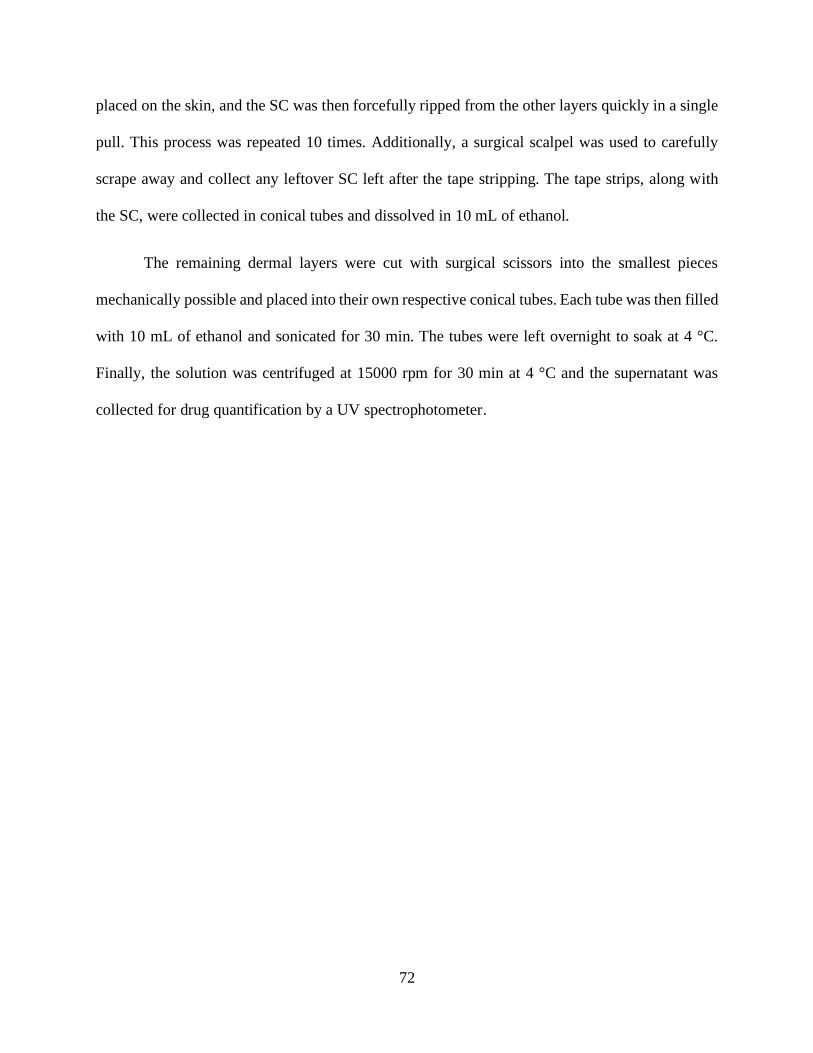
72
placed on the skin, and the SC was then forcefully ripped from the other layers quickly in a single
pull. This process was repeated 10 times. Additionally, a surgical scalpel was used to carefully
scrape away and collect any leftover SC left after the tape stripping. The tape strips, along with
the SC, were collected in conical tubes and dissolved in 10 mL of ethanol.
The remaining dermal layers were cut with surgical scissors into the smallest pieces
mechanically possible and placed into their own respective conical tubes. Each tube was then filled
with 10 mL of ethanol and sonicated for 30 min. The tubes were left overnight to soak at 4 °C.
Finally, the solution was centrifuged at 15000 rpm for 30 min at 4 °C and the supernatant was
collected for drug quantification by a UV spectrophotometer.

73
Results and Discussion
Formula Preparation and Characterization
Figure 3.1. The IL/navitoclax formulation was evaluated to determine size, polydispersity, zeta
potential, and maximum load.
The formulation displayed an average size of 213 nm, placing the formulation well within
the size expected to traverse through the SC layer to deliver navitoclax to the deeper layers of the
skin. Standard deviation remained within this boundary, and offered a consistent formulation
which can offer more consistent dosage [Figure 3.1].
Similar to the size evaluation, the measurement of the electrokinetic zeta potential was
proved to be equally displaying excellent stability. However, polydispersity index had some results

74
which initially displayed a low amount of dispersion, but if the formulation was left undisturbed
for more than 10 hr, the index would rise sharply, as seen in sample 3 of Figure 3.2. This study
was repeated to confirm results. Having a varied, time-dependent, polydispersity index indicates
that like most suspensions, one would be expected to shake well before use. However, shaking in
this case involves probe sonication. Therefore, in order to ensure consistency, the formulation
would have to be created or at the very least sonicated the day of application to ensure a consistent
and proper dosage of navitoclax to the target site. This precipitation seems to be unique to ionic
liquid when compared with H2O and may be due to the electrophoretic relationship between
navitoclax and IL.

75
Figure 3.2. Polydispersity of navitoclax loaded IL varies at different time points within a 10-hour
span. Increased time displayed higher diversity as well, displaying instability in the suspension.
Cell Internalization assay
Visual imaging of the coumarin 6 substitute was slightly modified using photo software to
offer a clearer picture of cell internalization; the color of coumarin 6 was changed from
orange/yellow to green to emphasize the integrative role associated with C6/IL [Figure 3.3].
Controls offered no green glow whatsoever, indicating negative presence of ulterior materials than
may have offered colorimetric interference. Standalone C6 had some residual flakes post-washing.
This may be due to the extracellular fluids holding the C6, or the adherence of the plate itself. This

76
may have allowed a small amount to stick, however very little C6 was able to penetrate the cells,
which can be indicated by the lack of color mixing on those particular plates. However, the IL/C6
plates clearly display a vast distinction, producing a teal color mixture of blue and green within
the cells, which is indicative of C6 entering almost all of the displayed cells. This supports the
notion that IL acts as a penetration enhancer, allowing vastly more potential drug penetration at
the cellular level.
Figure 3.3. Potential of ionic liquid to enhance cellular uptake of a therapeutic molecule. The
feasibility study was conducted with Coumarin 6, a hydrophobic small molecule with fluorescence
properties, as a model molecule. The images show enhanced uptake of coumarin 6 by the A375
cell when co-incubated with ionic liquid compared to free coumarin 6, upon 4 hr of incubation.

77
LDH Toxicity assay
LDH offers insight into the cells themselves, fluorescing only when the lactate
dehydrogenase successfully escapes the cell, and is a common indicator of cell death. Primary
concerns with enhanced cell penetration involved the possibility that cell death would also increase
due to this potential cellular damage involving the utilization of IL. Live cell percentages stayed
consistent in wells containing only IL[Figure 3.4]. These wells increased their live cell % over the
6-hr period, indicating little to no toxicity for standalone IL. However, standalone navitoclax and
the IL/navitoclax formulation both dropped at approximately the same level within the same time
period, mimicking each other with similar cell death rates. Enhanced cell permeation did not act
on an immediate time scale; however, it is important to take note that navitoclax is already highly
effective as an inhibitor. From these results, one can derive that IL does not enhance immediate
results and does not act as a toxic agent without navitoclax present. The LD50 is also extremely
similar to that of standalone navitoclax, which is promising considering the aim of this study is
more related to delivery than drug efficacy enhancement.

78
Figure 3.4. Cytotoxicity of ionic liquid, navitoclax and formulation was investigated with A375
cell upon incubation for various time points. Ionic liquid itself does not show any sign of toxicity
whereas both free navitoclax and ionic liquid containing navitoclax show 70% cell death after 6hr
of co-incubation. Data represent mean±SD, where n=6.
Ex Vitro Permeation Study
Ex Vitro permeation results displayed enhanced penetration in multiple layers of the skin,
with minimal penetration entirely through the skin into the bloodstream. The SC layer had a high
initial absorbance value in the PBS control when compared to the deeper epidermal layers, most
likely due to residual material being left over after centrifugation[Figure 3.5]. However, IL/C6
clearly displayed significantly higher absorbance readings when compared to standalone C6. PLB
and IL standalone both had similar values, emphasizing these individual controls similarities.

79
These increased absorbance values show that even in the outermost layer, absorbance was greatly
enhanced. Although the SC was a secondary target, promising results of enhanced penetration
bring value to overall therapy.
Figure 3.5. To investigate the diffusion ability of ionic liquid and potential as transdermal drug
delivery carrier, we have performed a diffusion study with pig skin. The results show absorbance
values of coumarin 6 within the stratum corneum is as much as 2 folds higher than PBS and free
coumarin 6. Data represent mean±SD, where n=6.
Deeper into the epidermal layers, IL/C6 toppled absorbance values by a significant
degree[Figure 3.6]. More importantly however, is the consistency when compared to standalone
c6, which had a widely diverse range in both the epidermis and SC layers. This higher consistency
lends credence to the notion that higher degrees of dose accuracy may be possible with this new
delivery method.

80
Figure 3.6. To investigate the diffusion ability of ionic liquid potential as transdermal drug
delivery carrier, we have performed a diffusion study with pig skin. The results show that
absorbance readings of coumarin 6 within the epidermis layer is as much as 3 folds higher than
PBS and free coumarin 6. Data represent mean±SD, where n=6.

81
Figure 3.7. To investigated diffusion ability of ionic liquid and potential as transdermal drug
delivery carrier, we have investigated diffusion study with pig skin. The results show that
concentration of coumarin 6 within the dermis layer is 2 folds higher than Stratum Corneum and
5 folds higher than epidermis. Data represent mean±SD, where n=6.
Due to the recent COVID-19 outbreak, testing of the scintillon vials has not been possible
due to lab restrictions. However, no visual observation of the C6 was present, indicating from the
picture of the skin below [Figure 3.8] that if there were significant amounts of C6 that were able
to traverse into the bloodstream, the distinctive color and glow would have been somewhat
noticeable. Additionally, our recent collaboration with Harvard involved a similar study performed
with IL/C6 and displayed similar results [Figure 3.7]. Sadly, the planned animal studies ended
with the euthanasia of all mice meant for this study. One noteable limitation is that this unfortunate
event left us with only coumarin 6 as a penetrative agent, which has a significantly lower molecular
weight over navitoclax, and a lower hydrophobicity. According to previous studies[4], these

82
limitations should not be of consequence, but without proper testing, one cannot draw this
conclusion with absolute certainty.
Figure 3.8. The photograph shows coumarin 6 in different layers of the skin after diffusion.
Conclusions
IL mediated transdermal transportation enhancement has shown promising results in the
delivery of navitoclax or other similar molecules to the target site of epidermal layers, without
traversing significantly into the bloodstream. This targeted drug delivery system has the possibility
to offer stronger and more effective therapies to those who are afflicted with this debilitating and
potentially deadly cancer. This type of application also offers the potential to increase patient
compliance due to the non-invasive method and offers an avenue previously unattainable through
conventional routes of drug administration. Looking back at the archaic methods of treatment
involving an invasive and scarring surgery, the application of a simple ionic liquid, gel, or salve
may be the potential lifesaving tool that will increase patient compliance and stop this disease from
reaching the more deadly stages of cancerous growth, spread, and infection. The formulation can
DermisEpidermis
Stratum Corneum
Hypodermis
After diffusion

83
be optimized with further studies in animal model and treatment feasibility studies in other skin
related diseases such as scleroderma.
References
[1] Tarver, Talicia. Cancer Facts & Figures 2018. Journal of Consumer Health on the
Internet, 2018. 16 (3), 2012, 366 – 367. https://doi: 10. 1080/15398285.2012.701177.
[2] Zitelli, John A; Mohs, Frederic; Larson, Paul; Stephen, Snow. “Mohs Micrographic
Surgery for Melanoma.”Dermatologic Clinics, 1989, 7 (4), 833 – 844.
https://doi.org/10.1016/S0733-8635(18)30571-0
[3] Boyer, John D.; Zitelli, John A.; Brodland, David G.; D’Angelo, Gina. Local control of
primary Merkel cell carcinoma: Review of 45 cases treated with Mohs micrographic surgery
with and without adjuvant radiation. Journal of the American Academy of Dermatology, 2002. 47
(6)., 885 – 892. https://doi.org/10.1067/mjd.2002.125083
[4] Qi, Qin M.; Mitragotri, Samir. Mechanistic study of transdermal delivery of
macromolecules assisted by ionic liquids. Journal of Controlled Release. 2019, 311 -312 (162 –
169). https://doi.org/10.1016/j.jconrel.2019.08.029
[5] Guy GP, Thomas CC, Thompson T, Watson M, Massetti GM, Richardson LC. Vital
signs: Melanoma incidence and mortality trends and projections—United States, 1982–2030.
MMWR Morb Mortal Weekly Report. 2015;64(21):591-596.
[6] Mourant, Judith R.; Johnson, Tamara M.; Los, Gerrit; Bigio, Irving J. Non-invasive
measurement of chemotherapy drug concentrations in tissue: preliminary demonstrations of in

84
vivo measurements. Physics in Medicine & Biology, IOPscience, 1999. 44 (5).
http://dx.doi.org/10.1088/0031-9155/44/5/322
[7] Bajetta, Emilio; Del Vecchio, Michele; Bernard-Marty, Chantal; Vitali, Milena; Buzzoni,
Roberto; Rixe, Olivier; Nova, Paola; Aglione, Stefania; Taillibert, Sophie; Khayat, David.
Metastatic melanoma: Chemtherapy. Seminar in Oncology. 2002. 29 (5) 427 – 445.
https://doi.org/10.1053/sonc.2002.35238
[8] C., Lerman; B., Rimer; B., Blumberg; S., Cristinzio; P. F.; Engstrom; N., MacElwee.
Effects of coping style and relaxation on cancer chemotherapy side effects and emotional
responses. Europe PMC. 1990, 13 (5), 308 – 315.
[9] Didolkar, M. S.; Kanter, P. M.; Baffi, R. R.; Schwartz, H. S.; Lopez, R. Comparison of
regional versus systemic chemotherapy with Adriamycin. Annals of Surgery. 1978, 187 (3), 332
– 336. https://doi.org/10.1097/00000658-197803000-0002
[10] Bae, You Han; Park, Kinam. Targeted drug delivery to tumors: Myths, reality and
possibility. Journal of Controlled Release, 2011, 153 (3) 198 – 205.
https://doi.org/10.1016/j.jconrel.2011.06.001
[11] Nitsche, JM.; Kasting, GB.; A microscopic multiphase diffusion model of viable
epidermis permeability. Biophysical journal, 2013. 104 (10), 2307 – 2320.
https://doi.org/10.1016/j.bpj.2013.03.056
[12] Miller, D. L.; Smith, N. B.; Bailey, M. R.; Czarnota, G. J.; Hynynen, K.; Makin, I. R. &
Bioeffects Committee of the American Institute of Ultrasound in Medicine. Overview of
therapeutic ultrasound applications and safety considerations. Journal of ultrasound in

85
medicine: official journal of the American Institute of Ultrasound in Medicine. 2012, 31 (4), 623
– 634. https://doi.org/10.7863/jum.2012.31.4.623
[13] Bolla, P. K.; Meraz, C. A.; Rodriguez, V. A.; Deaguero, I.; Singh, M.; Yellepeddi, V.
K.; Renukuntla, J. Clotrimazole Loaded Ufosomes for Topical Delivery: Formulation
Development and In-Vitro Studies. Molecules 2019, 24 (17),
3139. https://doi.org/10.3390/molecules24173139.
[14] Cleary, J.M., Lima, C.M.S.R., Hurwitz, H.I. et al. A phase I clinical trial of navitoclax, a
targeted high-affinity Bcl-2 family inhibitor, in combination with gemcitabine in patients with
solid tumors. Invest New Drugs 32, 937–945 (2014). https://doi.org/10.1007/s10637-014-0110-9
[15] Roberts, A. W., Seymour, J. F., Brown, J. R., Wierda, W. G., Kipps, T. J., Khaw, S. L.,
Carney, D. A., He, S. Z., Huang, D. C., Xiong, H., Cui, Y., Busman, T. A., McKeegan, E. M.,
Krivoshik, A. P., Enschede, S. H., & Humerickhouse, R. (2012). Substantial susceptibility of
chronic lymphocytic leukemia to BCL2 inhibition: results of a phase I study of navitoclax in
patients with relapsed or refractory disease. Journal of clinical oncology : official journal of the
American Society of Clinical Oncology, 30(5), 488–496.
https://doi.org/10.1200/JCO.2011.34.7898

86
DISSERTATION CONCLUSIONS
Overall, the culmination of these transdermal drug delivery systems presented in these studies has
proved to be both an exciting and fruitful endeavor. Penetration of nano-scale molecules using an
enhanced lipid-based carrier was successful in the transportation of one of the most widely used
antifungal agents in history in a safe and more effective manner has essentially improved the
outcomes of one of the most popular antifungal combatants used in conventional medical
application. This ability to improve one of the most affordable treatments may have a significant
impact in the safe, effective, and easy-to-use tools in a physicians arsenal. This improvement has
the ability to save many lives if taken to the commercial market.
Contrarily, a rare, large-molecule, experimental, anti-melanoma drug has been shown to be able
to traverse the SC layer of the skin in large amounts and attack the target site without the use of
systemic circulation. This revolutionary way of penetrating the skin and cells may be a new avenue
in fighting one of the most common cancers in the United States. This form of therapeutic relief
may circumvent the plethora of side-effects associated with more popular forms of chemotherapy
and may soon change the future of how certain cancers are dealt in the clinic. The association of
an IV needle and cancer is now slightly closer to becoming an archaic evocation of scientific
progress.

87
CURRICULUM VITA:
Isaac Giovanni Deaguero holds a Bachelors of Science in Biology from the University of Texas at
El Paso(2014). He is a Veteran of the Unites States Navy(2003-2008), where he served as a Fire
Control Technician onboard two nuclear submarines and attained the rank of Petty Officer Second
Class (E-5). During his graduate studies, he taught various courses including pharmaceutical
compounding, global health for biomedical engineers, and biomedical device design & regulation.
Published Works
Hossain, R. F., Deaguero, I. G., Boland, T., and Kaul, A. B. (2017). Biocompatible, large-format,
inkjet printed heterostructure MoS2-graphene photodetectors on conformable substrates. npj 2D
Mater. Appl. 28, 1–10.
Bolla, P.K.; Meraz, C.A.; Rodriguez, V.A.; Deaguero, I.; Singh, M.; Yellepeddi, V.K.;
Renukuntla, J. Clotrimazole loaded ufosomes for topical delivery: Formulation development and
in-vitro studies. Molecules 2019, 24, 3139.
IG Deaguero, MN Huda, V Rodriguez, J Zicari, T Al-Hilal, AZM Badruddoza, M
Nurunnabi. Nano-vesicle based anti-fungal formulation shows higher stability, skin diffusion,
biosafety and anti-fungal efficacy in vitro. Pharmaceutics 2020, 12, 516.
Contact Information: [email protected]
This dissertation was typed by Isaac Giovanni Deaguero