title studies on the peritoneal absorption of … · pathway ill . the ... of malignant tumors...
TRANSCRIPT

Title STUDIES ON THE PERITONEAL ABSORPTION OFPARTICULATE MATTER
Author(s) ITANI, KANICHI
Citation 日本外科宝函 (1959), 28(3): 802-824
Issue Date 1959-04-01
URL http://hdl.handle.net/2433/206817
Right
Type Departmental Bulletin Paper
Textversion publisher
Kyoto University

802
STUDIES ON THE PERITONEAL ABSORPTION OF PARTICULATE MATTER
Bv
I《ANICHIITANI
From the '.:'.ncl Surgical Diγision, Kyoto Uniγersity Medical只chool(Director: Prof. Dr. YAR!"I>SA AovAGIJ <ReceiYed for publication Feb. 7, 1959)
CONTENTS
I. HISTOLOGICAL OBヨERVATI0:¥1OF THE PERITO¥TEUM PARTICIPATI:¥TG THE
ABSORPTION [[. THE CHAr-'GES OF LYMPHATIC ABSORPTION AFTER DESTRUCTI00! OF MAIN
PATHWAY Ill. THE '.¥!IC:WSCO?IC' F2ATURE3 OF DIAPHRAD!λTIC AつPEぐTI¥! THE !NFL-
AW¥!ED PERITO.:¥TEAL CλVITIES
I. HISTOLOGICAL OBSERVλTIO::¥ OF THE PERITO::¥EU:¥I
PARTICIPATING THE ABSORPTION
Absorption through the peritoneum is of considerable interest clinical!~’ in
understanding the resolution of e町usion,the spreacl of infection and the metastasis
of malignant tumors occurring in the peritoneal ca ¥'it.'"
Since v.REcKLINGHAUSEN (1863) discovered that injected particles into physiolo-
gical peritoneal cavity of a rabbit ¥Yere rapid !~’ ab>'orbul through the diaphragm,
numerous studies about this have been made b~・ KLEIN, BuRDON, SANDERrnN (1872),
MuscATELLO (1895), GLOBER (1901), KUTTNER (1903), :HAcCALLUM (1903), BuxTON
(1906), WALTER (1921), BOLTON (1921), :¥1AGNUS (1923), HIGGINS, BAIN (1930),
HIGGIN3, BEAVER, LEMON (1931)' ALLEN,γOGT (1937) and SIMER (1948).
However, the只cliteratures were unsatisfactor.¥・ from the morphological point of
view.
Under Prof. Dr. KmARA, man~· investigators have s~·stcmaticalb· stL1clied this
problem and consequently discovered following facts that particles injected into the
peritoneal cavity penetrated the intercellular cement ~mbstances of the diaphragmatic
peritoneal endothelium ancl moved through the sieve-like constitution to the endo・
thelium of the lymphatic vesぽ ls. This sieve-like constitution, which was formed
IJ:-・ both collagen and reticulum白ber日 inthe subenclothelial connecti¥'e tissues of the
diaphragm, was named macula cribriformis bγProf. Dr. KmARA.
Thereafter, this structure was discovered on the parietal pleura, pericardium,
mediastinal pleura and omcntum, in which absorption of the particles into lymphatics
was observed. In other word円, maculacribriformis formed prae・lymphva~cular fluid
path in the subendothelial connecsi¥'c ti山 ues.
・whereas, :¥IATSUDA (1951) postulated that India ink injected into the plem・o-
peritoneal cavit≫ of some kirnいof vertebrates was absorbed in ¥'arious sites of

PERITONEAL ABSORPTION OF PARTICULATE :¥IATTER 803
serow.; surfac:e as follows; membrang, subvertebralis, mesenterium and serosa of
intestine in a wels; serosg, of stけ iac:hin newts; membrana subvertebralis, mesen-
terium awl serosa of large bowel in to:J.rls; mesenterium and 日erosaof the intestine
in a lizard and a tっrtoise;serosa of the stomach and the diaphragm in fowl; mainlsア
the diaphragm and the omentum, slightly mesenterium and mesoovalium in rabbits.
The subendothelial strnc:ture of these absorption sites is mesh-work composed of both
collagen and reticulum fibers.
This sttdy was underbkei1 to investigate whether macula cribriformis could be
detedable in the peritoneal cavity of mammalia besides diaphragm and omentum by accepted method.
:¥IETHOD AND MATERIAL
1) Animals. Albino rabbits weighing approximately 1.5-2.0 kg were used.
2) Technical procedures. India ink, (finelsァ dividedcarbon particles disolved in
5% glucose solution) was injected into peritoneal cavity through a fine vinyl tube inserting upper abdominal wall. Inje巴tedvolume of the h’e was 25 cc per kg of bodyweight.
Animals were bled to death at 15 minutes to 48 hours after injection of India ink. Routine gross ancl microscopic studies were performed.
For the purpose of morphological study of the parietal peritoneum, diaphragm,
omentum and mesenterium, care was taken to peel them out from the underlying tissues as thinly as possible and they were stretched out over a clean glass slide.
After being dried in the room temperature, these materials were 白xedby 10% neutral formalin and usually stained with Bielschowsky-Maresch silver mE>thod, occasionalhア thevwere stained with hematoxγlin and eosin or J¥Iay心iemsastain.
Liver, Lgll. sternalis cranialis, mediae and caudalis, lymph nodes in the peri咽
toneal cavity --especially, Lgll. cardiacae, Lgll. art. pancreaticolienalis, Lgll. art.
hepaticae, Lgll. mesentericae craniales, Lgll. a. mesentericae caudalis, -ー Lgll.iliacae,Lgll. renalis, Lgll. poplitae and Lgll.axillares were examined in all animals as
control materials for the Part II and III. The~, were embeded in paraffin after
fixation and sections were stained with hematoxylin and eosin.
The lymph nodes in thorax --Lgll. oesophagicae, Lgll. intercostales dorsales, Lgll. mediastinales v. cavae craniales dextrae, Lgll. aortae and Lgll. aortae thoracales
caudales lat.ー- were examined only macroscopically.
EXPERIMENT AL RESULT
After washing out India ink in the abdominal cavity of a sacrificed rabbit, definite necropsy examination was performed.
The features of peritoneal absorption were aぉfollows.
1) Diaphragm (Fig. 1, 2); Absorption from peritoneal diaphragmatic surface
was conspicuous in all animals. In the p昨日 tendinea of diaphragm. India ink
formed radial black ぉtripe只 between tendon bundle日, which extcnclecl in pars
muscularis, becoming obscure gradually.
804 日本外科宝函第28巻第3号
The cell boundaries of the diaphragmatic I】eritoneum were outlined here and
there b~ · carln India ink was visible excer〕ttwo animals which had the longer intervals from
injection to deg.th. After entering into subdiaphragmatic lymphatics, the particles
reached the I~·mphatic-venous connection main!~· through the lymphatic trunks
running up behind the sternum and parU_1,・ through the lymphatic vessels running
up in the mediastinum.
And the dorsal mediastinum had not the lymphatics containing the particles.
Silve2・ stained specimen revealed that macula cribriformis, which was mesh of
reticulum fibrils with collagen frame, was distributed in both pars tendinea and
pars muscularis of the diaphragm. The former was arranged regularly in parallel
with tendon bundles and the latter was distrilmtd irregularly between muscle
bundles forming like beehive. The macula cribriformis were ks日 in number and
deformed with sparse reticulum fibrils in the marginal area of the diaphragm.
Divided carbon particles of India ink were adherent to fine reticulum fibrils of the
macula cribriformis, through which the particles were gathered in the subendothelial
lymphatics.
2) Omentum (Fig. 3) : Though omentum was greyish coloured at 15 minutes
after injection, it was found microscopically that absorption from omental surface
wa日 γet下・Yigorous at 2 to 4 hours after injection. In these cases, carbon particles
were mostly accumulated in the milk.¥・ spots, where the particles surrounded the
venulc日 andemerged in the lymphatics. After 24 hours or more, free carbon
particles on the membrane decreased and macrophages ingulfing them were eminent.
The omentum was composed of wavy collagen的 ers,which were woven each
other, and straight elastic fibers. In the meshes of collagen白bers,fine fibrils were
proved, but they did not form a true net work as macula cribriformis, nor revealed the argyrophilic nature.
In the milky spots, wav.\’ collagen fibers and白nefibrils mentioned above were
WOγen densel.¥・ each other with abundant distribution of blood and lymphatic vessels,
and cellular elements were numerous. Carbon particles \\明・e scattered between
thc.;e fibei's, but were not exclusive!\アadherentto the fine fibrils.
3) ::.'dcscnterium (Fig. 4, 5, 6.) : The mesenterium of the small intestine w部
not coloured with India ink within 1 hour. After two hours or more, some specks,
light!,\’coloured greγish, appeared along the mesenteric vessels near the attachment
of the intestine. These只l〕cclrncould not be rubbed out with gauze. And there was
no speck in the thinner part aloof from the vessels. After 24 to 48 hours, these
spe~ks '"ere more deeply colourd by the accumulating phagocytes which ingulfed
the particles. Microscopical!.¥-, the carbon particles accumulated in these specks
were aclher‘ent to the mesenter‘ic surface, part of which entered into the subendo-
thelial tissue. L~ rnphatic absorption from these sites coulcl be proved. A re,γvenules were surround巴di町、 theparticles. but not containing them.
The me日sn tc ri um o l・the large intestine, espじじiall.\、 ぉigmoicl,hact greyish coloured
Sp巴~ks in some instances 11101℃ G¥'idently and rapidly than the small intestine. In

PERITONEAL ABSORPTION OF PARTICULATE MATTER 805
them, microscopic observation was similar to that of the small intestine.
Silver stained specimen revealed that the mesenterium was composed of the
wavy collagen fibers which were woven each other, more densely near the me!-'en-
teric vessels. In the meshes of the collagen fibers were found fibers like down, not
so argyrophlic as the reticulum fibrils, along which carbon particles were not accumulated.
A pair of diverticulum of peritoneum by the rectum were always filled up
with India ink, but no lymphatic vessel was coloured in adjacent region.
4) The serosa of digestive organ (Fig. 7, 8, 9): The narrowing rings of the
caecum in three cases, which were sacri負ced15 minutes, 1 hour and 2.5 hours each
after injection, were slightl;,ァ colouredwith India ink. Some of the particles were
proved in the subserous tissue, where the particles were arranged near the peculiar type of cells, seeming mesothelial.
Although e出cientabs0rption from these sites via blood stream or lymphatics
could not be detectable, this fact seemed very interesting from phylogenetic point of view, as mentioned later.
B~, the silver impregnation method, the subserous tissue of this region was consisted of wavy collagen fibers which were not woven each other and not rami-fied with reticulum fibrils.
5) The ventral peritoneum (Fig. 10) was not coloured with India ink in an~’
animal and neither blood nor lymphatic vessels containing carbon particles were
visible. Histologically, this part of peritoneum was composed of broad collagen fibers which were stained yellowish brown by Bielschowsky’s method and were
parallel with :VI. transversus abdominis. The reticulum fibrils were not found anywhere.
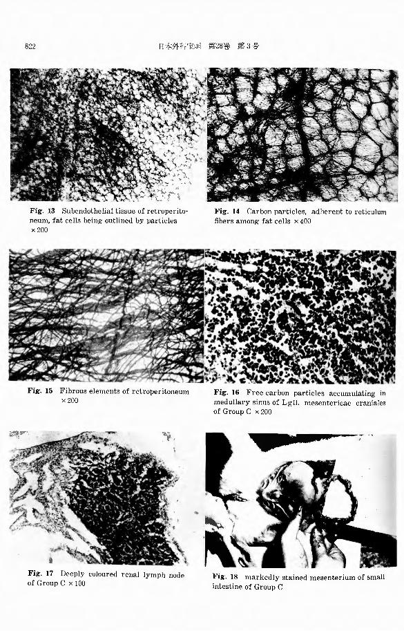
6) In the retroperitoneum (Fig. 11, 12, 13, 14, 15), most of the cases had greyish coloured specks on the fat tissues near the caudal pole of the kidne~·. The
stretched specimen of this colom℃~l parts, staining with hematoxylin and eosin,
revealed that injected carbon particles gathered in the interstitium of fat cells and
difined the contour of these cells. But blood and lymphatic vessels containing the
particles were not seen. By the silver impregnation method, it becomes evident that the carbon particles
seen among fat cells were constantly found at the sites of reticulum fibrils, and that subendothelial tissue of the retroperitoneum, except fat tissue, was composed
of wavy collagen fibers with various thickness, which were similar to mesenterium
or omentum and quite di百erentfrom ventral peritoneum. ::.¥Iacula cribriformis was
not found anywh~re of the retroperitoneum, although fine fibers barelyア argyrophilic,
were seen as well as mesenterium.
DISCUSSION
It is beyond dispute to conclude that particulate matter injcctcc1 into peritoneal cavity was absorbed cxclush・cly from peritoneal diaphragmatic a'-pect into lyrnpha-
tic path.

806 日本外科宝函第28巻第3号
"¥bsorption through the rest of the parietal peritoneum and the mesenteric
folds does not appe1r to be of much sig.,1ificance quantitativel~·.
The extent of lymphatic absorption th1-,n1江hthe omentum, however, has for a
1011g time besn debatable. Some investigators ha vεbeen doubtful against the exi~←
tence of omntal lymphatics. Ho\\アever, Tm (1937), SIMER and CASPARIS (1948)
have de;110九日trntclclc'=td~· a fairhア rich distribution of b'mphatics, generall>・ as回 c-
iatd with the blood ve山 els,in the omentum of cats, clogs, rabbits and men.
On the other hand, TAGUCHI (1943) maintained particulate matter was also
absorbed into the blood vessels on the omentum. In our experiments, absorption
from omental surface was ver≫ conspicuous in about 2 hours after injection, when
the carbon particles accumulating chief!~· in the milky spots were arranged along
the wall of venules or filled up the small l>・mphatics. But the fa.ct that the
particles reaching the liver were few similarlγin group B (Devastation of the
diaphragm) and group C (Devastation of the diaphragm and the extirpation of
the omentum), reveaL:::l little imp:wtance of alJsGtptio孔 throC1ghthe omentum.
As SIMER mentioned, the action of the macrophages which are present throu・
ghout the omentum se巴me:!to form an efficient barrier against invasion of partic-
ulate matter.
Though TsuBoucm (1950) po~tulatecl that the maculas were also distributed
in the thinner part of the membrane aloof from the blood vessels, we could not
confirm the cxi~ぇtence of the special structure, in which collagen fibers ramified
reticulum fib,.ils and formed true net vwrks like a sieve.
In rq-ard to the mesenterium, only J'dATSUDA (1951) postulated that the
particle『 1nssdthrough the cement substance between endothels of the membrane
and were absotbed into venules or lymphatics, with coloureJ lymph nodes, at two
to three hours after injection.
In O~li' e¥: I〕2rimc:1ts,the ab:sorption through the mesente1・ium could not alwa¥・s
be re:;ognize::l, but barely in a few instances. Greγish specks, locating along the
blood vessels near the attachment of the small intestine, especially ileum, the
sigmoi〔land the cae巴um,were consi日tcclof accumulating carbon particles, part of
which en t ereゴintoper行 ascular fat tissue or ingulfed H’ macrophages. It seemed
that a few particles 1¥・crc believd tοenter into blood stream through the definite
part of the mesenterium in some inst~mces. However, neither l≫mphatics nor
lymph no:les in the membrane were containing the particles.
It was revcaleオthat the membra11c was com po時 clof wav>・ collagen fibers,
wea,・ing each other, ar;d of fine down-likeれbnN・ Andthe network of collagen was
thick near the bloocl ve只同ls where the argyrophilic fibers were found. But no particle was adherent to these fibers.
In other wonls, special structure for absorption i. e. macula cribriformis or
milky日potscould not lie found anywhere on the mescnterium‘
It was br,mght to light br our experiment円 thatsome particles penetrating the
retrop2ri toneum cけuld reach into the 1℃trnperitoneal ca\'it~-. The gr℃.Yish specl、日
1¥・iiich indicated the alJsりrption 日ite日日℃Ic mo日tlyfound over the fat tissue討 near

PERITONEAL ABSORPTION OF PARTICULATE MATTER 807
the kidney.
:¥1icroscopic observation rcyc九lc:lthat the particles were either accumulated in
the subendothelial tissue with definite boundaries or arranged along the reticulum
fibrils among fat cells. After entering into fat tissue, the particles are presumably
absorbed l巧’ thetwo paths, venules and lymphatics. The vcnules distributing in
the fat tissue were often surrounded bγthe particles. On the other hand, it could
bs a proof for l~·mphatic absorption that retroperitoneal l:;.アmphnodes, Lgll. renalis
an〔lLgll. iliacae, were sometimes coloured with India ink, when Lgll. poplitae and
Lgll.axillares were never containing the particles neglecting the supply by the blood strea町1.
The structural elements of the retroperitoneum resembled those of the mesen-terium, quitel:;.ァ di町ering from those of the ventral peritoneum, and had not the
macula cribriformis an vw here.
As for the serous surface of the digestive organs, MATSUDA (1951) postulated after p1ηlo.;;enetic inve3tigation that absorption through the membrane into lymp-
hatics and blood vessels took place in all kinds of vertebrates, except mammalia,
and that the structure of the同 siteswas meshes of collagen fibers, partly ramify-
ing with reticulum白brils.
From this point of view, the gr町 ishcolouring, locating in the narrowing rings of the caecum which was first demonstrated by our experimemts seemed to be a phylogenetic remain.
Presumablγ,the peculiar type of cells in the sites, seeming mesothelial, which does not formeヨcapillaries,take definite role to fix the carbon particles. In other
words, the particles in the sul】endothelialtissue are arranged within a tissue space
walled b;-・ endothel-like cells incompletely. But the stites are not an e宵ective
pathway for the particles where the collagen fibers are neither woven each other,
nor ramified with reticulum fibrils. When the inflammation has progr邸 seclintrap-eritoneally, the gres包hcolouring in the narrowing rings became broad and dense
with accumulation of polymorphnuclear日, mom川 ・tesand lymphocytes simultaneously.
SU:VIMARY
The carbon particles which are injected into peritoneal cavity are absorbed
through the serous surface as follows : 1) The injected particles are removed exclusi\℃ly by diaphragmatic lymphat-
ics through conspicuously distributed macula cribriformis.
2) On the omentum, some particles are absorbed into venules and lymphatics chiefi;-γdistributing in the milky spots. We can not confirm the existence of
macula cribriformis in the membrane.
3) A small dose of particle is absorbed at the narrowing rings of caecum,
picturing dotted or complete circle around the bowel. It seemed to be a phylogen-
etic remain of absorption structure, which is active throughout serous surface of
vertebrates besides mamrnalia.
4) 1¥bsorption of particles tal、csplace, slightly in dose, in the I℃ti叩 eritoneum
808 日本外科宝函第28巻第3号
over the fat tissue near the ki<lnc~._
5) In the mesenterium, it is believed that a few p乳rticles,forming ・greyish
specks near the atbchment of the intestine, enter into blood stream through the
venule wall.
6) The macula cribriformis could not be detectable an~·where in the mesen-
terium, the parietal peritoneum, except diaphragm, and the serosa of digestive
organs.
IL THE CH.Al'~GES OF LY::¥IPHATIC ABSORPTION AFTER
DESTRCCTION OF :¥IAIN PATHWAY
As the foregoing paragraph had shown, particles injected into peritoneal cavit~’
were cwlusivel~· absorbe:l through the macula cribriformis in the diaphragm. This group of cxpci、ime日tswas aimed to investigate the changes appearing on
the absorption of particles after destruction of main path\\’a~· in the diaphragmatic
aspect.
::¥IETHOD AND l¥IA TERIAL
1) 人nimals:)Jbino rabbits weighing approximately 1.5 2.0 kg.
2) Te市nicalproc:edure: Two series of experiments were performed. The two
広roupsof rabbits were designated B and C and operated under ether anesthesia as fol lows;
Group B: Dernstatio i of peritoneal diaphragmatic surface b~· silver nitrate.
Group C : Extirpation of omentum and devastation of peritoneal diaphragm-atic surface b:-・日ilver nitrate.
The dc,・asted diaphragm was adherent looselv to the liver surface after 24
hours and adhesion bernme more tightly, unable to peel the membrane, as the time
lapsed. B:-・ this way, acti,・c diaphragmatic surfaces for absorption \\℃re closed almost completely.
Animals "℃ re injected with India ink intraperitoneally after various postoper-ativc survivals and then bled to death at 0.5 to 4 hours.
Following lyアmphnodes were examined microscopically in all animals; Lgll. cardiacae, Lgll. art. pancreatico-lienalis, Lgll. art. hepaticae, Lgll. mesentericae
craniales, Lgll. renales, Lgll. iliacac, Lgll. poplitae and Lgll. axillares. The ly・mph
nodes of animals used in Part I were examined as control materials.
The stretched specimen of the par匂tal peritoneum and the mesenterium ''℃ I℃
fixed with neutral 10% formalin and stained by・ Bielschowsky-Maresch’s method.
1'~XPERIMENTAL RESULT
The features appearing in the lymph nodes of control animals are shown in
Table 1, where black colm1red l~·mph nodes are shown as十ft,gTcy・ish coloured lymph
node日 as十十, lymph nodes 仁川itコi11i11g a little carbon particles, proved o川ymicros-
copically, as十, andlymph nodes free from carbon particle as一(Table1, 2, 3).
809 PERITONEAL ABSORPTION OF PARTICULATE MATTER
一+件併骨僻掛件
d
n
一
--
nL
円
、
山
&
&
・
・
・
・
s
Hιno’
H
一r
r
r
r
r
r
r
r
r
r
r
m侃刈》抗一h
h
h
h
h
h
h
h
h
h
h
h出
台
以
th一54
5
5
n
3リ
Tム一
0
1
4
3
4
8
1
/
1
0
2
I
h
ベ
2
4
1
5
0
、
「il
lli-
-
叫
一
N
b
一12
3
4
5
6
9
2
3
4
5
AU
一つJndqoqoqd
qJ
4aoooonD00
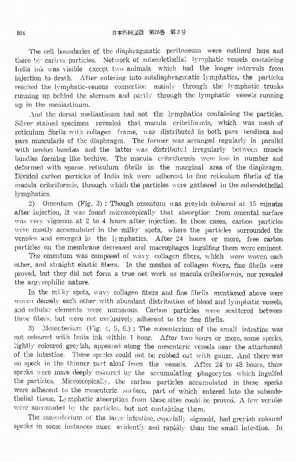
Carbon Particles Reaching Livers and Lymph Nodes of Sacrificed Rabbits in Group A. (Control Animals)
I. L. I A. L. I Pp. L. L
R
L
M
L
L
L
P
L
PU
Table 1
+
Lgll. cardiacae. P. L.; Lgll. art. pancreatico・lienalis.Lgll. art. hepaticae. M. L. ; Lgll. mesentericae craniales‘
Lgll. renales. I. L.; Lgll. iliacae. Lgll. axillares. Pp. L. ; Lgll. poplitae.
+
+
+ +
+
十
H
甘++
++
+
+
++
十
+
++川廿+件+H
廿
++十
i Liver
川廿+掛
C. L.;
L. L.;
R. L.;
A. L.;
Rabbit
Pp.L.
n
oD
1D
10
h
L
YE-‘
,G
e
-
c
L
Fb n
R
ρ
、a
s
L
gA
、J
-J
oe---
S
M
-7
er-
h
可-A
’ruuEVEL
-
hm一L
一朴特+
H
廿
品目-
h
一11111--
-
mh
-L
一一件特朴++++
vu只u
-IA
一
L
y
-J1li--一
Ill11Illi--
Il--
d
b
一L
一
制
m一
仁
一
一
一
朴
一
+
+
一
泊三1町
一、
111111
1
1--
昨日市一-
w
一-+一一一+
T
一++
μ・叫庁「|11一111111
11111ill-
gD
-
一
nf
-
一
十
mo--凶
n
一L・
・
&
・
芯
日比
n
-anoh一nr
r
r
r
r
r
r
r
r
即日
ω一円
qhAO抗一h
h
h
h
h
h
h
h
h
h
nE
抗一刻品-
mth一
5
3
5
ヨぬ-凶:1
.E一41
0
4
1
0
3
1
1
1
eM4ETA
-
一
凶
肝
-
一
ーれ
Di
一11ILIli--
1011111lil--
a(E
一
E
-
-
-
J
B
-sn
n
一
内
川
ベ
薗
’
’
A
n
v
、,一---
--
-
-
-
CM
-
一
C
M
C
M
V
A
q
u
川
均
一
mm.u・t
-ddd
d
d
d
d
d
h
d
,引印
Ermu口
刀
日
一
ar
-e’I引
1・H’一
5
G
G一此
fhah
-353
5
U
7
幻
Han
YE
--
FE
、
-
A.L.
+
+
+H
廿++
+
+
-lit
++
+
+
+
+
一件朴
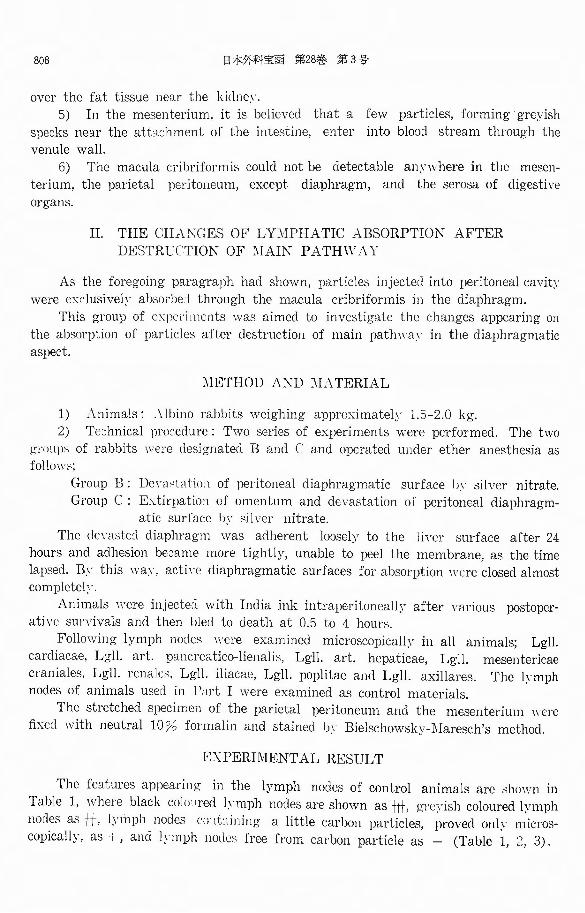
Table 2
Rabbit
-一-9
0
3
4
2
7
2
4
6
7
N
一3
4
4
4
5
5
6
7
7
7
ee
L
H
a
t比
1EE企
g
p
nゆ
.HI
-- a
L
4LrL
、ムTう
l
i
ω
L
similar to
++
In brief, while the lymph nodes in the epigastric region, Lgll. cardiacae, Lgll.
art. pancreatico・lienalisand Lgll. art. hepaticae, were containing carbon particles
at the early stage, Lgll. mesentericae craniales, which are large lymph nodes
mases filled with chyle, were not containing the particles with one exception,
sacrificed in 48 hours after injection.
In the retroperitoneal cavit:--, Lgll. renales and Lgll. iliacae
particles were less than the l~·mph nodes in the epigastric region.
and Lgll. axillares never contained the particle日.
In Group B and C, the features of epigastric lymph nodes \\℃1℃
十*
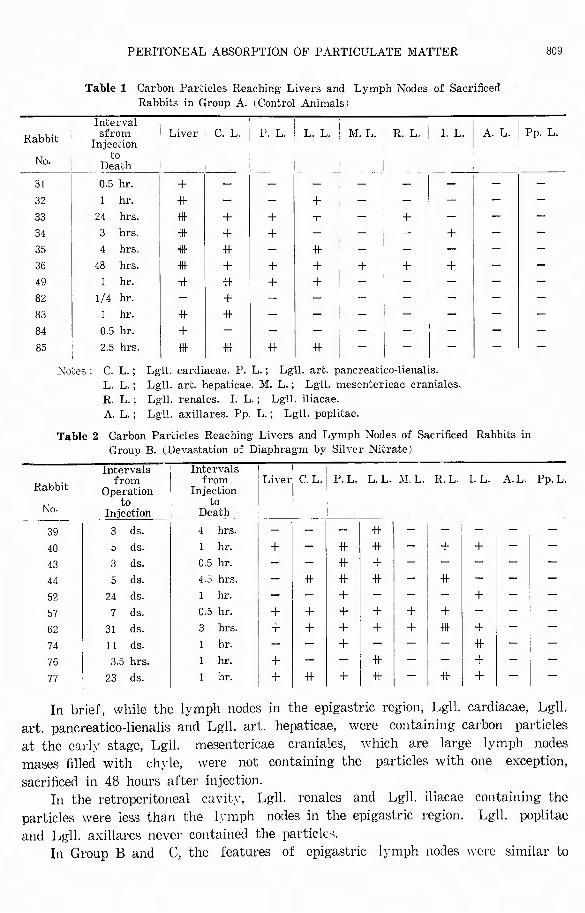
Table 3 Carbon Particles Reaching Livers and Lymph Nodes of Sacrificed Rabbits in Group C. 1 Dc\"as~aciりn of Diaphragm and Extirpation of Omentum)
Intervals from
Operation N I to 0・ i Injection 1
45 I 4 dS.
46 4 ds. 47 7 ds. 48 I 1 ds. 54 I zg ds. 60 ! 30 ds. 65 I 26 ds. 66 I zg as. 69 I 57 <ls.
73 I 12 <ls.
1s I 2 s hrs. 79 1 d.
第3号第28巻日本外科宝函810
Pp.L. A.L I. L.
件
+
+
十
+
掛
R.L.
+仲
+仲
+
*
Liver C. L. • P. L. L. L.' :¥I. L
+
+
+
H廿
H廿
+
+
仲
+
+
+
++
-It
+
け廿
H廿
H廿
#十
+
件
+
+
品川+
十件川市
十
件
+
+
+
++
+
lnしerva1sfrom
Injection LU
Death
hr. hrs. hrs. hr. hr. hrs. hr. hr. hr. hr. hr. hr.
-Aq
u
q
υ
t
i
t
i
n
J
1
4
1‘
1ょ
tA
-1
-A
Rabbit
++
those of control animals.
However, it seemed to be of co11siclerable importance that the carbon particles
were found in Lgll. mesentericae craniales more evidently and rapidlγ. The part・-
icles appeared in the medullary sinuses were not phagocytosed yet (Fig. 16).
The retroperitoneal !;;mph nodes were also coloured with India ink at the early
stage after injection. Above all, the cases, having 1 or 2 months of sunivals
after operation and perfect closure of diaphragmatic aspect, had the c;cepl~· coloured
lymph no:les and lymphatics which chained the retroperitoneal b・rnph nodes and
entere:l into hiatus aorticus of the diaphragm (Fig. 17).
The fact that Lgll. poplitae and Lgll. axillares never
confirmed them reaching the lymph nodes, mentioned a!JD¥'C,
lymphatics.
l¥scites wa只 foundin varying deょl℃c, which was transparent and not 日tink~·.
Injected India ink appeared to be mingled with ascites homogenousl~· , but in some
cases formed clots or membranous substances over mesenteric folds.
The omentum of Group B, which often covered the operated ''刊mdor the liver
lobe, were di百uselJγstaine::lwith the cl同, havingdistinguished milk:-’ spots, in some
cases which had been injeded in 3 to 5 claJ’日 afteroperation. ・whereas, the memb-
ranes of cases which had the intervals of two weeks or more after operation were
slightly coloured and microscopicall:iア had collagen fibers revealing the features of fibrinoid swelling.
In both Group B and C, it could be proved absorption through the peritoneum
except diaphragm and omentum took place, though ven・ slight!γ. Grc_1i:;h日pe::l、只 appeareclin the definite 11a1・tsof serous surface, that is. along
bloodγC山 clメ neat、theathchmcnt川・ theintc:;tinc, oγer fat ti:;日uc:;11Gt r the kidney
and the narrowing rings of the caecum.
the particles
brought by
contained
should be

PERITONEAL ABSORPTION OF PARTICULATE MATTER 811
Though the spe:-:ks emerge:l more evidently and rapidl~r than in control anim-als, it seeme:l th~t the features were not induced b>' promoting absorption from these sites, but were induced by accumulating m':lcrophages and increase of fibrin fixing the particles in situ (Fig. 18).
DISCUSSION
SAKAMOTO (1933) mentioned in his work concerning lymphatic system of rctbbit日 thatcarbon p.irticles were found in the epigastric l~ · mph nodes at the early shge after injection, while they were slightly found in L~ll. mesentericae craniales in four da>・s after injection.
This results were similar to those of our experiments. It seemed to be one of compensative features resulting from diaphragmatic
closure that Lgll. mesentericae craniales is conhining free carbon particles in me・dullary sinuses in only half a日 hourafter inje::tion. This phenomenon is found in the cases of Group D and E, in which absorption of particles through the diaphragm is impaired signi白cantly(Table 4, 5).
As L:sll. axillares and poplitea are never containing particles, the free ones appearing in the mefollary sinuses may be brought l≫’lymphatics.
However, l.vmphatics containing the particles could not be found throughout the mesenterium in these cases. Presumably the particles may reach the lymph noゴeswith retrograde flow after entering into lymphatics near the diaphragm.
Retroperitoneal lymphatic system of rabbits is poor. Lgll. renales and Lgll. iliacae were slightly coloured in one or two days after inje::tion in control animals, a,nd no l>・mphatics containing the particles was proved.
While these nodes subje:::t to Group B and C are often coloured with the dye evidentl>・ and rapi<ll>・・ The more perfect closure of diaphragmatic aspect continues, the more evident colouring of the nodes and lymphatics are found. We are of opinion that these features are analogous to those of Lgll. mesentericae craniales.
The p::trticles may reach the retroperitoneal lymph nodes through the retrope-ritoneum. However, we could not confirm whether accumulating particles forming spe::ks on the retroperitoneum are brought to these nodes.
Comparing with Group B and C, the latter included more cases with coloured superior mesenteric nodes than the former. It is not be able to maintain that the di百erenceis induced b~’ extirpation of omentum.
The fact that scarecely star like cell ingulfing the particles is detectable in both groups shows only a small amount of absorption through the omentum.
Besides diaphragm and omentum, greyish specks were often found in the definite parts of peritoneum, i. e. mesenterium near the attachment of the intestine, retroperitoneum ne'lr the kidney and narrowing rings of the caecum, more evidentl~アand rapidly than in control animals.
Microscopic observation revealed that some of particles entered into subserous tissue but many particles were merely scattered over the membrane, probabl~「 fixed
by fibrin, and ingulfed by macrophages.

812 日本外科宝函第28巻第3号
Though the particles penetrating the retroperitoneum are expected to increase
when the function of retroperitoneal lymph nodes is prosperous, it can not be dete・
rmined by this series of experiments.
SU:¥lMARY
1) When the diaphragmatic aspect has been blocked by operative procedure,
Lgll. mesentericae craniales, Lgll. renales and Lgll. iliacae are often containing
carbon particles more rapid]~' and evidently. This seemed to be a compensative feature appearing in lymphatic absorption.
2) Small amount of particles are absorbed into subserous tissue at the definite
parts of mesenterium, retroperitoneum and caecum as in control animals.
III. THE :¥1ICROSCOPIC FEATURES OF DIAPHRAGJ¥IATIC ASPECT
IN THE INFLAl¥DIED PERITONEAL CAVITIES
It is well known clinically that an inflammatory reaction occurring in the
peritoneal cavity tends to localize in situ. OPIE (1929) reported experiments in which hemolytic streptococci, injected into
the peritoneal cavities of rabbits, appeared within 10 minutes in the blood stream. If 24 hours prior to the injection of these bacteria, a sterile inflammatory irritant,
such as aleuronat, had been injected into the peritoneal cavities, the organisms
were prevent from reaching the blood stream. While MENKIN (1929) showed that if the inflammation, induced by aleuronat,
had been in progress as long as 2 hours when blue trypan was injected, the appe-arance of blue trypan in the retrosternal lymph nodes was conspicuously less than in the lymph nodes of control animals.
BANGHAM (1953) suggested, thereafter, that lymphatic absorption was accelated at first, and then impeded in the acute peritonitis.
These literatures mentioned that the phenomenon was induced by humoral and cellular mechanism in the inflammed peritoneal cavity, but did not described mor-phologic change of main absorption path, i. e. diaphragm.
From this point of view, I tend to investigate the change of macula cribrifo・rmis appearing in the diaphragm with peritonitis.
METHOD AND MATERIAL
1) Animals. Albino rabbits, weighing approximately 1.5 2.0 kg.
2) Technical procedures. The two series of experiments were performed. The two groups of rabbits used were designated D and E.
The animals subject to Group D were injected with terpentine oil, 0.1 g per kg of bodyweight diluted by physiological saline intraperitoneally.
In Group E, extirpation of omentum ・were performed under ether anesthesia.
After these procedures, India ink was injected into peritoneal cavities at various intervals. Animals survived for 0.5 to 5 hours prior to death.
The stretched specimens of diaphragm, omentum and other parts of peritoneum

PERITONEAL ABSORPTION OF PARTICULATE MATTER 813
were fixed in 10% formalin and stained by Bielschowsky-1¥'Iaresch’s method. The retrosternal lymph nodes and the liver were used to compare the amounts
of carbon particles passing through the diaphragm.
Table 4 Carbon Particles Reaching Livers and Lymph ~odes of Sacrificed Rabbits in Group D. (Sterile Peritontiis Induced by Terpentine Oil)
Rabbit I II:j:~泣 !. 1Inl1:詰; !…iveイC.L. 1 P. L. ! L. L. I :VI. L. . L. ¥ I. L. : A. L. l Pp.
No. ! ~f i of I i i I I i I ,
; to ' 旬 I I ! I I I ! !
next Inj. Death I I I I I 1 !
:: I ~ ::: ! :~:; \ ~1*1-lttlttl 件:~ i 1 ~:~· I 1 ~:: I ~ I ~ I仲|ー|ー|+
6s I 4 ds・ j i.s hr. Iー|特 I+ I - I + Iー ! +
I s. i hr. Iー|ー | ー|+
回 I 14 ds. I 1 hr. i + I一 |+|一 I+ I * l件
' s. ! hr. Iー|ー|ー I- I +
EXPERIMENT AL RESULT
Group D (Table 4) : Acute in自ammatory change, such as plentiful ascites, transparent and with terpentine odour, and turbidity of peritoneum were seen in varying degree. Necrotic spots induced by terpentine oil occasionally with hemor-rhagic change, were detectable on the retroperitoneum, the omentum and the ~erosa of digestive organs in some cases.
Clumps or precipitates of India ink were conspicuous on the peritoneal aspect of the diaphragm, having no adhesion with the liver surface.
Comparing with the control animals, the radial black str匂esamong tendon bundles became obscure and the subserous lymphatics containing India ink were more sparsely.
In the case of No. 67, in which the inflammation of peritoneal cavit~· had progressed for two hours, the macula cribriformis kept fine structure of reticulum 的 rilsand was covered partly by fibrin meshes with numerous mononuclears. Of this case, the retrosternal lymph nodes, the liver and the spleen were fully contain-ing carbon particles (Fig. 20) .
However, in the case of No. 63, in which the inflammation of peritoneal cav・ity had progressed for five hours, the carbon particles reaching the retrosternal lymph nodes and the liver were strikingly less than in No. 67, notwithstanding normal feature of macula cribriformis.
When ten hours or more passed after injection of irritant, the collagen fibers of the diaphragm were severely altered in microscopic observation. The collagen change of the nature was so-called fibrinoid swelling. And fine reticulum fibrils, structuralel ements of the macula cribriformis, were replaced by thick fibers and

第 3号
the~ loosed argyrophilic nature. As a result of these changes, the characteristic features revealed were concentric luminal narrowing of macula cribriformis and no inflammaton・ cellular remnant円. Few particles were accumulating over the memb-rane (Fig. 23, 24, 25, 26, 27). This significant feature of macula cribriformis \\’a~ found eyen in tw< 1 \γeeks after injection of irritant.
In these cases, the retrosternal l~·mph nodes either failed to show the presence of India ink, or occasionally showed it onl~· traces.
In control animals, the Ii ¥'el・日 were d.¥Td with
日1・e¥ish brown in accordance ''’ith the lapse of the time. Namely, at 30 minutes after injection, the li¥'Cl・had ~·et physiologic colour, at
1 hour, it was gre~·ish, and, at 3 hours, it was dark山℃\iSh.日icroscopically,injected carbon particles wc1℃ not seen at 15 minutes, however,
at 30 minutes or more, Kup百er乍 cells.full~· ingulfing carbon particle日, wereseen in the lobules of the li,・cr. whereas, sinusoid cells ¥1・ere containg a little carbon
particles (Fig. 19).
The particles reaching the Ii¥℃1・
through the diaphragm. However, the Ji,・ers of this group \\’ere containing few carbon particles in the
lobules, except a case in the earlγstage of inflammtion (Fig. 21). It could not be determined in this series that absorption of
through the omentum is increased or decreased. But the collagen fibers of the membrane which was in the inflammecl periton-
eal cavit>・ for sevral hours or more became thick and the particles accumulating on the milk~· spots 11℃I℃ less than in control animals.
Inclia ink purplish In・ow11 to
passing
particles
to them
the
proportional
第28巻
seemed to be
日1二外科宝夜、l811
Table 5 Carbon Particles Reaching Livers and Lymph :¥'odes of Sacrificed Rabbits in Group E. 1 Extirpation of Omentum)
: Inter、als Intervals 1 ' I from from I Operation i Injection
N I to ! to 0・ I Injection i Death
37 I 3 ds. I 1 hr. 38 I 5 ds. I 3 hr. 41 ' 5 ds. I 0.5 hr. 42 I 3 ds. I 5 hrs. 53 I 26 ds. I 1 hr. 71 ・ 7 ds. I 1 hr. 72 I 1 d. I 1 hr. 75 I 3 hrs. I 1 hr.
Li,e1・ C. L. [ P. L. L. L. l M.L.: R. L. i I. L.1 A. L. Pp. L.
_I _! i ; ! ;件|仲|+ |ー I- I -
++
++ H甘い廿
H廿
一特川廿+
++
+
一件++
一件++H
廿
+日廿
一+++
Rabbit
++
Group E (Table 5): i¥c;citcs was detectable in v孔I''."ill父・ c1egree, which was trarトsparent and not stinky. Injected India ink mingled with ascites homogenously, but in a few cases, partly formed clots in recc出 ormembranous substances oyer serous surface.
Diaphragmatic colour叙lwith wa日 notadherent to the liver and was surface

PERITONEAL ABSORPTION OF PARTICULATE MATTER 815
radial black stripes among ternlon bundles and subendothelial lymphatics containing
the p'.:lrticles. By the silver impregnation method, macula cribriformis mostly kept fine net work of reticulum fibrils.
But in a few cases, they showed concentric luminal narrowing due to fibrinoid swelling of fibrous elements and diminution of argyrophilic nature of reticulum
fibrils. Sometimes it could be seen that白brinmeshes with numerous mononuclears
blo巴kedthe mg,cula cribriformis (Fig. 28, 29). The retrosternal lymph nodes were fairly conspicuous staining with India ink. However, the amounts of particles
reg,ching the livers were clearl:-' less than in control animals. In other words, at
1 hour after injection, merely a few star like cells ingulfing the particles were found in the lobules of cases, which had survived for 3 hours to 7 days after operation (Fig. 22).
The omental absorption of particles was also taken place through the venules, but the amount of them reaching the liver was only a little, as foregoing parag-raph had shown.
By this reg.son, it seemed that the sp'.:lrse distribution of stained Kup古er乍 cells
in the liver lobule was induced b>' the decrease of particles passing through the diaphragm. Namely, the removal of the p!:Lrticles was impeded by an inflammatory reaction postoperatively.
This view was supported h>・ the fact that man:-' Kupffer乍 cellsfully ingulfing the particles were seen in one case, which had 26 days prior to death and failed p'.)stoperative peritonitis already, at 1 hour after injection of the dye as well as control animals.
DISCUSSION
By measuring the clearance of p'.:lrticles, amorphous and spherical radioactive glass p'.:lrticles containing caesium,174 from peritoneal cavities in which inflammation
of varying intensity and maturity had been induced, BANGHAM (1953) showed in the rats, 1 hour after the injection of a small dose terpentine a peritonitis was
present, but no reduction in clearance of particles was found. After the same dose
had been in contact with the peritoneum for 24 hours, however, there was a sign-
ificant fall. In severe hemorrh'.:lgic peritonitis produced bγmassive injection of terpentine,
there was almost no movement of particles from peritoneal cavity to retrosternal
lymph nodes. In our experiments, the dose of terpentine oil injected intraperitoneally was far
less than that in Bangham’s work.
The carbon particles, which 'vere injected into inflammatory peritoneal cavity
at 1 hour after injection of irritant, soon appeared conspicuously in the retrosternal
lymph nodes, liver and spleen as control animals.
While the particles which were injected in 5 hours after injection of irritant,
were checked in reaching these organ日. And this inhibition was proved in one case
which had terpentine peritonitis for two weeks.
816 日本外科宝函第28巻 第3号
The decrease of the rate, though slightly, at which particles leave the periton-
eal cavities of Group E seemed to be induced by an inflammation after operativE
procedure. Concerning the reason whsア theabsorption of particulate matter from peritoneal
cavity was impedecl in the in司ammatoryprocess, Zinser and PRYDE (1952) comm-
ented that deposition of fibrin on the peritoneal surface impaired access to the
lymphatics, and J¥Irnkin also suggested the lymphatics were blocked by fibrin clots.
In our experiments, clumps or precipitates of India ink were conspicuous over
serous surface and負nefibrin meshes with many macrophages were adherent to
macula cribriformis. The inhibition proved in postoperative peritonitis or in the
earls’ stage of terpentine peritonitis seemed to be induced by increase of fibrin in
the peritoneal cavity.
However, we are of opinion, in addition to these factors, that concentric lum-
inal narrowing of macula cribriformis, which is induced bs, fibrinoid swelling of
collagen fibers in the peritoneal diaphragmatic aspect,mas' be an e古田tivebarrier
for the removal of particles through the pathway. This marked features of macula
cribriformis impair the absorption of the particles not only by diminution of effec-
tive area but also by changed nature of reticulum fibrils, which seemed to be of
important role for absorption.
Though YAMAMOTO (1956) mentioned these microscopic changes of macula
cribriformis could be induced as an allergic response to protein fraction of the des-
tructed tubercle bacilli or horse serum, they should be generally considered as a
state of defensive reaction resulting from an intraperitoneal inflammation.
SUMMARY
The removal of particulate matter from inflammed peritoneal cavities, induced
by a small dose of terpentine oil, is accelerated in the early stages, and is strikingly
impaired after several hours or more.
We tend to conclude that this defensive response, to some extent, depends on
the marked luminal narrowing of macula cribriforn1is appearing in the diaphragm.
The local白xationof particles may be performed bγincrease of fibrin over
serous surface in postoperative process and in the early stage of terpentine perito-
nitis, where morphological changes of macula cribriformis are slight or scarce.
I am much indebted to Emeritus Prof. Dr. Takusaburo Kihara and Dr. l¥fasakatsu Yama-moto, Assist.-Professor of the Kansai l¥feclical C司ollege,for their kind guidance throughout this experimental study.
REFERENCES
1) Allen: The Peritoneal Stomata; Anat. Rec. 67, 89, 1936. 2) Allen, Vogt: A Mechanism of Lymphatic Absorption fron Peritoneal Cavity; Am. J. Physiol.
119, 775, 1937. 3〕 Bangham,A. D.: The Effect of Inflammation and O七herFactors on The l¥fowment of Rad-
ioactive Glass Particles from The Peritoneal Caγity; Brit. J. Exp. Path. 34, I. 1953. 4) Baum, H.: Das Lymphgef忌sssystemdes Kaninchen; Anat .Anzeiger. 71, 1931. 5) Bolton, C.: Absorption from Peritoneal Cavity; J. Path. Bact. 24, 429, 1921. 6) Clark, E. P.: Growth and Deveelopement of Function in Blood Vessels and Lymphatics;

PERITONEAL ABSORPTION OF PARTICULATE MATTER 817
Ann. Int. Med. 3, 1047, J 936.
7) Courtice, F. C., Steinbeck, A. W.: Dye Absorption from Peritoneal Cavity; Aust. J. Exp. Biolog. Med. 28, 161, 1950.
8) Courtice, F. C., Steinbeck, A. W.: Absorption of Protein from The Peritoneal Cavity; J. Physiol. 114, 336, 1951.
9) Courtice, F. C., Harding, J., Steinbeck, A.羽T.: Removal of Free Red Cells from Cavity of Animals; Aust. J. Exp. Biolog .. '.¥fed. 31, 21:5. 1953.
10) Courties, F. C.: Physiologic Signi自canceof Lymphdrainage of Serous Cavity and Lungs; Physiol. Rev. 34, 419, 1954.
11) Cowan, I. I., Cron, R. S., Burgess, G. F.: Transport of Radioactive Colloidal Gold between Serous Cavities; Surg. Gyn. Obst. 98, 721, 1954.
12) Cunningham, R. S.: Studies in Absorpion from Serous Cavities; Am. J. Physiol. 248, 1922.
13) Cunningham, R. S.: The Physiology of The Serous Membranes; Physiol. Rev. 6, 242, 1296.
14) Drinker, C. K., Yoffy, J. M.: Lymphatics, Lymph and Lymphoid Tissue; Cambridge Mass.
Harvard University Press. 1941, p. 88.
15) Field, M. E., Drinker, C. K., White, J. C.: Lymphpressure in Sterile Inflammation; J. Exp. Med. 56, 363, 1932.
16) Florey, H. Wア., Carlton, H. 1¥1.: Nature of Omentum; J. Path Bact. 29, 97, 1926.
171 Florey, H. W.: Reaction of and Absorption by Lympt:i.tics "’ith Special Reference to Those of Diaphragm; Brit. J. Exp. Path. 8, 479, 1927.
18) Furui, K.: Tenrakuseni no Bunpu ni tsuite; Osaka Joshiidai宅 asshi,2, l, 1949.
19) Hahan, P. F., Miller, L. L., Robscheit, R.: Peritoneal Absorption. Red cells Labelled by
Radio Iron Hemoglobin more promptly from Peritoneal Cavity to Circulation; J. Exp.
Med. 80, 77, 1944. 20) Higgins, G. M., Beaver, M. G., Lemon, W. S.: Phrenic Neurectomy and Peritoneal Absorp-
tion; Am. J. Anat. 45, 137, 1930. 21) Jossifow, J. M.: Das Lymphgefasssystem des Kaninchen; Anat. Anzeiger 71, 1931.
22) Kihara, T.: Rinpakankei no Bunka ni tsuite; Folia AnaιJap. 14, 2, 1939. 23) Kihara, T.; Extravascul忌resSaftbahnsystem; Ketsueki-Gakkai Togikaihohoku. vol. 3.
(Symposium on Hematology) 1949. Nagaishoten. 24) MacCallum: On The Mechanism of Absorption of Granule Materials from The Peritoneum;
Bull. Johns Hopkins Hosp. 14, 105, 1903. 25) Magee, P. N., Palmer, A. A.: Absorption from Cavity in Rats and Mice Treated with
Cortisone; Brit. J. Exp. Path. 34, 458. 26) Matsuda, K.: Absorption of Cellular Element from The Peritoneal Surface in Sevral Kinds
of Vertebrates; Acta Hemat. Jap. 14, 3, 1951.
27) Maximow: Textbook of Histology. 5th. Ed. 1948. 28) Makee, F.羽T.,Stewart,羽T.B.: Passage of Radioactive Erythrocytes from The Peritoneal
Cavity into The Blood Stream during Experimental Ascites; J. Exp. Med. 91, 599, 1950.
29) Meigs, J. V.: Fibroma of The Ovary with Ascites and Hydrothorax, Meigs’Syndrome; Am. J. Obst. Gynec. 67, 962, 1954.
30) Menhle, H. A.: The Influence of Anesthetics to Absorption in Serous Cavities; Arch. Surg.
34, 839, 1937. 31) Menkin, V.: Studies on Inflammation. l) Fixation of Vital Dyes in Inflammed Areas; J.
Exp. '.¥Ted. 50, 171, 1939. 日IFixation of A Metal in Inflammed Areas; J. Exp. ::¥led. 51,
1930. Il!J Fixation of Foreign Protein at The Sites of Inflammation; J. Exp. l¥Ied. 52, 201, 1930. N) A Measure of The Permeability of Capillaries in an Inflammed Ar巴as;J. Exp. Med. 51, 285, 1930. V) The Mechanism of Fixation by The In臼ammatoryReaction; J. Exp.
Med. 53, 171. 1931. 32) Menkin, V.: Dynamics of Inflammation; Macmillan New York. 1940.
33) Moris, B.: Effect of Diaphragmatic l¥Io\アementon Absorption of Protein and of Red Cells from Cadty; Aust. J. Exp. Biol. Med. 31, 239, 1953.
34) Muscatello, G.: Uber Die Bau und Das Ausgangs wrmi:igen des Peritoneums; Arch. Path.
Anat. 142, 327, 1895.

818 日本外科宝函第28巻第3号
35) :¥Tairn, R. C.: Fluid Transfer from The Peritoneal to The Pleural Cavities in Rり:lents;
Brit. Exp. Path. 38, l, 1957. 36) Naito: Kyokan oyobi Ryosoku-Keirinpakankan Kessatヨuno Dobutヨuni oyobosu Eikyo; Arch.
Jap. Chir. 9, s. 1932. 37〕 Nishimura,S.: Myakkang・ai-tsuekiro no Denkenteki Kenkyu; Fol-ia Anat. Jap. 26, 4, 1951.
38〕 Nose,Z.: Kyokan oyobi Sa-Keirinpakan kanKessatヨuno t:i.me okoru Rinpado no Henka;
Areh. Jap. Chir. 9, J 932. 39) Opie, E. L.: Inflammation and Immunity; J. Immunol. 17, 329, 1929.
40) Sakamoto, I.: Tenjikunezumi to kato no Rinpakei ni tsuite; Kekkaku. 11, 1030, 1933.
41) Simer, P. H.: The Passage of Particulate Matter from The Peritoneal Cavity into The
Lymph Vessels of The Diaphragm; Anat. Rec. 101, 333, 1948.
42) Steinberg, B., Goldblatt, H.: Studies on Peritonitis; D Production of Experimental Peri-
tonitis and Sunival Following・ Intraperitoneal Injection of Bacillus Colli; Arch. Int. Med.
39, 446, 1927. ID Passage of Bacteria from Peritoneal Ca,・ity int'.> Blood and Lymph;
Arch. Int. Med. 39, 449, 1927.
43) Suzuki, T.: Histologic Study on Lymphatic Apparatus in Human Adipose Tissue; Acta
Schol. Med. Univ. Kyot'.l・30,17t, 1952.
44) Taguchi, ?1'! : Fukumaku Ibutヨu・Kyushuni kansuru Jikkent:iki-Kenkyu; Sanfujinka Kiyo.
26, 10, 1943. ・15) Takisawa: !) Fukumaku no seni no Hairtsu; Folia Anat. Jap. 23, 3, 1948. Ill Fukumaku
no Basho ni yoru Kozo no Sai; Idb. 25, 4, 1950.
46) Tei, J.: Shoshu Yoyakul王ani okeru Fukumaku no Yukeiryushi Kyushu ni tsuite; Kyoto
Igakkai Zasshi. 34, IO, 1937.
」7) Tei J.: Hito oyobi Honyudobutsu ni okeru Daimo oyobi Shomo no Rinpa kan ni tsuite; Arch.
Jap. Chir. 14, 6, 1937.
48) Tei, J.: Okakumakufukumaku ni okeru Stoma to Rinpakan no Kankei ni tsuite; Arch. Jap.
Chir. 14, 4, 1937.
49) Teshima, G.: Fukumaku Ibutsukyushu ni atari Rinpakan ni arawareru Shogensho; Arch. Jap. Chir. 9, 3, 1932.
50) Tsubouchi, M.: Mubi-ryoseirui no Kyofukumaku ni arawareru KraterfリrmigerStomata no
Chiken-hoi; Folia Anat. Jap. 24, 2, 1949.
51) Tsubouchi, M.: Honyudobutsu no Fukumaku-naihikasoshiki ni miidashita Shijyo-ko:w(Macula
Cribriformis); Folia Anat. Jap. 25, 1. 1950.
52) Yamamoto, l¥f.: Studies on Th ~ Experiment:il Incidence of Pericostal Tuberclosis; Arch. Jap. Chir. 25, 495, 1956.
53) Webb, R. L.: Peritoneal Reactions in The White Rat, With Especial Reference to The Mast Cells; Am. J. Anat. 49, 327, 1931.
54) Zinsser, H. H., Pryde, A. W.: Experimental Study of Peritoneal Cavity of The Dog; Ann. Surg. 136, 818, 1952.

PERITONEAL ABSORPTION OF PARTICULATE MATTER 819
平日文抄録
腹膜腔の異物小粒子吸収に関する実験的研究
井
京都大学医学部外科学教室第2講座 (指導:青柳安誠教授)
幹
D 崎乳動物の腹膜佐に注入された異物小粒子が横
隔膜腹膜面を通過してリンパ管内に吸収されることは
古くから知られていた.木原教授はこの部分に穆原線
維を枠としP 網此線維を網目とする特殊な線維構造を
見出し,これが衆膜内皮からリンパ毛細管までの脈管
外通液路を形成し,異物小粒子の移動路となっている
ことをみとめ,筒状斑と命名した.
私は家兎腹腔墨汗注入法によって墨汗粒子が主とし
て,横隔膜筒状斑を通じて吸収されることを再確認す
るとともに横隔膜以外の腹膜の吸収状況および線維構
造を精査し,次の部分からも僅かながら墨粒子力:衆膜
下組織に移行し盲腸祭膜以外では小静脈或はリンパ毛
細管に吸収されることを確めた.すなわち ①大網,
とくに乳斑部.②腸間膜,腸管附着部附近の血管係廊
周囲.③後壁腹膜,腎周囲脂肪組織を蔽う部分. 宝;盲
腸絞翻輪部.には種々の程度に墨斑がみられるが,大
網以外は毎常出現するとは限らずp 墨染の度合も微弱
である.大網p 腸間膜.後壁腹膜は交織した謬原線維
とその聞に介在する羽毛状の好銀性の乏しい細線維か
ら成っているが筒状斑のごとく真の網目を形成してい
るところはない.
ノ、
’Cl"
盲腸絞腕輪部の墨斑は鳥類以下の脊椎動物に一般的
にみられる胃腸管衆膜の異物吸収能の発生学的な名残
りと考えられるがp 特殊な網状構造はみとめ得ない.
ID横隔膜腹膜を硝酸銀で腐蝕し肝表面との問に癒
着を生ぜしめ,通液路を遮断した家兎では腹腔内リ ン
パ節,とくに腸間膜根リンパ節,後腹膜怪の腎動脈リ
ンパ節,腸骨リ ンパ節などの機能が代償的に冗進し,
注入墨粒子によって早期に且,著明に着色される.
III)少量のテルベンチン油を注入して腹膜炎を惹起
せしめた家兎では,墨粒子の横隔膜からリンパ跨への
移行が,数時間後には著明に減少する.起炎物質注入
後10時間以上経過すると横隔膜筒状斑は線維成分の膨量
化によって小孔状と化し,吸収面積が著るしく狭くな‘
っているのを観ることができる.このことは炎症に伴
う,自衛反応の一種と解することができょう.
テルベンチン泊腹膜炎で筒状斑の変化が未だ現われ
ない期間p あるいは手術後(大網切除)にも横隔膜吸
収の減少をみるがp これは PrydeMenkinなども指
摘したように主として祭膜面の線維素増加によるもの
と考えられる.

820 日本外科宝函第28巻第3号
Fig. 1 Normal macula cribriformis Pars
tendinea diaphragmatica×200
r P' Y;トー・
Fig. 3 -Omentum, composed of collagen-fibers
and fine fibrils, but no formation of macula
×200
ぞ晴、、、 、ー 今魚、、.、,..ゐ喝 柏、-、v ..勾
‘ 、 色 、白鳥 _ .. 、‘\。 ,.〕一、円
. ,..事、金 h・‘聡内#叫,.色ムみ帯る・.~、ー ヤ 司 、
守珍よ ~"-~ c
ゅ‘・.
‘ >
Fig・. 5 Mesenterium of small intestine, carbon
particles aceumulating in subserous tissue
x 200
Fi3・. 2 :'formal macula cribriformis near
Vv. diaphragmatica x 200
Fig. 4 l¥Iesenterium of small intestine ×200
Fig・. 6 Mesenterium of caectm×200

PERITONEAL ABSORPTION OF PARTICULA TEMATTER
Fig. 7 Linear colouring appearing o、・ernarrowing ring of caecum
821
Fig. 8 Carbon particles entering into subserous tissue of narrowing ring of caecum×400
a
ma AU
VA e
nb m
u
ρしe
a
ρL
SE&
0
0・M
VA u
ιtu c
u
r
st
、CM
J
ed
叫
U.mmN
』
E
LU引μ
調
F×
圃
白
河
ぜ
同
g
置
-
a,F
Fig. 10 Ventral parietal peritoneum ×200
Fig. 12 Subendothelial tissue of retroperitかneum×200
822 日本外科宝山第28巻第3号
.. ~':.. ;;''
Fig. 15 Fibrous elements of retroperitoneum ×200
ゐ;.
官民
嘩
Fig. 17 Deeply coloured renal lymph node of Group C x 100
ネ-一帯雨輔睡町議般論Fig. 14 Carbon particles, adherent to reticulum fibers among fat cells x 400
・・圃・・・h』ーーーー,-~ ~ 可’.......- ・

PERITONEAL ABSORPTION OF PARTICULATE MATTER 823
吹,~,) .1 ~· 咽 ・ ~ - c~i:1i__; :~·~.思~1!•/ e吋 1 ' e;、 v 五M コ. .,.,弓a・R民司.......,司.......,.. ",睡司胴・w ••
Fig. 19 Normal liver, 1 hour after injection of Fig. 20 Liver of Group D, india ink being
India ink, appearing many star like cells ingulf- injected I hour after injection of irritant
ing carbon particles x 200 x 200
Fig. 21 Liver of Group D, India ink being
injected IO hours after injection of irritant
x 200
Fig 23 Marked luminal narrowing of macula
cribriformis, Group D
Fig. 22 Liver of Group E, India ink being injected 1 day after extirpation of omentum x200

824 日本外科宝函第28巻第3号
宮以
Fig. 25 '.¥larked luminal narrowing of macula
cribriformis, Group D ×200
Fig・. 26 Marked luminal narrowing of macula
cribriformis, Group D ×200
Fig. 27 Luminal narrowing of macula
cribriformis, Group D ×200
Fig・. 28 Slight luminal narrowing and dimin-
ution of reticulum fibrils of macula cribrifo・
rmis, Group E ×200
Fig. 29 Slight luminal narrowing of macula
cribriformis, Group R ×200