tissue engineering - university of rochestersa.rochester.edu/jur/issues/fall2003/krebs.pdf · 42 43...
TRANSCRIPT

40 41
C
en
gi
ne
er
in
g
ongenital and acquired diseases of the heart such as heart valve degradation, great artery dys-
function, and coronary artery blockage are the leading causes of morbidity and mortal-ity in the United States and other developed countries today.1,2,3 A large fraction of the total costs that the United States spends on health care can be attributed to tissue loss or organ failure, especially with accumulating costs from heart damage and heart failure, totaling almost $100 billion yearly.4 These costs are not only of monetary value, but more importantly, of value in human life and quality of life. Thus, there has been an urgent demand for more effective pharma-ceuticals to treat heart disease, as well as new tissue engineering methods to repair and replace damaged cardiac tissue.
TRANSPLANTATION
Transplantation is a highly success-ful therapy for incurable end-stage heart disease, but the need for organs far out-numbers the number of donor hearts avail-able. Consequently, 15% of the potential transplant candidates die before receiving a heart. Actual numbers show that only one to two thousand hearts are available each year for transplantation, which is far below the actual need of about 15,000 patients requiring transplantation yearly.5 Mechanical devices such as artificial hearts, internal defibrillators, ventricular-assist devices, intra-aortic balloon pumps, and intravenous oxygenators can often lead to complications. Some risk factors include: thrombosis (blood clotting), limited dura-bility, increased susceptibility to infections,
and the need for multiple operations due to a lack of growth, requiring replacement of the device. As a result, these devices cannot completely perform all the functions of a normal organ. Therefore, these methodolo-gies offer only temporary relief as bridging devices for patients awaiting permanent transplantation.6
The use of mechanical devices is not the only treatment for end-stage heart disease. Implementing xenotransplantation (i.e. transplantation from an animal source such as a pig) as a long-term therapy for tissue, vessel, or valve replacement remains speculative because of immunological barri-ers and high potential for biological hazards such as pig endogenous retrovirus (PERV).7 Much remains to be accomplished in these critical areas of heart transplantation in terms of making viable cells that will orga-nize themselves into functional myocardial tissues capable of being transplanted into patients.
Over 175,000 prosthetic cardiac valves are implanted annually throughout the world, 60,000 in the United States alone.8,9,10,11 Presently, there are only two primary choices for cardiac valve replace-ment that are clinically used, either mechan-ical valves or xenogenic/allographic tissue valves. Xenogenic options include stented porcine aortic valves or bovine pericardial valves, both of which are preserved in glu-teraldehyde, a potentially harmful chemical. Allographic valves are usually transplanted from a fresh cadaver or cryopreserved speci-men to the patient.12,13 Mechanical valves made of polymers, metals, and carbons are very durable with life expectancies of over
25 years. However, their drawbacks include the need for life-long anticoagulation thera-py, which could lead to serious hemorrhagic complications, associated bleeding disorders, and flow dynamics that are significantly dif-ferent from those of human heart valves. Xenogenic valves display much better flow dynamic properties and they do not require anticoagulation therapy, but their lifetime is significantly shorter (approximately 7-10 years) than mechanical valves.14,15,16,17 This is due to calcific (calcium buildup) and non-calcific deterioration. The inabil-ity of the dead cells to maintain normally low intracellular calcium levels results in calcium-phosphate crystals building up on the valves.18 Although these bio-prostheses mimic natural valves, their cells are nonvi-able, thus incapable of a normal turnover of extra-cellular matrix production. Also, the mechanical properties of the cusps of the replacement valves are much different from those of natural valves,19 which limits their functionality. On the horizon lies the tissue-engineered valve which has the potential to be nonobstructive, nonthrombogenic, and nonhemorrhagic, enabling the valve to last the lifetime of the patient.
In addition to the large number of car-diac valve replacements required each year, there are also over 600,000 vascular grafts (i.e. bypass surgeries) required to replace damaged or blocked blood vessels.20,21 Damage or blockage usually results in the formation of a thrombus and thus, an obstruction of the vessel to normal blood flow. Synthetic materials such as Dacron can be used in stents for large arteries like the aorta, but as the vessel diameter becomes
Volume 2 Issue 1 Fal l 2003 jur.rochester.edu
Tissue EngineeringAdvances in Cardiac Tissue Engineering and Cardiac Tissue Replacement Modalities
Nicholas Krebs, 2002Advisor: Scott Seidman and Kevin Davis, Ph.D.Department of Biomedical Engineering
With the advancements in tissue engineering, the possibility of artificial organ replacment is brought closer to becoming a reality.
42 43
kr
eb
s
significantly smaller (< 6 mm diameter), synthetic materials are no longer useful, because of the risk of thrombosis.22,23,24,25 For vessels this size, physicians will usually use an autologous (of self) vein or arterial graft to bypass the thrombus or obstruction. However, with these procedures, there is an increased need for multiple surgeries and higher cost for the patient. Thus, there is a need for engineered small vessel prostheses, preferably naturally occurring or synthesized naturally, to reconstruct, bypass, or replace small vessels.26
As a result of organ shortages, sub-opti-mal prosthetic/bio-prosthetic materials for repair and/or replacement of damaged car-diac tissue and organs, and immunological problems associated with xenotransplanta-tion (transplantation from animal sources), there continues to be extensive developments in the emergent field of tissue engineering, which ultimately should increase the pool of available tissues and organs available for transplantation.
TISSUE ENGINEERING
Tissue engineering is an interdisciplin-ary field where researchers throughout the
country have worked in parallel to create a vast array of living tissue and organ replace-ments for therapeutic and regenerative pur-poses.27 Tissue engineering aims to provide scientists, and ultimately patients, with via-ble tissue that has the ability to grow, repair, and remodel itself in vivo (inside the body) to improve tissue function where tissue has been lost through trauma or disease.28 Tis-sue engineering avoids complications such as immunological responses (e.g. rejection), as well as viral infections through the use of autologous cells.29
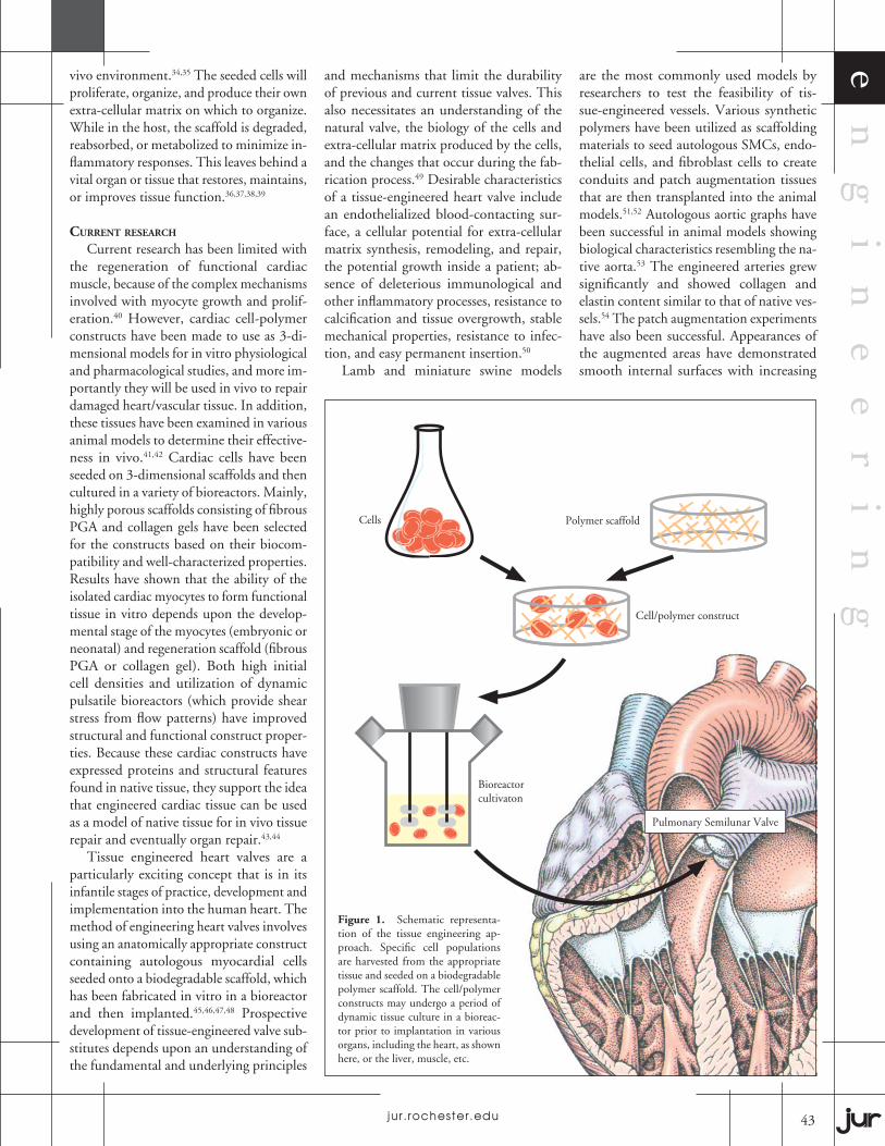
There are three ways to approach creat-ing new engineered tissues: the injection of isolated cells, the development of cell encap-sulation systems, and the use of stem cells or biopsied cells and scaffolds (both natural and biosynthetic) to implant constructs of tissue grown in vitro (in a test tube).30 The reabsorbable scaffold is made into the shape of the tissue being developed, such as heart valves or vascular tissue; the scaffolding materials must be biocompatible and must meet both the nutritional and biological needs for the specific cell population being grown. In the case of engineering cardiovas-cular tissues, myoblasts or myocytes (muscle cells) are seeded with fibroblasts onto bio-compatible polymer scaffolds to grow the intended tissue.31 (Figure 1)
Polymer scaffolds can be constructed from natural or synthetic biomaterials. Although natural biomaterials better simu-late environments in vivo, they do impose limitations such as large variations in the biological tissues that have been extracted for the scaffolding, as well as restrictions with the versatility of designing devices with specific biomechanical properties. Advances in the field of polymer chemistry have al-lowed scientists to develop several synthetic polymers that can be precisely altered for use as scaffolding materials. The desired scaffold is first seeded with autologous, allogeneic, or xenogeneic cells, potentially derived from either biopsies or stem cells. Next, the cells are placed into a bioreactor to stimulate tissue growth in vitro before
implantation. Specifically, a bioreactor is a device that produces a dynamic in vitro mi-croenvironment for guided tissue growth. It simulates the microenvironment that would be found in the body for normal tissue for-mation and growth. This device allows scientists to control the flow and mixing of cell culture media, which can enhance the transfer of nutrients, wastes, and regulatory molecules to and from the growing tissue. Additionally, bioreactors provide mechani-cal simulations of flow conditions found in the body, which enhances the formation of tissue constructs by providing appropriate shear stresses and strains to the growing cells. For example, pulsatile flow bioreactors have been used to construct trileaflet heart valves and blood vessels. These constructs under pulsed flow conditions exhibited greater burst strengths, better suture reten-tion, and higher collagen content than the nonpulsed constructs.32 Currently, the use of xenogenetic and allogenetic tissues with open-celled constructs is limited because of host immune responses against the im-planted tissue.33 As a result, the majority of the tissue-engineered experiments using polymer/cell construct technology have used autologous cells.
The delivery of cells to a polymer is called seeding. Seeding may take a static form by directly applying the cells to the scaffold, or it may take a dynamic form, where agitation is used to apply the cells. Dynamic seeding is accomplished through the use of a pulsatile flow bioreactor, and it results in a higher seeding density and more uniform cell distribution. With a bioreactor mechanical forces such as stretch, pressure, and shear forces are employed, while gas and nutrient exchange is accomplished through constant changing of the culturing medium. The construct-bioreactor system allows the user to control the in vitro culture conditions such as cell population, seeding density, biochemical signals, and physical forces. The effects of these parameters on engineered tissue structure and function can be quantified and examined in the in
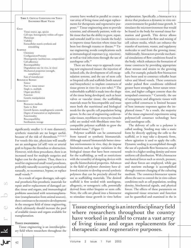
TABLE 1. CRITICAL CONDITIONS FOR TISSUE-ENGINEERED HEART VALVES
CELLS
Tissue source, age, species Cell type, heterogeneity within cell types Phenotype Viability Extracellular matrix synthesis and remodeling
SCAFFOLD
Chemistry Configuration/Porosity Heterogeneity (architecture, composition) Cell adherence Biocompatibility Degradation rate (in vivo, in vitro) Mechanical properties (strength, compliance) Ease of manufacture
BIOLOGICAL SIGNALS
Source Pure vs. tissue extract Single vs. multiple Organ specificity Function Genetic manipulation
CONSTRUCT
Bioreactor medium Static vs. flow Growth factors, morphogenetic controls Degree of maturation at implantation Functionality Biocompatibility Sterilization, storage
Volume 2 Issue 1 Fal l 2003 jur.rochester.edu
Tissue engineering is an interdisciplinary field where researchers throughout the country have worked in parallel to create a vast array of living tissue and organ replacements for therapeutic and regenerative purposes.
42 43
en
gi
ne
er
in
gvivo environment.34,35 The seeded cells will proliferate, organize, and produce their own extra-cellular matrix on which to organize. While in the host, the scaffold is degraded, reabsorbed, or metabolized to minimize in-flammatory responses. This leaves behind a vital organ or tissue that restores, maintains, or improves tissue function.36,37,38,39
CURRENT RESEARCH
Current research has been limited with the regeneration of functional cardiac muscle, because of the complex mechanisms involved with myocyte growth and prolif-eration.40 However, cardiac cell-polymer constructs have been made to use as 3-di-mensional models for in vitro physiological and pharmacological studies, and more im-portantly they will be used in vivo to repair damaged heart/vascular tissue. In addition, these tissues have been examined in various animal models to determine their effective-ness in vivo.41,42 Cardiac cells have been seeded on 3-dimensional scaffolds and then cultured in a variety of bioreactors. Mainly, highly porous scaffolds consisting of fibrous PGA and collagen gels have been selected for the constructs based on their biocom-patibility and well-characterized properties. Results have shown that the ability of the isolated cardiac myocytes to form functional tissue in vitro depends upon the develop-mental stage of the myocytes (embryonic or neonatal) and regeneration scaffold (fibrous PGA or collagen gel). Both high initial cell densities and utilization of dynamic pulsatile bioreactors (which provide shear stress from flow patterns) have improved structural and functional construct proper-ties. Because these cardiac constructs have expressed proteins and structural features found in native tissue, they support the idea that engineered cardiac tissue can be used as a model of native tissue for in vivo tissue repair and eventually organ repair.43,44
Tissue engineered heart valves are a particularly exciting concept that is in its infantile stages of practice, development and implementation into the human heart. The method of engineering heart valves involves using an anatomically appropriate construct containing autologous myocardial cells seeded onto a biodegradable scaffold, which has been fabricated in vitro in a bioreactor and then implanted.45,46,47,48 Prospective development of tissue-engineered valve sub-stitutes depends upon an understanding of the fundamental and underlying principles
and mechanisms that limit the durability of previous and current tissue valves. This also necessitates an understanding of the natural valve, the biology of the cells and extra-cellular matrix produced by the cells, and the changes that occur during the fab-rication process.49 Desirable characteristics of a tissue-engineered heart valve include an endothelialized blood-contacting sur-face, a cellular potential for extra-cellular matrix synthesis, remodeling, and repair, the potential growth inside a patient; ab-sence of deleterious immunological and other inflammatory processes, resistance to calcification and tissue overgrowth, stable mechanical properties, resistance to infec-tion, and easy permanent insertion.50
Lamb and miniature swine models
are the most commonly used models by researchers to test the feasibility of tis-sue-engineered vessels. Various synthetic polymers have been utilized as scaffolding materials to seed autologous SMCs, endo-thelial cells, and fibroblast cells to create conduits and patch augmentation tissues that are then transplanted into the animal models.51,52 Autologous aortic graphs have been successful in animal models showing biological characteristics resembling the na-tive aorta.53 The engineered arteries grew significantly and showed collagen and elastin content similar to that of native ves-sels.54 The patch augmentation experiments have also been successful. Appearances of the augmented areas have demonstrated smooth internal surfaces with increasing
Figure 1. Schematic representa-tion of the tissue engineering ap-proach. Specific cell populations are harvested from the appropriate tissue and seeded on a biodegradable polymer scaffold. The cell/polymer constructs may undergo a period of dynamic tissue culture in a bioreac-tor prior to implantation in various organs, including the heart, as shown here, or the liver, muscle, etc.
Polymer scaffold
Bioreactor cultivaton
Cell/polymer construct
Cells
Pulmonary Semilunar Valve
Volume 2 Issue 1 Fal l 2003 jur.rochester.edu

44 45
kr
eb
s
tissue and extra-cellular matrix formation, in addition to having near reabsorption of the polymer.55 These models have not yet been utilized with human patients in vivo due to the need for further studies to assess the biological functioning of these vessels in both short-term and long-term implanta-tion. However, the feasibility of culturing autologous implantable arteries has been demonstrated.56,57 (Figure 2)
Doctor John E. Mayer M.D. at Chil-dren’s Hospital and Dr. Joseph P. Vacanti M.D. at Massachusetts General Hospital in Boston have developed a pulsate bioreac-tor for the in vitro formation of normally functioning constructs with an architecture that mimics the naturally occurring valve environment. This system develops ad-equate mechanical properties (e.g. ability to withstand hemodynamic stresses) by providing physiological pressure and flow of nutrient/cytokine medium to the devel-oping valve construct. In addition, long term in vitro developments have resulted in contamination-resistant systems.58 This group has created tri-leaflet heart valve scaf-folds from various biodegradable polymers, and has successfully implanted them into lambs for up to four months (Fig. 2). The polymers that have been tested include polyglycolic acid (PGA), poly-4-hydroxy-butyrate (P4HB), and polyhydroxyalkano-ate (PHA), of which the latter has proved to be the most biocompatible as a result of its synchronous opening and closing abil-ity in the pulsate bioreactor and its abil-ity to accommodate cell attachment and growth.59,60 PHA has mechanical properties
such as elasticity and mechanical strength that far exceed the other polymer systems mentioned above.61,62,63 When autologous cardiac cell lines were seeded onto the PHA, they mostly viable, and formed a connec-tive tissue matrix between the inside and the outside of the porous heart valve scaf-fold.64 After being implanted into lambs at a segment of the pulmonary artery and extracted at various time intervals, no signs of thrombus formation were evident on the constructs. An acellular construct was used as a control and showed no signs of tissue growth or extracellular matrix production.65 These early results show a promising and adequate future for the development of tis-sue-engineered heart valves; the goal being the development of clinically feasible valve replacements.
The use of tissue engineering techniques to create heart valve substitutes from autolo-gous cell lines and biodegradable polymer scaffolds is growing and results in animal models have been promising thus far. Tissue engineering offers a new approach to the de-velopment of heart valves in that it allows for the creation of structures that are viable and have the capacity for self repair. This allows for greater durability without the side effects of anticoagulation therapy, thrombus for-mation, inflammation, immune rejection, infection, and hemorrhaging.66 Continued progress in this area of tissue engineering with heart valves strongly depends upon: (a) understanding the fundamentals behind valve development and function, including extracellular matrix biology, valvular cell biology, response to injury and how these
fundamentals relate to the progressive evo-lution of structure; (b) the understanding that has been developed over the past fifty years concerning failure modes of different types of valve replacement structures; (c) understanding the structural conditions of favorable valve alternatives, including the composition and organization of the collagen, elastin, and glycosaminoglycans in the engineered valve; (d) understand-ing the basic role/relationship between the host tissue and implanted tissue; and (e) the mechanisms of physiological repair process-es.67 Therefore, tissue-engineered valves will hopefully offer a more permanent solution to the problem.
Ideal biological grafts should possess a continuous endothelial layer and dif-ferentiated smooth muscle cells, as well as sufficient mechanical integrity and elastic properties to allow for structure retention and tolerance to arterial pressure. Shear stresses in fluid dynamics have a tremen-dous impact on vascular cell morphology, proliferation, orientation and both the or-ganization and composition of extracellular matrices.68 Thus, there have been increased interests in researching and defining shear stresses and pressures present in bioreactors for tissue conditioning and guided tissue formation, especially with tissue engineer-ing applications involving cardiovascular structures. This is because the artery must withstand both transmural pressures act-ing perpendicular to the vessel wall and the shear stresses acting along the wall surfaces.69 Shear stresses that accompany dynamic conditioning have shown posi-tive effects on the generation of functional blood vessels. The mechanical properties of the engineered vessels are derived from the smooth muscle cells (SMCs) and extracel-lular matrix proteins they produce. These mechanical properties are not derived from the scaffolding material (i.e. PGA), because most biodegradable scaffolding materials utilized in repairing/replacing vessels fragment and degrade to less than 15% of their initial mass after 5 weeks in culture.70 In addition, researchers have shown that the flow positively influences cell to cell interactions.71
FUTURE RESEARCH
An important area of future work for the generation of small diameter vessels is to repopulate tissue-engineered grafts with autologous cells in vivo instead of
Figure 2 Tissue engineered pulmonary valve leaflet in a lamb heart.
Volume 2 Issue 1 Fal l 2003 jur.rochester.edu

44 45
Nicholas Krebs graduated from the University of Rochester in 2003 with a B.S. in Biomedical Engineering. He also completed his Take Five program in African-American music and literature. Currently, Nick is employed as a research assistant at the Massachusetts General Hospital under Dr. Joseph Vacanti, professor of surgery at Harvard Medical School and director of the Tissue Engineering and Organ Fabrication Laboratory. Nick plans to at-tend medical school and pursue a career in surgery or internal medicine.
en
gi
ne
er
in
g
in vitro. In doing so, it has been proposed that endothelial progenitor cells be seeded onto polymer scaffolds to allow for a non-thrombogenic barrier, because in previous attempts at tissue-engineered vessels, in vitro culturing of SMCs and endothelial cells has resulted in thrombosis shortly af-ter implantation.72 Some problems plagu-ing synthetic vascular graphs that must be overcome to make vascular grafts a reality are platelet adhesion and decreased compli-ance (ability to stretch) compared with the adjacent arterial tissue, which have both led to the ideas discussed above.73
Many attempts have been made to develop and produce cardiac tissue that is long lasting, biocompatible, and effective. To overcome the mechanical and biologi-cal limitations posed by synthetic implants, research has begun to focus on the develop-ment of a naturally derived biomaterial for the fabrication of cardiac tissue, heart valve tissue, and vascular grafting tissue. Further advances in tissue engineering using autolo-gous cells are necessary before widespread applicability to multiple organ systems can
become a reality. Current in vitro bioreac-tor environments are mere approximations to the complex biochemical and physical environments that cells are situated in during organ development and repair in vivo. Likewise, the synthetic polymers that are used as scaffolding materials for cell growth are imperfect approximations to extracellular matrices. Thus, the develop-ment of synthetic or natural scaffolding templates for cell culturing, that mimic the architecture and surface biochemistry of the target tissue will better enable the autologous cells to develop into functional and effective replacement tissues and or-gans. To develop and engineer complete organ systems, techniques to promote complex vascularization of capillary beds in the tissue of the organ will be a neces-sity to promote the mass transfer of oxygen and nutrients throughout the tissue. Stem cell biology holds an enormous potential for artificial organ and tissue development for transplantation. However, at the cur-rent time, techniques to isolate multipotent and pluripotent stem cells from adult tissues
remains extremely difficult and complex.74 Therefore, advances in molecular immunol-ogy, tissue engineering and stem cell biology will offer even better therapeutic methods for treating organ and cardiac failure in the future. Finally, interdisciplinary cooperation between engineers, scientists, and physicians will allow for discoveries and innovations to be rapidly applied for the development of clinically useful, tissue-engineered cardio-vascular implants, and ultimately, a tissue-engineered heart that would provide a viable alternative to transplantation and ultimately ease the suffering of hundreds of thousands of individuals worldwide.
Volume 2 Issue 1 Fal l 2003 jur.rochester.edu
...a tissue-engineered heart that would provide a viable alternative to transplantation and ultimately ease the suffering of hundreds of thousands of individuals worldwide.