tirf microscopy‐based monitoring of drug permeation across
TRANSCRIPT

Drug Delivery
TIRF Microscopy-Based Monitoring of Drug Permeation Acrossa Lipid Membrane Supported on Mesoporous SilicaPaul Joyce,* Silver J�emetsa, Simon Isaksson, Shakhawath Hossain, Per Larsson,Christel Bergstrçm, and Fredrik Hççk*
Abstract: There is an urgent demand for analytic approachesthat enable precise and representative quantification of thetransport of biologically active compounds across cellularmembranes. In this study, we established a new means tomonitor membrane permeation kinetics, using total internalreflection fluorescence microscopy confined to a � 500 nmthick mesoporous silica substrate, positioned underneatha planar supported cell membrane mimic. This way, wedemonstrate spatiotemporally resolved membrane permeationkinetics of a small-molecule model drug, felodipine, whilesimultaneously controlling the integrity of, and monitoring thedrug binding to, the cell membrane mimic. By contrasting thepermeation behaviour of pure felodipine with felodipinecoupled to the permeability enhancer caprylate (C8), weprovide evidence for C8-facilitated transport across lipidmembranes, thus validating the potential for this approach tosuccessfully quantify carrier system-induced changes to cellu-lar membrane permeation.
The cellular exchange and transport of small moleculebioactives are fundamental processes for biological activitythat are strictly regulated by the plasma membrane.[1] Under-standing how small molecules traverse the cellular membraneis therefore critical for the design and formulation develop-ment of novel drug candidates, where promotion of efficientcellular delivery is required to ensure desired therapeuticactivity and response.[2] However, simulating and predictingthe transport of small molecules across biological membranes
in vitro presents a significant challenge due to the lack offacile, cost-effective and biologically relevant approaches thatdeliver rapid and precise estimations of drug translocation.
In the context of oral drug delivery, small moleculetherapeutics must readily absorb across the intestinal epithe-lium via transcellular and/or paracellular pathways to reachthe systemic circulation,[3] or be hosted within a carrier systemthat promotes permeability, and thus bioavailability.[4] Tosimulate this process and screen drug candidates/carriersystems for permeability, static in vitro cellular models, suchas the Caco-2 cellular monolayer assay, have long beenregarded as the benchmark approach.[5] However, the cost-and time-exhaustive nature of cellular assays, limited biolog-ical relevancy,[6] and propensity for tight junction and/or cellmonolayer disruption, lead to complications in obtaining andinterpreting drug permeability kinetics with high accuracy.[7]
To overcome the hurdles associated with cellular assays,recent focus has been attributed to developing representativecell-membrane mimics as a simplified approach for in vitroquantification of transcellular drug permeation.[8] In thesesystems, phospholipid membranes are adsorbed onto a porouspolymeric support between a donor and acceptor cell,allowing for the elucidation of time-dependent changes indrug concentration within the acceptor cell.[9] While sup-ported artificial membrane assays ignore paracellular trans-port, several studies have demonstrated successful correla-tions between in vitro and in vivo pharmacokinetics, thushighlighting their ability to predict in vivo intestinal perme-ability.[10] However, the formation and integrity of the lipidmembrane is neither monitored nor controlled using thisapproach, which increases susceptibility for random drug fluxdue to membrane disintegration.[11]
The aim of the present work was to overcome thislimitation by combining the supported membrane approachwith an analytical microscopy technique to validate bilayerintegrity throughout drug permeation studies, while simulta-neously allowing for the detection and quantification of drugpermeation. This was accomplished using a � 500 nm thickmesoporous silica thin films (MSTF)[12] serving as substratefor a supported lipid bilayer (SLB),[13] thereby enablingconcurrent fluorescence recovery after photobleaching(FRAP) analysis of membrane integrity and total internalreflection fluorescence (TIRF) spatiotemporal monitoring ofdrug permeation into the MSTF.
Formation of the supported lipid bilayer (SLB) wasmonitored with TIRF microscopy (Figure 1A), through theinclusion of rhodamine-labelled tracer vesicles (Rh-POPC),[14] on both non-porous (planar) and MSTF substrateswith a pore size of � 7 nm.[12] The rate of POPC vesicle
[*] P. Joyce, S. J�emetsa, S. Isaksson, Prof. F. HççkDepartment of Physics, Chalmers University of TechnologyGothenburg SE-412 96 (Sweden)E-mail: [email protected]
S. Hossain, P. Larsson, C. BergstrçmDepartment of Pharmacy, Uppsala UniversityUppsala SE-751 23 (Sweden)
S. Hossain, P. Larsson, C. BergstrçmThe Swedish Drug Delivery Forum, Department of Pharmacy,Uppsala UniversityUppsala SE-751 23 (Sweden)
Supporting information and the ORCID identification number(s) forthe author(s) of this article can be found under:https://doi.org/10.1002/anie.202011931.
� 2020 The Authors. Angewandte Chemie International Editionpublished by Wiley-VCH GmbH. This is an open access article underthe terms of the Creative Commons Attribution Non-CommercialNoDerivs License, which permits use and distribution in anymedium, provided the original work is properly cited, the use is non-commercial and no modifications or adaptations are made.
AngewandteChemieCommunications
How to cite:International Edition: doi.org/10.1002/anie.202011931German Edition: doi.org/10.1002/ange.202011931
1Angew. Chem. Int. Ed. 2020, 59, 1 – 6 � 2020 The Authors. Angewandte Chemie International Edition published by Wiley-VCH GmbH
These are not the final page numbers! � �
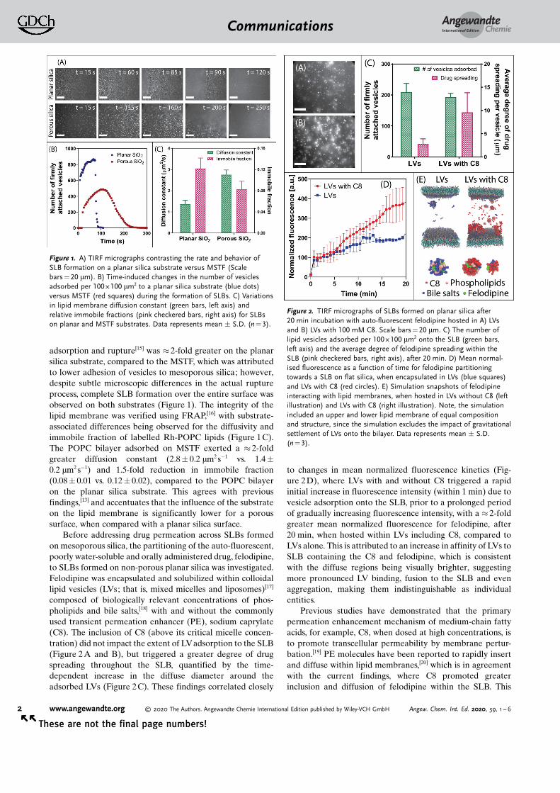
adsorption and rupture[15] was � 2-fold greater on the planarsilica substrate, compared to the MSTF, which was attributedto lower adhesion of vesicles to mesoporous silica; however,despite subtle microscopic differences in the actual ruptureprocess, complete SLB formation over the entire surface wasobserved on both substrates (Figure 1). The integrity of thelipid membrane was verified using FRAP,[16] with substrate-associated differences being observed for the diffusivity andimmobile fraction of labelled Rh-POPC lipids (Figure 1C).The POPC bilayer adsorbed on MSTF exerted a � 2-foldgreater diffusion constant (2.8� 0.2 mm2 s�1 vs. 1.4�0.2 mm2 s�1) and 1.5-fold reduction in immobile fraction(0.08� 0.01 vs. 0.12� 0.02), compared to the POPC bilayeron the planar silica substrate. This agrees with previousfindings,[13] and accentuates that the influence of the substrateon the lipid membrane is significantly lower for a poroussurface, when compared with a planar silica surface.
Before addressing drug permeation across SLBs formedon mesoporous silica, the partitioning of the auto-fluorescent,poorly water-soluble and orally administered drug, felodipine,to SLBs formed on non-porous planar silica was investigated.Felodipine was encapsulated and solubilized within colloidallipid vesicles (LVs; that is, mixed micelles and liposomes)[17]
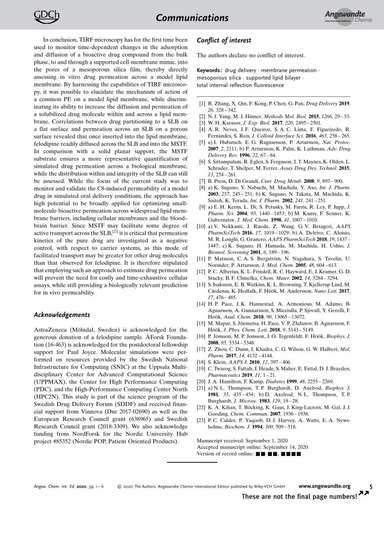
composed of biologically relevant concentrations of phos-pholipids and bile salts,[18] with and without the commonlyused transient permeation enhancer (PE), sodium caprylate(C8). The inclusion of C8 (above its critical micelle concen-tration) did not impact the extent of LVadsorption to the SLB(Figure 2A and B), but triggered a greater degree of drugspreading throughout the SLB, quantified by the time-dependent increase in the diffuse diameter around theadsorbed LVs (Figure 2C). These findings correlated closely
to changes in mean normalized fluorescence kinetics (Fig-ure 2D), where LVs with and without C8 triggered a rapidinitial increase in fluorescence intensity (within 1 min) due tovesicle adsorption onto the SLB, prior to a prolonged periodof gradually increasing fluorescence intensity, with a � 2-foldgreater mean normalized fluorescence for felodipine, after20 min, when hosted within LVs including C8, compared toLVs alone. This is attributed to an increase in affinity of LVs toSLB containing the C8 and felodipine, which is consistentwith the diffuse regions being visually brighter, suggestingmore pronounced LV binding, fusion to the SLB and evenaggregation, making them indistinguishable as individualentities.
Previous studies have demonstrated that the primarypermeation enhancement mechanism of medium-chain fattyacids, for example, C8, when dosed at high concentrations, isto promote transcellular permeability by membrane pertur-bation.[19] PE molecules have been reported to rapidly insertand diffuse within lipid membranes,[20] which is in agreementwith the current findings, where C8 promoted greaterinclusion and diffusion of felodipine within the SLB. This
Figure 1. A) TIRF micrographs contrasting the rate and behavior ofSLB formation on a planar silica substrate versus MSTF (Scalebars = 20 mm). B) Time-induced changes in the number of vesiclesadsorbed per 100 � 100 mm2 to a planar silica substrate (blue dots)versus MSTF (red squares) during the formation of SLBs. C) Variationsin lipid membrane diffusion constant (green bars, left axis) andrelative immobile fractions (pink checkered bars, right axis) for SLBson planar and MSTF substrates. Data represents mean � S.D. (n = 3).
Figure 2. TIRF micrographs of SLBs formed on planar silica after20 min incubation with auto-fluorescent felodipine hosted in A) LVsand B) LVs with 100 mM C8. Scale bars = 20 mm. C) The number oflipid vesicles adsorbed per 100 � 100 mm2 onto the SLB (green bars,left axis) and the average degree of felodipine spreading within theSLB (pink checkered bars, right axis), after 20 min. D) Mean normal-ised fluorescence as a function of time for felodipine partitioningtowards a SLB on flat silica, when encapsulated in LVs (blue squares)and LVs with C8 (red circles). E) Simulation snapshots of felodipineinteracting with lipid membranes, when hosted in LVs without C8 (leftillustration) and LVs with C8 (right illustration). Note, the simulationincluded an upper and lower lipid membrane of equal compositionand structure, since the simulation excludes the impact of gravitationalsettlement of LVs onto the bilayer. Data represents mean � S.D.(n = 3).
AngewandteChemieCommunications
2 www.angewandte.org � 2020 The Authors. Angewandte Chemie International Edition published by Wiley-VCH GmbH Angew. Chem. Int. Ed. 2020, 59, 1 – 6� �
These are not the final page numbers!

was also supported by coarse-grained molecular dynamicssimulations which indicated a clear increase in the interactionwith the lipid membrane for LVs with C8 compared to LVsalone, as shown in the right and left illustration in Figure 2E.These simulations additionally showed that felodipine parti-tioned closely to phospholipid molecules within LVs contain-ing lipid only, with limited drug detachment and partitioningtowards the SLB, while LVs with C8 present promoteda significant (two-fold) enhancement in the probability offelodipine detachment from the vesicle into the SLB.
The C8-provoked felodipine partitioning was hypothe-sized to correlate with enhanced felodipine permeation acrossthe model lipid membrane. Since it is not possible to observepermeation across a SLB formed on planar silica, the lipidbilayer was instead supported on a mesoporous silicasubstrate as described above, allowing for time-dependentdrug diffusion across the SLB into the silica pores to bequantified, using TIRF-confined evanescent wave illumina-tion restricted to the porous film underneath the SLB. Tocorrelate permeation with membrane partitioning, the angleof incidence, q, of the TIRF light source was controlled toeither induce total internal reflection (TIR) at the MSTF-SLBinterface, q1, or using a higher angle of incidence, q2, for TIRat the silica (cover slip)-MSTF interface (Figure 3A). Theevanescent wave created by light reflecting at the interfacebetween a material with a higher index of refraction (silica
substrate) and a material with a lower index of refraction(MSTF) decays exponentially into the material of lowerrefractive index.[21] Subsequently, by increasing the angle ofincidence above the critical angle required for TIR at thesilica-MSTF interface, it was possible to limit the depth ofpenetration of the evanescent wave to � 100–200 nm into theMSTF pores, as verified by the fluorescence intensity of Rh-POPC decreasing and the fluorescence intensity of felodipineincreasing as a function of increasing angle of incidence,measured after 20 min incubation with felodipine in LVs withC8 (Figure 3 B). Moreover, visual observations of TIRFmicrographs are indicative of LVs and C8 vesicles adsorbedon the SLB surface at q1 and submicron-sized felodipinepatches observed within the MSTF at q2 (Figure 3C). Theoccurrence of submicron (up to micron) sized patches offelodipine within the MSTF was attributed to the tendency forthe drug to partition in lipid aggregates and the strongadhesion between lipids and the silica surface within theporous film. Further, the existence of micron sized disorderedmesoporous phases,[13] which according to observations madefor mesoporous silicon scaffolds[22] may exist as independentdiffusional systems that hamper complete connectivitythroughout the film, could also promote the formation ofsmall-scale aggregates of water insoluble felodipine andlipids.
Figure 3. A) Schematic representation of the TIRF experimental set up: a SLB was adsorbed onto a MSTF, whereby the angle of incidence wasadjusted so that total internal reflection occurs at the silica–MSTF interface. This approach allows for identification of felodipine molecules thatare transported across the SLB into the pores of the thin film. B) Fluorescent intensity of the SLB, detected via rhodamine-labelled tracer lipids(green dots, left axis), and felodipine (red squares, right axis) as a function of angle of incidence. C) TIRF micrographs highlighting the change influorescence intensity and appearance of the SLB and felodipine at angles q1 and q2, as indicated in (B). Scale bars = 20 mm.
AngewandteChemieCommunications
3Angew. Chem. Int. Ed. 2020, 59, 1 – 6 � 2020 The Authors. Angewandte Chemie International Edition published by Wiley-VCH GmbH www.angewandte.org
These are not the final page numbers! � �

A small increase in fluorescence intensity was observedwhen the pure drug was added to buffer alone. In contrast,felodipine fluorescence intensity within the MSTF pores was� 5-fold greater when administered with LVs, compared tothe pure drug, which is attributed to the solubilizing capacityof the colloidal phases formed by the phospholipid speciesand bile salts (felodipine was added at 80 % its solubilitylimit). The ability for C8 to further promote permeation offelodipine was shown to correlate well with partitioningstudies performed on the planar silica SLB, with a time-dependent increase in felodipine fluorescence intensity withinthe MSTF being observed, leading to a mean normalizedfluorescence intensity of 1048� 315 AU after 20 min (Fig-ure 4C). In particular, analysis of the number and intensity ofsubmicron felodipine patches within the MSTF after 20 mindemonstrates a � 5-fold increase in drug permeation whenadministered with LVs and C8, compared to LVs alone(Figure 4A and B). Changes in normalized fluorescenceintensities due to felodipine permeation across the SLBdisplayed kinetics with multiple phases observed uponaddition of LVs including C8 (Figure 4C), where a lag phasewas followed by a rapid and sustained increase in fluorescenceintensity. This contrasts with the changes in normalizedfluorescent intensity when felodipine-encapsulated LVs withC8 were added to an SLB on planar silica (Figure 2D), wherea rapid increase in fluorescence intensity was observed within
60 s. Thus, the presence of a lag phase for LVs with C8indicates that C8 must first bind to and insert within the lipidmembrane prior to promoting transport of felodipine acrossthe lipid membrane, into the pores of the MSTF.
Importantly, the integrity of the SLB was not significantlydisrupted during (or after) exposure to felodipine, whenencapsulated within LVs including C8 (Figure 5). That is,monitoring of Rh-POPC fluorescence at the MSTF-SLBinterface (q1), concurrently to felodipine permeation,revealed only a minor time-dependent reduction in fluores-cence intensity (Figures 5A and C), being attributed to Rh-POPC molecules within the SLB interchanging with lipidswithin LVs. Further, FRAP analysis post-exposure to felodi-pine demonstrated essentially complete fluorescence recov-ery within 30 s after photobleaching (Figure 5B), validatingthe presence of an intact SLB throughout the drug perme-ation process. A small increase in diffusion constant was evenobserved following felodipine exposure, from 2.8� 0.2 mm2 s�1
to 3.5� 0.7 mm2 s�1, which is consistent with previous findingsthat demonstrated the ability for saturated fatty acids (e.g.C8) to facilitate lipid packing and membrane fluidity.[23]
Figure 4. A) TIRF micrographs at various time points highlightingfelodipine permeation across the SLB into the pores of the MSTF,when solubilized within LVs without a permeation enhancer (top row)and with C8 (bottom row). Scale bars = 20 mm. B) Fluorescent intensitydistributions for felodipine patches within MSTF when hosted in LVs(blue bars) and LVs with C8 (red bars). C) Mean normalized fluores-cence as a function of time for felodipine within MSTF in TRIS buffer(i.e. pure drug) (green triangles), LVs (blue squares) and LVs with C8(red circles). Data represents mean � S.D. (n =3).
Figure 5. A) TIRF micrographs highlighting the time-dependentchanges in visual appearance of the rhodamine-labelled SLB whenexposed to felodipine encapsulated within LVs in the presence of C8.Scale bars = 20 mm. B) TIRF micrographs revealing fluorescence recov-ery of the rhodamine-labelled SLB after photobleaching. FRAP wasperformed on the SLB following exposure to felodipine hosted withinLVs with C8. Thus, the ability for fluorescence recovery within 30 safter photobleaching indicates the presence of an intact SLB. C) Meannormalized fluorescence as a function of time for the rhodamine-labelled SLB during exposure to felodipine hosted within LVs in thepresence of C8. The TIRF angle was set at the MSTF-SLB interfacewhile monitoring Rh-SLB fluorescence. D) Variations in lipid mem-brane diffusion constant (green bars, left axis) and relative immobilefractions (pink checkered bars, right axis) for SLBs pre-exposure tofelodipine hosted within LVs in the presence of C8 (t = 0 min) andpost-exposure (t = 20 min). Data represents mean � S.D. (n= 3).
AngewandteChemieCommunications
4 www.angewandte.org � 2020 The Authors. Angewandte Chemie International Edition published by Wiley-VCH GmbH Angew. Chem. Int. Ed. 2020, 59, 1 – 6� �
These are not the final page numbers!
In conclusion, TIRF microscopy has for the first time beenused to monitor time-dependent changes in the adsorptionand diffusion of a bioactive drug compound from the bulkphase, to and through a supported cell-membrane mimic, intothe pores of a mesoporous silica film, thereby directlyassessing in vitro drug permeation across a model lipidmembrane. By harnessing the capabilities of TIRF microsco-py, it was possible to elucidate the mechanism of action ofa common PE on a model lipid membrane, while discrim-inating its ability to increase the diffusion and permeation ofa solubilized drug molecule within and across a lipid mem-brane. Correlations between drug partitioning to a SLB ona flat surface and permeation across an SLB on a poroussurface revealed that once inserted into the lipid membrane,felodipine readily diffused across the SLB and into the MSTF.In comparison with a solid planar support, the MSTFsubstrate ensures a more representative quantification ofsimulated drug permeation across a biological membrane,while the distribution within and integrity of the SLB can stillbe assessed. While the focus of the current study was tomonitor and validate the C8-induced permeability of a modeldrug in simulated oral delivery conditions, the approach hashigh potential to be broadly applied for optimizing small-molecule bioactive permeation across widespread lipid mem-brane barriers, including cellular membranes and the blood–brain barrier. Since MSTF may facilitate some degree ofactive transport across the SLB,[22] it is critical that permeationkinetics of the pure drug are investigated as a negativecontrol, with respect to carrier systems, as this mode offacilitated transport may be greater for other drug moleculesthan that observed for felodipine. It is therefore stipulatedthat employing such an approach to estimate drug permeationwill prevent the need for costly and time-exhaustive cellularassays, while still providing a biologically relevant predictionfor in vivo permeability.
Acknowledgements
AstraZeneca (Mçlndal, Sweden) is acknowledged for thegenerous donation of a felodipine sample. �Forsk Founda-tion (16-463) is acknowledged for the postdoctoral fellowshipsupport for Paul Joyce. Molecular simulations were per-formed on resources provided by the Swedish NationalInfrastructure for Computing (SNIC) at the Uppsala Multi-disciplinary Center for Advanced Computational Science(UPPMAX), the Center for High Performance Computing(PDC), and the High-Performance Computing Center North(HPC2N). This study is part of the science program of theSwedish Drug Delivery Forum (SDDF) and received finan-cial support from Vinnova (Dnr 2017-02690) as well as theEuropean Research Council grant (638965) and SwedishResearch Council grant (2018-3309). We also acknowledgefunding from NordForsk for the Nordic University Hubproject #85352 (Nordic POP, Patient Oriented Products).
Conflict of interest
The authors declare no conflict of interest.
Keywords: drug delivery · membrane permeation ·mesoporous silica · supported lipid bilayer ·total internal reflection fluorescence
[1] R. Zhang, X. Qin, F. Kong, P. Chen, G. Pan, Drug Delivery 2019,26, 328 – 342.
[2] N. J. Yang, M. J. Hinner, Methods Mol. Biol. 2015, 1266, 29 – 53.[3] W. H. Karasov, J. Exp. Biol. 2017, 220, 2495 – 2501.[4] A. R. Neves, J. F. Queiroz, S. A. C. Lima, F. Figueiredo, R.
Fernandes, S. Reis, J. Colloid Interface Sci. 2016, 463, 258 – 265.[5] a) I. Hubatsch, E. G. Ragnarsson, P. Artursson, Nat. Protoc.
2007, 2, 2111; b) P. Artursson, K. Palm, K. Luthman, Adv. DrugDelivery Rev. 1996, 22, 67 – 84.
[6] S. Sittampalam, R. Eglen, S. Ferguson, J. T. Maynes, K. Olden, L.Schrader, T. Shelper, M. Ferrer, Assay Drug Dev. Technol. 2015,13, 254 – 261.
[7] B. Press, D. Di Grandi, Curr. Drug Metab. 2008, 9, 893 – 900.[8] a) K. Sugano, Y. Nabuchi, M. Machida, Y. Aso, Int. J. Pharm.
2003, 257, 245 – 251; b) K. Sugano, N. Takata, M. Machida, K.Saitoh, K. Terada, Int. J. Pharm. 2002, 241, 241 – 251.
[9] a) E. H. Kerns, L. Di, S. Petusky, M. Farris, R. Ley, P. Jupp, J.Pharm. Sci. 2004, 93, 1440 – 1453; b) M. Kansy, F. Senner, K.Gubernator, J. Med. Chem. 1998, 41, 1007 – 1010.
[10] a) V. Nekkanti, J. Rueda, Z. Wang, G. V. Betageri, AAPSPharmSciTech 2016, 17, 1019 – 1029; b) A. Delrivo, C. Aloisio,M. R. Longhi, G. Granero, AAPS PharmSciTech 2018, 19, 1437 –1447; c) K. Sugano, H. Hamada, M. Machida, H. Ushio, J.Biomol. Screening 2001, 6, 189 – 196.
[11] P. Matsson, C. A. S. Bergstrçm, N. Nagahara, S. Tavelin, U.Norinder, P. Artursson, J. Med. Chem. 2005, 48, 604 – 613.
[12] P. C. Alberius, K. L. Frindell, R. C. Hayward, E. J. Kramer, G. D.Stucky, B. F. Chmelka, Chem. Mater. 2002, 14, 3284 – 3294.
[13] S. Isaksson, E. B. Watkins, K. L. Browning, T. Kjellerup Lind, M.C�rdenas, K. Hedfalk, F. Hççk, M. Andersson, Nano Lett. 2017,17, 476 – 485.
[14] H. P. Pace, J. K. Hannestad, A. Armonious, M. Adamo, B.Agnarsson, A. Gunnarsson, S. Micciulla, P. Sjçvall, Y. Gerelli, F.Hççk, Anal. Chem. 2018, 90, 13065 – 13072.
[15] M. Mapar, S. J�emetsa, H. Pace, V. P. Zhdanov, B. Agnarsson, F.Hççk, J. Phys. Chem. Lett. 2018, 9, 5143 – 5149.
[16] P. Jçnsson, M. P. Jonsson, J. O. Tegenfeldt, F. Hççk, Biophys. J.2008, 95, 5334 – 5348.
[17] Z. Zhou, C. Dunn, I. Khadra, C. G. Wilson, G. W. Halbert, Mol.Pharm. 2017, 14, 4132 – 4144.
[18] S. Klein, AAPS J. 2010, 12, 397 – 406.[19] C. Twarog, S. Fattah, J. Heade, S. Maher, E. Fattal, D. J. Brayden,
Pharmaceutics 2019, 11, 1 – 21.[20] J. A. Hamilton, F. Kamp, Diabetes 1999, 48, 2255 – 2269.[21] a) N. L. Thompson, T. P. Burghardt, D. Axelrod, Biophys. J.
1981, 33, 435 – 454; b) D. Axelrod, N. L. Thompson, T. P.Burghardt, J. Microsc. 1983, 129, 19 – 28.
[22] K. A. Kilian, T. Bçcking, K. Gaus, J. King-Lacroix, M. Gal, J. J.Gooding, Chem. Commun. 2007, 1936 – 1938.
[23] P. C. Calder, P. Yaqoob, D. J. Harvey, A. Watts, E. A. News-holme, Biochem. J. 1994, 300, 509 – 518.
Manuscript received: September 1, 2020Accepted manuscript online: September 14, 2020Version of record online: && &&, &&&&
AngewandteChemieCommunications
5Angew. Chem. Int. Ed. 2020, 59, 1 – 6 � 2020 The Authors. Angewandte Chemie International Edition published by Wiley-VCH GmbH www.angewandte.org
These are not the final page numbers! � �

Communications
Drug Delivery
P. Joyce,* S. J�emetsa, S. Isaksson,S. Hossain, P. Larsson, C. Bergstrçm,F. Hççk* &&&&—&&&&
TIRF Microscopy-Based Monitoring ofDrug Permeation Across a LipidMembrane Supported on MesoporousSilica
A unique angle: A new approach formonitoring the real-time permeation ofdrug molecules, from the bulk phase,across a lipid membrane was establishedby supporting a lipid bilayer on meso-porous silica thin films. Through the useof TIRF microscopy, the angle of inci-dence of illumination light could be con-trolled to ensure the resulting evan-escence was restricted within the thinfilm, and thus, only drug permeating thebilayer was resolved.
AngewandteChemieCommunications
6 www.angewandte.org � 2020 The Authors. Angewandte Chemie International Edition published by Wiley-VCH GmbH Angew. Chem. Int. Ed. 2020, 59, 1 – 6� �
These are not the final page numbers!